HAL Id: hal-02540640
https://hal.umontpellier.fr/hal-02540640
Submitted on 11 Apr 2020
HAL is a multi-disciplinary open access
archive for the deposit and dissemination of
sci-entific research documents, whether they are
pub-lished or not. The documents may come from
teaching and research institutions in France or
abroad, or from public or private research centers.
L’archive ouverte pluridisciplinaire HAL, est
destinée au dépôt et à la diffusion de documents
scientifiques de niveau recherche, publiés ou non,
émanant des établissements d’enseignement et de
recherche français ou étrangers, des laboratoires
publics ou privés.
International Journal of Radiation Biology Is static
magnetic field exposure a new model of metabolic
alteration? Comparison with Zucker rats
Miryam Elferchichi, Jacques Mercier, Annick Bourret, René Gross,
Anne-Dominique Lajoix, Hatem Belguith, Hafedh Abdelmelek, Mohsen Sakly,
Karen Lambert
To cite this version:
Miryam Elferchichi, Jacques Mercier, Annick Bourret, René Gross, Anne-Dominique Lajoix, et al..
International Journal of Radiation Biology Is static magnetic field exposure a new model of metabolic
alteration? Comparison with Zucker rats. International Journal of Radiation Biology, Informa
Health-care, 2011, 87 (5), pp.483-490. �10.3109/09553002.2011.544371�. �hal-02540640�
Full Terms & Conditions of access and use can be found at
https://www.tandfonline.com/action/journalInformation?journalCode=irab20
International Journal of Radiation Biology
ISSN: 0955-3002 (Print) 1362-3095 (Online) Journal homepage: https://www.tandfonline.com/loi/irab20
Is static magnetic field exposure a new model of
metabolic alteration? Comparison with Zucker rats
Miryam Elferchichi, Jacques Mercier, Annick Bourret, René Gross,
Anne-Dominique Lajoix, Hatem Belguith, Hafedh Abdelmelek, Mohsen Sakly &
Karen Lambert
To cite this article: Miryam Elferchichi, Jacques Mercier, Annick Bourret, René Gross, Anne-Dominique Lajoix, Hatem Belguith, Hafedh Abdelmelek, Mohsen Sakly & Karen Lambert (2011) Is static magnetic field exposure a new model of metabolic alteration? Comparison with Zucker rats, International Journal of Radiation Biology, 87:5, 483-490, DOI: 10.3109/09553002.2011.544371 To link to this article: https://doi.org/10.3109/09553002.2011.544371
Published online: 10 Jan 2011.
Submit your article to this journal
Article views: 52
View related articles
Is static magnetic field exposure a new model of metabolic alteration?
Comparison with Zucker rats
MIRYAM ELFERCHICHI
1,2,3, JACQUES MERCIER
2,3, ANNICK BOURRET
2,3,
RENE
´ GROSS
4, ANNE-DOMINIQUE LAJOIX
4, HATEM BELGUITH
1,
HAFEDH ABDELMELEK
1, MOHSEN SAKLY
1, & KAREN LAMBERT
2,31Faculte´ des Sciences de Bizerte, Laboratoire de Physiologie Inte´gre´e, Jarzouna, Tunisia,2INSERM, ERI25, ‘‘Muscle and Pathologies’’, Montpellier,3Universite´ Montpellier I, Montpellier, and4CNRS UMR5232, UM1, Centre for Pharmacology and Innovation in Diabetes, Montpellier, France
(Received 5 May 2010; Revised 24 November 2010; Accepted 25 November 2010)
Abstract
Purpose: The aim of this study was to investigate if the metabolic alterations observed after static magnetic field (SMF) exposure participates in the development of a pre-diabetic state. A comparison study using the insulin resistant animal model, the Zucker rat and the SMF-exposed Wistar rat was carried out.
Materials and methods: Zucker rats were compared to Wistar rats either exposed to a 128 mT or 0 mT SMF (sham exposed) and analysed. This moderate-intensity SMF exposure of Wistar rats was performed for 1 h/day during 15 consecutive days. Results: Wistar rats exposed to the SMF showed increased levels of carbohydrate and lipid metabolites (i.e., lactate, glycerol, cholesterol and phospholipids) compared to sham-exposed rats. Zucker rats displayed a normoglycemia associated with a high insulin level as opposed to Wistar rats which presented hyperglycemia and hypoinsulinemia after exposure to the SMF. During the glucose tolerance test, unexposed Zucker rats and Wistar rats exposed to the SMF exhibited a significantly higher hyperglycemia compared to sham-exposed Wistar rats suggesting an impairment of glucose clearance. In muscle, glycogen content was lower and phospholipids content was elevated for both unexposed Zucker rats and Wistar rats exposed to the SMF compared to Wistar rats sham control.
Conclusions: This study provides evidence that the metabolic alterations following exposure to a static magnetic field of moderate intensity could trigger the development of a pre-diabetic state.
Keywords: adaptive response, biochemistry, E-M fields
Introduction
According to their frequency, electric and magnetic fields (EMF) are classified into static, extremely low frequency, intermediate frequency and radiofre-quency fields. Static magnetic fields (SMF) are characterised by a frequency of 0 Hertz (Hz) and a field which does not vary with time (Repacholi and Greenebaum 1999). SMF are naturally present everywhere as the earth is surrounded by fields that vary between 25 and 65 mT (Feychting 2005). Superimposed on the earth’s magnetic field are man-made static magnetic fields resulting in an increase of people exposed to SMF. Moreover, SMF are widely used in the treatment of muscu-loskeletal pain relief (Pilla 2006), refractory
neuro-pathic pain (Weintraub and Cole 2004) and symptomatic diabetic neuropathy (Weintraub et al. 2003).
Compelling evidence exists that SMF modulate living systems (Repacholi and Greenebaum 1999, Havas 2000, World Health Organisation [WHO] 2006, Okano 2008). Thus, SMF can affect a wide range of biological systems and tissues as a result of moderate SMF (Rosen 2003a, Dini and Abbro 2005, Amara et al. 2006, Chater et al. 2006) to high intensity SMF (Iwasaka and Ueno 1994, High et al. 2000, Kotani et al. 2002, Ueno and Shigemitsu 2007). SMF can interact with moving charges or ferromagnetic materials and biological molecules with particular magnetic properties (haemoglobin, free radicals) presumably via two mechanisms: The
Correspondence: Mrs Karen Lambert, PhD, INSERM ERI25, Baˆtiment Crastes de Paulet, Hoˆpital A. de Villeneuve, 34 295 Montpellier Cedex 5, France. Tel:þ 33(4) 6741 5230. Fax: þ 33(4) 6741 5231. E-mail: karen.lambert@univ-montp1.fr
Int. J. Radiat. Biol., Vol. 87, No. 5, May 2011, pp. 483–490
ISSN 0955-3002 print/ISSN 1362-3095 online! 2011 Informa UK, Ltd. DOI: 10.3109/09553002.2011.544371
radical pair mechanism and the membrane transdu-cer mechanism. The former hypothesis assumes that SMF increase the lifetime of reactive oxygen species (ROS) and thus their cellular concentrations. These effects are amplified by combined exposure to toxic agents (Amara et al. 2009). Several studies have investigated the effect of SMF on oxidative stress caused by an imbalance between ROS generation and antioxidant capacity of the cell and the consequences of this stress (Zhang et al. 2003, Bekhite et al. 2010). As recently reviewed, there is ROS modulation by moderate SMF exposure with large variations de-pending on the models studied, the intensity of exposure and the tissues targeted (Okano 2008). The second hypothesis is based on reports that SMF can change the biophysical properties of membranes (Rosen 2003b, Genius 2008) leading to an alteration in calcium homeostasis, an increase in membrane rigidity and/or activation of at least nine different signaling networks (Wang et al. 2009). These two hypotheses give insights as to how SMF have the potential to induce metabolic alterations.
However, not much is known about the possible effects of SMF on in vivo metabolism. For instance, Gorczynska and Wegrzynowicz (1991) and Chater et al. (2006) observed a temporary diabetic-like response (i.e., increased blood glucose) in rats exposed to constant magnetic fields. On the other hand, Bellossi (1992) and Bellossi et al. (1996) and O¨ cal et al. (2008) have demonstrated a reduction in blood glucose levels after exposure to pulsed magnetic fields and alternating magnetic fields, respectively. Finally, a recent study illustrated that in addition to type 1 diabetes characterised by insufficient insulin production, and type 2 diabetes where the insulin produced is ineffectively used, a third type of diabetes may be environmentally exacerbated or induced by exposure to electromag-netic fields (Havas 2008).
Thus, we hypothesised that the metabolic altera-tions immerging after SMF exposure could partici-pate in the development of a pre-diabetic state. In an attempt to test this hypothesis, we compared the effects of SMF exposure of the Wistar rat to Wistar rat sham controls and the Zucker rat, which is a well-known insulin resistant animal model.
Materials and methods Animals and protocol
Male Wistar rats (n¼ 12) (Pasteur Institute, Tunis, Tunisia) and male Zucker rats (Janvier, Le Genest-Saint-Isle, France) were housed in a temperature-controlled room at 258C under a 12 h/12 h light/dark cycle, with free access to a standard diet and water. Wistar rats were randomly divided into the following
groups: exposed rats (n¼ 6) to SMF (128 mT; 1h/ day) for 15 consecutive days and sham-exposed control rats (n¼ 6) placed in the Lake Shore Electro-magnet tic processor (1h/day) for 15 consecutive days but not exposed to SMF (0 mT). Animals were cared for in compliance to the Tunisian code of practice for the ‘‘Care and Use of Animals for Scientific Purposes’’. The experimental protocols were ap-proved by the Faculty Ethics Committee. Faculte´ des Sciences de Bizerte, Tunisia.
Exposure system
We used an electromagnet (Model EM4-HVA, Lake Shore Cryotronic Inc., Westerville, OH, USA) charged by a magnet power supply (Model 647, Lake Shore Cryotronic Inc., Westerville, OH USA) con-taining an air gap of 11 cm (Figure 1). This apparatus incorporates water-cooled coils and precision yokes that assure precise cap alignment and excellent field stability and uniformity when high power is required to achieve the maximum field capability for the electromagnet. SMF intensity was measured and standardised over the total floor area of the Plexiglas cage at 128 mT. SMF uniformity in the active exposure volume was+ 0.2% over 1 cm3
. The experimental cage measured 206 10 6 20 cm. The two bobbins of the Lake Shore electromagnet were separated by a 12.1 cm gap. Exposed and sham control rats (n¼ 2/each time) were placed in the cage at the center of the uniform field area and exposed, or not, to 128 mT SMF. This intensity was chosen according to previous data of our laboratory which revealed that 128 mT was the minimal intensity for inducing alterations of physiological parameters (Abdelmelek et al. 2000, 2001, 2006, Chater et al. 2006).
Intraperitoneal glucose tolerance test (IPGTT)
Two days before being sacrificed, rats underwent an intraperitoneal glucose tolerance test (IPGTT), as previously described (Metz et al. 2005). Briefly, after 4 h of fasting, a glucose solution (2 g/kg body weight) was administered intraperitoneally (i.p.). Blood was collected 0, 20, 40, 60 and 90 min after i.p. glucose administration for consequent mea-surements of glucose and insulin plasma levels. Biochemical analysis
Zucker rats and Wistar rats, exposed or sham exposed, were sacrificed by decapitation while in a post prandial state. Blood samples were immediately centrifuged and plasma aliquots were frozen and stored at 7808C until further use. Plasma glucose concentration was measured using the enzymatic 484 M. Elferchichi et al.
method (Sigma 510, St-Quentin Fallavier, France), triglyceride and glycerol content were quantified by the Serum Triglycerides Determination Kit (Sigma TR0100, St-Quentin Fallavier, France). Insulin con-centration was determined by radioimmunoassay following manufacturer’s instructions (SRI-13K, La-bodia, Paris, France). We used a colorimetric enzy-matic test for cholesterol analysis (CHOD-PAP, Biomagrheb 20111, Ariana, Tunisia). For lactate assay, a 50ml blood sample aliquot was immediately mixed with 200ml of ice-cold 7% perchloric acid and centrifuged at 1500 g for 10 min at þ48C. The supernatant was analysed enzymatically for lactate content according to the method of Gutmann and Wahlefeld (Gutmann and Wahlefeld 1974). Phospho-lipids were analysed according to the method devel-oped by Shibuya et al. (1967). All reagents used were obtained from Sigma (St-Quentin Fallavier, France). Tissue sampling
Immediately after sacrificing each rat, the soleus (SOL; oxidative muscle) and the extensor
digitor-um longus (EDL; glycolytic muscle) of the hindlimb were removed, frozen in liquid nitrogen and stored at 7808C until use for various enzymatic activity measurements. Quadriceps and liver biopsies were carried out in order to quantify tissular glycogen, phospholipids, triglycerides and glycerol levels.
Enzymatic activities
Citrate synthase (CS) activity was measured at 412 nm and 308C for 2.5 min as suggested by Srere (1969). Also, 3-hydroxyacyl-coenzyme A-dehydro-genase (HADH) and lactate dehydroA-dehydro-genase (LDH) activities were measured at 340 nm during 10 min and 2.5 min, respectively. Results are expressed in micromoles per minute per g of tissue weight (mmol/ min/g).
Muscle and liver glycogen contents. Muscle and liver glycogen contents were measured on portions of quadriceps and liver using the procedure described by Lo et al. (1970). Briefly, liver and muscle were
Figure 1. Model EM4-HVA Electromagnet dimensions (Front view) (A) and magnetic field propagation (B). B (T)¼ Magnetic induction.
boiled in 30% potassium hydroxide (KOH) satu-rated with Na2SO4 for 30 min to become soluble,
and glycogen was then precipitated from the solution by addition of a 1.2 volume of 95% ethanol. Samples were centrifuged for 30 minutes at 840 g and pellets were resuspended in H2O. Assays were conducted
on aliquots in triplicate against appropriate blanks at 490 nm. Results were determined from a standard curve generated at the same time and expressed in mg glycogen.g tissue71.
Data presentation and statistical analysis. Data were reported as the mean+ standard error of the mean (SEM). Statistical significance of the differences between mean values was assessed by Student’s t-test. Differences within groups for the IPGTT values were assessed by analysis of variance (ANOVA) method followed by Bonferroni post hoc tests. The level of significance was set at p5 0.05.
Results
Metabolic parameters
Basal metabolic parameters related to carbohydrate and lipid metabolism are reported in Table I. In post prandial state, we observed an increase in glycemia for SMF-exposed Wistar rats and a normoglycemia in unexposed Zucker rats compared to sham-exposed Wistar rats. However, insulin concentration showed a marked difference between hyperinsuline-mic Zucker rats and SMF-exposed Wistar rats which on the contrary are hypoinsulinemic. Unexposed Zucker rats and Wistar rats exposed to SMF displayed a significant increase of plasma lactate levels compared to sham-exposed Wistar rats (p5 0.01). Additionally, both unexposed Zucker and Wistar SMF-exposed rats presented enhanced plasmatic concentrations of glycerol, cholesterol (p5 0.01) and phospholipids (p 5 0.01) as opposed to triglycerides (TG) levels which were significantly increased only in the Zucker rat group whereas the
SMF-exposed Wistar rat group showed no alteration in TG levels compared to sham-exposed animals. Intraperitoneal glucose tolerance test responses (IPGTT) To investigate whole body glucose metabolism, the intraperitoneal glucose tolerance test (IPGTT) was performed on fasted animals two days prior to their sacrifice (Figure 2A). Intraperitoneal administration of glucose resulted in an increase of plasma glucose and insulin concentrations in all groups. ANOVA analysis showed that overall, unexposed Zucker rats and Wistar rats exposed to SMF had higher glucose levels than sham-exposed animals. Therefore, the period of hyperglycemia was longer for SMF-exposed Wistar and Zucker rats than in sham-exposed rats suggesting an impairment of glucose clearance. These higher glucose levels detected in SMF-exposed Wistar rats were accompanied by
Table I. Basal metabolic parameters in sham exposed (C), Static Magnetic Field exposed (SMF), and Zucker (Z) rats.
C SMF Z Glycemia (mg/dl) 166+ 4 206+ 6* 152+ 8 Insulin (ng/ml) 4.7+ 0.1 1.7+ 0.5* 11.3+ 0.6* Lactate (mM) 1.4+ 0.1 3.2+ 0.4** 2.8+ 0.2** Triglycerides (mg/dl) 66+ 11 47+ 9 116+ 15* Glycerol (mg/dl) 14+ 3 23+ 5* 44+ 16** Cholesterol (g/l) 1.0+ 0.1 1.3+ 0.1** 1.8+ 0.1** Phospholipids (mg/ ml) 1.0+ 0.1 1.6+ 0.1** 2.06 + 0.6** Data represent the means+ SEM of six animals per group. *p5 0.05; **p 5 0.01 significantly different from sham exposed (C).
Figure 2. (A) Glucose response to an IPGTT in sham exposed (C), Zucker (Z) and SMF-exposed rats (SMF). (B) Insulin response to an IPGTT in sham exposed (C) and SMF-exposed rats (SMF). (C) Insulin response to an IPGTT in sham exposed (C) and Zucker (Z). Error bars indicate the standard error of the mean (SEM) for n¼ 4–6 independent experiments. *p 5 0.05 vs. C, **p5 0.01vs. C.
insulin levels that were similar to those observed in sham-exposed animals (Figure 2B), while in Zucker rats insulin response was higher than those of sham-and SMF-exposed Wistar rats (Figure 2C).
Muscular and hepatic biopsies parameters
Since skeletal muscle and liver play a crucial role in glucose and lipid metabolism, we analysed some of their metabolic parameters (Table II). In muscle, SMF-exposed Wistar rats and unexposed Zucker rats displayed both a decrease in glycogen content and an increase in phospholipid content without alteration in triglyceride and glycerol levels com-pared to the Wistar rat sham control. In liver, phospholipid concentrations increase only in SMF-exposed rats without any modification of glycerol and triglyceride levels for both Wistar sham-exposed and Zucker rat groups. Hepatic glycogen content was unaffected in Zucker rats whereas in Wistar rats SMF exposure induces a 25% decrease compared to Wistar sham control rats.
Enzymatic activities in oxidative and glycolytic muscle biopsies
To further analyse possible metabolic effects of SMF, we studied the activities of glycolytic and oxidative enzymes in both oxidative and glycolytic muscles. We consequently tested CS, HADH and LDH activities in the soleus (SOL; oxidative muscle) and the extensor digitorum longus (EDL; glycolytic muscle) of both animal groups (Figure 3). For the three enzymes tested, we obtained different meta-bolic responses between animal groups dependent on muscle type. In fact, although SMF-exposed Wistar rats had a lower CS activity in their EDL muscle, this activity was higher in unexposed Zucker rats for both muscles. On the other hand, SMF exposure to Wistar rats increased LDH activity (Figure 3B) only in the EDL muscle but no differences in LDH activity occurred in either muscle for Zucker rats compared to the sham-exposed group. Finally, HADH activity (Figure 3C) re-mained unchanged in both EDL and SOL muscles after Wistar rat SMF exposure, whereas its activity was higher only in the SOL muscles of unexposed Zucker rats.
Discussion
The major finding of this study is that metabolic disorders following exposure to a 128 mT static magnetic fields in Wistar rats were similar to those observed in unexposed Zucker rats.
In the present study, our moderate intensity exposure level is well above the level of natural environmental SMF exposure or the SMF intensity used for pain relief but well below the intensity employed for magnetic resonance imaging. How-ever, this moderate intensity is powerful enough to induce important metabolic alterations favouring the development of a pre-diabetic state involving some characteristics of both type 1 and type 2 diabetes.
Glucose tolerance testing allows the investigation of carbohydrate metabolism. We firstly note that the glucose levels of both SMF exposed Wistar and unexposed Zucker rats are higher compared to sham-exposed Wistar rats. Secondly, insulin response to glucose charge was higher in Zucker rats, whereas SMF exposure did not induce any noted difference compared to sham-exposed Wistar rats. These data suggest impairment in glucose clearance and/or insulin alteration. Indeed, Li et al. (2005) found an alteration in insulin binding to its hepatocyte receptors after pulsed electric field exposure. They also noted conformational changes in the insulin molecule itself, associated with an 87% reduction in the insulin binding capacity to its receptors com-pared with control groups. Recently, Chen et al. (2010) showed that electromagnetic pulse (EMP) exposure decreased the bioactivity of insulin in type 1 diabetic mice due to a decreased binding affinity between insulin and its receptor. This mechanism could involve a conformational insulin alteration due to EMP exposure as already noticed by Budi et al. (2008). These studies underline sensitivity of insulin to magnetic field exposure. Further investigations are needed to determine the effect of SMF exposure on insulin structure. Despite the relevance of these speculations, we cannot exclude the possible invol-vement of stress. Stress often increases plasma glucose levels while insulin release is strongly inhibited by norepinephrine (Avignon and Monnier 2001). Indeed, this is supported by the finding of Abdelmelek et al. (2006) who reported higher norepinephrine levels in skeletal muscle of rats after
Table II. Muscular and hepatic parameters in sham exposed (C), Static Magnetic Field exposed (SMF), and Zucker (Z) rats.
C Liver SMF Z C Muscle SMF Z
Glycogen (mg of gly/g tissue) 33+ 3 25+ 2* 35+ 5 5.7+ 0.5 3.2+ 0.3** 0.8+ 0.1** Phospholipides (mg/g tissue) 17+ 0.6 20+ 0.3** 15+ 0.5 13+ 0.7 18+ 1.2** 22+ 3**
Triglycerides (mg/dl) 103+ 12 118+ 10 121+ 12 20+ 2 22+ 1 17+ 4
Glycerol (mg/dl) 99+ 14 110+ 11 116+ 11 19+ 2 21+ 1 16+ 4
Data represent the means+ SEM of six animals per groups. *p 5 0.05; **p 5 0.01 significantly different from sham exposed rats (C).
SMF exposure at 128 mT. Navakatikyan et al. (1994) had previously measured serum insulin levels after daily 23 h exposure to magnetic fields of 50 Hz at 10, 50, and 250 mT for 11 days. Serum insulin levels were decreased for medium and high-flux magnetic densities when catecholamine levels were increased. Moreover, it is important to note that hyperglycemia could also be due to alterations in other hormones implicated in glucose homeostasis since Gorczynska and Wegrzynowicz (1991) found an increase in glucagon, cortisol, thyroid hormones and growth hormone levels after magnetic field exposure suggesting a diabetic-like state.
Regarding the level of blood lactate, we noticed a strong hyperlactatemia in both unexposed Zucker and Wistar SMF-exposed rats. Previous studies have found a relation between hyperlactatemia and lactate
exchange alterations in the etiology of insulin resistance (Vettor et al. 1997, Lombardi et al. 1999). These alterations were due to both impaired lactate metabolism (Vettor et al. 2000, Miller et al. 2002) and impaired lactate exchange in skeletal muscle (Py et al. 2001, 2002). Thus, the hypergly-cemia observed after SMF exposure, could be explained by a reduced glucose uptake due to high lactate levels. The tissues mainly responsible for glucose uptake are skeletal muscles and liver, thus we aimed to evaluate glucose storage in these tissues. Muscular glycogen was reduced in both unexposed Zucker rats and Wistar SMF-exposed rats, whereas a decrease in hepatic glycogen was only observed in Wistar rats following SMF exposure, in accordance with previous findings (Chater et al. 2006). This reduction could be caused either by a decrease in glucose uptake and insulin level or an increase glycogenolysis due to epinephrine (Abdelmelek et al. 2006).
Investigation of enzymatic activity in SMF-ex-posed rats seems to indicate a shift from oxidative to glycolytic metabolism consistent with previously published studies (Abdelmelek et al. 2006, Chater et al. 2006). It is important to consider that SMF exposure preferably affects glycolytic muscles and favours lactate production. However, unexposed Zucker rats presented an increased muscular oxida-tive capacity as previously found (Pujol et al. 1993, Dourmashkin et al. 2005).
Since glucose metabolism strongly interacts with lipid metabolism, lipid parameters were also mea-sured. Not counting triglyceride levels which re-mained unchanged after SMF exposure, a large increase in glycerol, cholesterol and phospholipids levels was noticed in both groups (unexposed Zucker rats and Wistar SMF-exposed rats) compared to sham-exposed Wistar animals. An excess level of circulating lipid is often associated with cardiovas-cular diseases and participates in the dysregulation of glucose metabolism (Boden and Shulman 2002, Savage et al. 2007).
Conclusion
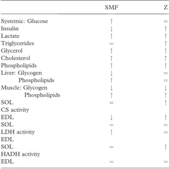
We propose a block diagram (Table III) which reveals the main similarities between Wistar rats exposed to static magnetic fields and unexposed Zucker rats. These data suggest that the metabolic alterations observed in Wistar rats following SMF exposure were similar in many ways to those obtained in Zucker rats. Our study provides evidence that a 128 mT static magnetic field exposure might favour the development of a pre-diabetic state or at least the emergency of some characteristics found in type 1 and type 2 diabetes. Thus, it seems that in addition to lifestyle and
Figure 3. Citrate synthase activity (A), Lactate dehydrogenase activity (B), and Hydroxyl-acyl CoA-desydrogenase activity (C) in Soleus (SOL) and Extensor Digitorum Longus (EDL) in sham exposed (C), SMF-exposed rat (SMF), and Zucker (Z) groups. Error bars indicate the standard error of the mean (SEM) for n¼ 6 independent experiments.*p5 0.05 vs. C, **p 5 0.01vs. C.
genetic predisposition, experimental magnetic ex-position at moderate intensity fields may be another factor promoting metabolic disorders. These results warrant further investigations to understand the mechanism and signalling pathways involved in these alterations.
Acknowledgements
We would like to thank Marie Hokayem for the critical review of the manuscript.
Declaration of interest: The authors report no conflicts of interest. The authors alone are respon-sible for the content and writing of the paper.
References
Abdelmelek H, Chater S, Sakly M. 2001. Acute exposure to magnetic field depresses shivering thermogenesis in rat. Biomedizinische Technik-Band 46-Ergiinzungsband 2:164– 166.
Abdelmelek H, Chater S, Smirani R, M’Chirgui A, Ben Jeddou C, Ben Salem M, Sakly M. 2000. Effects of 50Hz sinusoidal waveform magnetic field on dehydrated rat body. Millennium International Workshop on Biological Effects of Electromag-netic fields. pp 474–479.
Abdelmelek H, Molnar A, Servais S, Cottet-Emard JM, Pequignot JM, Favier R, Sakly M. 2006. Skeletal muscle HSP72 and norepinephrine response to static magnetic field in rat. Journal of Neural Transmission 113:821–827.
Amara S, Abdelmelek H, Garrel C, Guiraud P, Douki T, Ravanat JL, Favier A, Sakly M, Ben Rhouma K. 2006. Effects of subchronic exposure to static magnetic field on testicular function in rats. Archive of Medical Research 37: 47–952. Amara S, Garrel C, Favier A, Ben Rhouma K, Sakly M,
Abdelmelek H. 2009. Effect of static magnetic field and/or cadmium in the antioxidant enzymes activity in rat heart and skeletal muscle. General Physiology and Biophysics 28:414– 419.
Avignon A, Monnier L. 2001. Insulin sensitivity and stress. Diabetes and Metabolism 27:233–238.
Bekhite MM, Finkensieper A, Abou-Zaid FA, El-Shourbagy IK, Omar KM, Figulla HR, Sauer H, Wartenberg M. 2010. Static electromagnetic fields induce vasculogenesis and chondro-osteogenesis of mouse embryonic stem cells by reactive oxygen species-mediated up-regulation of vascular endothelial growth factor. Stem Cells and Development 19:731–743.
Bellossi A, Pouvreau-Quillien V, Rocher C, Ruelloux M. 1996. Effect of pulsed magnetic fields on cholesterol and triglyceride levels in rats study of field intensity and length of exposure. Z Naturforsch [C] 51:603–606.
Bellossi A. 1992. Effects of a 12 Hz and 460 Hz pulsed magnetic field on the weight of AKR mice. Biotherapy 4:277–283. Boden G, Shulman GI. 2002. Free fatty acids in obesity and type 2
diabetes: Defining their role in the development of insulin resistance and beta-cell dysfunction. European Journal of Clinical Investigation 32:14–23.
Budi A, Legge FS, Treutlein H, Yarovsky I. 2008. Comparative study of insulin chain-B in isolated and monomeric environ-ments under external stress. The Journal of Physical Chemistry B 112:7916–7924.
Chater S, Abdelmelek H, Pequignot JM, Sakly M, Rhouma KB. 2006. Effects of sub-acute exposure to static magnetic field on hematologic and biochemical parameters in pregnant rats. Electromagnetic Biology and Medicine 25:135–144.
Chen YB, Li J, Qi Y, Miao X, Zhou Y, Ren D, Guo GZ. 2010. The effects of electromagnetic pulses (EMP) on the bioactivity of insulin and a preliminary study of mechanism. International Journal of Radiation Biology 86:22–26.
Dini L, Abbro L. 2005. Bioeffects of moderate-intensity static magnetic fields on cell cultures. Micron 36:195–217. Dourmashkin JT, Chang GQ, Gayles EC, Hill JO, Fried SK,
Julien C, Leibowitz SF. 2005. Different forms of obesity as a function of diet composition. International Journal of Obesity (London) 29:1368–1378.
Feychting M. 2005. Health effects of static magnetic fields – a review of the epidemiological evidence. Progress in Biophysics and Molecular Biology 87:241–246.
Genius SJ. 2008. Fielding a current idea: Exploring the public health impact of electromagnetic radiation. Public Health 122: 113–124.
Gorczynska E, Wegrzynowicz R. 1991. Glucose homeostasis in rats exposed to magnetic fields. Investigative Radiology 26:1095–1100.
Gutmann I, Wahlefeld M. 1974. L (þ) lactate determination with lactate dehydrogenase and NAD. Methods of enzymatic analysis. New York: Academic Press. pp 1464–1472. Havas M. 2000. Biological effects of non-ionizing electromagnetic
energy: A critical review of the reports by the US National Research Council and the US National Institute of Environ-mental Health Sciences as they relate to the broad realm of EMF bioeffects. Environmental Reviews 8:173–253. Havas M. 2008. Dirty electricity elevates blood sugar among
electrically sensitive diabetics and may explain brittle diabetes. Electromagnetic Biology and Medicine 27:135–146.
High WB, Sikora J, Ugurbil K, Garwood M. 2000. Subchronic in vivo effects of a high static magnetic field (9.4 T) in rats. Journal of Magnetic Resonance Imaging 12:122–139. Table III. Comparison between metabolic alterations observed in
Static Magnetic Field exposed (SMF) and Zucker (Z) rats.
SMF Z Systemic: Glucose " ¼ Insulin # " Lactate " " Triglycerides ¼ " Glycerol " " Cholesterol " " Phospholipids " " Liver: Glycogen # ¼ Phospholipids " ¼ Muscle: Glycogen # # Phospholipids " " SOL ¼ " CS activity EDL # " SOL ¼ ¼ LDH activity " ¼ EDL SOL ¼ " HADH activity EDL ¼ ¼
": Increase; #: Decrease; ¼ : No effect; CS activity: Citrate synthase activity; LDH activity: Lactate deshydrogenase activity; HADH activity: 3-hydroxyacyl-coenzyme A-dehydrogenase activ-ity; SOL: soleus oxidative muscle; EDL: Extensor digitorum longus glycolytic muscle.
Iwasaka M, Ueno S. 1994. Enzymatic oxidation-reduction processes under magnetic fields up to 8 T (abstract). Journal of Applied Physics 75:7181.
Kotani H, Kawaguchi H, Shimoaka T, Iwasaka M, Ueno S, Ozawa H, Nakamura K, Hoshi K. 2002. Strong static magnetic field stimulates bone formation to a definite orientation in vitro and in vivo. Journal of Bone and Mineral Research 17:1814–1821. Li L, Dai Y, Xia R, Chen S, Qiao D. 2005. Pulsed electric field exposure of insulin induces anti-proliferative effects on human hepatocytes. Bioelectromagnetics 26:639–647.
Lo S, Russell JC, Taylor AW. 1970. Determination of glycogen in small tissue samples. Journal of Applied Physiology 28:234– 236.
Lombardi AM, Fabris R, Bassetto F, Serra R, Leturque A, Federspil G, Girard J, Vettor R. 1999. Hyperlactatemia reduces muscle glucose uptake and GLUT-4 mRNA while increasing (E1alpha)PDH gene expression in rat. American Journal of Physiology 276:E922–929.
Metz L, Vermaelen M, Lambert K, Broca C, Sirvent P, Raynaud E, and Mercier J. 2005. Endurance training increases lactate transport in male Zucker fa/fa rats. Biochemical and Biophy-sical Research Communications 331:1338–1345.
Miller BF, Fattor JA, Jacobs KA, Horning MA, Navazio F, Lindinger MI, Brooks GA. 2002. Lactate and glucose interac-tions during rest and exercise in men: Effect of exogenous lactate infusion. The Journal of Physiology 544:963–975. Navakatikyan MA, Antioch V, et al. 1994. Endocrine effects of
alternating magnetic fields 50 Hz. 16th Annual Meeting Bioelec-tromagn. Soc., June 12–17, Copenhagen, Denmark. p. 147. O¨ cal I, Kalkan T, Gu¨nay _I. 2008. Effects of alternating magnetic
field on the metabolism of the healthy and diabetic organisms. Brazilian Archives of Biology and Technology 51:523–530. Okano H. 2008. Effects of static magnetic fields in biology: Role of
free radicals. Frontiers in Bioscience 13:6106–6125. Pilla AA. 2006. Mechanisms and therapeutic applications of
time-varying and static magnetic fields. In: Barnes FGB, editor. Handbook of biological effects of electromagnetic fields. 3rd ed. Boca Raton, FL: CRC Press. pp 351–412.
Pujol A, Lefaucheur L, Ecolan P, Picon L, Penicaud L. 1993. Fiber type composition and enzyme activities of muscles in two models of obese rats. Comparative Biochemistry and Physiol-ogy B 106:269–272.
Py G, Lambert K, Milhavet O, Eydoux N, Prefaut C, Mercier J. 2002. Effects of streptozotocin-induced diabetes on markers of skeletal muscle metabolism and monocarboxylate transporter 1 to monocarboxylate transporter 4 transporters. Metabolism 51:807–813.
Py G, Lambert K, Perez-Martin A, Raynaud E, Prefaut C, Mercier J. 2001. Impaired sarcolemmal vesicle lactate uptake and skeletal muscle MCT1 and MCT4 expression in obese Zucker rats. American Journal of Physiology Endocrinology and Metabolism 281:E1 308–1315.
Repacholi MH, Greenebaum B. 1999. Interaction of static and extremely low frequency electric and magnetic fields with living systems: Health effects and research needs. Bioelectromag-netics 20:133–160.
Rosen AD. 2003a. Effect of a 125 mT static magnetic field on the kinetics of voltage activated Naþ channels in GH3 cells. Bioelectromagnetics 24:517–523.
Rosen AD. 2003b. Mechanism of action of moderate-intensity static magnetic fields on biological systems. Cell Biochemistry and Biophysics 39:163–173.
Savage DB, Petersen KF, Shulman GI. 2007. Disordered lipid metabolism and the pathogenesis of insulin resistance. Environmental Reviews 87:507–520.
Shibuya I, Honda H, Maruo B. 1967. Simplified colorimetry without incineration of phosphorus in phosphatides. Agricul-tural and Biological Chemistry 31:111–114.
Srere P. 1969. Citrate synthase. Methods in Enzymology 3–5. Ueno S, Shigemitsu T. 2007. Biological effects of static magnetic
fields. In: Barnes FS, Greenebaum B, editors. Handbook of biological effects of electromagnetic fields: Bioengineering and biophysical aspects of electromagnetic fields. 3rd ed. Boca Raton, FL: CRC Press.
Vettor R, Lombardi AM, Fabris R, Pagano C, Cusin I, Rohner-Jeanrenaud F, Federspil G, Rohner-Jeanrenaud B. 1997. Lactate infusion in anesthetized rats produces insulin resistance in heart and skeletal muscles. Metabolism 46:684–690. Vettor R, Lombardi AM, Fabris R, Serra R, Pagano C, Macor C,
Federspil G. 2000. Substrate competition and insulin action in animal models. International Journal of Obesity and Related Metabolic Disorders 24:S22–24.
Wang Z, Sarje A, Che PL, Yarema KJ. 2009. Moderate strength (0.23–0.28 T) static magnetic fields (SMF) modulate signaling and differentiation in human embryonic cells. BMC Genomics 10:356–378.
Weintraub MI, Cole SP. 2004. Pulsed magnetic field therapy in refractory neuropathic pain secondary to peripheral neuro-pathy: Electrodiagnostic parameters – pilot study. Neuroreh-abilitation and Neural Repair 18:42–46.
Weintraub MI, Wolfe GI, Barohn RA, Cole SP, Parry GJ, Hayat G, Cohen JA, Page JC, Bromberg MB, Schwartz SL. 2003. Static magnetic field therapy for symptomatic diabetic neuropathy: A randomized, double-blind, placebo-controlled trial. Archives of Physical Medicine and Rehabilitation 84:736– 746.
World Health Organisation (WHO). 2006. Environmental Health Criteria 232. Static fields. Geneva, Switzerland: WHO. Zhang QM, Tokiwa M, Doi T, Nakahara T, Chang PW,
Nakamura N, Hori M, Miyakoshi J, Yonei S. 2003. Strong static magnetic field and the induction of mutations through elevated production of reactive oxygen species in Escherichia coli soxR. International Journal of Radiation Biology 79:281– 286.