HAL Id: hal-00642197
https://hal.inria.fr/hal-00642197
Submitted on 17 Nov 2011
HAL is a multi-disciplinary open access
archive for the deposit and dissemination of
sci-entific research documents, whether they are
pub-lished or not. The documents may come from
teaching and research institutions in France or
abroad, or from public or private research centers.
L’archive ouverte pluridisciplinaire HAL, est
destinée au dépôt et à la diffusion de documents
scientifiques de niveau recherche, publiés ou non,
émanant des établissements d’enseignement et de
recherche français ou étrangers, des laboratoires
publics ou privés.
To cite this version:
Hervé Goëau, Pierre Bonnet, Alexis Joly, Nozha Boujemaa, Daniel Barthélémy, et al.. The
Image-CLEF 2011 plant images classification task. ImageImage-CLEF 2011, Sep 2011, Amsterdam, Netherlands.
�hal-00642197�
3 Telabotanica, France, [email protected], http://www.tela-botanica.org/
Abstract. ImageCLEFs plant identification task provides a testbed for the system-oriented evaluation of tree species identification based on leaf images. The aim is to investigate image retrieval approaches in the con-text of crowdsourced images of leaves collected in a collaborative manner. This paper presents an overview of the resources and assessments of the plant identification task at ImageCLEF 2011, summarizes the retrieval approaches employed by the participating groups, and provides an anal-ysis of the main evaluation results.
Keywords: ImageCLEF, plant, leaves, images, collection, identifica-tion, classificaidentifica-tion, evaluaidentifica-tion, benchmark
1
Introduction
Convergence of multidisciplinary research is more and more considered as the next big thing to answer profound challenges of humanity related to health, bio-diversity or sustainable energy. The integration of life sciences and computer sci-ences has a major role to play towards managing and analyzing cross-disciplinary scientific data at a global scale. More specifically, building accurate knowledge of the identity, geographic distribution and uses of plants is essential if agri-cultural development is to be successful and biodiversity is to be conserved. Unfortunately, such basic information is often only partially available for pro-fessional stakeholders, teachers, scientists and citizens, and often incomplete for ecosystems that possess the highest plant diversity. A noticeable consequence, expressed as the taxonomic gap, is that identifying plant species is usually im-possible for the general public, and often a difficult task for professionals, such as farmers or wood exploiters and even for the botanists themselves. The only way to overcome this problem is to speed up the collection and integration of raw observation data, while simultaneously providing to potential users an easy and efficient access to this botanical knowledge. In this context, content-based visual identification of plant’s images is considered as one of the most promising
The task was organized as a classification task over 70 tree species with visual content being the main available information. Additional information only in-cluded contextual meta-data (author, date, locality name) and some EXIF data. Three types of image content were considered: leaf scans, leaf photographs with a white uniform background (referred as scan-like pictures) and unconstrained leaf’s photographs acquired on trees with natural background. The main orig-inality of this data is that it was specifically built through a citizen sciences initiative conducted by Telabotanica5, a French social network of amateur and
expert botanists. This makes the task closer to the conditions of a real-world application: (i) leaves of the same species are coming from distinct trees living in distinct areas (ii) pictures and scans are taken by different users that might not used the same protocol to collect the leaves and/or acquire the images (iii) pictures and scans are taken at different periods in the year.
2
Task resources
2.1 The Pl@ntLeaves dataset
Building effective computer vision and machine learning techniques is not the only side of the taxonomic gap ptoblem. Speeding-up the collection of raw ob-servation data is clearly another crucial one. The most promising approach in that way is to build real-world collaborative systems allowing any user to enrich the global visual botanical knowledge [14]. To build the evaluation data of Im-ageCLEF plant identification task, we therefore set up a citizen science project around the identification of common woody species covering the Metropolitan French territory. This was done in collaboration with TelaBotanica social net-work and with researchers specialized in computational botany.
Technically, images and associated tags were collected through a crowd-sourcing web application [14] and were all validated by expert botanists. Several cycles of such collaborative data collection and taxonomical validation occurred. Scans of leaves were first collected over two seasons, between July and September 2009 and between June and September 2010 thanks to the work of active contributors from TelaBotanica social network. The idea of collecting only scans during this first period was to initialize the training data with limited noisy background and to focus on plant variability rather than mixed plant and view conditions
4
http://www.imageclef.org/2011
5
Fig. 1. List of tree species included in the Pl@ntLeaves dataset
variability. This allowed to collect 2228 scans over 55 species. A public version of the application6 was then opened in October 2010 and additional data were
collected up to March 2011. The new collected images were either scans, or photographs with uniform background (referred as scan-like photos), or uncon-strained photographs with natural background. They involved 15 new species from the previous set of 55 species. The Pl@ntLeaves dataset used within Im-ageCLEF finally contained 5436 images with 3070 scans, 897 scan-like photos and 1469 photographs. Figure 2 displays samples of these 3 image types for 4 distinct tree species. The full list of species is provided in Figure 1.
2.2 Pl@ntLeaves metadata
Each image of Pl@ntLeaves dataset is associated with the following meta-data: – Date upload date of the image
– Type (acquisition type: scan, scan-like or photograph)
6
Fig. 2. Illustration of the 3 image type categories for 4 species
– Content content type: single leaf, single dead leaf or foliage (several leaves on tree visible in the picture)
– Taxon full taxon name (sub-regnum, regnum, class, division, order, family, genus, species)
– VernacularNames French or English vernacular names – Author name of the author of the picture
– Organization name of the organization of the author
– Locality locality name (a district or a country division or a region) – GPSLocality GPS coordinates of the observation
These meta-data are stored in independent xml files, one for each image. Figure 3 displays an example image with its associated xml data.
Additional but partial meta-data information can be found in the image’s EXIF, and might include the camera or the scanner model, the image resolution and
Fig. 3. An image of Pl@ntLeaves dataset and its associated metadata
dimension, the optical parameters, the white balance, the light measures, etc.
2.3 Pl@ntLeaves variability
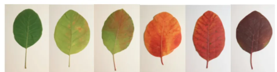
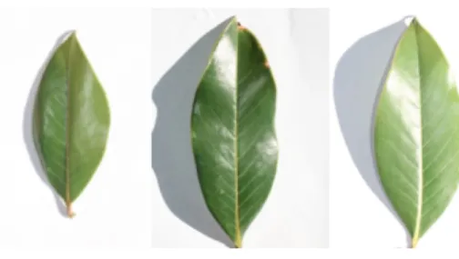
The main originality of Pl@ntLeaves compared to previous leaf datasets, such as the Swedish dataset [16] or the Smithsonian one [9], is that it was built in a collaborative manner through a citizen sciences initiative. This makes it closer to the conditions of a real-world application: (i) leaves of the same species are coming from distinct trees living in distinct areas (ii) pictures and scans are taken by different users that might not used the same protocol to collect the leaves and/or acquire the images (iii) pictures and scans are taken at different periods in the year. Intra-species visual variability and view conditions variabil-ity are therefore more stressed-out which makes the identification more realistic but more complex. Figures 4 to 9 provide illustrations of the intra-species vi-sual variability over several criteria including leaf’s color, leaf’s global shape, leaf’s margin appearance, number and relative positions of leaflets and number of lobes. On the other side, Figure 10 illustrates the light reflection and shadows variations of scan-like photos. It shows that this acquisition protocol is actu-ally very different than pure scans. Both share the property of a limited noisy background but scan-like photos are much more complex due to the lighting conditions variability (flash, sunny weather, etc.) and the unflatness of leaves. Finally, the variability of unconstraint photographs acquired on the tree and with
Fig. 4. Color variation of Cotinus coggygria Scop. (Eurasian smoketree)
Fig. 5. Global shape variation of Corylus avellana L. (European Hazel)
natural background is definitely a much more challenging issue as illustrated in Figure 11.
3
Task description
The task was evaluated as a supervised classification problem with tree species used as class labels.
3.1 Training and Test data
A part of Pl@ntLeaves dataset was provided as training data whereas the remain-ing part was used later as test data. The trainremain-ing subset was built by randomly selecting 2/3 of the individual plants of each species (and not by randomly splitting the images themselves). So that pictures of leaves belonging to the same individual tree cannot be split across training and test data. This prevents iden-tifying the species of a given tree thanks to its own leaves and that makes the task more realistic. In a real world application, it is indeed much unlikely that a user tries to identify a tree that is already present in the training data. Detailed statistics of the composition of the training and test data are provided in Table 1.
Fig. 6. Leaf’s margin variation of Quercus ilex L. (Holm oak)
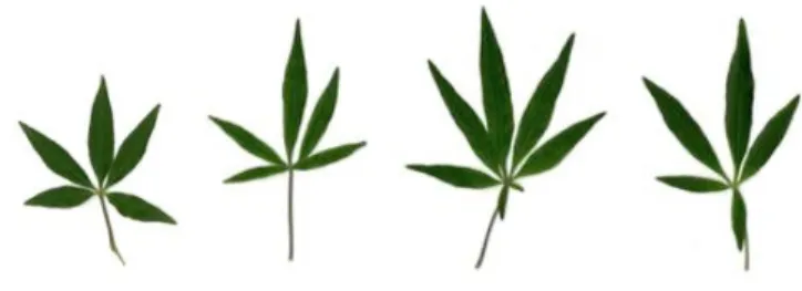
Fig. 7. Number of leaflets variation of Fraxinus angustifolia Vahl (Narrow-leafed Ash) Nb of pictures Nb of individual plants Nb of contributors
Scan Train 2349 151 17 Test 721 55 13 Scan-like Train 717 51 2 Test 180 13 1 PhotographTrain 930 72 2 Test 539 33 3 All Train 3996 269 17 Test 1440 99 14
Table 1. Statistics of the composition of the training and test data
3.2 Task objective and evaluation metric
The goal of the task was to associate the correct tree species to each test image. Each participant was allowed to submit up to 3 runs built from different meth-ods. As many species as possible can be associated to each test image, sorted by decreasing confidence score. Only the most confident species was however used in the primary evaluation metric described below. But providing an extended ranked list of species was encouraged in order to derive complementary statistics (e.g. recognition rate at other taxonomic levels, suggestion rate on top k species,
Fig. 8. Leaflets relative position variation of Vitex agnus-castus L. (Judas Tree)
Fig. 9. Number of lobes variation of Ficus carica L. (Common Fig)
etc.).
The primary metric used to evaluate the submitted runs was a normalized clas-sification rate evaluated on the 1st species returned for each test image. Each test image is attributed with a score of 1 if the 1st returned species is correct and 0 if it is wrong. An average normalized score is then computed on all test images. A simple mean on all test images would indeed introduce some bias with regard to a real world identification system. Indeed, we remind that the Pl@ntLeaves dataset was built in a collaborative manner. So that few contribu-tors might have provided much more pictures than many other contribucontribu-tors who provided few. Since we want to evaluate the ability of a system to provide correct answers to all users, we rather measure the mean of the average classification rate per author. Furthermore, some authors sometimes provided many pictures of the same individual plant (to enrich training data with less efforts). Since we want to evaluate the ability of a system to provide the correct answer based on a single plant observation, we also decided to average the classification rate on each individual plant. Finally, our primary metric was defined as the following average classification score S:
S = 1 U U X u=1 1 Pu Pu X p=1 1 Nu,p Nu,p X n=1 su,p,n (1)
diflora (Southern Magnolia)
Fig. 11. Variability of unconstrained photographs of Acer platanoides (Norway Maple)
U : number of users (who have at least one image in the test data) Pu : number of individual plants observed by the u-th user
Nu,p : number of pictures taken from the p-th plant observed by the u-th user
su,p,n: classification score (1 or 0) for the n-th picture taken from the p-th plant
observed by the u-th user
It is important to notice that while making the task more realistic, the nor-malized classification score also makes it more difficult. Indeed, it works as if a bias was introduced between the statistics of the training data and the one of the test data. It highlights the fact that bias-robust machine learning and computer vision methods should be preferred to train such real-world collaborative data. Finally, to isolate and evaluate the impact of the image acquisition type (scan, scan-like, photogragh), a normalized classification score S was computed for each type separately. Participants were therefore allowed to train distinct classifiers, use different training subsets or use distinct methods for each data type.
4
Participants and techniques
A total of 8 groups submitted 20 runs, which is a successful participation rate for a first year pilot task on a new topic. Participants were mainly academics, spe-cialized in computer vision and multimedia information retrieval, coming from all around the world: Australia (1), Brazil (1), France (2), Romania (1), Spain
USP run2 & IFSC USP run1) are mainly based on a new shape boundary anal-ysis method they introduced recently [11]. It is based on the complex network theory [10]. A shape is modeled into a small-world complex network and it uses degree and joint degree measurements in a dynamic evolution network to com-pose a set of shape descriptors. This method is claimed to be robust, noise tolerant, scale invariant and rotation invariant and proved to provide better per-formances than Fourier shape descriptors, curvature-based descriptors, Zernike moments and multiscale fractal dimensions.
LIRIS (4 runs) [2] This participant also used a classification scheme based on shape boundary analysis. The main originality however is that they used a model-driven approach for the segmentation and shape estimation. Their four runs differ in the parameters of the method.
UAIC (3 runs) [3] This participant was the only one trying to benefit from metadata associated to the images (location, date, author, etc.). They submitted therefore 3 runs to evaluate the contribution of metadata compared to using vi-sual content only. Their first run (UAIC2011 Run01) is based on vivi-sual content only, the second one (UAIC2011 Run02) uses only metadata based features in the classification process, and the third one uses both (UAIC2011 Run03).
SABANCI (1 run) [4] This participant submitted only one run based on a supervised classification approach using support vector machine (SVM) and a combination of 3 visual features: a new texture feature they introduced in ??, a shape boundary feature based on FFT coefficients [] and some basic global color features. This is the only method combining leaf boundary information with other texture end color features.
INRIA (2 runs) [5] This participant submitted two runs based on two rad-ically different methods. Their second run (inria imedia plantnet run2) is based on a shape boundary feature, called DFH [17], that they introduced in 2006. Their first run (inria imedia plantnet run1) is more surprising for a supervised classification task of leaves since it is based on local features matching with rigid geometrical models . Such generalist method is usually more dedicated to large-scale retrieval of rigid objects and this is the only participant who used such approach.
fer (kmimmis run1 and kmimmis run4), simple edge and corner point detection with 1-NN vote label transfer (kmimmis run2), simple edge and corner point detection with 10-NN vote label transfer (kmimmis run3).
Over all runs, the most frequently used class of methods is shape boundary analysis. 8 runs among 20 are based on some boundary shape features.This is not surprising since state-of-the-art methods addressing leaf-based identification in the literature are mostly based on leaf segmentation and shape boundary fea-tures [12, 15, 13, 17, 11]. On the other side, it was a good news that the majority of the runs were based on other various approaches so that more relevant con-clusions can be expected.
5
Results
5.1 Global analysis
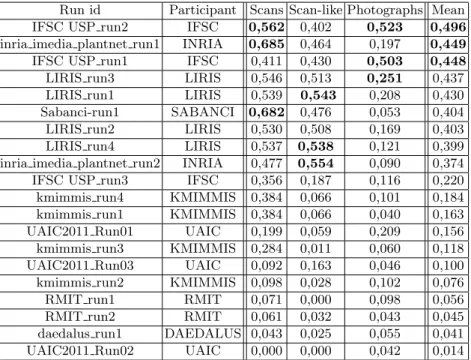
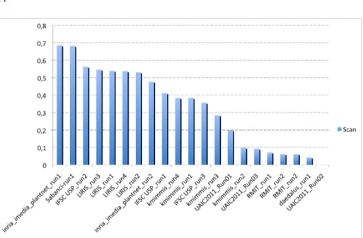
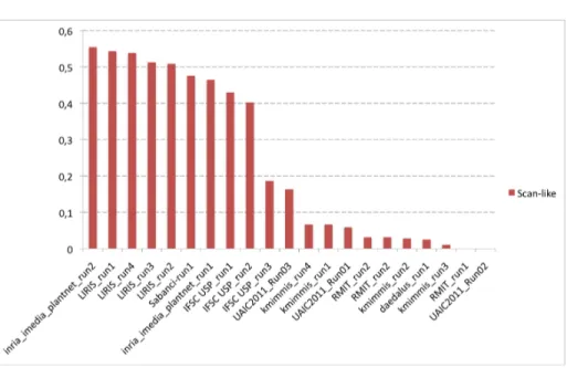
Figures 12, 13 and 14 present the normalized classification scores of the 20 sub-mitted runs for each of the three image types. Alternatively, Figure 15 presents the overall performances averaged over the 3 image types. Table 2 finally presents the same results but with detailed numerical values.
A first global remark is that, as expected, the performances are degrading with the complexity of the acquisition image type. Scans are more easy to identify than scan-like photos and unconstrained photos are much more difficult. This is can be easily seen in Figure 15 where the relative scores of each image type are highlighted by distinct colors.
A second global remark is that no method provide the best score for all image types. None of the run even belongs to the top-3 runs of all image types (as shown in Table 2). This is somehow disappointing from the genericity point of view but not surprising regarding the nature of the different image types. One could expect that scans and scan-like photos lead to similar conclusions but this is actually not the case. The only runs that give quite stable and good perfor-mances over the three image types are the two runs of IFSC based on complex network shape boundary analysis method (IFSC USP run1 & IFSC USP run2). This justifies their excellent ranking when averaging the classification scores over
A third important remark is that shape boundary analysis methods do not provide the best results on the scan images whereas they are usually consid-ered as being state-of-the-art on such data. They all provide good classification scores between 48% and 56% but they are consistently outperformed by two more generic image retrieval approaches (as shown in Figure 12). The best score is achieved by INRIA’s run using large-scale matching of local features with rigid geometrical models (inria imedia plantnet run1, 68% classification rate). This suggests that modeling leaves as part-based rigid and textured objects might be an interesting alternative to shape boundary approaches that do not characterize well margin details, ribs or limb texture. Second best score on scans is obtained by the run of SABANCI (Sabanci-run1) which uses a supervised classification approach based on support vector machine (SVM) and a combina-tion of 3 global visual features. This suggests that combining shape boundary features with other color and shape textures is also a promising direction. Global conclusions on the scan-like photos are quite different (Figure 13). The best score is obtained by INRIA’s run (inria imedia plantnet run2) purely based on a global shape boundary feature (DFH [17]). It is followed closely by the four runs of LIRIS also based on boundary shape features but using a model-driven approach for the segmentation and the shape estimation. Then come the two runs of INRIA and SABANCI that ranked first on the scan images (the first one based on rigid objects matching and the second one training combined fea-tures) and finally the shape boundary method of IFSC. One conclusion is that shape boundary methods appear to provide more stable results for scans and scan-like photos. On the other side the rigid object matching method of INRIA degrades much more from scans to scan-like pictures. This can be explained by the fact that it is more discriminant regarding the leave’s morphology but less robust to light reflections and shadows. These lighting variations might also explain the degrading performances of the combined features used by SABANCI.
5.2 About using metadata
Using metadata to help the identification, and more particularly geo-tags, is definitely something that has to be studied. We were therefore very enthusias-tic to see the runs of UAIC, aimed at evaluating the potential contributions of Pl@ntLeaves metadata. Unfortunately, results clearly show that adding meta-data degrades their identification performances. Metameta-data alone even give a null
IFSC USP run3 IFSC 0,356 0,187 0,116 0,220 kmimmis run4 KMIMMIS 0,384 0,066 0,101 0,184 kmimmis run1 KMIMMIS 0,384 0,066 0,040 0,163 UAIC2011 Run01 UAIC 0,199 0,059 0,209 0,156 kmimmis run3 KMIMMIS 0,284 0,011 0,060 0,118 UAIC2011 Run03 UAIC 0,092 0,163 0,046 0,100 kmimmis run2 KMIMMIS 0,098 0,028 0,102 0,076 RMIT run1 RMIT 0,071 0,000 0,098 0,056 RMIT run2 RMIT 0,061 0,032 0,043 0,045 daedalus run1 DAEDALUS 0,043 0,025 0,055 0,041 UAIC2011 Run02 UAIC 0,000 0,000 0,042 0,014 Table 2. Normalized classification scores for each run and each image type. Top 3 results per image type are highlighted in bold
classification success rate. Besides technical details that could probably slightly improve such species filtering based on metadata, it still shows that Pl@ntLeaves metadata might be intrinsically not very useful for identification purposes. The main reason is probably that the geographic spread of the data is limited (French mediterranean area). So that most species of the dataset might be identically and uniformly distributed in the covered area. Geo-tags would be for sure more useful at a global scale (continent, countries). But at a local scale, the geo-graphical distribution of plants is much more complex. It usually depends on localized environmental factors such as sun exposition or water proximity that would require much more data to be modeled.
5.3 Performances per species
To evaluate which species are more difficult to identify than others, we averaged the performances over the runs of all participants (for each specie). It is how-ever difficult to understand precisely the score variations. They can be due to morphological variations but also to different view conditions or other statistical bias in the data such as the number of training images. Figure 16 presents the obtained graph for the scan images only (in order to limit view conditions bias). The only global trend we discovered so far is that single leaves are on average
Fig. 12. Normalized classification scores for scan images
easier to identify than compound leaves.
5.4 Performances per image
To qualitatively assess which kind of images causes good results and which one makes all methods failed, we sorted all test pictures by the number of runs in which they were correctly identified. The obtained ranking confirms that scan images are much easier for the identification. 99% of 100-top ranked images are actually scans (with 11 to 17 successful runs). Figure 17 displays the 4 best identified images (with 17/20 successful runs). They all are very standard leaf images similar to the one found in books illustrations. On the other side, 260 images were not identified by any run with a majority of unconstrained photos (63 scans, 27 scan-like photos, 168 unconstrained photographs). The scans and scan-like photos belonging to this category of most difficult images are very interesting. As illustrated in Figure 18, most of them correspond to outlier leaves with defaults or unusual morphology (ill or torn leaves, missing leaflets, etc.). Figure 18 displays 8 of them.
6
Conclusions
This paper presented the overview and the results of ImageCLEF 2011 plant identification testbed. A challenging collaborative dataset of tree’s leaf images
Fig. 13. Normalized classification scores for scan-like photos
was specifically built for this evaluation and 8 participants coming from different countries submitted a total of 20 runs. A first conclusion was that identification performances are close from mature when using scans or photos with uniform background but that unconstrained photos are still much more challenging. More data and evaluation are clearly required to progress on such data. Another im-portant conclusion was that sate-of-the-art methods based on shape boundary analysis were not the best ones on leaf scans. Better performances were no-tably obtained with a local features matching technique usually more dedicated to the large-scale retrieval of rigid objects. On the other side, shape boundary analysis methods remain better on scan-like photos due to their better robust-ness to light reflections and shadow effects. This suggests that combining shape boundary features with part-based rigid object models might be an interesting direction. Adding texture and color information also showed some improvements. On the contrary, using additional metadata such as geo-tags was not concluding on the evaluated dataset. Probably because the geographic spread of the data was limited.
Acknowledgement
This work was funded by the Agropolis fundation through the project Pl@ntNet (http://www.plantnet-project.org/) and the EU through the CHORUS+ Coor-dination action (http://avmediasearch.eu/)
Fig. 14. Normalized classification scores for photographs
References
1. In: Working notes of CLEF 2011 conference (2011) 2. In: Working notes of CLEF 2011 conference (2011) 3. In: Working notes of CLEF 2011 conference (2011) 4. In: Working notes of CLEF 2011 conference (2011) 5. In: Working notes of CLEF 2011 conference (2011) 6. In: Working notes of CLEF 2011 conference (2011) 7. In: Working notes of CLEF 2011 conference (2011) 8. In: Working notes of CLEF 2011 conference (2011)
9. Agarwal, G., Belhumeur, P., Feiner, S., Jacobs, D., Kress, J.W., R. Ramamoorthi, N.B., Dixit, N., Ling, H., Mahajan, D., Russell, R., Shirdhonkar, S., Sunkavalli, K., White, S.: First steps toward an electronic field guide for plants. Taxon 55, 597–610 (2006)
10. Albert, R.Z.: Statistical mechanics of complex networks. Ph.D. thesis, Notre Dame, IN, USA (2001)
11. Backes, A.R., Casanova, D., Bruno, O.M.: A complex network-based approach for boundary shape analysis. Pattern Recognition 42(1), 54 – 67 (2009)
12. Belhumeur, P., Chen, D., Feiner, S., Jacobs, D., Kress, W., Ling, H., Lopez, I., Ramamoorthi, R., Sheorey, S., White, S., Zhang, L.: Searching the worlds herbaria: A system for visual identification of plant species. In: ECCV, pp. 116–129 (2008) 13. Bruno, O.M., de Oliveira Plotze, R., Falvo, M., de Castro, M.: Fractal dimension
applied to plant identification. Information Sciences 178(12), 2722 – 2733 (2008) 14. Goeau, H., Joly, A., Selmi, S., Bonnet, P., Mouysset, E., Joyeux, L.: Visual-based
plant species identification from crowdsourced data. In: Proceedings of ACM Mul-timedia 2011 (2011)
Fig. 15. Normalized classification scores averaged over all image types
15. Neto, J.C., Meyer, G.E., Jones, D.D., Samal, A.K.: Plant species identification using elliptic fourier leaf shape analysis. Computers and Electronics in Agriculture 50(2), 121 – 134 (2006)
16. S¨oderkvist, O.J.O.: Computer Vision Classification of Leaves from Swedish Trees. Master’s thesis, Link¨oping University, SE-581 83 Link¨oping, Sweden (September 2001), liTH-ISY-EX-3132
17. Yahiaoui, I., Herv, N., Boujemaa, N.: Shape-based image retrieval in botanical collections. In: Advances in Multimedia Information Processing - PCM 2006, vol. 4261, pp. 357–364 (2006)
Fig. 16. Mean classification score per species averaged over all participant runs - scan test images only
Fig. 18. Top: 4 of the most difficult test scans (0/20 successful runs) - Bottom: 4 of the most difficult scan-like test photos (0/20 successful runs)


![[PDF] Formation d’introduction à Common Lisp pour débutant | Formation informatique](data:image/gif;base64,R0lGODlhAQABAIAAAP///wAAACH5BAEAAAAALAAAAAABAAEAAAICRAEAOw==)