HAL Id: hal-02912888
https://hal.univ-grenoble-alpes.fr/hal-02912888
Submitted on 23 Nov 2020HAL is a multi-disciplinary open access archive for the deposit and dissemination of sci-entific research documents, whether they are pub-lished or not. The documents may come from teaching and research institutions in France or abroad, or from public or private research centers.
L’archive ouverte pluridisciplinaire HAL, est destinée au dépôt et à la diffusion de documents scientifiques de niveau recherche, publiés ou non, émanant des établissements d’enseignement et de recherche français ou étrangers, des laboratoires publics ou privés.
Influenza Nucleoproteins on Importins-α
Amélie Donchet, Emilie Vassal-Stermann, Francine Gérard, Rob W H
Ruigrok, Thibaut Crépin
To cite this version:
Amélie Donchet, Emilie Vassal-Stermann, Francine Gérard, Rob W H Ruigrok, Thibaut Crépin. Dif-ferential Behaviours and PreDif-ferential Bindings of Influenza Nucleoproteins on Importins-α. Viruses, MDPI, 2020, 12 (8), pp.834. �10.3390/v12080834�. �hal-02912888�
Viruses 2020, 12, 834; doi:10.3390/v12080834 www.mdpi.com/journal/viruses
Article
Differential Behaviours and Preferential Bindings
of Influenza Nucleoproteins on Importins-α
Amélie Donchet, Emilie Vassal-Stermann, Francine C. A. Gérard †, Rob W. H. Ruigrok
and Thibaut Crépin *
Centre National de la Recherche Scientifique (CNRS), Institut de Biologie Structurale (IBS),
University Grenoble Alpes, Commissariat à l'Energie Atomique et aux Energies Alternatives (CEA), 38044 Grenoble, France; amelie.donchet@ibs.fr (A.D.); emilie.stermann@ibs.fr (E.V.-S.);
francine.baraggia@grenoble-inp.fr (F.C.A.G.); rob.ruigrok@ibs.fr (R.W.H.R.)
* Correspondence: thibaut.crepin@ibs.fr; Tel.: +33-(0)476-209-439
† Current address: Laboratoire des Matériaux et du Génie Physique (LMGP), University Grenoble Alpes, CNRS, Institut Polytechnique de Grenoble (Grenoble INP), F-38000 Grenoble, France.
Received: 30 May 2020; Accepted: 27 July 2020; Published: 30 July 2020
Abstract: Influenza viruses are negative single-stranded RNA viruses with nuclear transcription
and replication. They enter the nucleus by using the cellular importin-α/-β nuclear import machinery. Influenza nucleoproteins from influenza A, B, C and D viruses possess a nuclear
localization signal (NLS) localized on an intrinsically disordered extremity (NPTAIL). In this paper,
using size exclusion chromatography (SEC), SEC-multi-angle laser light scattering (SEC-MALLS) analysis, surface plasmon resonance (SPR) and fluorescence anisotropy, we provide the first
comparative study designed to dissect the interaction between the four NPTAILs and four
importins-α identified as partners. All interactions between NPTAILs and importins-α have high association and
dissociation rates and present a distinct and specific behaviour. D/NPTAIL interacts strongly with all
importins-α while B/NPTAIL shows weak affinity for importins-α. A/NPTAIL and C/NPTAIL present
preferential importin-α partners. Mutations in B/NPTAIL and D/NPTAIL show a loss of importin-α
binding, confirming key NLS residues. Taken together, our results provide essential highlights of this complex translocation mechanism.
Keywords: influenza nucleoprotein; nuclear transport; importin-α; nuclear localization signal;
influenza-host interaction; surface plasmon resonance; fluorescence anisotropy
1. Introduction
Influenza viruses are part of the Orthomyxoviridae family and form the genus Influenzavirus. Up to now, the International Committee officially recognizes four types (respectively types A, B, C and D) in its Taxonomy of Viruses (ICTV), but because of actual large-scale meta-transcriptomic approaches new specimens are regularly discovered, making this classification a constantly evolving entity. While influenza viruses were thought to be uniquely warm-blooded organism pathogens, a study has recently uncovered their existence in fish and amphibians in China [1]. A first criterion of distinction between the actual four types of Influenzavirus is based on their tropism. Influenza type A viruses (IAV) are viruses from water fowl but some of the viruses can also infect a range of hosts from chickens to swine and humans; type B (IBV) almost exclusively infect humans; type C (ICV) can infect both human and swine whereas type D (IDV) has a broad host tropism but limited to mammals. Influenza viruses are negative-sense single-stranded RNA viruses with segmented genomes. The RNA segments are packaged in ribonucleoproteins (RNPs), a complex architecture made by a single RNA molecule encapsidated by multiple copies of the nucleoprotein (NP) and the heterotrimeric RNA-dependent RNA polymerase (RdRp) bound to the strictly conserved 3′ and 5 ‘ end [2,3]. The
number of RNPs varies depending on the type: IAV and IBV genomes consist of eight RNA segments,
i.e., there are eight different RNPs, but only seven for ICV and IDV viruses. This is related to the
inherent organisation of the viral spikes, respectively the haemagglutinin (HA) plus the neuraminidase (NA) for A and B or a single hemagglutinin-esterase-fusion (HEF) polypeptide with both concatenated activities for C and D.
Unlike most RNA viruses, influenza viruses transcribe and replicate their genome in the nucleus of infected cells. In general, the access to the nuclear compartment is through the highly regulated nuclear pore complex (NPC) [4–7]. Several pathways are involved in cytoplasmic-nuclear trafficking. Small molecules (< 30–60 kDa) are able to diffuse passively through the NPC but bigger molecules often require an active mechanism to be imported [8–10]. The main active transport is based on the importins-α/β (coupled to the small GTPase Ran) pathway. Importins-α and -β can both interact with the cargos but only the last ones mediate directly the translocation, and importin-α:cargo complexes must first interact with importin-β [11,12]. Cargos are recognized by importins-α/β through nuclear localization signals (NLSs), usually made of basic or hydrophobic motifs [13,14]. Additionally to the interaction with the NLS motifs, recent karyopherins-β/cargo-protein structures have demonstrated that these complexes are dependent on multiple interactions, mediated through intricate 3D interfaces which involve residues well beyond the NLS peptide stretch [15–17].
The human genome encodes for seven importins-α and at least twenty importins-β [9,18,19], meaning that the importins-α/β pathway is a difficult problem. Nuclear import of influenza virus proteins has been of a major interest for a long time [20,21]. Considering the neo-synthetized components of the replication machinery, two subunits (PA and PB1) of the heterotrimeric RdRp are imported as a sub-complex by the importin-β RanBP5 [17,22–24] whereas the third subunit (PB2) and NP interact with importins-α [25–28]. Because of its role in the RNPs formation, NP is critical for influenza replication but also shown to be the major contributor to the nuclear transport of the RNPs
[29]. The NLS of NPs are located in intrinsically disordered tails of the proteins, either in Nterminal for
A/ and B/NP or in Cterminal for C/ and D/NP (Figure 1). A minor putative NLS has been located in a
loop of the core of A/NP, between residues 198 to 216, but its role remains unclear [30,31]. A/NP is known to interact with several importin-α isoforms, including the universal -α1, -α3, -α5 and possibly -α7 [32–36]. In recent years, our group has worked on the mechanisms of the interaction between two different NPs and the human nuclear trafficking machinery [37,38], in particular the tails containing
the NLS of influenza B (B/NPTAIL) and D (D/NPTAIL) NPs. We showed that the NLS of B/NPTAIL is
highly extended from residues 30 to 71, around a single basic Lysine-Arginine (44-KR-45) patch, with
a low affinity of 844 nM for the human importin-α7. In the case of the Cterminal D/NPTAIL, the NLS was
composed of two basic KR patches, respectively 514-KR-515 and 532-KR-533. Both contribute to the
nuclear trafficking and we estimated that the affinity of D/NPTAIL for the human importin-α7 was at
100 nM by surface plasmon resonance (SPR). The literature also contains several studies detailing the relation between A/NP and C/NP and importins-α [21,29,31,33,39–43] (Table 1). However, recent work highlighted that each importin-α is unique, with its own features and preferential partners [28,35,44,45].
Figure 1. Influenzavirus nucleoproteins. The schematic representations of Influenzavirus
nucleoproteins are based on this structural analysis [37]. The schema respects the size of the proteins. The flexible tails with the NLSs are represented with simple lines whereas the cores are represented with filled boxes.
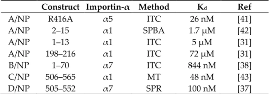
Table 1. Binding of influenza nucleoproteins or their peptides to importins-α according to the
literature. Abbreviations: ITC, isothermal titration calorimetry; SPBA, solid phase binding assay; MT, microscale thermophoresis; SPR, surface plasmon resonance.
Construct Importin-α Method Kd Ref
A/NP R416A α5 ITC 26 nM [41]
A/NP 2–15 α1 SPBA 1.7 µM [42] A/NP 1–13 α1 ITC 5 µM [31] A/NP 198–216 α1 ITC 72 µM [31] B/NP 1–70 α7 ITC 844 nM [38] C/NP 506–565 α1 MT 48 nM [43] D/NP 505–552 α7 SPR 100 nM [37]
Because we were confronted with a wide range of values (from 26 nM to 72 µM) obtained using different techniques and a plethora of different partner constructs, we wanted to establish a
systematic comparative study of the in vitro interaction between the flexible extremities (NPTAILs) of
all types of influenza NPs and the different human importin-α isoforms described in the literature. Our strategy was design, with a specific focus upon the short NLS sequences, and built with the objective of avoiding the inherent oligomerization of NPs, while guaranteeing the architecture of the tails in a unique globular environment. Using size exclusion chromatography experiments, we show
that all NPTAILs interact with importins-α in solution and then used surface plasmon resonance
measurements to determine the affinity and binding kinetics of these interactions. In the current
paper, we show that D/NPTAIL interacts with all importins-α isoforms with strong affinity. Both
A/NPTAIL and C/NPTAIL interact with varying affinities with the corresponding importins-α. On the
other hand, B/NPTAIL shows a completely different behaviour, with even faster association and
dissociation rates and a low affinity with all the human importins-α we measured.
2. Materials and Methods
2.1. Molecular Biology and Constructs
Human importin-α1 (residues 69 to 529), -α3 (residues 64 to 521) and -α5 (residues 66 to 538) DNA coding sequences were bought at GENEART GmbH (Thermo Fischer Scientific, Regensburg, Germany) and subcloned into pETM11 bacterial expression vector (EMBL) without their importin-β (IBB) domains. Human importin-α7 (residues 58 to 536) cloning was described in [26]. NLS-bearing NPTAILs from strains A/WSN/1933 (residues 1 to 21), B/Memphis/13/03 (residues 1 to 71),
C/Ann-Arbor/1/50 (residues 508 to 565) and D/bovine/France/2986/2012 (residues 505 to 552) were ordered
from GeneART, fused to a biotinylation sequence in Cterminal (Figure 2, and details of the sequences
are provided in Table S1). The NPTAILs were fused to the yellow fluorescent protein (YFP) either in
Cterminal (C/NPTAIL and D/NPTAIL) or in Nterminal (A/NPTAIL, B/NPTAIL) according to their native
localization; at the other YFP extremity, the biotinylation sequence was added (Figure 2). NPTAILs-YFP
constructs were subcloned into pETM11 bacterial expression vector. All constructs were expressed as N-terminally His-tagged proteins. Sequencing was performed by Eurofins Genomics (Köln, Germany). The control peptide was a synthetic peptide (GENPEP, St-Jean-de-Védas, France) corresponding to a GLA domain-derived polypeptide of the human coagulation factor X [46].
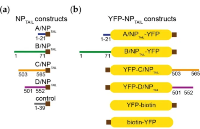
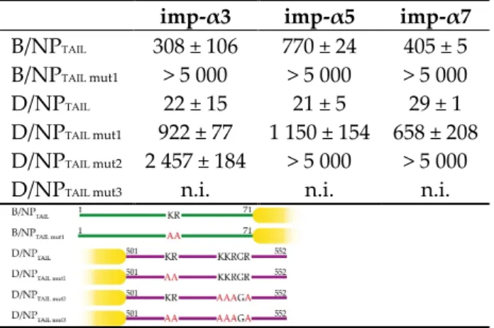
Figure 2. Constructs. (a) Influenzavirus nucleoproteins tails were expressed and purified
independently fused with a biotinylation sequence (brown squares) in their Cterminal end. The details of the sequences are provided in Table S1. (b) The corresponding tails were fused to the yellow fluorescent protein (YFP) in order to integrate then in the context of a globular protein. The yellow filled boxes represent the YFP and the brown squares indicate the position of the biotinylation sequence.
2.2. Expression and Purification
Escherichia coli BL21 RIL (DE3) cells (Life Technologies, Thermo Fischer Scientific, Courtaboeuf,
France) were used for protein expression. Cultures were supplemented with biotin (12.5 μg.mL−1,
Sigma, Saint-Quentin-Fallavier, France) to biotinylate the proteins in cellulo, induced for 12 h by adding 0.3 mM isopropyl-β-D-thiogalactopyranoside (IPTG; Euromedex, Souffelweyersheim, France) at 18 °C and collected by centrifugation. Pellets were resuspended in 50 mM Tris-HCl pH 7.5, 300 mM NaCl, 2 mM β-mercaptoethanol (β-ME; Roth, Lagny-sur-Marne, France) and cOmplete™, ethylenediaminetetraacetic acid (EDTA)-free protease inhibitor cocktail (Roche, Meylan, France) before sonication. All purifications included a nickel affinity chromatography (resin Ni-NTA; Qiagen, Les Ulis, France), a Tobacco Etch Virus (TEV) protease cleavage, a second nickel affinity chromatography and a size-exclusion chromatography (SEC). SEC was performed on a NGC system (Bio-Rad, Marnes-La-Coquette, France) with a S75 10/300 GL column (GE-Healthcare, Dutscher, Brumath, France). The final buffer composition for all proteins was 10 mM Hepes pH 7.5, 150 mM
NaCl. NPTAILs-YFP protein concentrations were determined by measuring the absorbance at 280 nm
and using the theoretical extinction coefficients (ε) 28 942 M−1.cm−1 for A/NPTAIL-YFP and C/NPTAIL
-YFP, of 27 452 M−1.cm−1 for B/NPTAIL-YFP, D/NPTAIL-YFP and YFP control. ε at 280 nm used for ΔIBB
importins-α protein concentrations were 50 607 M−1.cm−1 for importin-α1, 42 190 M−1.cm−1 for
importin-α3, 52 285 M−1.cm−1 for importin-α5 and 46 785 M−1.cm−1 for importin-α7. The concentrations
were then obtained using the Beer–Lambert law.
2.3. Interaction Assays by Size Exclusion Chromatography
All size exclusion chromatography (SEC) experiments were performed in the same buffer
(10 mM Hepes pH 7.4, 150 mM NaCl) using a SuperdexTM 200 increase 10/300GL column
(GE-Healthcare) for YFP-NPTAIL fusion proteins and a SuperdexTM 75 10/300GL column (GE-Healthcare)
for all the other NPTAILs. Samples were diluted to be used at 30 µM for YFP-NPTAIL fusion proteins, 60
µM for NPTAILs and 25 µM importin-α7 in 300 μL. They were incubated for one hour at room
temperature before injection on a NGC system (Bio-Rad).
2.4. SEC-MALLS-RI Analysis
Size exclusion chromatography (SEC) followed by multi-angle laser light scattering (MALLS) and refractometry (RI) analysis allow the determination of the molecular mass of a protein or a complex in solution that is independent of its dimensions and shape [47]. SEC was performed with a
Hepes pH 7.5, 150 mM NaCl. Analytical runs were performed at 20 °C with a flow rate of 0.5
mL.min−1. Multi-angle laser light scattering (MALLS) detection was performed with a
DAWN-HELEOS II detector (Wyatt Technology, Toulouse, France) using a laser emitting at 690 nm and protein concentration was measured on-line with the use of differential refractive-index measurements, with an Optilab T-rEX detector (Wyatt Technology) and a refractive-index increment,
dn/dc of 0.185 mL.g−1. Data were analyzed and weight-averaged molar masses were calculated using
the ASTRA software (Wyatt Technology Corp., Santa Barbara, CA, USA).
2.5. Surface Plasmon Resonance
Surface plasmon resonance (SPR) experiments were carried out at 25 °C on a Biacore T200 (GE
Healthcare Life Sciences Europe GmbH, Velizy-Villacoublay, France). Avidin (50 µg.mL-1 in 50 mM
acetate buffer pH 4.5; Sigma) was first immobilized on a Serie S CM5 sensor chip (GE-Healthcare) surface through amine coupling chemistry according to the manufacturer instructions, until a
coupling level of about 17,000 resonance units (RUs) was reached. The biotinylated NPTAIL-YFP (1–10
µg.mL−1) or biotinylated YFP control (1–10 µg.mL−1) were then captured in surfactant-supplemented
HBS-N running buffer (10 mM Hepes pH 7.4, 150 mM NaCl, 0.05 % Tween 20; GE Healthcare) at a
flow rate of 30 µL.min−1 until a coupling level of 100-200 RU was obtained. A flow-cell with
immobilized avidin only was used as negative control while the flow-cell with the avidin:
biotinylated NPTAIL–YFP complex was used as active flow-cell. For kinetic measurements,
importins-α were serial-diluted in surfactant-supplemented HBS-N running buffer. Each concentration was injected in triplicate on both the reference and active flow-cells. Analyte injection and following buffer injection times were set between 55-180 s and 55-200 s respectively, depending on the
examined partners, at a flow-rate of 30 µL.min−1. Regeneration was required and achieved by pulse
injection of 5 M NaCl. All sensorgrams were reference-subtracted. Data were analyzed with Steady State Analysis using the Biacore T200 Evaluation software (GE Healthcare) under a Langmuir 1:1
binding model, assuming that the binding is equivalent and independent for all binding site. Chi2
values for the fitting were kept below 1 for most of the analysis with a maximum of 2.5, and T-value means for the dissociation constant above 10.
2.6. Fluorescence Anisotropy
Fluorescence anisotropy experiments were performed on a Clariostar microplate reader (BMG Labtech, Champigny-sur-Marne, France) set and dedicated to fluorescence anisotropy measurements, with excitation and emission wavelengths fixed at 482 and 530 nm, respectively. Importin-α1 was serially diluted in buffer (10 mM Hepes pH 7.4, 150 mM NaCl) and mixed in dilution
series with the YFP-fused NPTAILS. YFP construct final concentration was set at 25 nM. Measurements
were done in 384-well plates at room temperature. Blank substraction was done using the condition
with YFP-fused NPTAILS alone. Data were normalized using the equation:
𝐵𝐵 =(𝐴𝐴𝐴𝐴𝐴𝐴𝐴𝐴 − 𝐴𝐴𝐴𝐴𝐴𝐴𝐴𝐴)(𝐴𝐴𝐴𝐴𝐴𝐴𝐴𝐴 − 𝐴𝐴𝐴𝐴𝐴𝐴𝐴𝐴) (1)
where B corresponds to the YFP-NPTAIL bound fraction (value between 0 and 1), Aexp is the anisotropy
value observed for one given importin-α1 concentration, and Amin and Amax stand for the
fluorescence anisotropy values of the free YFP-NPTAIL and bound YFP-NPTAIL, respectively. Titrations
were then fitted to the GraphPad Prism model “Single binding site with Hill slope (h)”.
3. Results
3.1. Biochemical and Biophysical Analysis of the Interaction between NPTAILS and Importin-α7
In all experiments with NP and its partners, measurements are difficult because all NPs tend to form oligomers depending on the buffer concentrations [41,48], and therefore, we used only the
disordered NPTAILs. We designed our study with a specific focus upon these short sequences that do
different NPTAILs for importins-α, we first use the corresponding peptides, in size exclusion
chromatography (SEC) experiments combined with surface plasmon resonance (SPR) measurements to determine the affinity and binding kinetics. The first method was used to confirm the ability of the tails to interact with its partner in solution, whereas the second method was used for the determination of the kinetic parameters of the interactions. In SPR, one partner (called the ligand) is immobilized onto a biosensor surface. The interacting partner (called the analyte) diluted in a buffer is then continuously flowed across the biosensor surface, where it binds to the ligand. The binding is measured as a change in resonance units (RUs) on the biosensor surface. Measuring the increase in
binding over time for a given analyte set of concentrations gives the association rate (kon) up to the
point where the system is at equilibrium (i.e., as many association events are observed as dissociation events). Ceasing flow and changing to buffer alone then allows the analyte to wash off the ligand.
Measuring the decrease in bound partner over time gives the dissociation rate (koff). The equilibrium
dissociation constant (Kd) is calculated from the kinetic association and dissociation rates (koff/kon).
When both association and dissociation rates are too fast, it is possible to use the equilibrium part of the curves to extract the dissociation constant, without relying on the rates. For the SPR experiments, we chose to immobilize the tails on the biosensor surface by using the importins-α as the analytes.
The immobilization of the tails was done by fusing a biotinylation sequence in Cterminal (Figure 2a),
with a capture strategy based on a biotin: avidin interaction. The corresponding constructs were
recombinantly produced in bacteria (with 12.5 μg.mL−1 biotin in the growth media) and purified. We
first tested their ability to interact with importin-α7 by SEC experiments. They all formed a stable complex in solution with importin-α7 (Figure S1). However, for SPR measurements, the kinetic
parameters could not all be established because of technical issues. In particular, for B/NPTAIL, the
inability to stabilize the SPR signal because of a putative mass transfer phenomenon made it impossible to exploit the raw data. In consequence, we adapted our strategy.
3.2. Biochemical and Biophysical Analysis of the Interaction between NPTAILS Fused to Yellow Fluorescent
Protein (YFP) and Importin-α7
We decided to increase the size of the tails by fusing them to a known protein, unrelated to the influenza virus life cycle. A larger ligand should allow the experiment to be performed with lower ligand densities, resulting in lower mass transport rates. We chose the yellow fluorescent protein because of its small size and compact folding, with an unchanged capture strategy on the biosensor
surface. A YFP construct was fused in Nterminal for C/NPTAIL and D/NPTAIL and in Cterminal for A/NPTAIL
and B/NPTAIL, to respect their position in the wild-type proteins (Figure 2b). The recombinant proteins
were produced, purified (Figure S2) and used in SEC experiments to test their ability to interact with importin-α7. We confirmed that adding the YFP has no visible effect on the interaction between the NPTAILs and the human import factor in solution. For each tail, a peak corresponding to the complex
was observed prior those of each separate samples (Figure 3). On the contrary, when the control YFP was mixed with importin-α7, a double peak corresponding to the sum of each sample injected separately was observed (Figure S3).
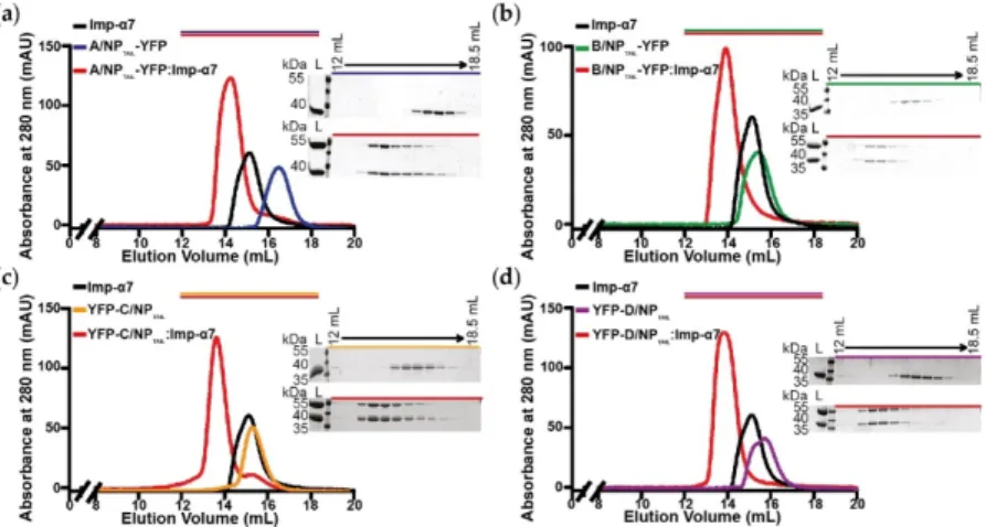
Figure 3. Analysis of the complexes between YFP-NPTAILs and human importin-α7 in solution. Each YFP-NPTAIL fusion protein (30 µM) was injected on a SuperdexTM 200 increase 10/300GL column, alone or in the presence of importin-α7 (25 μM). The figure shows the superimposition of the SEC profiles obtained for (a) A/NPTAIL-YFP, (b) B/NPTAIL-YFP, (c) YFP-C/NPTAIL and (d) YFP-D/NPTAIL. The colors correspond to the code used on Figure 2b with all the complexes in red and the importin-α7 alone in
black. For each panel, the 12 % SDS-PAGE corresponding to the elution of the YFP-NPTAIL fusion
protein alone (top) and its complex with importin-α7 (bottom) are shown. The control experiment is shown on Figure S3.
Using the corresponding samples, we measured the kinetic parameters of the different
interactions by SPR (Figure 4; Tables 2 and 3 respectively for affinities (Kd) and kinetic parameters
values (kon and koff)). Gradient concentration of importin-α7 were injected into the SPR flowing
channels at a speed of 30 µL.min-1 for 50 - 180 s to reach equilibrium. As shown in Figure 4, a binding
signal was generated immediately after injection of importin-α7 which dissociated from the sensor
chip very quickly, suggesting that importin-α7 bound to the immobilized NPTAILs in a quick “in and
out” manner. Equilibrium analysis was then chosen to derive affinity binding constants (Kd) for the
interaction between the immobilized NPTAILs and the importin-α7 in solution. Our reference was the
data obtained for the interaction between D/NPTAIL and importin-α7 previously measured using the
same method and the same strategy [37] but with a previous generation instrument. Using a Biacore
3000, we had measured a Kd of 99 nM ± 9 nM for this interaction and 29 nM ± 1 nM using a new
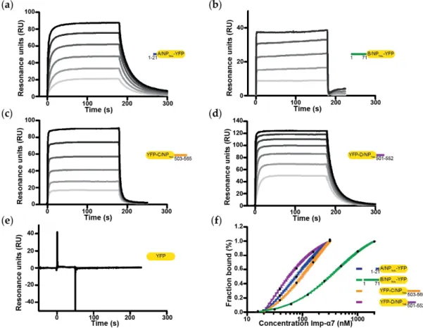
generation Biacore T200. This time, we were able to fully exploit the SPR data obtained with all NPTAILs. In particular, we showed that the architecture of the NLS (mono- or bipartite NLS) has no
importance for importin-α7; the affinity for the non-conventional monopartite A/NPTAIL was better
than for the bipartite C/NPTAIL, with Kd of 73 ± 4 nM and 146 ± 8 nM respectively, but weaker than
D/NPTAIL. B/NPTAIL appeared to be the lowest value with a Kd of 405 ± 5 nM, but comparable to the
value obtained using isothermal titration calorimetry [38]. Despite the nanomolar affinities, all of these interactions displayed a highly dynamic behaviour. Indeed, kinetics parameters show extremely high on rates, suggesting that very little energy is needed to form the interaction, but also very high off rates. Both association and dissociation rates were close to the upper detection limits of
the apparatus, respectively around 10-6 M-1.s-1 and 0.1 s-1 and even above in the case of the B/NPTAIL
Figure 4. Affinity between YFP-NPTAILs and human importin-α7 by surface plasmon resonance (SPR). The figure shows the sensorgrams of the interaction between importin-α7 and (a) A/NPTAIL-YFP, (b) B/NPTAIL-YFP, (c) YFP-C/NPTAIL, (d) YFP-D/NPTAIL and (e) the YFP control. For each panel, the gradation of grey represents the different concentrations of importin-α7 used for each titration, from the lowest (light-grey) to the highest (dark-grey). The concentrations range from 19.5 nM to 625 nM for (a) and (c), from 125 nM to 2 µM for (b), from 19.5 nM to 1.25 µM for (d) and from 80.5 nM to 10.3 µM for (e). (f) Steady state analysis was performed on Biacore evaluation software, the resulting fitting normalized and plotted on a log10 scale.
Table 2. Affinity between wild-type influenza nucleoprotein tails and human importins-α obtained
by SPR. The apparent dissociation constants (Kd) are expressed in nM; n.i. means ‘no interaction’.
imp-α3 imp-α5 imp-α7
A/NPTAIL 301 ± 21 128 ± 20 73 ± 4
B/NPTAIL 308 ± 106 770 ± 24 405 ± 5
C/NPTAIL 59 ± 9 287 ± 31 146 ± 8
D/NPTAIL 22 ± 15 21 ± 5 29 ± 1
YFP control n.i. n.i. n.i.
Table 3. Kinetic parameters of the interaction between wild-type influenza nucleoprotein tails and
human importins-α obtained by SPR. The association rate constants (kon) are expressed in M-1.s-1 and
the dissociation rate constants (koff) values are expressed in s-1; n.i. means ‘no interaction’.
imp-α3 imp-α5 imp-α7
kon (.106) koff (.10-1) kon (.106) koff (.10-1) kon (.106) koff (.10-1)
A/NPTAIL 1.31 ± 0.03 2.81 ± 0.04 1.0 ± 0.6 1.6 ± 0.1 1.6 ± 0.1 0.7 ± 0.1
B/NPTAIL > 10 > 10 > 10 > 10 > 10 > 10
C/NPTAIL 2.4 ± 1.1 1.3 ± 0.4 6.1 ± 3.6 2.6 ± 0.4 2.6 ± 0.4 2.5 ± 0.1
D/NPTAIL 2.8 ± 0.2 0.07 ± 0.01 1.1 ± 0.1 5.0 ± 0.2 5.0 ± 0.2 1.04 ± 0.03
3.3. Systematic Analysis of the Interactions between NPTAILs and Importins-α
Once the strategy for measuring the kinetics of all NPTAILs for the importin-α7 was established,
we decided to expand it to other members of the importins-α family. The human genome encodes seven isoforms of importin-α, divided into three subfamilies known as α1, α2 and α3: the α1 subfamily contains importin-α1 and -α8; the α2 subfamily contains importin-α3 and -α4; and the α3 subfamily contains importin-α5, -α6 and -α7 [18,49]. We chose to work with one representative isoform of each subfamily (importin-α1 and importin-α3, respectively for the α1 and α2 subfamilies) plus a second isoform for the α3 subfamily (i.e., importin-α5). Furthermore, all these isoforms are described in the literature regarding the influenza virus infection. We produced and purified the recombinant isoforms without their importin-β binding (IBB) domain. The case of importin-α1 is shown below. Using SEC-MALLS-RI experiments, we first confirmed that both ΔIBB importins-α3 and -α5 are monomeric in solution (Figure S4), before we used them in our SPR experiments to determine their respective kinetics (Tables 2 and 3). Importin-α5 shows a similar interaction profile
as importin-α7, with Kd D/NPTAIL < A/NPTAIL < C/NPTAIL < B/NPTAIL but with a higher amplitude in the
values (from 21 to 770 nM). On the contrary, the architecture of the NLS seems essential for
recognition by importin-α3. The two bipartite NLS tails (i.e., C/NPTAIL and D/NPTAIL) have a higher
affinity than the two monopartite (i.e., A/NPTAIL and B/NPTAIL) which both show identical values.
Based on these results, we further deciphered the interactions by introducing point mutations in the
positive charge patches of the NLSs which we had analyzed in the past (i.e., B/NPTAIL and D/NPTAIL)
[37,38]. We show that for bipartite NLS, the second basic patch could be stronger in relation to the stability of the cargo: importin-α than the first patch (Table 4). Based on the affinities of each single
patch mutants, D/NPTAIL seems to present a cooperative behaviour in importin-α binding. On the
other hand, importin-α7 seems to be more prompt in compensating for mutations in the first patch whereas importin-α3 looks more permissive to mutations in the second. The results obtained with
B/NPTAIL mutant confirm our nuclear magnetic resonance (NMR) observations on the major
contribution of the whole tail, from residues 30 to 71, in the interaction with importins-α, and not
only the regions surrounding the KR motif [38]. Regarding the B/NPTAIL mut1-YFP, even if we were not
able to quantify precisely the equilibrium dissociation constants from the raw data, probably because importin-α require a longer injection period to saturate all the ligand binding sites in this condition,
the sensorgrams show clear concentration dependent-responses in contrast to the YFP-D/NPTAIL mut3
construct (Figure S5).
Table 4. Effects of mutations in B/NPTAIL and D/NPTAIL on the affinities for the human importins-α. The Kd obtained by SPR are expressed in nM; n.i. means ‘no interaction’.
imp-α3 imp-α5 imp-α7
B/NPTAIL 308 ± 106 770 ± 24 405 ± 5
B/NPTAIL mut1 > 5 000 > 5 000 > 5 000
D/NPTAIL 22 ± 15 21 ± 5 29 ± 1
D/NPTAIL mut1 922 ± 77 1 150 ± 154 658 ± 208
D/NPTAIL mut2 2 457 ± 184 > 5 000 > 5 000
D/NPTAIL mut3 n.i. n.i. n.i.
3.4. The Case of the Importin-α1
The SEC-MALLS-RI experiments showed a different behaviour of importin-α1 in solution
(Figure S4). Whereas the experimental molecular mass for ΔIBB importins-α3, -α5 and –α7 were
similar to the theoretical values, the results obtained with ΔIBB importin-α1 suggest a propensity for dimerization of this construct. The corresponding sample was eluted precociously from the S200
column with an apparent molecular mass of 88 kDa rather than the theoretical 50 kDa (Figure S4a).
Dimerization was reported previously [50] with a monomer–dimer Kd estimated to be 8 µM and the
equilibrium could be shifted to the monomeric form by adding NLS-mimicking peptides. In a second SEC-MALLS-RI set of experiments but using a Superdex 75 column, we showed that with the
different NPTAIL peptides the peaks corresponding to the importin-α1 were shifted to the smaller
molecular mass (Figure 5a). The largest effect is observed with D/NPTAIL suggesting that a
quasi-quantitative estimation of the interaction could be extracted from these results, with apparent Kd
D/NPTAIL < C/NPTAIL ≤ A/NPTAIL < B/NPTAIL.
When we tried to measure the corresponding kinetics by SPR, we could not obtain a stable equilibrium, probably due to the competition between the monomer-dimer transition of importin-α1 and its interaction with the immobilized tails. This experimental artifact is inherent to the nature of the analyte, which despite the purification steps may dimerize in time and significantly affect affinity
measurements by SPR. We then found an alternative method; with the YFP-NPTAILs we could measure
the fluorescence anisotropy. Fluorescence anisotropy is a measure of light emitted by a fluorophore, unequal along different axes of polarization. This measure depends on rotational behaviour of this fluorescent molecule in solution. Depending on the fluorophore environment, fluorescence anisotropy will vary from a low value because of its high rotational rate when the molecule is free, to a high value in case of its interaction with a partner. This feature allows the determination of an apparent binding constant when the fluorescent molecule is kept at a constant concentration upon titration of its partner. By titration of our ΔIBB importin-α1 construct in presence of the different YFP-NPTAILs (Figure 5b), the corresponding apparent Kd was determined (Table 5). The curve of the
B/NPTAIL could not be estimated because the Kd of the B/NPTAIL for importin-α1 was too close to the
Kd of the dimerization of importin-α1.
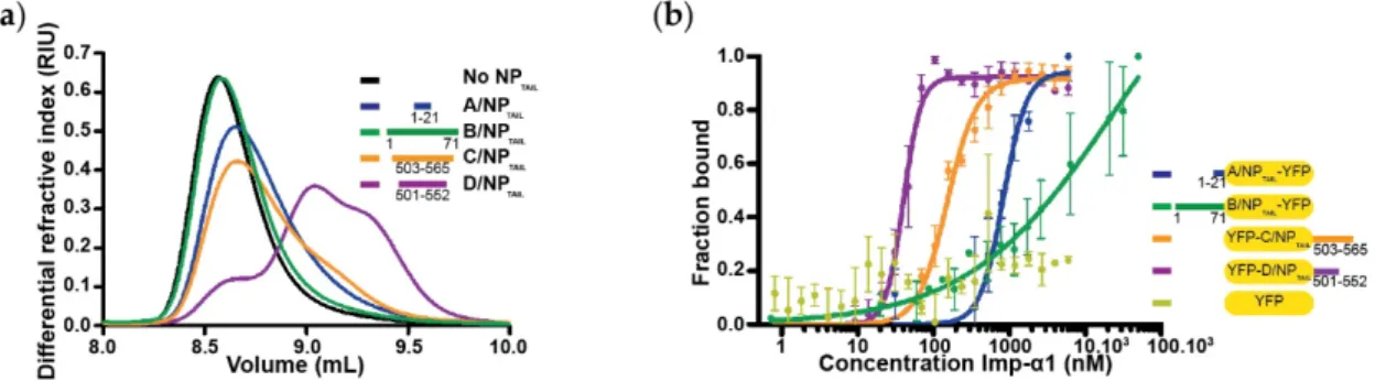
Figure 5. Binding of influenza nucleoproteins tails to importin-α1. (a) Dissociation of importin-α1
dimers by competition with NPTAIL using a SEC-MALLS-RI experiments. Human importin-α1 (45 μM)
alone or incubated with each NPTAIL (45 µM) was injected on a SuperdexTM 75 increase 10/300 G. Successful competition resulted in a delayed elution volume compared to the importin-α1 alone. Three distinct species can be identified from importin-α1:D/NPTAIL injection and two species from importin-α1:C/NPTAIL one. (b) YFP-NPTAILs: importin-α1 affinities by fluorescence anisotropy. Titration with each NPTAIL (25 nM) was carried out by fluorescence anisotropy with importin-α1 concentrations ranging from 0.7 nM to 50 µM for B/NPTAIL-YFP or from 0.8 nM to 6 µM for the other constructs. The titrations were normalized and fitted with GraphPad analysis model “specific binding with Hill slope”. The raw data are shown in Figure S6.
Table 5. Affinity between wild-type influenza nucleoprotein tails and human importin-α1 obtained
by fluorescence anisotropy. The apparent Kd are expressed in nM; n.d. and n.i. mean ‘not determined’
and ‘no interaction’, respectively.
A/NPTAIL B/NPTAIL C/NPTAIL D/NPTAIL YFP
control
4. Discussion
The interaction between viral proteins and cellular factors is of growing interest for understanding the evolution mechanisms of viruses, and RNA viruses in particular. These are major potential therapeutic targets, as these interfaces are less prone to mutations. It has been decades since the interactions between importin-α and the influenza virus nucleoprotein have been studied but numerous features and mechanisms are still unknown [32,33,35,51]. Importins-α are essential actors in cells and were diversified and highly conserved through evolution (Table S2). Each importin-α shows a high sequence identity in higher eukaryotes with a specific expression pattern and behaviour but also preferential bindings to cargo and functions.
In this paper, we provide a comparative characterization of the interactions between four importin-α isoforms with the tails from A/NP, B/NP, C/NP and D/NP. The affinities are between the nanomolar and the low micromolar range, with association and dissociation rates surprisingly high.
Two different behaviours were identified: i) A/NPTAIL, C/NPTAIL and D/NPTAIL present a dynamic
complex formation and release with high-affinity preferential binding and ii) B/NPTAIL shows even
faster association and dissociation rates with very low affinity.
The A/NPTAIL was isolated from a human strain of influenza A virus and interacted preferentially
with importin-α7. This is consistent with the importins-α expression pattern in the human respiratory track [52] as well as of previously reported work [31]. It is of note that PB2 isolated from human strains also preferentially bind to importin-α7, while avian PB2s tend to interact with importin-α3
[28]. The poor binding of A/NPTAIL to importin-α3 is consistent with the fact that importin-α3 tends
not to interact well with monopartite NLS; while A/NPTAIL is not a monopartite NLS per se, it possesses
only a short NLS motif. In contrast, C/NPTAIL presents a higher affinity for importin-α3. Importin-α3
has a low auto-inhibition and a unique intrinsic flexibility, acting as an adapter for unusual and complex NLS sequences or NLS close to globular domains [53,54]. Such NLSs could not associate
with a less flexible importin-α due to steric hindrance. The NLS sequence of C/NPTAIL is similar to a
bipartite NLS, with two basic patches, but separated by a long linker sequence (more than 20 residues). Such a sequence could require the flexibility of importin-α3 to achieve optimal binding.
D/NPTAIL is a textbook case of bipartite NLS, which binds tightly to importins-α, using both the minor
and major binding pockets [55–57]. D/NPTAIL strongly interacts with all tested importins with a high
affinity (20–41 nM), in the same range as other bipartite NLSs (Kd ≈ 10 nM) [28,58]. In other studies, it
has been shown that the in vitro affinities using purified proteins and peptides were in the nanomolar range whereas the interaction between NLSs and importins-α in cellulo is in the micromolar range. Addition of cytosol in in vitro assays led to the destabilization of importin: NLS complexes, even in the case of bacterial lysate, highlighting the contribution of non-specific interaction and competition in this cellular process [44,59]. Importins cycles between both compartments have been estimated to be fast, around 30 ms; nanomolar affinity leads to a residence time of several seconds or up to several minutes, which is far too long to allow optimal release in the nucleus, while micromolar affinity put it in the millisecond range [59].
The sensorgram shapes of C/NPTAIL and D/NPTAIL along with the affinities resulting from
D/NPTAIL mutations clearly suggest that the interaction follows a 1:1 binding model with
cooperativity. At the other end of the spectrum, B/NPTAIL behaviour towards importins-α is still
puzzling. It is possible that the B/NPTAIL is not properly processed in our settings. It is well known
that A/NP and B/NP nuclear import are regulated through post-translational modifications (PTM), in particular through phosphorylation [60,61]. A discrete PTM could be an activating signal for
nuclear import regulation [62–66]. The absence of the NPCORE in our experiment could also be
responsible for the lack of functionality of B/NPTAIL: three-dimensional context could matter for
importin specificity, with putative additional contacts and constrains, and for NLS docking [18,28,53,67]. Another hypothesis comes from the nuclear import receptor involved and the complex to be imported. While importin-α/β complex is largely believed to be the most common nuclear import pathway, this is still debated. Other adaptor proteins besides importin-α are known to interact with cargo and importin-β [68,69]. Co-import of neo-synthesized B/NP with another cargo is also a possibility, taking in account that RNP nuclear import functionality putatively relies on avidity rather
than affinity [70,71]. Therefore, it is not be excluded that B/NP uses another receptor/complex to gain access to the nucleus. Further investigations into other nuclear import receptors could shed light on successful B/NP nuclear translocation. One interesting feature of B/NP is its long, intrinsically
disordered Nterminal tail. Such a tail could be involved in the formation of liquid phase separation and
help with protein recruitment [72,73]. If B/NP exhibits such behaviour, it could be interesting to dissect it further as nucleoporins in the nuclear pore complex channel tend themselves to be involved in phase separation [74–76].
Our work focuses on human importins-α, as three out of the four NPTAILS were derived from
human viruses. However, due to the high conservation of these importins through evolution (Table S2), it is likely that extrapolation to other mammal importins can be achieved to some degree. Further work would be required to validate this completely.
While a lot of work is still required to fully understand the mechanistic and specificity of the interactions between influenza nucleoproteins and importins-α, we provide here this first
comparative study on the interaction of NPTAILs with importins-α1, -α3, -α5 and -α7. NPCORE
involvement, NP oligomerization, cellular environment, IBB domain contribution, and post-translational modification of both partners are other factors which need to be investigated in the future, in the hope of fully dissecting this host-pathogen interaction and trying to target and disrupt this essential hijacking mechanism.
5. Conclusions
While the interactions between influenza NPs and importins-α have been extensively studied, this has mainly been done through the prism of A/NP, with a large number of highly different methods and sometimes without the essential focus required on importins-α. Whereas each method and study brought useful characterization insights, it is a complex task to compare affinity from widely different assays, with significant changes in conditions, partners, settings and analysis. Here, we provide the first comparative study to dissect several interactions between influenza NPs and
importins-α using the same strategy and under the same conditions. We showed that A/NPTAIL,
C/NPTAIL and D/NPTAIL interact strongly with one or several importins-α. On the contrary, the human
specific B/NPTAIL interacts poorly with selected importins-α, raising new questions about this
essential interaction in the influenza virus cycle.
Supplementary Materials: The following are available online at www.mdpi.com/1999-4915/12/8/834/s1, Figure
S1: analysis of the complexes between NPTAILS and human importin-α7 in solution, Figure S2: SEC-MALLS-RI
analysis of the different YFP-fused tails used in that work, Figure S3: absence of interaction between YFP and human importin-α7 in solution, Figure S4: SEC-MALLS-RI analysis of the different importins-α used in that work, Figure S5: interaction between human importin-α7 and YFP-NPTAILs mutants, Figure S6: interaction
between human importin-α1 and YFP-NPTAILs, Table S1: sequences, Table S2: sequence identity between the
importins-α used in the studies.
Author Contributions: T.C. conceived and designed the experiments; A.D., E.V-S. and F.C.A.G. performed the
experiments; A.D., E.V.-S., F.C.A.G. and T.C. analysed the data; A.D. and T.C. wrote the original draft; A.D., E.V.-S., F.C.A.G., R.W.H.R. and T.C. edited the final manuscript. All authors have read and agreed to the published version of the manuscript.
Funding: This work was supported by the Agence Nationale pour la Recherche, grant number
ANR-14-CE09-0017.
Acknowledgments: We thank Justine Sensini and Louis Saliceti for their contribution in the samples
preparation. We are highly grateful to Jean-Marie Bourhis, Pascal Fender, Corinne Mercier, Carlo Petosa and Antoine Royant for discussions. We thank Darren J Hart for the plasmid to express the human importin-α7. This work used the platforms of the Grenoble Instruct-ERIC center (ISBG; UMS 3518 CNRS-CEA-UGA-EMBL) within the Grenoble Partnership for Structural Biology (PSB), supported by FRISBI (ANR-10-INBS-05-02) and GRAL, financed by the University Grenoble Alpes - Ecoles Universitaires de Recherche CBH-EUR-GS (ANR-17-EURE-0003).
References
1. Shi, M.; Lin, X.D.; Chen, X.; Tian, J.H.; Chen, L.J.; Li, K.; Wang, W.; Eden, J.S.; Shen, J.J.; Liu, L.; et al. The evolutionary history of vertebrate RNA viruses. Nature 2018, 556, 197–202.
2. Klumpp, K.; Ruigrok, R.W.; Baudin, F. Roles of the Influenza virus polymerase and nucleoprotein in forming a functional RNP structure. EMBO J. 1997, 16, 1248–1257.
3. Coloma, R.; Arranz, R.; de la Rosa-Trevin, J.M.; Sorzano, C.O.S.; Munier, S.; Carlero, D.; Naffakh, N.; Ortin, J.; Martin-Benito, J. Structural insights into Influenza A virus ribonucleoproteins reveal a processive helical track as transcription mechanism. Nat. Microbiol. 2020, 5, 727–734.
4. Feldherr, C.M.; Kallenbach, E.; Schultz, N. Movement of a karyophilic protein through the nuclear pores of oocytes. J. Cell Biol. 1984, 99, 2216–2222.
5. Davis, L.I. The nuclear pore complex. Annu. Rev. Biochem. 1995, 64, 865–896.
6. Mans, B.J.; Anantharaman, V.; Aravind, L.; Koonin, E.V. Comparative genomics, evolution and origins of the nuclear envelope and nuclear pore complex. Cell Cycle 2004, 3, 1612–1637.
7. Lin, D.H.; Hoelz, A. The structure of the nuclear pore complex (an update). Annu. Rev. Biochem. 2019, 88, 725–783.
8. Bonner, W.M. Protein migration into nuclei. II. Frog oocyte nuclei accumulate a class of microinjected oocyte nuclear proteins and exclude a class of microinjected oocyte cytoplasmic proteins. J. Cell Biol. 1975, 64, 431–437.
9. Chook, Y.M.; Suel, K.E. Nuclear import by karyopherin-betas: Recognition and inhibition. Biochim. Biophys. Acta 2011, 1813, 1593–1606.
10. Timney, B.L.; Raveh, B.; Mironska, R.; Trivedi, J.M.; Kim, S.J.; Russel, D.; Wente, S.R.; Sali, A.; Rout, M.P. Simple rules for passive diffusion through the nuclear pore complex. J. Cell Biol. 2016, 215, 57–76.
11. Gorlich, D.; Vogel, F.; Mills, A.D.; Hartmann, E.; Laskey, R.A. Distinct functions for the two importin subunits in nuclear protein import. Nature 1995, 377, 246–248.
12. Chook, Y.M.; Blobel, G. Karyopherins and nuclear import. Curr. Opin. Struct. Biol. 2001, 11, 703–715. 13. Lange, A.; Mills, R.E.; Lange, C.J.; Stewart, M.; Devine, S.E.; Corbett, A.H. Classical nuclear localization
signals: Definition, function, and interaction with importin alpha. J. Biol. Chem. 2007, 282, 5101–5105. 14. Lee, B.J.; Cansizoglu, A.E.; Suel, K.E.; Louis, T.H.; Zhang, Z.; Chook, Y.M. Rules for nuclear localization
sequence recognition by karyopherin beta 2. Cell 2006, 126, 543–558.
15. Bono, F.; Cook, A.G.; Grunwald, M.; Ebert, J.; Conti, E. Nuclear import mechanism of the EJC component Mago-Y14 revealed by structural studies of importin 13. Mol. Cell 2010, 37, 211–222.
16. Padavannil, A.; Sarkar, P.; Kim, S.J.; Cagatay, T.; Jiou, J.; Brautigam, C.A.; Tomchick, D.R.; Sali, A.; D’Arcy, S.; Chook, Y.M. Importin-9 wraps around the H2A-H2B core to act as nuclear importer and histone chaperone. Elife 2019, 8, doi:10.7554/eLife.43630.
17. Swale, C.; Da Costa, B.; Sedano, L.; Garzoni, F.; McCarthy, A.A.; Berger, I.; Bieniossek, C.; Ruigrok, R.W.H.; Delmas, B.; Crepin, T. X-ray structure of the human karyopherin RanBP5, an essential factor for Influenza polymerase nuclear trafficking. J. Mol. Biol. 2020, 432, 3353–3359.
18. Pumroy, R.A.; Cingolani, G. Diversification of importin-alpha isoforms in cellular trafficking and disease states. Biochem. J. 2015, 466, 13–28.
19. Oka, M.; Yoneda, Y. Importin alpha: Functions as a nuclear transport factor and beyond. Proc. Jpn. Acad. Ser. B Phys. Biol. Sci. 2018, 94, 259–274.
20. Martin, K.; Helenius, A. Transport of incoming Influenza virus nucleocapsids into the nucleus. J. Virol. 1991, 65, 232–244.
21. Neumann, G.; Castrucci, M.R.; Kawaoka, Y. Nuclear import and export of Influenza virus nucleoprotein. J. Virol. 1997, 71, 9690–9700.
22. Deng, T.; Engelhardt, O.G.; Thomas, B.; Akoulitchev, A.V.; Brownlee, G.G.; Fodor, E. Role of ran binding protein 5 in nuclear import and assembly of the Influenza virus RNA polymerase complex. J. Virol. 2006, 80, 11911–11919.
23. Hutchinson, E.C.; Orr, O.E.; Man Liu, S.; Engelhardt, O.G.; Fodor, E. Characterization of the interaction between the Influenza A virus polymerase subunit PB1 and the host nuclear import factor Ran-binding protein 5. J. Gen. Virol. 2011, 92, 1859–1869.
24. Swale, C.; Monod, A.; Tengo, L.; Labaronne, A.; Garzoni, F.; Bourhis, J.M.; Cusack, S.; Schoehn, G.; Berger, I.; Ruigrok, R.W.; et al. Structural characterization of recombinant IAV polymerase reveals a stable complex between viral PA-PB1 heterodimer and host RanBP5. Sci. Rep. 2016, 6, 24727.
25. Tarendeau, F.; Boudet, J.; Guilligay, D.; Mas, P.; Bougault, C.; Boulo, S.; Baudin, F.; Ruigrok, R.W.H.; Daigle, N.; Ellenberg, J.; et al. Structure and nuclear import function of the C-terminal domain of Influenza virus polymerase PB2 subunit. Nat. Struct. Mol. Biol. 2007, 14, 229–233.
26. Boivin, S.; Hart, D.J. Interaction of the Influenza A virus polymerase PB2 C-terminal region with importin alpha isoforms provides insights into host adaptation and polymerase assembly. J. Biol. Chem. 2011, 286, 10439–10448.
27. Hudjetz, B.; Gabriel, G. Human-like PB2 627K Influenza virus polymerase activity is regulated by importin-alpha1 and -alpha7. PLoS Pathog. 2012, 8, e1002488.
28. Pumroy, R.A.; Ke, S.; Hart, D.J.; Zachariae, U.; Cingolani, G. Molecular determinants for nuclear import of Influenza A PB2 by importin alpha isoforms 3 and 7. Structure 2015, 23, 374–384.
29. Cros, J.F.; García-Sastre, A.; Palese, P. An unconventional NLS is critical for the nuclear import of the Influenza A virus nucleoprotein and ribonucleoprotein. Traffic 2005, 6, 205–213.
30. Weber, F.; Kochs, G.; Gruber, S.; Haller, O. A classical bipartite nuclear localization signal on Thogoto and Influenza A virus nucleoproteins. Virology 1998, 250, 9–18.
31. Wu, W.; Sankhala, R.S.; Florio, T.J.; Zhou, L.; Nguyen, N.L.T.; Lokareddy, R.K.; Cingolani, G.; Pante, N. Synergy of two low-affinity NLSs determines the high avidity of Influenza A virus nucleoprotein NP for human importin alpha isoforms. Sci. Rep. 2017, 7, 11381.
32. O’Neill, R.E.; Palese, P. NPI-1, the human homolog of SRP-1, interacts with Influenza virus nucleoprotein. Virology 1995, 206, 116–125.
33. Wang, P.; Palese, P.; O’Neill, R.E. The NPI-1/NPI-3 (karyopherin alpha) binding site on the Influenza a virus nucleoprotein NP is a nonconventional nuclear localization signal. J. Virol. 1997, 71, 1850–1856. 34. Melen, K.; Fagerlund, R.; Franke, J.; Kohler, M.; Kinnunen, L.; Julkunen, I. Importin alpha nuclear
localization signal binding sites for STAT1, STAT2, and Influenza A virus nucleoprotein. J. Biol. Chem. 2003, 278, 28193–28200.
35. Gabriel, G.; Klingel, K.; Otte, A.; Thiele, S.; Hudjetz, B.; Arman-Kalcek, G.; Sauter, M.; Shmidt, T.; Rother, F.; Baumgarte, S.; et al. Differential use of importin-alpha isoforms governs cell tropism and host adaptation of Influenza virus. Nat. Commun. 2011, 2, 156.
36. Sasaki, Y.; Hagiwara, K.; Kakisaka, M.; Yamada, K.; Murakami, T.; Aida, Y. Importin alpha3/Qip1 is involved in multiplication of mutant Influenza virus with alanine mutation at amino acid 9 independently of nuclear transport function. PLoS ONE 2013, 8, e55765.
37. Donchet, A.; Oliva, J.; Labaronne, A.; Tengo, L.; Miloudi, M.; Gerard, F.C.A.; Mas, C.; Schoehn, G.; Ruigrok, R.W.H.; Ducatez, M.; et al. The structure of the nucleoprotein of Influenza D shows that all Orthomyxoviridae nucleoproteins have a similar NPCORE, with or without a NPTAIL for nuclear transport. Sci. Rep. 2019, 9, 600. 38. Labaronne, A.; Milles, S.; Donchet, A.; Jensen, M.R.; Blackledge, M.; Bourhis, J.M.; Ruigrok, R.W.H.; Crepin, T. Structural analysis of the complex between Influenza B nucleoprotein and human importin-alpha. Sci. Rep. 2017, 7, 17164.
39. Ozawa, M.; Fujii, K.; Muramoto, Y.; Yamada, S.; Yamayoshi, S.; Takada, A.; Goto, H.; Horimoto, T.; Kawaoka, Y. Contributions of two nuclear localization signals of Influenza A virus nucleoprotein to viral replication. J. Virol. 2007, 81, 30–41.
40. Ng, A.K.; Zhang, H.; Tan, K.; Li, Z.; Liu, J.H.; Chan, P.K.; Li, S.M.; Chan, W.Y.; Au, S.W.; Joachimiak, A.; et al. Structure of the Influenza virus A H5N1 nucleoprotein: Implications for RNA binding, oligomerization, and vaccine design. FASEB J. 2008, 22, 3638–3647.
41. Boulo, S.; Akarsu, H.; Lotteau, V.; Muller, C.W.; Ruigrok, R.W.; Baudin, F. Human importin alpha and RNA do not compete for binding to Influenza A virus nucleoprotein. Virology 2011, 409, 84–90.
42. Nakada, R.; Hirano, H.; Matsuura, Y. Structure of importin-alpha bound to a non-classical nuclear localization signal of the Influenza A virus nucleoprotein. Sci. Rep. 2015, 5, 15055.
43. Tang, Y.S.; Lo, C.Y.; Mok, C.K.; Chan, P.K.; Shaw, P.C. The extended C-terminal region of Influenza C virus nucleoprotein is important for nuclear import and ribonucleoprotein activity. J. Virol. 2019, 93, e02048-18, doi: 10.1128/JVI.02048-18.
44. Timney, B.L.; Tetenbaum-Novatt, J.; Agate, D.S.; Williams, R.; Zhang, W.; Chait, B.T.; Rout, M.P. Simple kinetic relationships and nonspecific competition govern nuclear import rates in vivo. J. Cell Biol. 2006, 175, 579–593.
45. Mackmull, M.T.; Klaus, B.; Heinze, I.; Chokkalingam, M.; Beyer, A.; Russell, R.B.; Ori, A.; Beck, M. Landscape of nuclear transport receptor cargo specificity. Mol. Syst. Biol. 2017, 13, 962.
46. Sumarheni, S.; Hong, S.S.; Josserand, V.; Coll, J.L.; Boulanger, P.; Schoehn, G.; Fender, P. Human full-length coagulation factor X and a GLA domain-derived 40-mer polypeptide bind to different regions of the adenovirus serotype 5 hexon capsomer. Hum. Gene Ther. 2014, 25, 339–349.
47. Wyatt, P.J. Submicrometer particle sizing by multiangle light scattering following fractionation. J. Colloid Interface Sci. 1998, 197, 9–20.
48. Labaronne, A.; Swale, C.; Monod, A.; Schoehn, G.; Crepin, T.; Ruigrok, R.W. Binding of RNA by the nucleoproteins of Influenza viruses A and B. Viruses 2016, 8, 247.
49. Mason, D.A.; Stage, D.E.; Goldfarb, D.S. Evolution of the metazoan-specific importin alpha gene family. J. Mol. Evol. 2009, 68, 351–365.
50. Miyatake, H.; Sanjoh, A.; Unzai, S.; Matsuda, G.; Tatsumi, Y.; Miyamoto, Y.; Dohmae, N.; Aida, Y. Crystal structure of human importin-alpha1 (Rch1), revealing a potential autoinhibition mode involving homodimerization. PLoS ONE 2015, 10, e0115995.
51. Resa-Infante, P.; Paterson, D.; Bonet, J.; Otte, A.; Oliva, B.; Fodor, E.; Gabriel, G. Targeting importin-alpha7 as a therapeutic approach against pandemic Influenza viruses. J. Virol. 2015, 89, 9010–9020.
52. Ninpan, K.; Suptawiwat, O.; Boonarkart, C.; Phuangphung, P.; Sathirareuangchai, S.; Uiprasertkul, M.; Auewarakul, P. Expression of importin-alpha isoforms in human nasal mucosa: Implication for adaptation of avian Influenza A viruses to human host. Virol. J. 2016, 13, 90.
53. Sankhala, R.S.; Lokareddy, R.K.; Begum, S.; Pumroy, R.A.; Gillilan, R.E.; Cingolani, G. Three-dimensional context rather than NLS amino acid sequence determines importin alpha subtype specificity for RCC1. Nat. Commun. 2017, 8, 979.
54. Smith, K.M.; Tsimbalyuk, S.; Edwards, M.R.; Cross, E.M.; Batra, J.; Soares da Costa, T.P.; Aragao, D.; Basler, C.F.; Forwood, J.K. Structural basis for importin alpha 3 specificity of W proteins in Hendra and Nipah viruses. Nat. Commun. 2018, 9, 3703.
55. Robbins, J.; Dilworth, S.M.; Laskey, R.A.; Dingwall, C. Two interdependent basic domains in nucleoplasmin nuclear targeting sequence: Identification of a class of bipartite nuclear targeting sequence. Cell 1991, 64, 615–623.
56. Fontes, M.R.; Teh, T.; Kobe, B. Structural basis of recognition of monopartite and bipartite nuclear localization sequences by mammalian importin-alpha. J. Mol. Biol. 2000, 297, 1183–1194.
57. Hodel, M.R.; Corbett, A.H.; Hodel, A.E. Dissection of a nuclear localization signal. J. Biol. Chem. 2001, 276, 1317–1325.
58. Lokareddy, R.K.; Hapsari, R.A.; van Rheenen, M.; Pumroy, R.A.; Bhardwaj, A.; Steen, A.; Veenhoff, L.M.; Cingolani, G. Distinctive properties of the nuclear localization signals of inner nuclear membrane proteins Heh1 and Heh2. Structure 2015, 23, 1305–1316.
59. Cardarelli, F.; Bizzarri, R.; Serresi, M.; Albertazzi, L.; Beltram, F. Probing nuclear localization signal-importin alpha binding equilibria in living cells. J. Biol. Chem. 2009, 284, 36638–36646.
60. Hutchinson, E.C.; Denham, E.M.; Thomas, B.; Trudgian, D.C.; Hester, S.S.; Ridlova, G.; York, A.; Turrell, L.; Fodor, E. Mapping the phosphoproteome of Influenza a and B viruses by mass spectrometry. PLoS Pathog. 2012, 8, e1002993.
61. Zheng, W.; Li, J.; Wang, S.; Cao, S.; Jiang, J.; Chen, C.; Ding, C.; Qin, C.; Ye, X.; Gao, G.F.; et al. Phosphorylation controls the nuclear-cytoplasmic shuttling of Influenza A virus nucleoprotein. J. Virol.
2015, 89, 5822–5834.
62. Terry, L.J.; Shows, E.B.; Wente, S.R. Crossing the nuclear envelope: Hierarchical regulation of nucleocytoplasmic transport. Science 2007, 318, 1412–1416.
63. Bedford, M.T.; Clarke, S.G. Protein arginine methylation in mammals: Who, what, and why. Mol. Cell 2009, 33, 1–13.
64. Nardozzi, J.; Wenta, N.; Yasuhara, N.; Vinkemeier, U.; Cingolani, G. Molecular basis for the recognition of phosphorylated STAT1 by importin alpha5. J. Mol. Biol. 2010, 402, 83–100.
65. Wang, Y.E.; Pernet, O.; Lee, B. Regulation of the nucleocytoplasmic trafficking of viral and cellular proteins by ubiquitin and small ubiquitin-related modifiers. Biol. Cell. 2012, 104, 121–138.
66. Ptak, C.; Wozniak, R.W. SUMO and nucleocytoplasmic transport. Adv. Exp. Med. Biol. 2017, 963, 111–126. 67. Friedrich, B.; Quensel, C.; Sommer, T.; Hartmann, E.; Kohler, M. Nuclear localization signal and protein
context both mediate importin alpha specificity of nuclear import substrates. Mol. Cell. Biol. 2006, 26, 8697– 8709.
68. Jullien, D.; Gorlich, D.; Laemmli, U.K.; Adachi, Y. Nuclear import of RPA in Xenopus egg extracts requires a novel protein XRIPalpha but not importin alpha. EMBO J. 1999, 18, 4348–4358.
69. Narayanan, U.; Ospina, J.K.; Frey, M.R.; Hebert, M.D.; Matera, A.G. SMN, the spinal muscular atrophy protein, forms a pre-import snRNP complex with snurportin1 and importin beta. Hum. Mol. Genet. 2002, 11, 1785–1795.
70. Hodges, J.L.; Leslie, J.H.; Mosammaparast, N.; Guo, Y.; Shabanowitz, J.; Hunt, D.F.; Pemberton, L.F. Nuclear import of TFIIB is mediated by Kap114p, a karyopherin with multiple cargo-binding domains. Mol. Biol. Cell 2005, 16, 3200–3210.
71. Tome-Amat, J.; Ramos, I.; Amanor, F.; Fernandez-Sesma, A.; Ashour, J. Influenza A virus utilizes low-affinity, high-avidity interactions with the nuclear import machinery to ensure infection and immune evasion. J. Virol. 2019, 93, e01046-18, doi: 10.1128/JVI.01046-18.
72. Schuster, B.S.; Reed, E.H.; Parthasarathy, R.; Jahnke, C.N.; Caldwell, R.M.; Bermudez, J.G.; Ramage, H.; Good, M.C.; Hammer, D.A. Controllable protein phase separation and modular recruitment to form responsive membraneless organelles. Nat. Commun. 2018, 9, 2985.
73. Guseva, S.; Milles, S.; Jensen, M.R.; Salvi, N.; Kleman, J.P.; Maurin, D.; Ruigrok, R.W.H.; Blackledge, M. Measles virus nucleo- and phosphoproteins form liquid-like phase-separated compartments that promote nucleocapsid assembly. Sci. Adv. 2020, 6, eaaz7095.
74. Milles, S.; Mercadante, D.; Aramburu, I.V.; Jensen, M.R.; Banterle, N.; Koehler, C.; Tyagi, S.; Clarke, J.; Shammas, S.L.; Blackledge, M.; et al. Plasticity of an ultrafast interaction between nucleoporins and nuclear transport receptors. Cell 2015, 163, 734–745.
75. Zilman, A. Aggregation, phase separation and spatial morphologies of the assemblies of FG nucleoporins. J. Mol. Biol. 2018, 430, 4730–4740.
76. Dormann, D. FG-nucleoporins caught in the act of liquid-liquid phase separation. J. Cell Biol. 2020, 219, e201910211.
© 2020 by the authors. Licensee MDPI, Basel, Switzerland. This article is an open access article distributed under the terms and conditions of the Creative Commons Attribution (CC BY) license (http://creativecommons.org/licenses/by/4.0/).
![Figure 1. Influenzavirus nucleoproteins. The schematic representations of Influenzavirus nucleoproteins are based on this structural analysis [37]](https://thumb-eu.123doks.com/thumbv2/123doknet/13580340.422017/3.892.306.581.925.1039/influenzavirus-nucleoproteins-schematic-representations-influenzavirus-nucleoproteins-structural-analysis.webp)