HAL Id: tel-02157954
https://tel.archives-ouvertes.fr/tel-02157954
Submitted on 17 Jun 2019HAL is a multi-disciplinary open access archive for the deposit and dissemination of sci-entific research documents, whether they are pub-lished or not. The documents may come from teaching and research institutions in France or abroad, or from public or private research centers.
L’archive ouverte pluridisciplinaire HAL, est destinée au dépôt et à la diffusion de documents scientifiques de niveau recherche, publiés ou non, émanant des établissements d’enseignement et de recherche français ou étrangers, des laboratoires publics ou privés.
New activity-based probes to detect matrix
metalloproteases
Monika Kaminska
To cite this version:
Monika Kaminska. New activity-based probes to detect matrix metalloproteases. Biomolecules [q-bio.BM]. Université Paris-Saclay, 2018. English. �NNT : 2018SACLS538�. �tel-02157954�
New activity-based probes to
detect matrix metalloproteases.
Thèse de doctorat de l'Université Paris-Saclay
préparée
à l'Université Paris-Sud
au CEA Saclay
École doctorale n°569 : innovation thérapeutique,
du fondamental à l'appliqué (ITFA)
Spécialité de doctorat: chimie thérapeutique
Thèse présentée et soutenue à Gif-sur-Yvette, le 14 Décembre 2018, par
Monika Kaminska
Composition du Jury: Pr. Erwan Poupon
(Université Paris Saclay) Présidente
Pr. Rebecca Deprez-Poulain
(Université de Lille) Rapporteur
Dr. Vincent Aucagne DR2
(Centre de Biophysique Moléculaire, Orléans, CNRS) Rapporteur
Dr. Sonia Cantel, MC/HDR
(Université de Montpellier) Examinateur
Dr. Laurent Devel, HDR
(CEA Saclay) Directeur de thèse
NNT : 20 18 S A C LS 53 8
Acknowledgments
The thesis presented in this manuscript was completed at the The French Alternative Energies and Atomic Energy Commission (CEA Saclay) unit of the Molecular engineering of proteins (SIMOPRO) thanks to funding from Excellence in Research on Medication and Innovative Therapeutics (Labex Lermit).
Firstly, I would like to express my very profound gratitude to my family, my parents and brother who were always with me despite the distance, for their faith in me, their love, their invaluable support and for standing beside me throughout my studies and all of my life. This accomplishment would not have been possible without them. Dziękuję!
I would like to dedicate special thanks to my thesis supervisor Dr. Laurent Devel for his optimism, patience, motivation, and for sharing his immense knowledge. During the past three years I always could rely on his help and support whenever I encountered an obstacle or had a question relating to my research.
I would like to thank Pr. Rebecca Deprez-Poulain and Dr. Vincent Aucagne for having agreed to be the reviewers of this manuscript. I would like also thank Pr. Erwan Poupon and Dr. Sonia Cantel for having accepted to be part of the jury in my thesis’ defense.
I am very grateful to everyone in the SIMOPRO unit, especially to the members that I had the pleasure of sharing my daily routine with; Bob, Steven, Carole, Mylene, Fabrice, Andrzej, Pavel, Evelyne. I really appreciate the warm welcome and kindness that they showed me. I could not have completed this work without their help. I am so thankful to Dr. Sarah Bregant for her scientific and emotional support, especially during the period in which I was writing this manuscript. I deeply appreciate the kindness and friendliness displayed by Livia, Alessia and Fabien. During every workday in the laboratory they were always encouraging towards me, willing to listen, and I could count on their help concerning every problem. I would also really like to thank Kiarach who offered me a large amount of support, especially during the last months of my PhD.
I could not imagine passing these last few years without my dear friends. I am extremely grateful that I have had the opportunity to meet such wonderful people who were motivated to build our CEA students/postdocs/researchers' crew and international family together. Thanks to: Ewa, Livia, Manu, Alessia, Oscar, Fabien, Martin, Malik, Aninda, Anshuman, Hector, Abdullah, Jorge, Liza, Vittore, Bianca, Mohcine, Anouar, Blake.
Abbreviations
ABP Activity-Based probe
ACE Angiotensin-converting enzyme
ACN Acetonitrile
ADAM A Disintegrin And Metalloproteinase
ADAMTS A Disintegrin And Metalloproteinase with Thrombospondin Motifs
AfBP Affinity-based probes
APS ammonium persulfate
BIC Bicarbonate
cDNA Complementary DNA
ClHOBt 6-Chloro-1-Hydroxybenzotriazole di-hydrate
COPD chronic obstructive pulmonary disease
DC dendritic cell DCM Dichloromethane DIC Diisopropylcarbodiimide DIEA N,N-Diisopropylethylamine DMAP 4-Dimethylaminopyridine DMF N,N-Dimethylformamide DMF Dimethylformamide DSC N,N’ disuccinimidyl carbonate
ECM Extracellular matrix
EDC. HCl N-(3-Dimethylaminopropyl)-N′-ethylcarbodiimide hydrochloride
EDTA Ethylenediaminetetraacetic acid
EGF Epidermal growth factor
ESI-MS Electrospray Mass spectrometry
FACS Fluorescence-activated cell sorting
DIEA N,N-Diisopropylethylamine
Fmoc Fluorenylmethyloxycarbonyl
GM-CSF Recombinant murine granulocyte-macrophage colony stimulating factor
GPI Glycosylphosphatidyl inositol
hCA Human Carbonic anhydrase
HCCA 4-hydroxycinnamic acid
HEPES 4-(2-hydroxyethyl)-1-piperazineethanesulfonic acid
hMMP Human matrix metalloproteinases
hsa Human serum albumin
iDC Immature dendritic cells
IFN Favour interferon-
IMDM Iscove's Modified Dulbecco's Medium
LC/MS Liquid chromatography/Mass spectrometry
LDAI The ligand-directed acyl imidazole
LDSP Ligand-directed N-Sulfonyl pyridone
LDT Ligand-directed tosyl
MALDI-TOF Matrix-assisted laser desorption/ionization Time of Flight MAP-Kinase Mitogen activating protein kinase
Mca-MAT Metoxycoumarin- Matrixine substrate
MME Murine macrophage elastase
MMP Matrix metalloproteinases
mRNA Messenger RNA
MT-MMP Membrane-type matrix metalloproteinases
NEP Neutral endopeptidase
NHS N-Hydroxysuccinimide
NMR Nuclear magnetic resonance
PA Activators of plasminogen
PCA Principal Component Analysis
PDB Protein data bank
PDGF Platelet-derived growth factor
PEA3 Activator that enhances the expression of poliovirus genes
PEG Polyethylene glycol
RP-HPLC Reverse phase- high performance liquid chromatography
SDS Sodium dodecyl sulphate
SDS-PAGE Sodium dodecyl sulphate- Polyacrylamide gel electrophoresis
SP spacer N-sulfonyl pyridone
TACE Tumour necrosis factor-alpha converting enzyme belonging to ADAM family
TEMED N, N, N ', N'-tetramethylethylenediamine
TGF-β Transforming growth factor
TIMP-2 Tissue inhibitors of metalloproteinases
TIMPS Tissue inhibitors of metalloproteinases
TIS Triisopropylsilane
TNFα Tumour necrosis factor α
tPA Tissue type activator
uPA Urokinase
TABLE OF CONTENTS
CHAPTER I: LITERATURE REVIEW
...1
1.
GENERAL CONSIDERATIONS ON PROTEASES ... 3
2.
MATRIX METALLOPROTEINASES ... 5
2.1. Discovery and MMPs classification. ... 5
2.2. MMPs belong to the metzincin family ... 7
2.3. MMPs possess a modular structure ... 8
2.4. Regulation of MMPs activities ... 10
2.4.1. Transcription ... 11
2.4.2. Extracellular activation and cysteine switch concept ... 12
2.4.3. Intracellular activation ... 13
2.4.4. Cell Surface activation involving TIMP-2 ... 13
2.4.5. Inactivation by endogenous inhibitors ... 14
3.
BIOLOGICAL FUNCTIONS OF MMPS ... 15
3.1. The physiological function of MMPs. ... 16
3.2. Pathological function of MMPs ... 16
3.3. The specific case of macrophage elastase (MMP12) ... 17
4.
INHIBITORS OF MMPS: CHEMICAL TOOLS TO INVESTIGATE MMPS BIOLOGICAL
FUNCTION. ... 19
4.1. Structure-based design of MMP inhibitors: from broad-spectrum to selective inhibitors ... 19
4.1.1. RXP470.1 as a highly selective inhibitor of MMP12 ... 22
4.1.2. RXP 470.1 as a chemical tool to study MMP12 in vivo ... 25
4.1.2.1. RXP470.1 target MMP12 within atherosclerotic plaques. ... 25
4.1.2.2 RXP470.1 can target extracellular MMP12 during viral infection. ... 26
5.
CHEMICAL PROBES FOR DETECTING MMPS ACTIVATION IN COMPLEX
PROTEOMES ... 28
5.1. Imaging of MMPs activity with substrate- and ligand-derived probes ... 28
5.2.1. The concept of activity-based protein profiling ... 30
5.2.2. Affinity-based probes (AfBP) directed to MMPs ... 32
5.2.3. Targeting amino acids within metalloproteases active sites. ... 35
5.2.4. Traceless affinity labelling of native proteins ... 38
5.2.5. Comparison between LDT/LDAI and LDSP on hCA. ... 40
BIBLIOGRAPHY CHAPTER I...43
CHAPTER II: RESULTS
………..………..53
6.
CONCEPTION AND IN VITRO EVALUATION OF ACYL IMIDAZOLE PROBES
TARGETING RECOMBINANT HMMP12 ... 55
6.1. Rational design of the probes ... 55
6.2. Synthesis and characterisation of probes 9 and 10 ... 59
6.3. Choice of the labelling conditions. ... 61
6.4. Stability of Probes 9 and 10 in labelling buffer. ... 63
6.5. Determination of Probe 9 and 10 affinity constants ... 64
6.6. Labelling of recombinant hMMP12 catalytic domain by probe 9 and 10 ... 64
6.7. Determination of residue(s) identity covalently modified within hMMP-12 S3’ region ... 69
6.8. Impact of a labelling within hMMP12 S3’ region on its proteolytic activity. ... 72
7.
LABELLING OF OTHERS MMPS ... 74
7.1. A favorable structural context for extending the ligand-directed acyl imidazole chemistry to other MMPs ... 74
7.2. Evaluation of Probe 10 on a set of seven hMMPs ... 75
7.3. Conception and validation of a broad-spectrum acyl imidazole probe directed towards hMMPs . 76 7.4. Attempts to determine labelled residues within the S3’ region of hMMP-13. ... 81
8.
TARGETING HMMP12 ACTIVE FORM IN COMPLEX PROTEOMES ... 82
8.1. Labelling of hMMP12 in presence of serum albumin. ... 82
8.2. Labelling of hMMP12 in liver extract ... 83
9.
LABELLING OF ENDOGENOUS MMP12 ... 86
9.1. Labelling of human dendritic cells with acyl imidazole probe 10 and 11. ... 86
9.2.1. Validation of mMMP12 labelling with acyl imidazole probe 10 and 11 ... 89
9.2.2. Labelling of endogenous mMMP12 secreted by dendritic cells ... 92
9.2.3. Labelling of mMMP12 secreted by thioglycollate-stimulated mouse macrophages... 94
10.
COMPARISON BETWEEN LIGAND-DIRECTED TOSYL AND ACYL IMIDAZOLE
CHEMISTRY. ... 98
11.
NEW ACTIVITY-BASED PROBES FOR MASS SPECTROMETRY PROFILING OF
METALLOPROTEASES: PRELIMINARY DATA. ... 100
12.
TOWARDS SUBSTRATE-DERIVED ACYL IMIDAZOLE PROBES? ... 103
BIBLIOGRAPHY CHAPTER II...105
CHAPTER III: GENERAL DISCUSSION
...107
CHAPTER IV: MATERIALS AND METHODS
...113
13.
PROBES SYNTHESIS AND ANALYTICAL CHARACTERIZATION ... 115
13.1. General information. ... 115
13.1.1. Materials ... 115
13.1.2. Instrumentation ... 115
13.2. Synthetic protocols and compounds characterisation ... 117
13.2.1. Solid phase synthesis of pseudo peptides 1 and 2. ... 117
13.2.2. Incorporation of 4-Imidazole acetic acid and access to compounds 3 and 4. ... 122
13.2.3. Synthesis of precursors 5, 6, 7, and 8 ... 126
13.2.4. Synthesis of ligand-derived acyl imidazole probes 9 to 14. ... 131
13.2.5. Synthesis of substrate-derived acyl imidazole probes 15, 16, 17, and 18 ... 139
13.2.6. Synthesis of Tosyl probe 19... 149
13.3. Stability of probe 9 and 10 ... 151
14.
MMPS PRODUCTION AND CHARACTERIZATION ... 152
14.1. Production of mMMP12 catalytic domain ... 152
14.3. MMPs catalytic activity ... 155
14.3.1. General principle of the enzymatic assay with fluorogenic substrate. ... 155
14.3.2. MMPs activity ... 156
14.3.3. MMPs Titration ... 157
14.4. Analysis of MMP catalytic domain by SDS-PAGE ... 161
14.4.1. Principle of one-dimensional electrophoresis in denaturing conditions ... 161
15.
INHIBITORY POTENCY OF PROBES VS MMPS. ... 165
16.
LABELLING OF MMPS WITH PROBES ... 167
16.1. Production of complex proteomes ... 168
16.1.1. Murine sources of complex proteomes ... 168
16.1.2. Human sources of complex proteomes ... 174
16.2. Procedures of labelling ... 175
16.3. RP-HPLC samples analysis and labelling yield determination ... 179
16.4. Samples analysis by SDS-Page followed by In gel fluorescence imaging and western blot.... 182
17.
PROTEOMICS ... 185
17.1. Determination of the position of MMP12 labelling by RXP 470.1 derived probes. ... 185
17.2. Digestion by chymotrypsin and MALDI analysis ... 189
BIBLIOGRAPHY CHAPTER IV...192
I. FIGURES...193
II. TABLES...197
III. SCHEMES...198
IV. ANNEXES...199
A. LIST OF COMPOUNDS...201
B. ARTICLE...205
ABSTRACTS...215
1
CHAPTER I
3
1. General considerations on proteases
By cleaving peptides bonds, proteases (proteinases, peptidases or proteolytic enzymes) play many vital
roles in living organisms like apoptosis, autophagy, digestion and activation/deactivation of many different biological processes. So far, more than 500 proteolytic enzymes have been identified in the human proteome, corresponding to 2% of all genes. Proteolytic enzymes are a subclass of hydrolase class and are identified by the EC 3.4 code.
Proteases differentiate according to four different criteria: I) the position along the peptide sequence on which they intervene, II) the optimal pH at which they work, III) their cell compartmentalization and IV) their mechanism of hydrolysis.
The proteases that hydrolyse peptide bonds in the middle of protein sequence are named endopeptidases (e.g. elastase, trypsin, pepsin, chymotrypsin, papain). Those that detach the terminal amino acids from the protein chain are exopeptidases and can be further distinguished between aminopeptidases and carboxypeptidase processing the N-terminal and C-terminal side of peptides sequence respectively (Figure 1A).
Figure 1. Proteases standard nomenclature. A) distinction between Amino peptidase, Endo peptidase and Carboxypeptidase as function to the position of cleavage along peptide sequence. B) Enzyme Subsite (S3-S2-S1-S1'-S2'-S3') binds to a corresponding residue in the substrate sequence (P3-P2-P1-P1'-P2'-P3'-). The numbering is defined as function to the position of the scissile bond.
The active site residues in protease are composed of contiguous pockets termed subsites 1. Each subsite
pocket binds to a corresponding residue in the substrate sequence. According to this definition, amino acid residues from substrate sequence are numbered from either side of scissile bond:
...-P3-P2-P1-P1'-P2'-P3'-Amino peptidase
Endo peptidase
Carboxy peptidase
N-ter
C-ter
Peptide bond length: 1.3 Å
Scissile bond S2’ S3’ S1’ S1 S2 S3 A) B)
4
... while the corresponding subsites within the protease active site are labelled as ...-S3-S2-S1-S1'-S2'-S3'- ...(Figure 1B).
Based on the pH in which proteases show maximum activity, basic (or alkaline), neutral and acid proteases
can be distinguished. Further, depending on their compartment in which they are located three groups are identified: extracellular, intracellular and transmembrane proteases.
Considering their catalytic mechanism seven groups of proteases are identified: aspartic- (first described in
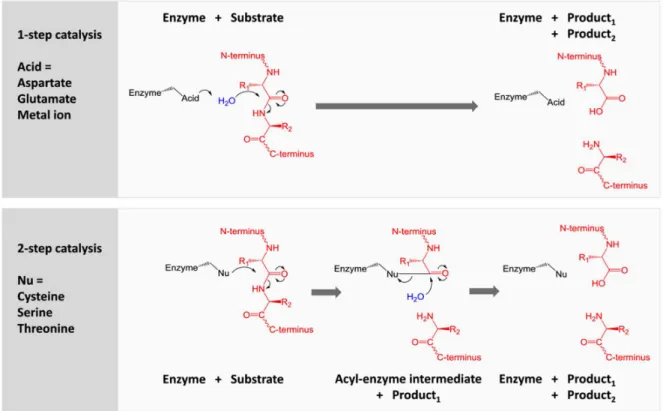
1993), cysteine- (1993), serine (1993) metallo- (1993), threonine- (1997), glutamic- (2004) and asparagine peptidase (2010). The cysteine, serine and threonine proteases possess a nucleophilic residue within their active site (thiol or alcohol function) that directly participates in peptide bond hydrolysis. In the case of aspartic and glutamic proteases, these enzymes benefit from the presence of acid residues (glutamic or aspartic acid) that activates a molecule of water participating in hydrolysis. In addition to a similar residue (Glutamic acid), Metalloproteases possess a zinc ion that acts as a Lewis acid. For each class of proteases, their environment in active site amino acids rules their catalytic mechanism (Figure 2).
Figure 2. One or two-step mechanism as function to environment in amino acids within protease active site. In the case of cystein, serine and threonine proteases a two-step mechanism takes place. Thus, the nucleophilic residue (thiol or alcohol group) performs a nucleophilic attack onto the carbonyl function, which results in the formation of a covalent adduct between the protease and the substrate protein
(acyl-5
enzyme intermediate). Noteworthy, two other residues within the active site (acid and basic residues) form a charge-relay network to polarize and activate the nucleophile: catalytic triad. The acyl-enzyme intermediate is then hydrolysed by an activated molecule of water.
In a one-step mechanism, a water molecule is activated by an acid residue that acts as a general base. Concomitantly, the activated water molecule proceeds to a nucleophilic attack on peptide bond carbonyl function leading to its hydrolysis. In the case of metalloprotease, a zinc ion (catalytic ion) binds to the oxygen carbonyl function thus increasing the electrophilic character of this latter.
The case of asparagine peptidase is very specific as they cleave themselves by an autoproteolytic reaction involving a glutamine residue.
2. Matrix Metalloproteinases
2.1. Discovery and MMPs classification.
In 1962, Gross and Lapiere described an enzyme responsible for the degradation of extracellular matrix proteins (ECM). Particularly, this enzyme displayed a collagenolytic activity during metamorphosis of
tadpole tail fin tissue explants 2. The identification of this protease named collagenase and matrix
metalloprotease 1 (MMP-1) later on, consituted the first milestone into a field that grew exponentionnaly in the following years, resulting notably in the characterisation of 23 MMPs in human. This name MMPs was based on the initial assumption that these proteases only cleaved the protein components of the extracellular matrix.
Since the pioneered works of Gross and Lapiere, researches on MMPs have focused on various aspects such as biochemistry (purification, activation, inhibition, structure), molecular biology (cloning of MMPs and
regulation of gene expression) and MMP biology in normal and pathological process 3. Among the most
significant steps, one can mention I) the first isolation of human collagenase from rheumatoid synovium 4,
II) the identification, isolation an sequencing of MMP-2 (gelatinase A) and MMP-3 in the 1970s 5, III) the
identification and characterisation of TIMP-1 (Tissue Inhibitor of metalloproteinase) as endogenous
metalloproteases inhibitors 6, IV) the emergence in the early 1990s of cysteine switch concept associated
to MMP activation 7 and V) the first crystal structure of collagenase catalytic domain in 1994 8, that paved
the way for the rational design of MMPs inhibitors.
Historically, the first members of the MMP family were named and classified according to several criteria: their sequence homology, the nature of the matrix extracellular components that they degraded in vitro and the number of years elapsed from the discovery of the first family member. In this respect, MMPs were initially divided into four subgroups: I) collagenases (MMP-1, 8 and 13), II) gelatinases (MMP-2 and 9), III)
6
stromelysins (MMP-3, 10 and 11) and IV) matrilysin (MMP-7). Over the years, this classification has largely evolved with the discovery of new family members and additionnal observations made regarding MMP substrates specificity. Further, these proteins show high variability at the level of their quaternary structure, with differences in the structure of their subunits.
To be classified as an MMP family member, the protease should meet the following requirements: I) cDNA has sequence homology to MMP-1, II) activation by proteinases or organomercurials, III) catalysis
dependent on zinc at the active site, iv) processing of at least one ECM component, and v) inhibition by ethylenediaminetetraacetic acid (EDTA), 1,10 phenathroline, and one of the TIMPS.
Due to the specificity of the substrate and the mechanism of action, MMPs is now divided into six groups:
Collagenases: MMP-1 (Collagenase I, interstitial, fibroblast); MMP-8 (Collagenase II, neutrophilic) and
MMP-13 (Collagenase III) degrade type I, II, III, VII, VIII, X collagen, gelatine, IL-1b, L-selectin, proteoglycans, proMMP-2, proMMP-9 and fibronectin.
Gelatinases: MMP-2 (in other: Gelatinase A or neutrophilic) and MMP-9 (Gelatinase B) degrade
collagen type IV, V, VII, IX, fibronectin, proteoglycans, plasminogen, act synergistically with collagenases.
Stromelysins/ matrilysin: MMP-3 (Stromelysin-1, Transylin); MMP-10 (Stromelysin-2) and MMP-11
(Stromelysin-3) digest the base membrane collagen, proteoglycans and glycoproteins of the extracellular matrix; whereas MMP-7 (Matrilysin-1, others PUMP-1) and MMP-26 (Matrilysin-2) degrade type I and IV collagen, gelatine, laminin, elastin, fibronectin, proteoglycans, MMP, pro-TNFα, E-cadherin.
Membrane type MT-MMP (membrane-type matrix metalloproteinases) characterised by the presence
of a C-terminal transmembrane domain that maintains the enzyme in the cell membrane structure. This group includes: MT1-MMP (MMP-14), which degrades type I, II III collagen, gelatine, fibronectin, laminin, vitronectin, proteoglycans, pro-MMP-2 and pro-MMP-13; MT2-MMP (MMP-15), MT3-MMP (MMP-16), MT4-MMP (MMP-17) and MT5-MMP (MMP-24) that activate pro-MMP-2; MT6-MMP (MMP-25), which degrades gelatine. Some of these enzymes (MT4- and MT6-MMPs) are membrane-anchored through glycosylphosphatidyl inositol (GPI).
Other metalloproteinases not included in the above groups, 12 (macrophage elastase),
MMP-18 (Collagenase 4), MMP-19, -21, -27 and epilisin MMP-28 were classified as "other" 9,10.
In recent peptidase nomenclature, MMPs are included in the M10A subfamily described as a broad family
of endopeptidases where enzymatic activity is determined by Ca2+ and Zn2+ ions.
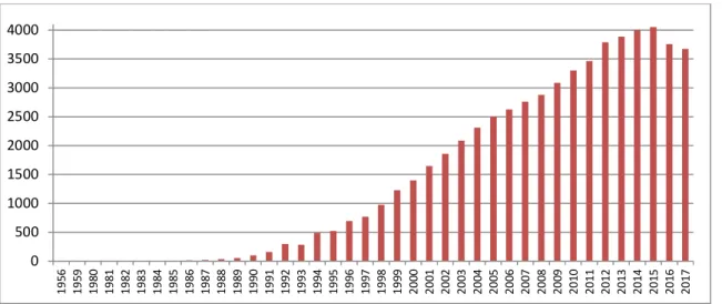
Although these proteases were discovered more than 50 years ago, researches on MMPs remain particularly active as attested by the increasing number of publications in this field since the mid-1980s.
7
Figure 3 Timeline – the number of publications about matrix metalloproteinases over the years.
2.2. MMPs belong to the metzincin family
MMPs belong to the metzincin family which is part of vast family of zinc metalloproteases. The metzincin superfamily is composed of five groups:
Astacins: subdivided into Meprin and BMP1 enzymes. They were originally named from the prototypical proteinases astacin which functions as a major collagenolytic enzyme in the digestive
tract of the crayfish Astacus astacus L. 11,12.
Adamalysins subdivided into ADAM (A Disintegrin and Metalloproteinase) and ADAMTS (A Disintegrin and Metalloproteinase with Thrombospondin Motifs), in the past called the snake
venom zinc endopeptidases.
Serralysins: proteolytic enzymes secreted into their environment by various pathogenic bacteria of
the genera Serratia 13, Pseudomonas 14, and Erwinia 15.
Pappalysins.
MMPs or Matrixines 16–18.
In the family of metzincines all members are characterised by the presence within their active site of
a catalytic zinc atom and a C-terminal consensus sequence HEXXHXXGXXH/D. This sequence contains
I) three zinc-binding residues: two conserved Histidine and a third one that can be either a Histidine or an Aspartate, II) a catalytic glutamate, and III) a strictly conserved glycine. Moreover, all metzincines share
a conserved methionine residue below the active site metal as part of a “Met-turn’’ 19,20.
0 500 1000 1500 2000 2500 3000 3500 4000 19 56 19 59 19 80 19 81 19 82 19 83 19 84 19 85 19 86 19 87 19 88 19 89 19 90 19 91 19 92 19 93 19 94 19 95 19 96 19 97 19 98 19 99 20 00 20 01 20 02 20 03 20 04 20 05 20 06 20 07 20 08 20 09 20 10 20 11 20 12 20 13 20 14 20 15 20 16 20 17
8
Figure 4. Metzincin superfamily members and structure of Matrixins family 21.
Each individual family distinguished by the residue following the third histidine zinc ligand in the motif, and the residues surrounding the methionine in the Met-turn.
2.3. MMPs possess a modular structure
In humans, 23 different MMPs and 24 encoding them genes have been identified. MMP-23 is encoded by two identical genes located on chromosome 1. As extracellular or membrane endopeptidases, metalloproteinases are multi-domain enzymes. All MMPs share a minimal structure composed of a signal peptide (S), a pro peptide domain (Pro) and a catalytic domain containing Zinc ion (Cat Zn).
In addition, subfamilies are categorized by other domains such as fibronectin-like repeats, C-terminal hemopexin-like domains, Ig-like domain and transmembrane domains.
Signal peptide directs MMPs to the secretory pathway.
Catalytic domain composed of approximately 170 acids, two zinc ions (one catalytic and one structural) and usually three structural calcium ions. As mentioned for all the proteases belonging to the metzincines
9
family, the catalytic domain contains a highly conserved HEXGHXXGXXH motif in a conserved AXMX sequence ("Met-turn") located below the active site. This methionine residue appears at the beginning of
a loop and is responsible for the proper structure around the catalytic zinc ion 22
Figure 5. Scheme of the zinc-binding motif and the Met-turn showing the common sequence motifs of the
metzincins. Variable segments are shown as dotted lines 20.
Pro-peptide domain is 80 amino acids long. It contains in its C-terminal part a conservative PRCGVDP motif with a cysteine residue, behaving like a fourth zinc ligand, preventing activation of the water molecule. MMPs activation requires to abolish the interaction between cysteine residue and catalytic zinc ion. This process is called the "cysteine switch" and takes place in two stages (see below 2.4.2 ).
Hemopexin domain possesses approximately 200 amino acids structured in four-blanded β-sheet. This domain plays a significant roles in substrate recognition, interaction with tissue inhibitors of
metalloproteinases (TIMPs), binding to the ECM or cell surface, MMPs internalization and degradation 22.
Linker region rich in proline, links the hemopexin domain (C terminal) to the catalytic domain. This flexible linkage helps to maintain the stable structure of the enzyme, and may also be important in recognition of some MMPs substrates.
Fibronectin domain insert in triplicate within the sequence of MMP-2 and 9 catalytic domains and mediates binding to gelatin substrate.
Transmembrane domain (type I or II) that can be prolonged by a very short cytoplasmic domain (Cyt) or by a signal domain allows the anchoring to cell membrane (MMP-14/16/23 and 24). MMP-17 and 25 anchors
10
Furin-like domain as a domain recognized by serine proteases allows the intracellular activation of MMPs. This motif is located between the prodomain and the catalytic domain.
Immunoglobulin-like motif, cystein-rich domain and pro peptide domain without cysteine switch-motif are characteristic of MMP-23.
Figure 6. Domain structure of matrix metalloproteinases family 23.
2.4. Regulation of MMPs activities
Since MMPs are able collectively to degrade all the protein constituents within extracellular matrix, their proteolytic activity has to be tightly controlled under normal conditions to prevent tissue destruction. Like other enzymes, the functioning of MMPs is regulated at different levels: from transcription and secretion to activation, inhibition and degradation (Figure 7).
11
Figure 7. Regulation of extracellular proteolysis 25.
2.4.1. Transcription
The first level of regulation of MMP activity is during translation and transcription of RNAs. This regulation is modulated by very specific signals which themselves are subjected to precise spatial and temporal control. Transcription of genes encoding MMPs is induced by growth factors: transforming growth factor (TGF-β), epidermal growth factor (EGF), platelet-derived growth factor (PDGF) and fibroblast growth factor (-FGF), hormones, inflammatory cytokines (interleukin-1, interleukin-6, tumour necrosis factor α TNF-α) and others. Most of them interact with protein activators of transcription AP-1 (protein transcription activator 1) and PEA3 (activator that enhances the expression of poliovirus genes) that lead to the initiation of transcription. Activation via these factors takes place through the phosphorylation cascade, dependent on the mitogen activating protein kinase (MAP-Kinase).
The activity of genes encoding some MMPs is also influenced by epigenetic factors such as DNA methylation and histone modifications. The expression level of MMP genes is also controlled by post-transcriptional
12
processes. TGF-β has been shown to increase the levels of MMP-2 and MMP-9 mainly by prolonging the half-life of these metalloproteinases in prostate cancer cells and human fibroblasts, and signals from α3β1 integrin stabilise MMP-9 mRNA. Similarly, cortisol enhances the stability of MMP-13 mRNA in rat
osteoblasts 26–28.
2.4.2. Extracellular activation and cysteine switch concept
Most of the MMPs are extracellularly secreted as inactive zymogen also called pro-MMPs that needs to be subsequently activated. Within the extracellular space, zymogens are activated through different mechanisms involving proteolytic enzyme (e.g. MMPs and serine proteases) and action of active forms of oxygen.
The N-terminal segment of zymogens possess a pro peptide with a molecular weight of 9-20 kD, depending on the type of metalloproteinases. This pro domain consists of three α helices connected by flexible loops. Contiguous to the third helix, a sequence with a cysteine residue (PRCGXPD) has been preserved in evolution, except in the case of MMP-23. As illustrated by Figure 8, the thiol group of the cystein residue
interacts with the catalytic zinc ion (Zn2+), maintaining the pro enzyme in its inactive state.
Figure 8. Activation of MMPs, the interaction of cysteine residue from the pro peptide with the catalytic zinc ion of
the catalytic domain 29.
The activation process is a two step-process that requires abolishing interactions between cysteine and zinc ion followed by pro domain removal upon proteolysis, resulting in active site unveiling. Various mechanisms can lead to such an activation:
1) Allosteric perturbation or posttranslational modifications that induces pro-MMP conformational changes
13
2) Modification of the thiol function integrity by physiological agents or non-physiological agents leading to destabilizing interaction between cysteine and zinc ion. This is often accompanied by an autolysis. 3) Direct cleavage of the pro domain by a other proteolytic activity.
The proteases responsible for pro domain processing are serine proteases (trypsin, chymotrypsin, plasmin) as well as other MMPs (-1, -2, -8, -9). In the case of activation by plasmin, the plasminogen Activator System (PAS) is required. The system consists of two activators of plasminogen (PA), urokinase (uPA) and a tissue type activator (tPA), that are membrane-associated. They both activate plasminogen into plasmin, that subsequently process proMMP at the cell surface. This mechanism is likely involved in the activation of
proMMP-3, proMMP-12 and proMMP-13 31–34.
The prodomain removal is commonly called in the literature the "cysteine switch" 35,36.
2.4.3. Intracellular activation
Most of MMPs are activated outside the cell, although it has been shown that for some members this activation can be carried out intracellularly in the Golgi apparatus. In this respect, It was demonstrated that proMMP-11 (stromelysin 3) is processed intracellularly by furin that specifically recognizes a RXKR or RRKR
sequence within the pro domain sequence 37. Besides MMP-11, MT-MMPs, MMP-23 and MMP-28 also
possesses a furin-like proprotein convertase recognition sequence 38. Once activated, MMPs are
extracellularly secreted.
2.4.4. Cell Surface activation involving TIMP-2
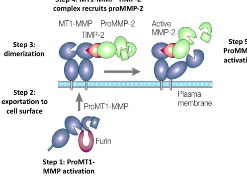
Contrary to what has been long considered, endogenous TIMP can also participate to MMPs activation. In the respect, the activation proMMP-2 by the combined action of MT-1 MMP and TIMP-2 constitutes one of the most characteristic examples. As illustrated by Figure 9, the proposed mechanism of activation is a multistep process.
First, MT1-MMP (blue) is activated intracellularly by furin and localizes to the cell surface through the transmembrane domain. A MT1-MMP dimers is formed through hemopexin-domain interactions. The amino-terminal domain of TIMP-2 (red) binds to the active catalytic domain of MT1-MMP. The MT1-MMP– TIMP-2 complex recruits proMMP-2 (green) by interactions of the carboxy-terminal domain of TIMP-2 with the hemopexin domain of proMMP-2. The 'receptor' MT1-MMP thereby brings proMMP-2 close to the
14
Figure 9. Schematic representation of two-step ProMMP-2 activation process by the combined action of
MT1-MMP (MT1-MMP-14) and TIMP-2 3.
2.4.5. Inactivation by endogenous inhibitors
The regulation of MMPs activity takes place not only through the mechanisms of their activation, but also in the processes of enzymes inhibition by protease inhibitors. Non-specific metalloproteinase inhibitors like α-2-macroglobulin and α-antiprotease are mainly responsible for the regulation of MMPs activity in plasma. Human α2-macroglobulin is a broad-spectrum proteinase inhibitor of tissue fluids and blood. It is a homo tetrameric macromolecule of 725 kDa which in able to inhibits almost all classes of endopeptidases by entrapping the whole enzyme. During the inactivation of MMPs by α macroglobulin, the first step of proteolysis leads to the formation of a covalent complex between these two partners, this complex is subsequently removed from circulation by endocytosis. Noteworthy, MMPs/α-macroglubolin complex is no longer able to degrade MMPs endogenous substrates but can always degrade synthetic substrates or interact with small ligands.
Natural and specific inhibitors of MMPs are proteins belonging to the group of tissue inhibitors of metalloproteinases (TIMPs). TIMP proteins are a family of 4 glycoproteins: TIMP-1, TIMP-2, TIMP-3 and
TIMP-4. They are characterized by a molecular weight ranging from 21 to 29 kD 39. TIMP 1, 2 and 4 forms
are soluble and they are detected in the blood serum, whereas TIMP-3 is insoluble and anchored in the
ECM 40. All TIMPs have similar folding with a N-terminal domain containing six cysteine residues assembled
in three disulphide bridges. Despite their structural similarity, TIMPs differ in their selectivity profile. TIMP1 Step 1: ProMT1-MMP activation Step 5: ProMMP2 activation Step 2: exportation to cell surface Step 3: dimerization Step 4: MT1-MMP–TIMP-2 complex recruits proMMP-2
15
inhibits all MMPs, particularly MMP-9, with the exception of membrane metalloproteases MT1-MMP,
MT3-MMPs, MT5-MMPs and MMP-19 41. In contrast, TIMP-3 specifically inhibits MMP-2, -3, -7 and -9.
As illustrated by Figure 10, the N-terminal segment of TIMP-1 structured around two disulphide bridges
between Cys1-Cy70 and Cys3-Cys99 interacts within the active MMP site through a hydrogen bonding network
and catalytic zinc atom chelation. Coordination of TIMP to the catalytic zinc displaces a zinc-bound water molecule present at the MMP active site, removing the nucleophile essential for peptide bond hydrolysis.
Figure 10. Crystallographic structure of MMP-3 catalytic domain (in grey) interacting with TIMP-1 (in red), (PDB code: 1UEA). Zoom of the N-terminal part of TIMP-1-Cys1 interacting within the catalytic domain of MMP-3, hydrogen bonds are shown in hatched form.
In vitro, this interaction is described of high affinity but remains reversible. Similar to MMPs, expression of TIMP genes is regulated by vasoconstrictor stimulators such as haemodynamic stimuli, oxidative stress, pro-and anti-inflammatory factors (cytokines or vasoactive agents). Disturbing the balance between MMPs pro-and
TIMPs is systematically associated with progressive pathological changes 42.
3. Biological functions of MMPs
Although latent forms of MMPs (pro-MMPs) have been shown to be involved in a few biological processes, a wide consensus in the literature attributes a functional role to MMPs under their active forms. MMPs have been originally described as proteases acting exclusively on extracellular matrix components with
consequences on tissue remodelling. It is now clear that MMPs are capable to process a wide range of
matrix and non-matrix substrates. Thus, MMPs degrade adhesion proteins, apoptosis mediators, receptors,
chemokines, cytokines, growth factors, and cell-to-cell linkage proteins. As a consequence, these proteases
are involved in numerous biological processes that go far beyond the simple remodelling of the extracellular matrix and should no longer be perceived as simple molecular scissors. To add another level of complexity to MMP biology, each MMPs does not present strict specificity for one single substrate. Accordingly, collagen
16
can be degraded by collagenases but also by MMP-14 (MT-MMP) and MMP-9. Another difficulty relies on
compensation phenomenon, very common in this group of enzymes 43. Further, MMPs are integrated into
a network of proteases. Thus, a single MMP can play a direct or indirect function during the evolution of biological process.
3.1. The physiological function of MMPs.
MMP are characterized by a broad spectrum of activity and are involved in various many different biological events including embryonic implantation, tissue morphogenesis, mechanisms of healing, control of the
immune response and cell death25,44,45.
As already mentioned, MMPs are able collectively to transform and degrade many components of the extracellular matrix (ECM) e.g. collagen, elastin, fibronectin, laminin, fibronectin, entactin, aggrecan,
osteonectin, vitronectin and tenascin 46. Degradation of ECM by MMPs can lead to the destruction of
adhesive interactions between the cell and the matrix, preventing the cell to receive signals from the environment and, consequently, introducing it into apoptosis. Since ECM is a reservoir of numerous growth factors, MMPs also participate in signals transfer from the ECM to the cells. MMPs regulate bioavailabity of VEGF (Vascular Endothelial Growth Factor) by cleaving matrix-bound isoforms of VEGF, a critical mediator
for specification, morphogenesis, differentiation, and homeostasis of vessels 47. Cutting off some ECM
proteins can also reveal inaccessible cryptic sites that can transmit signals or lead to products with completely new biological activity. For example, the migration of endothelium cells is triggered by a
MMP- 14-processed form of laminin 5 48,49.
3.2. Pathological function of MMPs
In the late 1960s MMPs overexpression was demonstrated in various pathological states such as cancer and
rheumatoid arthritis 3,50,51. Since these pioneered studies and beside their involvement in tumour
progression and metastasis 52,53, MMPs have been shown to play critical roles in pathologies with
inflammatory components including emphysema, asthma 54,55 and atherosclerosis 56–58, in cardiovascular
diseases 56–58, and in diseases of central nervous system 59.
Originally, MMPs were considered as solely overexpressed by cancer cells to support tumour growth and metastasis. However, in 1990 Paul Basset showed that Stromelysin-3 (MMP-11) activity could be also found
in stromal host cells surrounding the tumour, drastically upset this paradigm 60. Thus, MMP secretion is not
exclusively limited to cancer cells and host cells can also produce MMPs in response to changes in their environment. From such a study but also from those on MMPs knock out (KO) mice for which MMP genes have been invalidated, emerged the concept that MMPs could indeed support disease progress but could
17
targets while other are anti-targets. To add another degree of complexity, for one given MMP divergent functions can be observed depending on pathological stage. Retrospectively, these observations largely
explain the failure of clinical assays with broad-spectrum MMP inhibitors in cancer patients 63.
3.3. The specific case of macrophage elastase (MMP12)
In 1975, Z. Werb reported for the first time a proteolytic activity associated to murine macrophage. Under
thioglycolate-stimulation, murine macrophages secreted a proteinase able to degrade insoluble elastin 64.
A few years later, the same group isolated a protein that showed characteristic properties of zinc
metalloproteases, with inhibition by EDTA and TIMP-165. This enzyme was initially named murine
macrophage elastase (MME). In 1992 Shapiro’s group was able to isolate and identify MME cDNA and
definitely confirmed its membership to MMPs family66. As expected for MMPs, this enzyme of 54 kDa
possesses a pro domain, a catalytic domain with the characteristic sequence signature HEXXHXXGXXH and a haemopexin domain. Interestingly in solution, the pro domain turned out instable and only an active form
of 22 KDa corresponding to haemopexin domain removal was observed 66. Macrophage elastase was
further named MMP12.
The murine and human forms of MMP12 only possess 64% sequence identity. This is a rather surprising observation considering that percentages of identity between human and murine MMPs are usually around 95%. Similarly, percentages of identity between MMP12 and other MMPs vary between 33% and 49%, which is, again, a fairly low percentage. However, MMP12 coding sequence is located on 11q22 human chromosome, a location shared with many other MMPs.
As it is mainly expressed by macrophages, MMP-12 overexpression has been associated to inflammatory pathologies such as abdominal aortic aneurysm, atherosclerosis, rheumatoid arthritis, asthma, emphysema
or bronchitis, and chronic obstructive pulmonary disease (COPD) 54–56,58,67–70.
Like other MMPs, MMP12 can adopt ambivalent function depending on pathological context. As a consequence, MMP12 cannot be systematically considered as valuable therapeutic target. For instance, MMP12 expression in mouse melanoma cells resulted in reduction in early-stage tumour growth and a reduction in blood vessel formation, effects that seemed to be mediated by the ability of MMP12 to
generate angiostatin through cleavage of plasminogen 71. In patients with primary colorectal carcinomas,
MMP12 expression was associated with increased survival times 72. In those cases, MMP12 is clearly
a candidate anti-target.
Conversely, MMP-12 overexpression was associated to atherosclerotic plaque development and rupture.
In murine model of atherosclerosis, MMP-12-KO mice showed more stable plaques 61. MMP-12 seems to
18
cells 73. In human, the existence of a unique relationship between expression of MMP-12 and
atherosclerosis development remains to be established. However, the presence of MMP-12 has been linked
to instable atherosclerotic plaque 74,75
Shapiro’s group also demonstrated an antimicrobial activity for MMP12. Surprisingly in this case, the antimicrobial properties of MMP12 do not reside within its catalytic domain, but rather within the
heamopexin domain 76. More recently, the participation of MMP-12 in defence processes against viral
infections was also demonstrated77. Thus, during viral infection, active forms of MMP-12 secreted by
macrophages can translocate to the nucleus of virus-infected cells while a portion of these active forms remains outside of the cell (Figure 11).
Figure 11. In antiviral immunity, MMP12 can adopt a dual localisation associated to opposite functions 78.
Interestingly, this ubiquitous localisation is associated to opposite functions. Whereas nuclear MMP-12 active forms favour interferon- (IFN-) secretion essential for host protection, extracellular active forms cleave off the IFN- preventing an unchecked immune response. In this regard, a therapeutic intervention aiming to elevate systemic INF- level for improving antiviral response, should have to target selectively the extracellular active form of MMP-12 while sparing the intracellular one. Very recently, MMP12 was also
shown to downregulate INF- activity with consequences on resolution of acute inflammation 79.
Target
19
The case of MMP-12 is particularly enlightening and illustrates well the difficulty to unambiguously attribute to one given MMP a clear function during a pathological process, with an enzyme that potentially processes a wide diversity of substrates and can adopt opposite functions depending on its cell compartmentalization. This stresses the need to develop selective methods and chemical tools to document MMP spatial and temporal activation in various biological contexts. In other words, during the evolution of a pathological process it is critical to unambiguously determine which MMP(s) are present in their active form and which of them actually support disease development. Only these latter must be subjected to therapeutic intervention. This last point strongly suggests that highly selective MMPs inhibitor is the most favourable
therapeutic option 67.
4. Inhibitors of MMPs: chemical tools to investigate MMPs biological
function.
Originally, MMPs inhibitors (MMPIs) were developed without knowledge on MMP 3D-structure and directly derived from substrate sequences on which a zinc chelating moiety was conjugated. The first structure of
MMP-1 catalytic domain in complex with a synthetic inhibitor 8, allowed to take a step towards
structure-based design of synthetic inhibitors. Since this pioneered study, more than 200 MMPs structures (RX and NMR) have been deposited in the protein data bank (PDB) and several generations of inhibitors with improved selectivity profile have emerged within this well-defined structural context.
4.1. Structure-based design of MMP inhibitors: from broad-spectrum to
selective inhibitors
All the MMP catalytic domains share a marked sequence similarity, where the percentage of identical residues ranges from a minimum of 33% between MMP-21 and MMP-23, to a maximum of 86% between MMP-3 and MMP-10. The catalytic domain contains two zinc and three calcium atoms which are involved in the stabilization of the three-dimensional structure. Interestingly, no disulphide bridges are present. The second zinc atom (catalytic zinc) is located in the middle of the catalytic cleft crossing the protein from west to east (zinc ion in magenta, Figure 12).
20
Figure 12. Catalytic domain of hMMP12. A) In surface representation with S1’ cavity highlighted in red. B) In ribbon
representation withsheets in orange, -helix in cyan and S1’ loop in red.
In this standard orientation, peptide substrates of at least 6 residues bind on either side of the catalytic zinc ion in a conformation extending from the left (un prime subsites) to the right (prime subsites). The zinc ion, whose degree of oxidation is 2+ is in interaction with three histidine residues (Figure 12B). The -helix that
carries two of the three chelating histidine also contains the catalytic glutamate (Glu219 in the case of
MMP12). At the bottom of the catalytic zinc ion, a rather hydrophobic cavity open to the solvent on both
sides is present. This S1’ cavity constitutes an important element of variability between different MMPs with
outer wall of this cavity defined by a flexible S1 ' loop (in red in Figure 12B). Depending on MMPs, this loop
also called specificity loop varies in amino acid composition (number and nature of residues) 80. Thus, the
length of this loop can range from 9 (MMP-1, 9, 11 and 23) to 13 residues (MMP-17 and 25) resulting in S1’
cavity varying in volume. The volume of S1’ cavity volume is also influenced by the nature of the residue at
position 214 (with reference to MMP-1) on the -helix defining the rear wall of the S1' cavity. When this
residue is a leucine, which is the case for most MMPs, the S1’ cavity is relatively wide and deep. In the case
of MMP-1 and MMP-7, the presence in this position of an arginine (MMP-1) or a Tyrosine residue (MMP-7)
pointing to the interior of the S1’ cavity significantly reduces its internal volume. In an equivalent position,
a methionine and a glutamine residue also clog S1’ cavity of MMP-16 and MMP-11 respectively. Moreover,
21
comparison tools such as Principal Component Analysis (PCA) also revealed small topological differences
within S2 and S3 sub sites 81.
The particular topology of MMP active site has greatly influenced the design of synthetic inhibitors. Particularly, most synthetic MMPs inhibitors are competitive inhibitors that contain I) a zinc-binding group,
II) a P1’ hydrophobic side chain inserting within S1’ cavity, III) a peptide or pseudo peptide backbone
interacting within the catalytic cleft through hydrogen bonds network and IV) additional residues to bind further sub sites (Figure 13A).
Figure 13. A) Generic structure of the most representative families of MMPs inhibitors sharing a common
zinc-binding moiety (magenta), a hydrophobic P1’ side chain (red) and a peptide or pseudo peptide backbone to make
hydrogen bonds within catalytic site. B) Schematic representation of most standard zinc-chelating moieties found in structure of MMPs inhibitors.
Since the first generation of MMPs inhibitors whose structure was inspired from collagen sequence, several successive generations of inhibitors have emerged and can be divided in three big families. These families mainly differ in their binding within MMPs catalytic domains. The first family (Family I) comprises inhibitors interacting with the catalytic zinc atom and exploring only the prime subsites. The second family (Family II) clusters synthetic inhibitors whose structure was largely simplified with only a zinc-binding moiety and
a long P1’ hydrophobic chain inserting into the S1' cavity. The third family (Family III) is mainly constituted
by phosphinic pseudo peptides with a phosphinate function as zinc-binding group 82. By comparison with
Hydrophobic P1’side chain
Zinc-Binding Group Hydrogen bond donor or acceptor
S1’ cavity
A)
B)
S1’ cavity S1’ cavity
Hydroxamate Carboxylate Phosphinate
22
compounds from the two other series, phosphinic derivatives can probe the whole enzyme active site across prime and unprime subsites.
Among the factor that can modulate MMPIs selectivity, the chemical nature of the zinc-chelating moiety is critical. In this respect, the hydroxamate function as a bidentate ligand can be considered as a strong zinc chelator while the carboxylate or phosphinate functions as monodentate binder display lower avidity for
the zinc ion (Figure 13B) 83.Thus, inhibitors incorporating a hydroxamate function are very potent but
non-selective for MMPs. In this series of compounds, GM6001 (see annex A for structure) is even capable to
bind to zinc metalloproteases that do not possess any structural analogies with MMPs 84.
The incorporation of a weak binder such as carboxylate function within the structure of family II
compounds, combined with variation on P1’ side chain (variation in length and flexibility) resulted in the
identification of more selective MMPIs 82. Based on this approach, MMP-12 85 and MMP-13 86 selective
inhibitors were developed. Thus, by tuning the strength of interaction with the zinc ion, it was possible to
increase the contribution of P1’ side chain that could generate more specific interaction within S1’ pocket.
This concept has been pushed further through the development of MMPIs inhibitors that do not possess
any zinc binding-group and only interacted within the S1’ cavity. In this series highly selective MMP8 and
MMP-13 inhibitors were identified 87,88.
The phosphinic pseudo peptides also possess a weak zinc-chelating moiety and through its unique capacity to interact within the entire catalytic cleft offer further opportunities for selective interactions compared to inhibitors with a hydroxamate or carboxylate zinc-binding group. In this respect, phosphinic pseudo
peptides were considered as a good starting point for the development of MMPs selective inhibitors 80,89.
4.1.1. RXP470.1 as a highly selective inhibitor of MMP-12
Through the screening of focused libraries, RXP470.1, a pseudo tetra peptide was identified by our group as the first potent and highly selective inhibitor of MMP-12 (Figure 14) 90.
This phosphinic pseudo peptide, incorporates a bulky side chain in its P1’ position and two glutamate
residues in P2’ and P3’ positions. In vitro, this molecule was characterised by a high potency toward hMMP12
catalytic domain (Ki = 0.19 nM) and was 2-3 orders of magnitude less potent toward other examined MMPs. Further, RXP470.1 behaves as a very weak binder toward other metalloproteases such as TACE (Tumour necrosis factor-Alpha Converting Enzyme belonging to ADAM family), NEP (neutral endopeptidase) and ACE (Angiotensin-Converting Enzyme).
23
Figure 14. RXP470.1 structure and its selectivity profile towards a set of human metalloproteases. The 1/Ki (M) values are reported in logarithm scale. The higher is the bar the higher is the inhibitor potency. Each MMP is identified with a colour code and hMMP12 is represented by a black bar.
Our group recently reported a study combining crystallography and isothermal titration calorimetry in order to clarify the role of P1’, P2’, P3’ positions in the excellent affinity and selectivity of RXP470.1 for MMP-12 91.
Through the resolution of RXP470.1/hMMP12 complex crystal structure, RXP470.1 binding mode was first confirmed.
As illustrated by Figure 15, the phosphinate function interacts in a monodentate manner with catalytic zinc
ion. The P1’ isoxazol side chain inserts deeply into the S1’ cavity, potentially generating several Van der
Waals contacts with side chains of S1’ loop. Further, distance measurements between RXP470.1 and protein
backbones were compatible with hydrogen bonds. Surprisingly, no evident interactions involving the two
glutamate residues were observed within S2’ and S3’ regions.
Despite a multitude of interactions potentially generated upon RXP470.1 binding to hMMP12, calorimetric studies revealed that RXP470.1 binding is not characterized by a large enthalpy component. Indeed, the formation of RXP470.1/hMMP-12 complex is mostly entropically driven, which was ascribed to hydrophobic
24
Figure 15. Crystal structure of RXP470.1 in interaction with hMMP12 catalytic domain (PDB: 4GQL, 1.15 Å). hMMP-12 is represented in grey (cartoon representation), RXP470.1 in yellow stick, calcium and zinc ions are in green and magenta spheres respectively. Distances compatible with potential hydrogen bonds are highlighted with red dot lines. Catalytic glutamate is bleu stick.
This study has also revealed a pH dependence upon RXP470.1 binding, with a marked shift in association constant value (Ka) when pH raised from 6 to 7 (Figure 16A). This pH dependence was associated with a proton uptake from the buffer upon inhibitor binding (Figure 16B). This proton linkage was postulated to be necessary for neutralizing the two negative charges of ionized phosphinate (pKa= 1.5) and catalytic glutamate 219 (pKa= 4).
The analysis of crystallographic structure showed a very short distance separating one oxygen from the phosphinate (PO ext) and one from Glu219 (Figure 16C), a distance compatible with hydrogen bonding. Furthermore, calorimetric titration measurements were carried out on MMP-12 E219Q mutant and confirmed the involvement of Glu219 in the protonation phenomenon induced by RXP470.1 binding. The importance of zinc-chelating moiety in selectivity profile was very recently discussed through the development of RXP470.1 analogues within which the phosphinate function was replaced by
a hydroxamate or a carboxylate zinc-binding moiety 92. This study first showed that parent RXP470.1
remained the most selective compound towards MMP12 while its hydroxamate version was more potent but not selective. Furthermore, modifying the nature of the zinc-binding group seems to impact inhibitor positioning and dynamics within the active site, with potential impacts on its selectivity profile.
25
Figure 16. A) Change in RXP470.1 affinity toward MMP12 as function to pH, Assocation constant (Ka) is in logarithm scale and binding measurements were performed in single buffer (20 mM ACES). B) Schematic representation of proton uptake upon phosphinic inhibitor binding, C) Zoom on the catalytic zinc ion region catalytic, zinc is in
magenta; RXP470.1 in yellow stick and glutamate 219 in cyan 91.
Overall, these two recent studies suggest that at least two main parameters govern the excellent selectivity
of RXP470.1 toward MMP12: the nature of its zinc binding group and its long P1’ side chain interacting
within S1’ cavity. Importantly, these studies also stressed that it was possible to modulate RXP470.1 potency
as function to pH without affecting its selectivity profile.
4.1.2. RXP 470.1 as a chemical tool to study MMP-12 in vivo
Considering its excellent selectivity profile, RXP470.1 appeared as an ideal tool to study MMP-12 in preclinical models in which this protease was susceptible to play a critical role. In addition, it was previously demonstrated in the lab that phosphinic pseudo peptides were particularly stable in vivo with no apparent
toxicity 93–95.
RXP470.1 target MMP-12 within atherosclerotic plaques.
As mentioned above, certain MMPs are overexpressed within atherosclerotic plaques and can contribute to their destabilization through ECM degradation. However, the link between MMP activity and plaque rupture is not so easy to establish. Thus, it has been shown in hypercholesterolemic mice that MMP-3 and
26
MMP-9 had a plaque stabilizing effect while MMP-12 a detrimental one 61. In this context, RXP470.1 was
assessed in a mice models of atherosclerosis and its effect on plaques progression and morphology was
investigated 96.
To evaluate the effect of RXP470.1 an intervention protocol was chosen. In this respect, mice were fed a high-fat rodent diet for 8 weeks to develop mature atherosclerotic plaques. At this stage, RXP470.1 was administrated by using osmotic pumps over a period of four weeks. This mode of administration allowed circumventing rapid blood clearance of RXP470.1 while maintaining a constant plasmatic concentration of 200 nM.
Figure 17. Effect of RXP470.1 treatment on atheroma plaque size 96.
Within their brachiocephalic arteries, animals treated with RXP470.1 showed similar plaque sizes to those in control group after 8 weeks (Figure 17). Conversely, untreated animal (Ctrl 12 week) presented plaques of larger size. Thus, RXP470.1 was able to block plaques progression. In addition, RXP470.1 was also capable to modify plaques composition by decreasing their content in marker of instability (necrotic core, foam cells), thus inducing a stable phenotype. Within atherosclerotic plaque, RXP470.1 also significantly reduced elastinolytic activity. Importantly, effects of this inhibitor on plaque development and composition were comparable to those observed in MMP-12 KO mice.
Taken together, these data strongly suggested that RXP470.1 was able to target MMP12 in vivo and confirmed the detrimental role of MMP12 in plaque development and rupture.
RXP470.1 can target extracellular MMP12 during viral infection.
Recently, RXP470.1 was also shown to be able to block MMP-12 activity in model of viral infection 77. In
27
and further translocate to the nucleus where it activates interferon- transcription. Remarkably, a part of MMP-12 remains outside at the cell surface to process circulating interferon-, thus modulating antiviral response. In the perspective of antiviral therapy, RXP470.1 was evaluated for boosting systemic IFN-α level. Infected mice by coxsackievirus type B3 were exposed on continuous infusion of RXP470.1 using minipumps over 7 days.
Figure 18. A) A/J mice infected with coxsackievirus type B3 were treated with MMP-12 inhibitor RXP470.1 or saline by continuous minipump infusion for 7 d. ELISA detected plasma IFN-α levels after 96 h of virus infection. Uninfected control animals B) Effect of extracellular MMP-12 inhibition on body weight during coxsackievirus type B3 infection. C) Coxsackievirus replication, as detected by in situ hybridization of the viral RNA genome (+ sense stained blue) and
replication intermediates (− sense) in pancreas from infected mice 77.
This treatment resulted in highly elevated plasma IFN-α levels (Figure 18A), and reduced morbidity. Moreover, viral replication was also abolished on day 7, resulting in approximately 50% reduction in viral load (Figure 18C).
These data further validated the RXP470.1 capacity to target MMP12 in vivo. More importantly maybe, they also suggested a marked preference of this inhibitor for the extracellular portion of MMP-12, a property that can be explained by RXP470.1 global negative charge preventing its cellular internalisation.
The MMP12 targeting capacity of RXP470.1 was also demonstrated I) in a mouse model of rheumatoid
arthritis 97, II) in a model of cardiac dysfunction in which the critical role of MMP12 in inflammation
resolution was demonstrated 98, III) during lung tumor propagation 99 and IV) in an acute model of
inflammation 79.
In all these preclinical studies, RXP470.1 can be considered as a valuable chemical probe able to indirectly document the presence of MMP12 active form in vivo. By combining its use with models in which its privileged target has been invalidated (KO), it allows better deciphering the role of MMP12 in different pathological contexts.
28
More generally, selective MMPs inhibitors are particularly useful for validating or not MMPs as relevant target for therapeutic intervention. However, they cannot inform precisely about spatial and temporal activation of these proteases. Considering the MMPs ambivalent function during disease progress, this aspect may turn out critical to define the most appropriate therapeutic window for treating diseases. In this respect, imaging strategies as well as proteomic and chemical biology approaches allow to take a step in this direction.
5. Chemical probes for detecting MMPs activation in complex
proteomes
Existing methods to detect MMPs within complex tissues include genomic techniques to measure mRNA, immunoassays, gelatin zymography and in situ zymography. Although largely explored, these methods present evident limits since they cannot differentiate between TIMP-complexed MMPs (inactive) and active MMPs. To solve these limitations, several chemical probes has been developed. Based on their chemical structures they can be divided into two main categories: the ligand-derived and substrate-derived probes.
5.1. Imaging of MMPs activity with substrate- and ligand-derived probes
Substrate-derived probes are composed of characteristic sequence recognized by targeted protease. Most of them are quenched near-infrared fluorescent probes possessing a fluorophore whose fluorescent signal is abolished by a second fluorophore in close proximity (auto quenching) or by a quencher (Figure 19). The
intact probe isoptically silent and becomes highly fluorescent upon protease-mediated activation.
Figure 19. Schematic representation of MMPSense agents. Two quenched fluorophores are separated by a cleavable linker in the native state. MMPs recognize the cleavable linker and once the agent is cleaved, the fluorophores
29
produce signal. The agents also contain a pharmacokinetic modifier (PKM) selected to provide optimal attributes for in vivo imaging.
By nature, these probes can detect protease in very low amount due to signal amplification by enzyme turnover. Among the most representative substrate-derived probes developed in the literature, activatable
optical probes MMPSenseTM 680 allowed a real-time monitoring of MMPs activity in tumour 100 and within
atherosclerotic plaques 101.
Despite their high sensitivity and low background noise in vivo, these probes suffered from some disadvantages including long-time imaging and signal attenuation due to diffusion within the tissues. This last point has been partly overcome though the development of probes connected to cell-penetrating
peptides that induce their cell internalisation, thus limiting their tissue diffusion 102.
More questionable is their ability to selectively target MMPs in vivo 103. Indeed, such substrate-derived
probes can be processed by other proteases present locally at much higher concentration than that of MMP, yielding a signal that cannot be attributed unambiguously to targeted protease. In principle, the combined use of activatable probes with MMPs inhibitors may address this limitation. Nevertheless, considering the small repertoire of highly selective inhibitors, this strategy is not fully suitable for the unambiguous detection of MMPs. Finally, in pathological contexts where circulating MMP activity has been
evidenced 104–106, substrate-derived probes may turn out unstable within the blood stream leading to poor
target/non-target contrast when imaging diseased tissues.
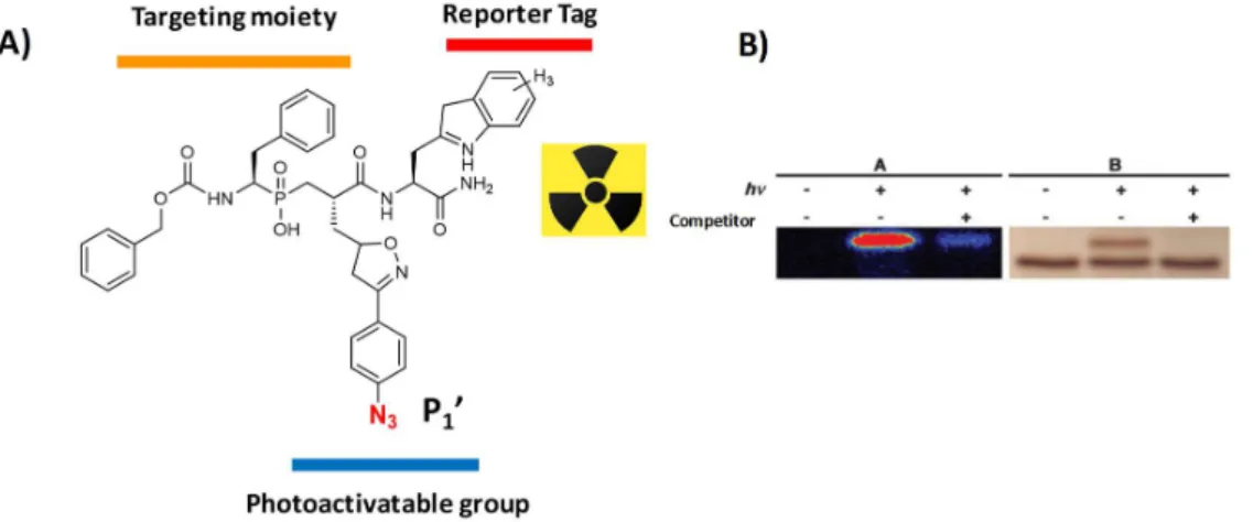
These stability issues can be overcome, at least in part, by probes possessing an inhibitor-derived scaffold. Such ligand-derived probes are composed of a MMP-targeting moiety conjugated to a reporter group for imaging modalities (Figure 20A). Among the most successful candidates, RP805 as a SPECT tracer is able to
selectively accumulate within tissues overexpressing MMPs under their active form 107.
Similarly, our group has recently developed a selective RXP470.1-derived tracer able to target MMP12 (Figure 20B) in a mouse model of aneurysm (manuscript in preparation).
Overall, although capable of visualizing in vivo MMPs activity, these probes do not provide any direct evidences as to the presence of MMPs under their active form. This can be only achieved through functional proteomic strategies, with activity-based protein profiling as the most effective one.
30
Figure 20. Structure of SPECT contrast agents targeting MMPs. A) Structure of RP805 composed of a broad-spectrum targeting moiety and a reporter tag B) RXP470.1-derived probes conjugated to a reporter Tag.
5.2. Activity-based profiling of MMPs
5.2.1. The concept of activity-based protein profiling
Activity-based protein profiling (ABPP) aim to analyse the functional state of proteins within complex biological samples. In the late 1990s, the Cravatt’s group was the first to report a proteomic method using chemical probes able react in a mechanism-based manner with enzyme under their functional state within
complex proteomes 109. A typical Activity-Based probe (ABP) is composed of I) a reactive warhead, which
reacts in a covalent manner with residues (electrophilic or nucleophilic residues) within enzyme active site, II) a targeting moiety that imposes selectivity upon the reactive group for a specific subset of enzymes and III) a detectable group for subsequent analyses (Figure 21).