HAL Id: hal-00509715
https://hal.archives-ouvertes.fr/hal-00509715
Submitted on 15 Aug 2010HAL is a multi-disciplinary open access
archive for the deposit and dissemination of sci-entific research documents, whether they are pub-lished or not. The documents may come from teaching and research institutions in France or abroad, or from public or private research centers.
L’archive ouverte pluridisciplinaire HAL, est destinée au dépôt et à la diffusion de documents scientifiques de niveau recherche, publiés ou non, émanant des établissements d’enseignement et de recherche français ou étrangers, des laboratoires publics ou privés.
Two-dimensional gel electrophoresis in proteomics: Past,
present and future.
Thierry Rabilloud, Mireille Chevallet, Sylvie Luche, Cécile Lelong
To cite this version:
Thierry Rabilloud, Mireille Chevallet, Sylvie Luche, Cécile Lelong. Two-dimensional gel electrophore-sis in proteomics: Past, present and future.. Journal of Proteomics, Elsevier, 2010, 73 (11), pp.2064-77. �10.1016/j.jprot.2010.05.016�. �hal-00509715�
Two-dimensional gel electrophoresis in proteomics: past, present and future Thierry Rabilloud1,2, Mireille Chevallet2, Sylvie Luche2,3 , Cécile Lelong 2,3
CNRS UMR5092, Biochemistry and Biophysics of Integrated Systems, CEA Grenoble, iRTSV/BSBBSI, 17 rue des martyrs, F-38054 GRENOBLE CEDEX 9 DSV/iRTSV/LBBSI, Biophysique et Biochimie des Systèmes Intégrés, CEA-Grenoble, 17 rue des martyrs, F-38054 GRENOBLE CEDEX 9, France
Université Joseph Fourier, UMR CNRS-CEA-UJF 5092, CEA-Grenoble, 17 rue des martyrs, F-38054 GRENOBLE CEDEX 9
Correspondence to
Thierry Rabilloud, iRTSV/BBSI CEA-Grenoble, 17 rue des martyrs, F-38054 GRENOBLE CEDEX 9 Tel (33)-4-38-78-32-12
Fax (33)-4-38-78-44-99
Abstract
Two-dimensional gel electrophoresis has been instrumental in the birth and developments of proteomics, although it is no longer the exclusive separation tool used in the field of proteomics. In this review, a historical perspective is made, starting from the days where two-dimensional gels were used and the word proteomics did not even exist. The events that have led to the birth of proteomics are also recalled, ending with a description of the limitations of two-dimensional gels in proteomics. However, the advantages of two-dimensional gels are also mentioned leading to a critical description of how and when to use them best in a proteomics approach. Taking support of theses advantages (robustness, resolution, and ability to separate entire, intact proteins), possible future applications of this technique in proteomics are also briefly mentioned.
1. From mute star maps to protein buzz: Two-dimensional electrophoresis and the birth of proteomics
SDS electrophoresis in its modern form was introduced in the early 70's [1] and soon became very widely used by protein biochemists. However, analysis of complex cellular extracts by this method made obvious the fact that the resolution was far from being sufficient to separate the many protein components of such extracts. To increase the resolution to a sufficient extent, it was necessary to couple two independent separations. At that time (and this is still the case) it was obvious that the separation best able to complement SDS electrophoresis was isoelectric focusing, and this was the coupling that researchers in protein separation tried to achieve. One of the first reports of successful two-dimensional electrophoresis coupling IEF to SDS PAGE was published in 1974 [2] but go relatively unnoticed by the community, probably because of two features. The first one was the inclusion of the sample within the IEF tube gel, which is a quite efficient but slightly cumbersome process. The second was the detection method chosen, i.e. the classical Coomassie blue staining in alcohol-acid mixtures. The poor sensitivity of the method led to nice but rather "empty" 2D images, and therefore not that impressive. The situation changed dramatically the year after, with the seminal paper of PH O'Farrell [3]. This paper coupled better technical choices (loading of samples on top of IEF gels), a very detailed description of the protocol, and detection by autoradiography, showing at one hundreds of protein spots on a single gel. Many researchers got very impressed by the results and underlying power of the method, so that 2D electrophoresis spread quite widely and quickly around the world. This is clearly shown by the citation index of the paper, gathering more than 1500 citations from 1976 to 1980. As soon as that time (late 70's very early 80's), the first applications of what was to become proteomics took off, with applications to cell biology [4, 5] and clinical biology [6], with the first ancestor of the human proteome project [7].
However, it must absolutely be stressed that protein identification at that time was quite an ordeal. The main methods were comigration with purified proteins [8] and blotting with antibodies [9, 10]. Thus, it was possible to say where was protein X (purified or with an available antibody) on the gels, but it was very difficult to know who was spot Y. For example, it took several years to know that cyclin/PCNA, identified as a cell cycle dependent protein [11] was indeed a subunit of DNA polymerase delta [12].
In the absence of readily-available protein identification tools, lot of attention was brought to the use of quantitative protein patterns as identifiers of cellular states via correlation analyses. It is often forgotten that statistical analysis in proteomics did not start a few years ago by getting inspired from transcriptomics, but long before the word proteomics was even coined, and in fact some 20-25 years ago. Indeed, computer analysis included both gel image analysis [13-16], but also strong data analysis with multivariate methods [17-22].
However, it was not by chance that one of the first 2D gel analysis system was name TYCHO [14], after the name of the famous astronomer (and alchemist) Tycho Brahe, as 2D maps were much like silent star maps for more than ten years (1977 to 1987). In addition to this dearly-resented lack of universal protein identification system, 2D
electrophoresis was plagued at that time with major reproducibility problems, and was much more an art than everything else. New practitioners of the field may not realise that the IEF gels were soft unsupported tube gels (typically 4% acrylamide) that were to be extruded from the glass tube containing them for the first dimension in order to be loaded on the second dimension slab gel. This was quite a difficult process, and a lot of non-linear deformation of the tube gel took place there. Ending with lengths variations of 10-20% was not uncommon, and this was highly detrimental to spot positional reproducibility. Moreover, IEF with carrier ampholytes is by itself a process that must be carefully controlled to achieve reproducibility. Carrier ampholytes-based pH gradients are not really stable, and prone to cathodic drift, i.e. progressive loss of the basic portion of the gradient, so that the migration profile (in Volts and Volt.hours) had to be carefully controlled. Moreover, the fine profile of the pH gradient is dependent on the precise composition of the carrier ampholytes, and this varies from batch to batch, leading to long-term irreproducibility of the IEF separations.
Despite these difficulties, exquisite experiments could be devised, as the demonstration of the expression of several actin forms in a single cell [23]. In addition, important dimensions of our current proteomic knowledge root from that period, such as the number of proteins present in a mammalian cell [24] or the knowledge that serum proteins are extensively modified [8].
The best was to come in the last 80's, with two key events for the development of two-dimensional electrophoresis in biology. The first key event was the introduction of immobilized pH gradients in the first dimension. By construction, immobilized pH gradients eliminate the reproducibility problems associated with carrier ampholytes-based pH gradients. However, initial attempts to adapt them for 2D electrophoresis, either in the tube gel mode [25] or via a gel slab [26] were not very successful, and the final solution, i.e. the now classical gel strips, was introduced later [27]. Further work was necessary to ensure good interfacing of the IPG strips with the SDS slab gel [28], but a stable protocol was soon available for the community [29, 30].
With the establishment of iPGs, 2D electrophoresis ceased to be a difficult art to convert only into good craftsmanship. There was a real quantum leap in reproducibility, going up to interlaboratory studies with a positive outcome [31, 32], something that was just unbelievable a few years before.
The real change of paradigm, however, came with the long-waited interface of 2D gels with protein microanalysis techniques. This was some years before mass spectrometry entered the scene, and obtained through the progress of Edman sequencing. The late 80's saw a blossoming of microsequencing applied to gel-separated proteins, either directly N-terminal sequencing [33], easy to perform and sensitive (only 1-2 picomoles required) but requiring a free N-terminus, something not often available in eukaryotic proteins. To bypass this limitation, internal sequencing protocols were developed, either from blots [34], [35] or directly from stained gels [36, 37]. This process of internal sequencing was more sample-consuming (around 50-100 picomoles required) and technically difficult, requiring microseparation of peptides on narrow-bore HPLC, collection of individual peptides by UV trace and sequencing of these individual peptides. In many studies, obtaining 50 picomoles of the protein(s) of interest was not an easy task, but was still achievable by using the high loading capacity of IPG gels [38-40], and devices to
pool the proteins extracted from several gel spots [41, 42]. 2. The take-off of proteomics
This possibility to be able to analyze spot of interest coming from 2D gels was a crucial change of paradigm, and the real start of proteomics. At those times where no complete genome was published yet, Edman sequencing provided enough information to look for homologs, or to devise oligonucleotides for screening DNA libraries. It was also the starting of brief golden age of 2D gel images databases [43-47]. To be useful to the community, 2D gel image databases must contain enough information, and not be just descriptive. Therefore, generic methods for obtaining information on proteins must be available. However, if these methods become easy and widespread, every laboratory can make its own identifications, and the added value of a 2D gel image database decreases. This is why the development and use of 2D gel databases culminated at this period, where protein identification was feasible but not very easy, i.e. in the early 90's.
Around the middle of the 90's, a quantum leap in protein identification arrived with the introduction of mass spectrometry-based methods. The first methods were based on peptide mass fingerprinting [48-51]. In order to work, these methods need a pure protein, or at least a mixture where one protein dominates by far the other ones in a quantitative point of view, something that 2D gels provide rather easily, especially when narrow pH ranges (here again available through the IPG technology) are used [52]. Compared to Edman sequencing, peptide mass fingerprinting brings less information on sequences, but some on putative post-translational modifications, and is by far more productive and less protein consuming. Even at these rather early days of peptide mass fingerprinting, the demand was in the low picomole range, i.e. at least one order of magnitude below the one of internal Edman sequencing, and the analysis time was counted in hours of work, not days, which allowed for a much improved productivity in proteomics.
Even though the coining of the word "proteomics" as simultaneous with the onset of practicable mass spectrometry methods in the field, it was not the true birth of proteomics. However, it was its first real blossoming, and numerous laboratories used this combination of 2D gel electrophoresis and peptide mass fingerprinting to carry out proteomics work in various areas of biology, and also for feeding databases [53-58] . At that time, many researcher, although conscious that some challenges remained [59] , were very confident that this combination of tools would be able to resolve complete proteomes [60], but it soon appeared that this would not be the case.
3. Touching the limits of 2D electrophoresis, and the creation of alternate methods. Because of this intensive and worldwide effort using 2D electrophoresis and mass spectrometry as the core tool, many data were accumulated and analyzed, and it soon became obvious that it was always the same types of proteins that were found again and again, and the same types that were always missing, i.e. the low abundance and the hydrophobic proteins [61, 62].
In order to improve the resolution of hydrophobic proteins, many efforts were devoted to improve protein solubilization under the conditions prevailing in the IEF dimension. This included changes in the chaotropes used in IEF [63] and also in the
detergents used for this step. Better solubilization for membrane proteins in 2D gels had been identified as a problem since the very beginning of the technique [64], and some improvements had been proposed over the years [65-67]. However, the increasing evidence of the problem prompted several groups to use various types of IEF-compatible detergents to alleviate this problem [68-71]. There was some success, i.e. demonstration of membrane proteins in 2D gels. However, it is fair to say that the problem of hydrophobic, and especially membrane proteins on 2D gels is largely unsolved [72-74]. Moreover, as this problem is clearly correlated with the IEF dimension and to the chemical conditions prevailing at this step (low ionic strength, no ionic detergents) [75], it is quite obvious that this will remain a built-in problem for all IEF-containing 2D electrophoresis systems. Thus, for analyzing membrane proteins, IEF-free separation systems were required. However, as the IEF/SDS combination is the only one which separation power matches the complexity of proteomes, this meant in turn that IEF-free separations would only lead to protein mixtures in the fractions arising from the separation, so that identification techniques able to cope with mixtures must be used. Fortunately enough, this had been worked for some years by developing the use of tandem mass spectrometry [76-80]. Thus, efficient IEF-free proteomic schemes could be devised, using peptide separations only, as in the shotgun approach [81, 82], or a combination of protein SDS electrophoresis followed by a peptide separation [83] [84]. In fact these setups proved able to analyze the hydrophobic proteins refractory to classical 2D gels. The problem of the low abundance proteins is also more acute in 2D gel-based proteomics than in these other setups for several reasons.
The first one is the simple fact that 2D gel-based proteomics is the only proteomic setup in which there is a readout before mass spectrometry. In other words, 2D gels are not gridded blindly with each and every piece of gel sent out for digestion and MS analysis. Thus, their performance, and especially their ability to detect and quantify low abundance proteins, is dependent on a protein visualization method after electrophoresis. Although modern high-sensitivity detection techniques (silver stain and fluorescence) are able to operate in the low nanogram range, with fluorescence-based detection being linear over several orders of magnitude, there is no detection method able to cope with the enormous dynamic range (i.e. quantitative ratio between the rarest protein expressed in a sample and the most abundant one) present in most biological samples [85] and even worse in biological fluids [86]. The second reason, going along the same line, is the distribution of the protein abundances, i.e. the proportion of proteins expressed at a rather high concentration vs. the proportion of proteins expressed at low concentrations. Such figures are much more difficult to obtain, but have been thoroughly investigated in yeast [87], and the results are appalling. Of the almost 4000 gene products that could be quantified, 130 gene products account for half of the protein content in yeast cells. The 10% most-expressed gene products account for 75% of the protein content, and the 2/3 less-expressed proteins account for only 10% of the protein content. Thus, being limited in the detection of low abundance proteins also means leaving a large proportion of cellular proteins out of he analysis.
There is an obvious countermeasure to alleviate this problem, which would be to load more sample, taking advantage of the huge capacity of 2D gels [38-40], and thus bringing many more proteins above the detection limit. But here the third
limitation comes to play to keep the usefulness of this high-loading approach in check. This limitation is linked to gel crowding and is linked to the protein abundance distribution. As 2D gels are able to resolve many modified forms of the proteins, the modified forms of the high abundance proteins will occupy precious separation space on 2D gels [59]. Consequently, loading much more sample will result mainly in completely obscuring some zones, leading in no added performance for the detection of the low abundance proteins in these zones [24]. A possible retort would be to use giant gels with a much increased resolution and capacity [88], but this technology is rather difficult to use due mainly to the fragility of the giant gels.
As this problem is linked mainly to the ability of 2D gels to separate modified forms, mainly through the IEF dimension, it is easy to understand that this problem is much more severe in this configuration than in IEF-free methods.
Last but not least, it should be stressed that 2D gels have a rather moderate overall yield [89], with important losses at the IEF stage and during equilibration between the IEF and SDS dimensions.
However, it shall not be derived from this section that 2D electrophoresis is the only proteomic method with a poor yield and unable to deal with low abundance proteins. All proteomics setups bump into the rare proteins problem, as exemplified by the fact that no proteomic method has proven able to analyze comprehensively a complex biological sample (even a bacterium). Apart from the problem of hydrophobic proteins, which is really a built-in weakness of 2D gels, 2D gels just perform differently from other proteomic setups. Although it is true that non-IEF proteomic setups perform better for low abundance proteins, it should be noted that on any sample type, 2D gels are still able to analyze protein that escape analysis by other methods, as shown by the examples of nucleolus proteomics [90, 91], or by the detection of cytokines in secreted proteins [92].
Instead, it should be analyzed that the weaknesses of 2D gels are now well-known because this is the oldest technique, used by a wide community of researchers who have done their homework and investigated honestly the limits of their tools. Because of their more recent introduction, other proteomic setups have not been subjected to the same testing and their limits are not as well- and widely-known yet. And despite these now well-known limitations, 2D gels still offer may attractive features that can be very useful in proteomics-based research.
4. Using the strengths of 2D gels in modern proteomic research
In this section, we will try to sort out the major strengths of 2D gels and to exemplify how they can be used for the benefit of proteomic research.
4.1 Robustness and technical confidence
In fact, because of the hindsight developed by the proteomics community on 2D gels, the drawbacks of 2D gels are well-known, but their advantages are also very well-known. One of the major advantages of 2D gels in proteomics lies in the robustness of the technique. As mentioned earlier, this robustness has been tested thoroughly [93], even in interlaboratory comparisons [31][32], and the influence of the various parameters on the intralaboratory reproducibility have also been investigated
[94]. In fact, the most critical variable nowadays are no longer in the 2D gel process per se, but rather upstream and downstream, i.e. sample preparation and image production and analysis.
As to image production, most often made now by protein stains, it has made enormous gains in reproducibility over the more than 30 years period in which scientists have run 2D gels. Modern detection methods, such as fluorescent stains [95, 96] colloidal Coomassie blue [97], and even modern silver staining [98], where development goes to an end-point, all show a modal coefficient of variation (CV) of ca. 20% (including the variation of the 2D gel process). In addition, most of these detection methods are fairly mass-spectrometry compatible [99], so that sequence coverages in the range of 25-50% are very common. This is due not only to the performances of the staining procedures themselves, but also to the fact the 2D electrophoresis produces a good separation and a concentration process of the proteins of interest into the spots, so that the mass spectrometry process is focused on the protein of interest and not polluted by contaminating proteins (if good laboratory practices excluding the airborne contaminants are used, of course). Classical silver staining methods do not perform as well as to compatibility with mass spectrometry [100], although specialized variants show better compatibility with mass spectrometry [101]. This problem has been shown to be correlated with the use of formaldehyde as the staining-developing agent [102], and consequently completely formaldehyde-free silver staining methods have been introduced [103]. Moreover, the technical variability has decreased with the use of multiplexed electrophoresis [104]. In this system, where different samples are differentially labelled with different fluorophores and then mixed before migration on a single 2D gel, it has been shown that reproducibility and precision were greatly increased compared to the standard system [105].
Thus, the main remaining factor for variability lies in the sample itself. Moreover, 2D electrophoresis, (and especially the IEF dimension) is very sensitive to many interfering compounds present in most biological samples, so that sample preparation must be adapted to each sample type.
However, the main source of variability is still the biological sample itself. This holds especially true at both ends of biological complexity, i.e. for mammalian or plant tissues or fluids on the one hand, and for bacteria on the other hands. In the former case (plants and mammals) the genetic heterogeneity and the poor experimental control of the physiological states are the major sources of variability. In the latter case (bacteria), the metabolic flexibility is the problem, as minor differences in the culture conditions will result in metabolic adaptation and thus to proteome changes that are easily detected [106]. However, when this parameter is well-controlled, the combination of the limited complexity of bacteria [85] with the resources of bacterial genetics makes the 2D gel/MS a very successful tool for studying bacteria at a proteomic scale for very various aspects [57, 107-112].
4.2. Parallelism and statistical confidence
Parallelism of 2D gels is an often overlooked, but quite important aspect in proteomics. It has often been stated that running 2D gels is a poorly automatizable process, needing 3-4 days of highly-qualified staff time. This is absolutely true, but it
is no less true that the same time and moderately higher effort are required to run not one but twelve or even twenty gels. Due to the poor reproducibility of 2D gels in their early days, the pioneers of the field foresaw that parallel running of 2D gels was a critical issue to succeed [113, 114]. This trend has survived over the years, and parallel running of gels is a very common practice, with some obvious benefits. The first one is that the criticism of poor confidence that can be raised toward more recent methods [115] does not apply to 2D gel-based proteomics, where the level of requirement from journals is much higher [116, 117]. Parallel running of biological replicates, i.e. the only way to gain statistical confidence in complex analyses, is routine business in 2D gel-based proteomics, and this is not always the case in other flavors of proteomic analyses.
Moreover, this possibility of making easily multiple comparisons, and not only binary ones, is of high value when complex, non-binary, biological situations must be handled, as in the example of plant-bacteria symbiosis [118]. It is also of high value when very large series of samples must be handled, as in toxicological studies [119-121].
4.3. Adequate use of 2D gels in classical proteomic research
When combining the above-mentioned advantages with the sensitivity to dynamic range issues exposed in section 3, it comes to attention that 2D gels offer a reliable analysis but are limited in the available range of proteins that can be analyzed at a single time, and thus in the optimal complexity of the sample.
This explains several trends that can be observed from the literature, and can be summarized rather easily as "the lower the complexity, the better the performance". Consequently, as stated earlier, 2D gel-based proteomics performs very well on bacteria and also, although at a lesser extent, on lower eukaryots such as yeast, where exquisite cell regulation experiments have been published [122-124]. However, when going up in genetic complexity and protein expression dynamic ranges [85], the figures of merit decrease rapidly, and 2D gel-based proteomics analyzes only a limited fraction of abundant and soluble proteins, leading to restrictions in the relevance of the observed events. This holds true for mammalian cells [125, 126], but also for biological fluids [127]. This does not mean, however, that 2D gel-based proteomics shall not be applied to this type of sample. When the biological question of interest can be answered, at least partially, within the type of proteins amenable to analysis by 2D gels, the operational advantages of 2D gels (robustness and reproducibility) operate at full strength, and allow to obtain very relevant results as shown on mammalian cells (e.g. [128-130]), but also on plants [131].
However, the best way to use 2D gel-based proteomics is either to focus the question, as exemplified on cell biology-oriented proteomics [132-135], or to lower the complexity of the sample.
In the field of clinical proteomics, this means not to analyze complete serum or plasma, but biological fluids of lower complexity, such as cerebrospinal fluid, which led to a very old success story of proteomics in this field [136][137], but has also been used in more recent research [138]. In this field of clinical proteomics, other fluids can be used, such as tumor interstitial fluid [139], but also cellular extracts,
with further confirmation of the putative marker by serum dosage [140, 141].
In the field of cell biology, lowering the complexity of the sample often means analyzing a cellular subfraction such as an organelle. This approach has shown to be efficient on several organelles, such as mitochondria [142-145], but one of the nicest examples might be found in the change of paradigm in the field of phagocytosis [146] and involving the ER, following the observation of the ER protein flotillin in the proteome of phagosomes [147]. Such benefits can be found with other, quite different types of samples, such as detergent-resistant domains [148] or plant cell walls [149].
Thus, the take-home message of this section is to adapt the complexity of the sample to the resolving power of the method, something that sounds trivial in the field of analysis but has not been made that often in the field of proteomics, and especially with 2D gel-based proteomics. In the field of classical proteomics, where proteins presence or change in amount is the parameter of interest, this is the price to pay to take advantage of the robustness and reproducibility of 2D gels. However, 2D gels also offer other, more subtle advantages that can be of even greater value for more precise studies.
4.4. Interface with other biochemical methods (e.g. antibody-based)
One of these "hidden" advantages is the easy and efficient interfacing of 2D gels with other biochemical techniques, and especially those based on antibodies. In fact, antibodies can be used in two formats in biochemistry. They can be used as analytical reagents, to detect and quantify the antigen, or they can be used as micropreparative reagents, to purify the corresponding antigen from a complex sample, a process called immunoaffinity or immunoprecipitation, and relying on the immobilization (direct or indirect) of the antibody on a solid support. Although of utmost interest in the field of proteomics, this immunopurification process is plagued by severe artefacts. These artefacts are mostly due to the spurious binding of unwanted proteins on other regions of the antibody than on the antigen-biding site, or directly on the bead itself [150], or to the contamination of the eluate with antibody-derived protein fragments, as the quantitative yield of this procedure is usually very low.
2D gels, however, can be used with antibodies in the other format, i.e. purely for detection purposes, in the classical blotting setup. In this scheme, the immunoblotting step is used to assign the protein(s) of interest on a reference 2D map, and a subsequent 2D gel is run to perform the identification and characterization of the protein(s) of interest. The overall value of this multistep process relies very heavily on three key parameters:
i) the specificity of the immunodetection. In this frame, it must be stressed that tricks that are completely impossible to use in a preparative scheme, such as competing proteins to increase the specificity of the process, can easily be used in a purely detection-centered process
ii) the reproducibility of 2D gels, to ensure the smooth transposition from gels to blots and vice versa, and even more
iii) the resolving power, as the major caveat would be comigrating proteins, where the antibody would identify the minor component and the subsequent proteomic analysis would identify the major, unrelated component.
Thus, when this scheme is used, its specificity should be assessed, ideally, and when possible, by proteomic identification of the determinant recognized by the antibody (when known), or at least via the use of zoom gels (narrow pH gradients), where the probability of comigration falls.
This blotting-using scheme has been used from the infancy of 2D gels [9]. While it has lost interest as a general protein identification scheme, it still has very valuable application in two principal fields.
The first one is the clinical field, where it can be of interest to know what antigens from a pathogen (including cancer cells) are recognized by the immune system of the patients. In this case, the 2D gel of the pathogen is probed with patients sera, and the recognized proteins are then identified by classical, mass spectrometry-based proteomics. Several examples involving bacteria [151, 152], fungi [153] or cancer cells [154]
The second one is the field of post-translational modifications, using the fact that more and more antibodies are available to detect, directly or indirectly, modified amino acids. The main examples of modified amino acids detected by this scheme are phosphotyrosine (e.g. in [155-157]) nitrotyrosine (e.g. in [158-160] ), but also other oxidative stress-induced modifications such a citrullination [161], protein carbonylation (e.g. in [162-164]), hydroxynonenal adducts [165] or changes in the thiol oxidation, using an immunodetectable, thiol-labelling agent [166]
Thus, this coupling of immunoblotting with the resolution of 2D gels and the analytical power of mass spectrometry allows to perform very efficient analyses of modified proteins, e.g. in a pathological context. However, this scheme is dependent on the availability of good antibodies against modified aminoacids, and only a supervised detection of modified amino acids is possible in this setup.
4.4. Antibody-free analysis of PTM
Besides the use of antibodies, 2D electrophoresis offers the possibility to detect modified form of proteins, mainly on the basis of changes in their pI. The first modification that has been tracked by this process is probably phosphorylation. To give a single example among a numerous literature, protein phosphorylation has been studied in GH cells [167], leading to the discovery of stathmin [168]. In this example, 2D gels have shown their ability to separate and quantify the various phosphorylated states of this protein [169].
In the case of phosphorylation, use of 2D gels relies on the use of radioisotopes, as exemplified above or in [170], or nowadays on enrichment procedures [171] or on the use of selective stains [172].
In another flavor of supervised but antibody-free PTM detection, the study of protein glutathionylation deserves mention through its elegant combination of radiolobelling, reducing vs. non-reducing 2D gels and mass spectrometry-based protein identification [173].
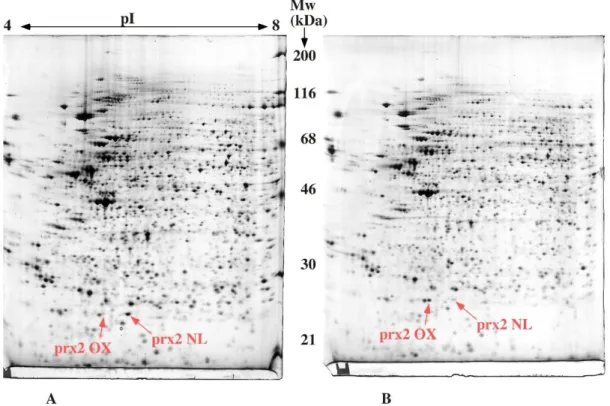
2D gels, however, offer another way of detecting post-translational modifications, in an unsupervised way, by taking advantage of the change in pI induced by many modifications (e.g. phosphorylation). In this scheme, the modified form is first visualized on 2D gels as an extra spot not migrating together with the principal spot, and then the name of the game is to identify the modified peptide(s) by classical mass spectrometry-based analysis. Quite often, the modified peptides are difficult to identify smoothly in mass spectrometry, either because the modification confers an extra negative charge (e.g. phosphorylation), removes a cleavage site (e.g. lysine acylation or methylation) resulting in extra-long peptides, or changes the hydrophobicity/hydrophilicity of the peptide (e.g. glycosylation) resulting in impaired extraction or separation of the peptide. In such cases, the ability of 2D gels to separate the modified form from the bulk of the unmodified one, coupled with their relatively high loading power, are decisive advantages to succeed. Due to these difficulties, there are not that many examples of such approaches in the literature. One can cite, however, the study of adiponection modifications [174], the evidencing of protein deamidation [175], a modification that is quite difficult to detect (low change in mass), for which no role is know, and that is likely to be more widespread than commonly thought, and cysteine overoxidation, especially in the case of peroxiredoxins [176]. This latter example is typical of the difficulties encountered in this approach. The oxidative stress-related modification of peroxiredoxins had been described earlier [177], but without the characterization of the modification, which required 50 picomoles of oxidized peroxiredoxin, due to the length of the peptide (more than 3000 Da) and decrease in flying ability due to the cysteine oxidation. This required to use the full loading capacity of 2D gels to meet such requirements (more than 5 mg of total proteins loaded on the gels). Interestingly enough, when things go to this level of difficulty, the certainty of the modification and of its importance, brought by the 2D gels, is a key argument to go on with the study.
4.5. Analysis of complete proteins
The above examples show how two key features of 2D gels may be used in modern proteomics.The first one is, as mentioned, the ability of 2D gels to serve as a micropreparative tool, a feature that is still used even in complex schemes [178]. The second, which is implicit but need to be strongly emphasized, is the unique ability of 2D gels to be a high-resolution method separating complete proteins, with all their modifications. This is of key importance in studies in which the filiation of peptides (i.e. which peptides belong to which protein) is an important feature.
Interestingly enough, one of the key applications of this feature is to find proteins that are degraded under certain conditions, e.g. apoptosis. In this scheme, a native cell extract is treated (or not) with the protease of interest. The two resulting lysates are separated by 2D gels, the differences are recorded and then characterized by mass spectrometry [179, 180]. This approach has been applied to several proteases involved in apoptosis, such as granzyme B [179], caspase 3 [180], caspase 6 [181] and caspase 7 [182]. Up to now, mostly the decrease of the target proteins has been analyzed, but the often-overlooked ability of 2D gels to separate rather low molecular weight proteins (e.g. in [183]) may allow to investigate the cleavage fragments and thus the cleavage sites.
be used for other purposes than studying protein degradation. It can be used to prepare a well-defined, pure protein for other purposes such as antibody production, [184], or to study the landscape of protein modifications on selected proteins [185-187].
5. Concluding remarks. What future for 2D gels in proteomics
From all of the above, it is tempting to try to predict the future uses of 2D electrophoresis in the future, although this prediction exercise is always difficult and risky. It can be analyzed, however, that in the present proteomics landscape, 2D electrophoresis has two main drawbacks and three main advantages. The two main drawbacks are its very low efficiency (to say the least), in the analysis of hydrophobic proteins, and its high sensitivity to the dynamic range and quantitative distribution issues. The three main advantages are its robustness, its parallelism, and its unique ability to analyze complete proteins at high resolution. When combining all these features, it becomes obvious that 2D gels will deliver their technical advantages when samples with a limited range of protein expression will be used (e.g. bacteria or cellular subfractions), in order to go beyond the classical "déjà vu" [125, 126], unless this class of proteins is of interest in the biological context (e.g. study of cellular stress).
However, one of the key areas where 2D gels should deliver in the future will be the study of modification landscapes, i.e. how protein modifications combine (or exclude mutually) to modulate protein activity in cells. So, as stated earlier [188], when we will have to go to the details of protein functions, it can be predicted that 2D gels will again deliver their full power.
References
[1] Laemmli UK. Cleavage of structural proteins during the assembly of the head of bacteriophage T4. Nature. 1970;227:680-5.
[2] MacGillivray AJ, Rickwood D. The heterogeneity of mouse-chromatin nonhistone proteins as evidenced by two-dimensional polyacrylamide-gel electrophoresis and ion-exchange chromatography. Eur J Biochem. 1974;41:181-90.
[3] O'Farrell PH. High resolution two-dimensional electrophoresis of proteins. J Biol Chem. 1975;250:4007-21.
[4] Garrels JI. Changes in protein synthesis during myogenesis in a clonal cell line. Dev Biol. 1979;73:134-52.
[5] Bravo R, Celis JE. A search for differential polypeptide synthesis throughout the cell cycle of HeLa cells. J Cell Biol. 1980;84:795-802.
[6] Anderson L, Anderson NG. High resolution two-dimensional electrophoresis of human plasma proteins. Proc Natl Acad Sci U S A. 1977;74:5421-5.
[7] Taylor J, Anderson NL, Scandora AE, Willard KE, Anderson NG. Design and Implementation of a Prototype Human Protein Index. Clinical Chemistry.
1982;28:861-6.
[8] Anderson NL, Anderson NG. Microheterogeneity of Serum Transferrin, Haptoglobin and Alpha-2hs Glycoprotein Examined by High-Resolution 2-Dimensional Electrophoresis. Biochemical and Biophysical Research Communications. 1979;88:258-65.
[9] Anderson NL, Anderson NG. 2 Dimensional Separation of Human-Serum Proteins, Use of Antibodies for Identification, and Optical Methods for Gel Intercomparison. Federation Proceedings. 1977;36:1063-.
[10] Anderson NL, Nance SL, Pearson TW, Anderson NG. Specific Antiserum Staining of Two-Dimensional Electrophoretic Patterns of Human-Plasma Proteins Immobilized on Nitrocellulose. Electrophoresis. 1982;3:135-42.
[11] Bravo R, Fey SJ, Bellatin J, Larsen PM, Arevalo J, Celis JE. Identification of a nuclear and of a cytoplasmic polypeptide whose relative proportions are sensitive to changes in the rate of cell proliferation. Exp Cell Res. 1981;136:311-9.
[12] Bravo R, Frank R, Blundell PA, Macdonald-Bravo H. Cyclin/PCNA is the auxiliary protein of DNA polymerase-delta. Nature. 1987;326:515-7.
[13] Garrels JI. Two dimensional gel electrophoresis and computer analysis of proteins synthesized by clonal cell lines. J Biol Chem. 1979;254:7961-77.
[14] Anderson NL, Taylor J, Scandora AE, Coulter BP, Anderson NG. The TYCHO system for computer analysis of two-dimensional gel electrophoresis patterns. Clin Chem. 1981;27:1807-20.
[15] Vincens P, Paris N, Pujol JL, Gaboriaud C, Rabilloud T, Pennetier JL, et al. Hermes - a 2nd Generation Approach to the Automatic-Analysis of Two-Dimensional Electrophoresis Gels .1. Data Acquisition. Electrophoresis. 1986;7:347-56.
[16] Vincens P, Tarroux P. Hermes - a 2nd Generation Approach to the Automatic-Analysis of Two-Dimensional Electrophoresis Gels .3. Spot List Matching.
Electrophoresis. 1987;8:100-7.
[17] Tarroux P. Analysis of Protein-Patterns During Differentiation Using 2-D Electrophoresis and Computer Multidimensional Classification. Electrophoresis. 1983;4:63-70.
[18] Anderson NL, Hofmann JP, Gemmell A, Taylor J. Global Approaches to Quantitative-Analysis of Gene-Expression Patterns Observed by Use of
Two-Dimensional Gel-Electrophoresis. Clinical Chemistry. 1984;30:2031-6.
[19] Rabilloud T, Vincens P, Tarroux P. A New Tool to Study Genetic Expression Using 2-D Electrophoresis Data - the Functional Map Concept. Febs Letters. 1985;189:171-8.
[20] Tarroux P, Vincens P, Rabilloud T. Hermes - a 2nd Generation Approach to the Automatic-Analysis of Two-Dimensional Electrophoresis Gels .5. Data-Analysis. Electrophoresis. 1987;8:187-99.
[21] Appel R, Hochstrasser D, Roch C, Funk M, Muller AF, Pellegrini C. Automatic classification of two-dimensional gel electrophoresis pictures by heuristic clustering analysis: a step toward machine learning. Electrophoresis. 1988;9:136-42.
[22] Pun T, Hochstrasser DF, Appel RD, Funk M, Villars-Augsburger V, Pellegrini C. Computerized classification of two-dimensional gel electrophoretograms by
correspondence analysis and ascendant hierarchical clustering. Appl Theor Electrophor. 1988;1:3-9.
[23] Bravo R, Fey SJ, Small JV, Larsen PM, Celis JE. Coexistence of 3 Major Isoactins in a Single Sarcoma-180 Cell. Cell. 1981;25:195-202.
[24] Duncan R, McConkey EH. How many proteins are there in a typical mammalian cell? Clin Chem. 1982;28:749-55.
[25] Hochstrasser D, Augsburger V, Funk M, Appel R, Pellegrini C, Muller AF. Immobilized Ph Gradients in Capillary Tubes and Two-Dimensional Gel-Electrophoresis. Gel-Electrophoresis. 1986;7:505-11.
[26] Gianazza E, Astruatestori S, Giacon P, Righetti PG. An Improved Protocol for Two-Dimensional Maps of Serum-Proteins with Immobilized Ph Gradients in the 1st Dimension. Electrophoresis. 1985;6:332-9.
[27] Gorg A, Postel W, Weser J, Gunther S, Strahler JR, Hanash SM, et al.
Horizontal Two-Dimensional Electrophoresis with Immobilized Ph Gradients in the 1st-Dimension in the Presence of Nonionic Detergent. Electrophoresis. 1987;8:45-51.
[28] Gorg A, Postel W, Weser J, Gunther S, Strahler JR, Hanash SM, et al.
Elimination of Point Streaking on Silver Stained Two-Dimensional Gels by Addition of Iodoacetamide to the Equilibration Buffer. Electrophoresis. 1987;8:122-4.
[29] Gorg A, Postel W, Gunther S. The current state of two-dimensional
electrophoresis with immobilized pH gradients. Electrophoresis. 1988;9:531-46. [30] Gorg A, Postel W, Gunther S. Methodology of Ipg-Dalt for the Analysis of Cell Lysates and Tissue Proteins. Electrophoresis. 1988;9:628-.
[31] Corbett JM, Dunn MJ, Posch A, Gorg A. Positional Reproducibility of Protein Spots in 2-Dimensional Polyacrylamide-Gel Electrophoresis Using Immobilized Ph Gradient Isoelectric-Focusing in the First Dimension - an Interlaboratory
Comparison. Electrophoresis. 1994;15:1205-11.
[32] Blomberg A, Blomberg L, Norbeck J, Fey SJ, Larsen PM, Larsen M, et al.
Interlaboratory Reproducibility of Yeast Protein-Patterns Analyzed by Immobilized Ph Gradient 2-Dimensional Gel-Electrophoresis. Electrophoresis. 1995;16:1935-45. [33] Matsudaira P. Sequence from picomole quantities of proteins electroblotted onto polyvinylidene difluoride membranes. J Biol Chem. 1987;262:10035-8.
[34] Aebersold RH, Leavitt J, Saavedra RA, Hood LE, Kent SBH. Internal Amino-Acid Sequence-Analysis of Proteins Separated by One-Dimensional or Two-Dimensional Gel-Electrophoresis after Insitu Protease Digestion on Nitrocellulose. Proceedings of the National Academy of Sciences of the United States of America. 1987;84:6970-4.
Analysis - Enzymatic Digestion for Less Than 10 Mu-G of Protein-Bound to Polyvinylidene Difluoride or Nitrocellulose Membranes. Analytical Biochemistry. 1992;201:255-64.
[36] Tempst P, Link AJ, Riviere LR, Fleming M, Elicone C. Internal
Sequence-Analysis of Proteins Separated on Polyacrylamide Gels at the Submicrogram Level - Improved Methods, Applications and Gene Cloning Strategies. Electrophoresis. 1990;11:537-53.
[37] Rosenfeld J, Capdevielle J, Guillemot JC, Ferrara P. In-gel digestion of proteins for internal sequence analysis after one- or two-dimensional gel electrophoresis. Anal Biochem. 1992;203:173-9.
[38] Hanash SM, Strahler JR, Neel JV, Hailat N, Melhem R, Keim D, et al. Highly Resolving 2-Dimensional Gels for Protein Sequencing. Proceedings of the National Academy of Sciences of the United States of America. 1991;88:5709-13.
[39] Bjellqvist B, Sanchez JC, Pasquali C, Ravier F, Paquet N, Frutiger S, et al. Micropreparative 2-Dimensional Electrophoresis Allowing the Separation of Samples Containing Milligram Amounts of Proteins. Electrophoresis. 1993;14:1375-8.
[40] Rabilloud T, Valette C, Lawrence JJ. Sample Application by in-Gel Rehydration Improves the Resolution of 2-Dimensional Electrophoresis with Immobilized Ph Gradients in the First-Dimension. Electrophoresis. 1994;15:1552-8.
[41] Lombardplatet G, Jalinot P. Funnel-Well Sds-Page - a Rapid Technique for Obtaining Sufficient Quantities of Low-Abundance Proteins for Internal Sequence-Analysis. Biotechniques. 1993;15:668-&.
[42] Gevaert K, Verschelde JL, Puype M, VanDamme J, Goethals M, DeBoeck S, et al. Structural analysis and identification of gel-purified proteins, available in the femtomole range, using a novel computer program for peptide sequence
assignment, by matrix-assisted laser desorption ionization reflectron time-of-flight mass spectrometry. Electrophoresis. 1996;17:918-24.
[43] Celis JE, Gesser B, Rasmussen HH, Madsen P, Leffers H, Dejgaard K, et al. Comprehensive two-dimensional gel protein databases offer a global approach to the analysis of human cells: the transformed amnion cells (AMA) master database and its link to genome DNA sequence data. Electrophoresis. 1990;11:989-1071. [44] Anderson NL, Esquer-Blasco R, Hofmann JP, Anderson NG. A two-dimensional gel database of rat liver proteins useful in gene regulation and drug effects studies. Electrophoresis. 1991;12:907-30.
[45] Rasmussen HH, van Damme J, Puype M, Gesser B, Celis JE, Vandekerckhove J. Microsequences of 145 proteins recorded in the two-dimensional gel protein database of normal human epidermal keratinocytes. Electrophoresis. 1992;13:960-9. [46] Appel RD, Sanchez JC, Bairoch A, Golaz O, Miu M, Vargas JR, et al. SWISS-2DPAGE: a database of two-dimensional gel electrophoresis images.
Electrophoresis. 1993;14:1232-8.
[47] Garrels JI, Futcher B, Kobayashi R, Latter GI, Schwender B, Volpe T, et al. Protein identifications for a Saccharomyces cerevisiae protein database.
Electrophoresis. 1994;15:1466-86.
[48] Henzel WJ, Billeci TM, Stults JT, Wong SC, Grimley C, Watanabe C. Identifying Proteins from 2-Dimensional Gels by Molecular Mass Searching of
Peptide-Fragments in Protein-Sequence Databases. Proceedings of the National Academy of Sciences of the United States of America. 1993;90:5011-5.
[49] James P, Quadroni M, Carafoli E, Gonnet G. Protein Identification by Mass Profile Fingerprinting. Biochemical and Biophysical Research Communications. 1993;195:58-64.
[50] Pappin DJ, Hojrup P, Bleasby AJ. Rapid identification of proteins by peptide-mass fingerprinting. Curr Biol. 1993;3:327-32.
[51] Yates JR, Speicher S, Griffin PR, Hunkapiller T. Peptide Mass Maps - a Highly Informative Approach to Protein Identification. Analytical Biochemistry.
1993;214:397-408.
[52] Tonella L, Hoogland C, Binz PA, Appel RD, Hochstrasser DF, Sanchez JC. New perspectives in the Escherichia coli proteome investigation. Proteomics. 2001;1:409-23.
[53] Celis JE, Gromov P, Ostergaard M, Madsen P, Honore B, Dejgaard K, et al. Human 2-D PAGE databases for proteome analysis in health and disease: http://biobase.dk/cgi-bin/celis. FEBS Lett. 1996;398:129-34.
[54] Parker KC, Garrels JI, Hines W, Butler EM, McKee AH, Patterson D, et al. Identification of yeast proteins from two-dimensional gels: working out spot cross-contamination. Electrophoresis. 1998;19:1920-32.
[55] Shaw AC, Rossel Larsen M, Roepstorff P, Holm A, Christiansen G, Birkelund S. Mapping and identification of HeLa cell proteins separated by immobilized
pH-gradient two-dimensional gel electrophoresis and construction of a two-dimensional polyacrylamide gel electrophoresis database. Electrophoresis. 1999;20:977-83. [56] Raymackers J, Daniels A, De Brabandere V, Missiaen C, Dauwe M, Verhaert P, et al. Identification of two-dimensionally separated human cerebrospinal fluid
proteins by N-terminal sequencing, matrix-assisted laser desorption/ionization--mass spectrometry, nanoliquid chromatography-electrospray ionization-time of flight-mass spectrometry, and tandem mass spectrometry. Electrophoresis. 2000;21:2266-83. [57] Buttner K, Bernhardt J, Scharf C, Schmid R, Mader U, Eymann C, et al. A comprehensive two-dimensional map of cytosolic proteins of Bacillus subtilis. Electrophoresis. 2001;22:2908-35.
[58] Porubleva L, Vander Velden K, Kothari S, Oliver DJ, Chitnis PR. The proteome of maize leaves: use of gene sequences and expressed sequence tag data for identification of proteins with peptide mass fingerprints. Electrophoresis.
2001;22:1724-38.
[59] Wilkins MR, Sanchez JC, Williams KL, Hochstrasser DF. Current challenges and future applications for protein maps and post-translational vector maps in proteome projects. Electrophoresis. 1996;17:830-8.
[60] Wilkins MR, Sanchez JC, Gooley AA, Appel RD, Humphery-Smith I,
Hochstrasser DF, et al. Progress with proteome projects: why all proteins expressed by a genome should be identified and how to do it. Biotechnol Genet Eng Rev. 1996;13:19-50.
[61] Wilkins MR, Gasteiger E, Sanchez JC, Bairoch A, Hochstrasser DF. Two-dimensional gel electrophoresis for proteome projects: The effects of protein hydrophobicity and copy number. Electrophoresis. 1998;19:1501-5.
[62] Corthals GL, Wasinger VC, Hochstrasser DF, Sanchez JC. The dynamic range of protein expression: A challenge for proteomic research. Electrophoresis.
2000;21:1104-15.
[63] Rabilloud T, Adessi C, Giraudel A, Lunardi J. Improvement of the solubilization of proteins in two-dimensional electrophoresis with immobilized pH gradients. Electrophoresis. 1997;18:307-16.
[64] Ames GFL, Nikaido K. 2-Dimensional Gel-Electrophoresis of Membrane Proteins. Biochemistry. 1976;15:616-23.
[65] Gianazza E, Rabilloud T, Quaglia L, Caccia P, Astruatestori S, Osio L, et al. Additives for Immobilized Ph Gradient Two-Dimensional Separation of Particulate
Material - Comparison between Commercial and New Synthetic Detergents. Analytical Biochemistry. 1987;165:247-57.
[66] Rabilloud T, Gianazza E, Catto N, Righetti PG. Amidosulfobetaines, a Family of Detergents with Improved Solubilization Properties - Application for Isoelectric-Focusing under Denaturing Conditions. Analytical Biochemistry. 1990;185:94-102. [67] Witzmann F, Jarnot B, Parker D. Dodecyl Maltoside Detergent Improves Resolution of Hepatic Membrane-Proteins in 2-Dimensional Gels. Electrophoresis. 1991;12:687-8.
[68] Chevallet M, Santoni V, Poinas A, Rouquie D, Fuchs A, Kieffer S, et al. New zwitterionic detergents improve the analysis of membrane proteins by
two-dimensional electrophoresis. Electrophoresis. 1998;19:1901-9.
[69] Taylor CM, Pfeiffer SE. Enhanced resolution of glycosyl phosphatidylinositol-anchored and transmembrane proteins from the lipid-rich myelin membrane by two-dimensional gel electrophoresis. Proteomics. 2003;3:1303-12.
[70] Luche S, Santoni V, Rabilloud T. Evaluation of nonionic and zwitterionic detergents as membrane protein solubilizers in two-dimensional electrophoresis. Proteomics. 2003;3:249-53.
[71] Babu GJ, Wheeler D, Alzate O, Periasamy M. Solubilization of membrane proteins for two-dimensional gel electrophoresis: identification of sarcoplasmic reticulum membrane proteins. Analytical Biochemistry. 2004;325:121-5.
[72] Santoni V, Molloy M, Rabilloud T. Membrane proteins and proteomics: Un amour impossible? Electrophoresis. 2000;21:1054-70.
[73] Rabilloud T. Membrane proteins and proteomics: love is possible, but so difficult. Electrophoresis. 2009;30 Suppl 1:S174-80.
[74] Rabilloud T, Vaezzadeh AR, Potier N, Lelong C, Leize-Wagner E, Chevallet M. Power and limitations of electrophoretic separations in proteomics strategies. Mass Spectrom Rev. 2009;28:816-43.
[75] Eravci M, Fuxius S, Broedel O, Weist S, Krause E, Stephanowitz H, et al. The whereabouts of transmembrane proteins from rat brain synaptosomes during two-dimensional gel electrophoresis. Proteomics. 2008;8:1762-70.
[76] Eng JK, McCormack AL, Yates JR. An Approach to Correlate Tandem Mass-Spectral Data of Peptides with Amino-Acid-Sequences in a Protein Database. Journal of the American Society for Mass Spectrometry. 1994;5:976-89.
[77] Yates JR, Eng JK, McCormack AL, Schieltz D. Method to Correlate Tandem Mass-Spectra of Modified Peptides to Amino-Acid-Sequences in the Protein Database. Analytical Chemistry. 1995;67:1426-36.
[78] Jonscher KR, Yates JR. Mixture analysis using a quadrupole mass filter quadrupole ion trap mass spectrometer. Analytical Chemistry. 1996;68:659-67. [79] Yates JR, McCormack AL, Schieltz D, Carmack E, Link A. Direct analysis of protein mixtures by tandem mass spectrometry. Journal of Protein Chemistry. 1997;16:495-7.
[80] Arnott D, Henzel WJ, Stults JT. Rapid identification of comigrating gel-isolated proteins by ion trap mass spectrometry. Electrophoresis. 1998;19:968-80.
[81] Washburn MP, Wolters D, Yates JR. Large-scale analysis of the yeast proteome by multidimensional protein identification technology. Nature Biotechnology.
2001;19:242-7.
[82] Wolters DA, Washburn MP, Yates JR. An automated multidimensional protein identification technology for shotgun proteomics. Analytical Chemistry.
2001;73:5683-90.
al. Analysis of the Plasmodium falciparum proteome by high-accuracy mass spectrometry. Nature. 2002;419:537-42.
[84] Schirle M, Heurtier MA, Kuster B. Profiling core proteomes of human cell lines by one-dimensional PAGE and liquid chromatography-tandem mass spectrometry. Mol Cell Proteomics. 2003;2:1297-305.
[85] Lu P, Vogel C, Wang R, Yao X, Marcotte EM. Absolute protein expression profiling estimates the relative contributions of transcriptional and translational regulation. Nat Biotechnol. 2007;25:117-24.
[86] Anderson NL, Anderson NG. The human plasma proteome: history, character, and diagnostic prospects. Mol Cell Proteomics. 2002;1:845-67.
[87] Ghaemmaghami S, Huh W, Bower K, Howson RW, Belle A, Dephoure N, et al. Global analysis of protein expression in yeast. Nature. 2003;425:737-41.
[88] Voris BP, Young DA. Very-high-resolution two-dimensional gel electrophoresis of proteins using giant gels. Anal Biochem. 1980;104:478-84.
[89] Zhou SB, Bailey MJ, Dunn MJ, Preedy VR, Emery PW. A quantitative
investigation into the losses of proteins at different stages of a two-dimensional gel electrophoresis procedure. Proteomics. 2005;5:2739-47.
[90] Scherl A, Coute Y, Deon C, Calle A, Kindbeiter K, Sanchez JC, et al. Functional proteomic analysis of human nucleolus. Molecular Biology of the Cell. 2002;13:4100-9.
[91] Andersen JS, Lyon CE, Fox AH, Leung AK, Lam YW, Steen H, et al. Directed proteomic analysis of the human nucleolus. Curr Biol. 2002;12:1-11.
[92] Chevallet M, Diemer H, Van Dorssealer A, Villiers C, Rabilloud T. Toward a better analysis of secreted proteins: the example of the myeloid cells secretome. Proteomics. 2007;7:1757-70.
[93] Challapalli KK, Zabel C, Schuchhardt J, Kaindl AM, Klose J, Herzel H. High reproducibility of large-gel two-dimensional electrophoresis. Electrophoresis. 2004;25:3040-7.
[94] Choe LH, Lee KH. Quantitative and qualitative measure of intralaboratory two-dimensional protein gel reproducibility and the effects of sample preparation, sample load, and image analysis. Electrophoresis. 2003;24:3500-7.
[95] Berggren K, Chernokalskaya E, Steinberg TH, Kemper C, Lopez MF, Diwu Z, et al. Background-free, high sensitivity staining of proteins in one- and two-dimensional sodium dodecyl sulfate-polyacrylamide gels using a luminescent ruthenium complex. Electrophoresis. 2000;21:2509-21.
[96] Luche S, Lelong C, Diemer H, Van Dorsselaer A, Rabilloud T. Ultrafast
coelectrophoretic fluorescent staining of proteins with carbocyanines. Proteomics. 2007;7:3234-44.
[97] Neuhoff V, Arold N, Taube D, Ehrhardt W. Improved staining of proteins in polyacrylamide gels including isoelectric focusing gels with clear background at nanogram sensitivity using Coomassie Brilliant Blue G-250 and R-250.
Electrophoresis. 1988;9:255-62.
[98] Chevallet M, Luche S, Rabilloud T. Silver staining of proteins in polyacrylamide gels. Nat Protoc. 2006;1:1852-8.
[99] Chevalier F, Centeno D, Rofidal V, Tauzin M, Martin O, Sommerer N, et al. Different impact of staining procedures using visible stains and fluorescent dyes for large-scale investigation of proteomes by MALDI-TOF mass spectrometry. J
Proteome Res. 2006;5:512-20.
[100] Scheler C, Lamer S, Pan Z, Li XP, Salnikow J, Jungblut P. Peptide mass fingerprint sequence coverage from differently stained proteins on two-dimensional
electrophoresis patterns by matrix assisted laser desorption/ionization-mass spectrometry (MALDI-MS). Electrophoresis. 1998;19:918-27.
[101] Shevchenko A, Wilm M, Vorm O, Mann M. Mass spectrometric sequencing of proteins from silver stained polyacrylamide gels. Analytical Chemistry. 1996;68:850-8.
[102] Richert S, Luche S, Chevallet M, Van Dorsselaer A, Leize-Wagner E, Rabilloud T. About the mechanism of interference of silver staining with peptide mass
spectrometry. Proteomics. 2004;4:909-16.
[103] Chevallet M, Luche S, Diemer H, Strub JM, Van Dorsselaer A, Rabilloud T. Sweet silver: A formaldehyde-free silver staining using aldoses as developing agents, with enhanced compatibility with mass spectrometry. Proteomics. 2008;8:4853-61.
[104] Unlu M, Morgan ME, Minden JS. Difference gel electrophoresis: a single gel method for detecting changes in protein extracts. Electrophoresis. 1997;18:2071-7. [105] Tonge R, Shaw J, Middleton B, Rowlinson R, Rayner S, Young J, et al.
Validation and development of fluorescence two-dimensional differential gel electrophoresis proteomics technology. Proteomics. 2001;1:377-96.
[106] VanBogelen RA, Schiller EE, Thomas JD, Neidhardt FC. Diagnosis of cellular states of microbial organisms using proteomics. Electrophoresis. 1999;20:2149-59. [107] Lange R, Henggearonis R. Identification of a Central Regulator of Stationary-Phase Gene-Expression in Escherichia-Coli. Molecular Microbiology. 1991;5:49-59. [108] Mahairas GG, Sabo PJ, Hickey MJ, Singh DC, Stover CK. Molecular analysis of genetic differences between Mycobacterium bovis BCG and virulent M-bovis. Journal of Bacteriology. 1996;178:1274-82.
[109] van Vliet AHM, Wooldridge KG, Ketley JM. Iron-responsive gene regulation in a Campylobacter jejuni fur mutant. Journal of Bacteriology. 1998;180:5291-8. [110] Arevalo-Ferro C, Hentzer M, Reil G, Gorg A, Kjelleberg S, Givskov M, et al. Identification of quorum-sensing regulated proteins in the opportunistic pathogen Pseudomonas aeruginosa by proteomics. Environmental Microbiology. 2003;5:1350-69.
[111] Eymann C, Dreisbach A, Albrecht D, Bernhardt J, Becher D, Gentner S, et al. A comprehensive proteome map of growing Bacillus subtilis cells. Proteomics. 2004;4:2849-76.
[112] Rodriguez-Ortega MJ, Norais N, Bensi G, Liberatori S, Capo S, Mora M, et al. Characterization and identification of vaccine candidate proteins through analysis of the group A Streptococcus surface proteome. Nat Biotechnol. 2006;24:191-7. [113] Anderson NG, Anderson NL. Analytical Techniques for Cell Fractions .21. 2-Dimensional Analysis of Serum and Tissue Proteins - Multiple Isoelectric-Focusing. Analytical Biochemistry. 1978;85:331-40.
[114] Anderson NL, Anderson NG. Analytical Techniques for Cell Fractions .22. 2-Dimensional Analysis of Serum and Tissue Proteins - Multiple Gradient-Slab Gel-Electrophoresis. Analytical Biochemistry. 1978;85:341-54.
[115] Hackett M. Science, marketing and wishful thinking in quantitative proteomics. Proteomics. 2008;8:4618-23.
[116] Celis JE. Gel-based proteomics: what does MCP expect? Mol Cell Proteomics. 2004;3:949.
[117] Wilkins MR, Appel RD, Van Eyk JE, Chung MC, Gorg A, Hecker M, et al. Guidelines for the next 10 years of proteomics. Proteomics. 2006;6:4-8.
[118] Natera SHA, Guerreiro N, Djordjevic MA. Proteome analysis of differentially displayed proteins as a tool for the investigation of symbiosis. Molecular
Plant-Microbe Interactions. 2000;13:995-1009.
[119] Anderson NL, EsquerBlasco R, Richardson F, Foxworthy P, Eacho P. The effects of peroxisome proliferators on protein abundances in mouse liver. Toxicology and Applied Pharmacology. 1996;137:75-89.
[120] Aicher L, Wahl D, Arce A, Grenet O, Steiner S. New insights into cyclosporine A nephrotoxicity by proteome analysis. Electrophoresis. 1998;19:1998-2003.
[121] Steiner S, Gatlin CL, Lennon JJ, McGrath AM, Aponte AM, Makusky AJ, et al. Proteomics to display lovastatin-induced protein and pathway regulation in rat liver. Electrophoresis. 2000;21:2129-37.
[122] Godon C, Lagniel G, Lee J, Buhler JM, Kieffer S, Perrot R, et al. The H2O2 stimulon in Saccharomyces cerevisiae. Journal of Biological Chemistry.
1998;273:22480-9.
[123] Lee J, Godon C, Lagniel G, Spector D, Garin J, Labarre J, et al. Yap1 and Skn7 control two specialized oxidative stress response regulons in yeast. Journal of Biological Chemistry. 1999;274:16040-6.
[124] Hu Y, Wang G, Chen GYJ, Fu X, Yao SQ. Proteome analysis of
Saccharomyces cerevisiae under metal stress by two-dimensional differential gel electrophoresis. Electrophoresis. 2003;24:1458-70.
[125] Petrak J, Ivanek R, Toman O, Cmejla R, Cmejlova J, Vyoral D, et al. Deja vu in proteomics. A hit parade of repeatedly identified differentially expressed proteins. Proteomics. 2008;8:1744-9.
[126] Wang P, Bouwman FG, Mariman EC. Generally detected proteins in comparative proteomics--a matter of cellular stress response? Proteomics. 2009;9:2955-66.
[127] Lescuyer P, Hochstrasser D, Rabilloud T. How shall we use the proteomics toolbox for biomarker discovery? J Proteome Res. 2007;6:3371-6.
[128] Sarto C, Deon C, Doro G, Hochstrasser DF, Mocarelli P, Sanchez JC.
Contribution of proteomics to the molecular analysis of renal cell carcinoma with an emphasis on manganese superoxide dismutase. Proteomics. 2001;1:1288-94. [129] Shin YK, Yoo BC, Chang HJ, Jeon E, Hong SH, Jung NS, et al. Down-regulation of mitochondrial F1F0-ATP synthase in human colon cancer cells with induced 5-fluorouracil resistance. Cancer Research. 2005;65:3162-70.
[130] Verhoeckx KCM, Bijlsma S, de Groene EM, Witkamp RF, van der Greef J, Rodenburg RJT. A combination of proteomics, principal component analysis and transcriptomics is a powerful tool for the identification of biomarkers for macrophage maturation in the U937 cell line. Proteomics. 2004;4:1014-28.
[131] Catusse J, Strub JM, Job C, Van Dorsselaer A, Job D. Proteome-wide characterization of sugarbeet seed vigor and its tissue specific expression.
Proceedings of the National Academy of Sciences of the United States of America. 2008;105:10262-7.
[132] Heriche JK, Lebrin F, Rabilloud T, Leroy D, Chambaz EM, Goldberg Y. Regulation of protein phosphatase 2A by direct interaction with casein kinase 2 alpha. Science. 1997;276:952-5.
[133] Hondermarck H, Vercoutter-Edouart AS, Revillion F, Lemoine J, El-Yazidi-Belkoura I, Nurcombe V, et al. Proteomics of breast cancer for marker discovery and signal pathway profiling. Proteomics. 2001;1:1216-32.
[134] Kanamoto T, Hellman U, Heldin CH, Souchelnytskyi S. Functional proteomics of transforming growth Factor-beta 1-stimulated Mv1Lu epithelial cells: Rad51 as a target of TGF beta 1-dependent regulation of DNA repair. Embo Journal.
[135] Rao RV, Poksay KS, Castro-Obregon S, Schilling B, Row RH, del Rio G, et al. Molecular components of a cell death pathway activated by endoplasmic reticulum stress. Journal of Biological Chemistry. 2004;279:177-87.
[136] Harrington MG, Merril CR, Asher DM, Gajdusek DC. Abnormal proteins in the cerebrospinal fluid of patients with Creutzfeldt-Jakob disease. N Engl J Med.
1986;315:279-83.
[137] Burkhard PR, Sanchez JC, Landis T, Hochstrasser DF. CSF detection of the 14-3-3 protein in unselected patients with dementia. Neurology. 2001;56:1528-33. [138] Lescuyer P, Allard L, Zimmermann-Ivol CG, Burgess JA, Hughes-Frutiger S, Burkhard PR, et al. Identification of post-mortem cerebrospinal fluid proteins as potential biomarkers of ischemia and neurodegeneration. Proteomics. 2004;4:2234-41.
[139] Celis JE, Gromov P, Cabezon T, Moreira JMA, Ambartsumian N, Sandelin K, et al. Proteomic characterization of the interstitial fluid perfusing the breast tumor microenvironment - A novel resource for biomarker and therapeutic target discovery. Mol Cell Proteomics. 2004;3:327-44.
[140] Roessler M, Rollinger W, Palme S, Hagmann ML, Berndt P, Engel AM, et al. Identification of nicotinamide N-methyltransferase as a novel serum tumor marker for colorectal cancer. Clin Cancer Res. 2005;11:6550-7.
[141] Roessler M, Rollinger W, Mantovani-Endl L, Hagmann ML, Palme S, Berndt P, et al. Identification of PSME3 as a novel serum tumor marker for colorectal cancer by combining two-dimensional polyacrylamide gel electrophoresis with a strictly mass spectrometry-based approach for data analysis. Mol Cell Proteomics. 2006;5:2092-101.
[142] Kruft V, Eubel H, Jansch L, Werhahn W, Braun HP. Proteomic approach to identify novel mitochondrial proteins in Arabidopsis. Plant Physiology.
2001;127:1694-710.
[143] Mitsumoto A, Takeuchi A, Okawa K, Nakagawa Y. A subset of newly
synthesized polypeptides in mitochondria from human endothelial cells exposed to hydroperoxide stress. Free Radical Biology and Medicine. 2002;32:22-37.
[144] Palacino JJ, Sagi D, Goldberg MS, Krauss S, Motz C, Wacker M, et al.
Mitochondrial dysfunction and oxidative damage in parkin-deficient mice. Journal of Biological Chemistry. 2004;279:18614-22.
[145] Chevallet M, Lescuyer P, Diemer H, van Dorsselaer A, Leize-Wagner E, Rabilloud T. Alterations of the mitochondrial proteome caused by the absence of mitochondrial DNA: A proteomic view. Electrophoresis. 2006;27:1574-83.
[146] Gagnon E, Duclos S, Rondeau C, Chevet E, Cameron PH, Steele-Mortimer O, et al. Endoplasmic reticulum-mediated phagocytosis is a mechanism of entry into\ macrophages. Cell\. 2002;110\:119-31\.
[147] Garin J, Diez R, Kieffer S, Dermine JF, Duclos S, Gagnon E, et al. The
phagosome proteome: insight into phagosome functions. J Cell Biol\. 2001;152\:165-80\.
[148] Mannova P, Beretta L. Activation of the N-Ras-PI3K-Akt-mTOR pathway by hepatitis C virus: Control of cell survival and viral replication. Journal of Virology. 2005;79:8742-9.
[149] Chivasa S, Ndimba BK, Simon WJ, Robertson D, Yu XL, Yu XL, et al. Proteomic analysis of the Arabidopsis thaliana cell wall. Electrophoresis. 2002;23:1754-65.
[150] Trinkle-Mulcahy L, Boulon S, Lam YW, Urcia R, Boisvert FM, Vandermoere F, et al. Identifying specific protein interaction partners using quantitative mass