HAL Id: tel-01958881
https://tel.archives-ouvertes.fr/tel-01958881
Submitted on 18 Dec 2018HAL is a multi-disciplinary open access archive for the deposit and dissemination of sci-entific research documents, whether they are pub-lished or not. The documents may come from teaching and research institutions in France or abroad, or from public or private research centers.
L’archive ouverte pluridisciplinaire HAL, est destinée au dépôt et à la diffusion de documents scientifiques de niveau recherche, publiés ou non, émanant des établissements d’enseignement et de recherche français ou étrangers, des laboratoires publics ou privés.
Unraveling the sperm transcriptome by next generation
sequencing and the global epigenetic landscape in
infertile men
Fadi Choucair
To cite this version:
Fadi Choucair. Unraveling the sperm transcriptome by next generation sequencing and the global epigenetic landscape in infertile men. Molecular biology. Université Côte d’Azur; Université libanaise, 2018. English. �NNT : 2018AZUR4058�. �tel-01958881�
THÈSE DE DOCTORAT
Exploration du transcriptome spermatique par le séquençage nouvelle
génération et le portrait épigénétique de l’infertilité masculine
Unraveling the sperm transcriptome by next generation sequencing and the global epigenetic landscape in infertile men
Fadi CHOUCAIR
INSERM U1065, C3M
Présentée en vue de l’obtention du grade de docteur en interactions
moléculaires et cellulaires
de l’Université Côte d’Azur et de l’Université Libanaise
Dirigée par : Valérie Grandjean / Mira
Hazzouri
Soutenue le : 6 Septembre 2018
Devant le jury, composé de :
Mme RACHEL LEVY, PR, UMRS 938, UPMC M. FABIEN MONGELARD, MC, CRCL, ENS Lyon Mme NINA SAADALLAH-ZEIDAN, PR, Université Libanaise
Mme Anne-Amandine CHASSOT, CR, iBV, Université de Nice
Mme VALÉRIE GRANDJEAN, CR, C3M, Université de Nice Mme MIRA HAZZOURI, PR, Université Libanaise
Exploration du transcriptome spermatique par le séquençage nouvelle
génération et le portrait épigénétique de l’infertilité masculine
Jury
Président du Jury
Mme Anne-Amandine CHASSOT, CR, iBV, Université de Nice Rapporteurs
Mme RACHEL LEVY, PR, UMRS 938, UPMC M. FABIEN MONGELARD, MC, CRCL, ENS Lyon Examinateurs
Mme NINA SAADALLAH-ZEIDAN, PR, Université Libanaise Mme VALÉRIE GRANDJEAN, CR, C3M, Université de Nice Mme MIRA HAZZOURI, PR,Université Libanaise
Invités
M. IMAD ABOU JAOUDE, M.D., Hôpital Abou Jaoude, Liban M. JOHNNY AWWAD, M.D., PR, American University of Beirut
ii
Exploration du transcriptome spermatique par le séquençage nouvelle
génération et le portrait épigénétique de l’infertilité masculine
Résumé
L’infertilité masculine est actuellement considérée comme un problème majeur qui pose une situation alarmante sur la santé publique. L’oligozoospermie, l’asthénozoospermie et la tératozoospermie sont les trois anomalies les plus connues des spermatozoïdes. Elles affectent, respectivement, la densité, la motilité et la morphologie des spermatozoïdes. Un spermatozoïde anormal est très souvent corrélé à des altérations génétiques et épigénétiques qui peuvent affecter considérablement le transcriptome. Dans ce sens, le sequençage aléatoire du transcriptome entier des spermatozoïdes ou RNA-seq constitue un outil puissant pour caractériser ces maladies. Jusqu’à présent, il n’existe aucune étude exploitant des données RNA-seq chez des hommes présentant de telles anomalies spermatiques. L’objectif principal de notre étude fût d’identifier des profils distincts des modifications du transcriptome de chaque phénotype d’infertilité pour ainsi révéler des gènes-signatures qui tamponnent une spermatogenèse pathologique
Pour ce faire, les transcriptomes des spermatozoïdes de 60 sujets infertiles atteints soit d’oligozoospermie, d’asthénozoospermie ou de tératozoospermie ont été comparés à ceux de 20 patients fertiles. Ces analyses supervisées nous ont conduit à identifier: (i) les gènes clés spécifiques aux différentes anomalies des spermatozoïdes (ii) les voies de signalisation associées, (ii) les différents longs ARNs non codants dérégulés dans ces anomalies.
Au niveau de l’oligozoospermie, les transcrits de spermatozoïdes dérégulés étaient associées à divers stades de la spermatogenèse, y compris le cycle cellulaire méiotique, l’assemblage du complexe synaptonémal, la cohésion des chromatides sœurs, les processus métaboliques de piRNA, le processus catabolique protéique dépendant de la voie de l’ubiquitine, à la réponse aux dommages de l'ADN et particulièrement le processus de fécondation. Quant à l’asthenozoospermia, la spermatogenèse, l’assemblage du cil, des voies métaboliques reliées à la spermatogenèse, la chimiotaxie et la physiologie des cellules immunitaires ont été significativement dérégulés.
De plus, ce qui nous a intéressé au plus était l’analyse des transcrits sous-exprimés qui a permis l’identification de nombreux transcrits associées aux modifications des histones.
Nous avons aussi mis en évidence une sous expression des gènes différentiellement exprimés qui définit la tératozoospermie. Cette sous expression est associée au système ubiquitine-protéasome, à l’organisation du cytosquelette, au cycle cellulaire, à la SUMOylation en réponse aux dommages de l'ADN et aux protéines de réparation ainsi qu’à de nombreux modulateurs épigénétiques.
Les gènes signature de l'oligozoospermie ont été liés au processus de fécondation et les composants de la matrice extracellulaire, tandis que ceux de la tératozoospermie sont liés à la spermatogenèse et la morphogenèse cellulaire, alors que les gènes signature de l'asthénozoospermie sont impliqués dans l'assemblage du ribosome et du flagelle.
En complément de cette étude, nous avons réalisé une étude très globale du paysage épigénétique du sperme des hommes infertiles. Nous avons, ainsi comparé les niveaux des espèces réactives de l’oxygène (ERO), de méthylation de l’ADN, ainsi que l’intégrité de la chromatine dans les spermatozoïdes de 30 individus infertiles avec ceux de 33 individus fertiles. Nos analyses montrent des niveaux élevés d’espèces réactives de l’oxygène chez les individus infertiles. Ces niveaux sont d’une part négativement corrélés avec les niveaux de méthylation globale de l’ADN et d’autre part négativement corrélés avec ceux de la 5-hydroxyméthylcytosine et de la 5-formylcytosine (intermédiaire dans le processus de déméthylation active). Ces derniers suggèrent qu’une infertilité associée au stress oxydatif conditionne l’épigénome du sperme.
En conclusion, l’ensemble de notre travail apporte des ressources précieuses et originales dans la compréhension des pathologies de sperme.
iii
Unraveling the sperm transcriptome by next generation sequencing and the
global epigenetic landscape in infertile men
Abstract
Male infertility is actually considered as a public alarming health problem. The sperm pathologies spectrum ranges between different phenotypes including oligozoospermia, asthenozoospermia and teratozoospermia depending on the sperm conventional parameters abnormalities. Abnormal sperm is characterized by genetic alterations and epigenetic alterations which can affect the transcriptome extensively. These alterations in RNA profiles are retrospectively indicative of aberrant spermatogenic events. RNA-seq is a powerful tool for comprehensive characterization of whole transcriptome. To date, RNA-seq analysis of sperm from infertile men has not been reported. Our objectives are: (i) recognize key clusters, key pathways and specific gene transcripts for different sperm abnormalities; (ii) catalog the spermatozoal lncRNAs in different sperm pathologies; (iii) identify signature genes which are mechanistically important in the cascade of events driving a pathological spermatogenesis; (iii) portray the global epigenetic landscape in sperm from infertile men. Expression data from 60 sperm samples from 3 groups of infertile men (oligozoospermia, asthenozoospermia, and teratozoospermia) were generated on Illumina HiSeq platform, compared to 20 fertiles, and the resulting gene expression patterns were analyzed for functional enrichment.
Our supervised analyses identified numerous differentially expressed genes between fertile and infertile men. In oligozoospermia, the deregulated spermatozoal transcripts were associated with various stages of spermatogenesis including meiotic cell cycle, synaptonemal complex assembly, sister chromatid cohesion, piRNA metabolic process, ubiquitin-dependent protein catabolic process, cellular response to DNA damage stimulus and interestingly fertilization. As for asthenozoospermia, spermatogenesis, cilium assembly, metabolic-related pathways, chemotaxis and immune cell physiology were most significantly differentially expressed. Interestingly, numerous transcripts associated with histone modifications were highly down-regulated.
With regards to teratozoospermia, we evidenced sperm-specific differentially expressed genes which are involved in the ubiquitin-proteasome, cytoskeleton organization, the cell cycle pathway, SUMOylation of DNA damage response and repair proteins, as well as many putative epigenetic modulators of gene expression.. We also attempted to identify distinct patterns of gene expression changes that were definite to the different abnormal sperm phenotypes in infertile men relative to controls. Signature genes of oligozoospermia were over-enriched by genes involved in fertilization and extracellular matrix components, while signature genes of teratozoospermia were enriched by genes involved in spermatogenesis and cellular components involved in morphogenesis, whilst signature genes of asthenozoospermia were enriched by genes implicated in ribosome and cilium assembly.
We complemented this work by a parallel epigenetic analysis of the global epigenetic landscape in infertile men. We compared the levels of reactive oxygen species (ROS), DNA integrity and global epigenetic parameters in sperm from 33 infertile subjects with abnormal semen parameters compared to fertile individuals. We pointed out that infertile men are characterized by strikingly high levels of reactive oxygen species (ROS) which were in part negatively correlated with the global DNA methylation, and positively correlated with the levels of 5-hydroxymethylcytosine and 5-formylcytosine (active demethylation intermediates). These findings suggest that male infertility associated with oxidative stress shapes the sperm epigenetic landscape.
In summary, this original work yielded a transcriptional portrait of sperm abnormalities and provided valuable resources that would further elucidate sperm pathologies.
iv
ACKNOWLEDGMENTS
As I come to close this chapter in my life, I would like to thank many unique people, with whom I was lucky to interact and who made my time at graduate school a wonderful experience.
First, I would like to express a deep gratitude to my supervisors, ‘my academic moms’, Mira and Valérie. Thank you both for enabling me to grow and believe in myself as an independent scientist, for patiently guiding me through different steps of this endeavor and for always being there when I needed.
Thank you Mira, for creating new opportunities for me, and providing endless intellectual and emotional support. Thank you Valérie, for providing invaluable support, insightful comments and ideas. I am very fortunate to have you both as my mentors.
I would also like to thank Khaled Hazzouri for generously sharing your time and knowledge: this has been an enriching learning experience.
I thank my dear collaborators and colleagues: Imad Abou Jaoude, Johnny Awwad, Youssef Gemayel, Elias Saliba, Nahida Azar, Rita Assaf, Christina Karam, Layal Hamdar, Aurore Assaker, Dalia Khalifeh and all members of the molecular biology lab at the Faculty of Public Health in the Lebanese University. Thank you for taking the time to help me with different aspects of my science.
I thank the members of my dissertation and examining committees: Rachel Levy, Fabien Mongelard, Nina Saadallah-Zeidan and Anne-Amandine Chassot. Thank you, for your time, support and insights.
vi
TABLE OF CONTENTS
LIST OF TABLES ... XI
LIST OF FIGURES ... XII
LIST OF ABBREVIATIONS ... XV
INTRODUCTION ... 1
CHAPTER I ... 3
MOLECULAR CONTROL O F SPERMATOGENESIS AND
POTENTIAL CAUSES OF MALE INFERTILITY ... 3
1.1. “Drawing” the molecular portrait of human spermatogenesis - a review of the past familiar faces and a glimpse into the new features ... 3
1.1.1. Molecular control of spermatogenesis ... 3
1.1.1.1 Introduction ... 3
1.1.1.2. Male germ cells development during embryogenesis ... 4
1.1.1.3. Male germ cells during the mitotic phase ... 5
1.1.1.4. Male germ cells during the meoitic phase ... 7
1.1.1.5. Post-meoitic phase/ spermiogenesis... 8
1.1.1.5.1. Acrosome biogenesis ...8
1.1.1.5.2. Manchette biogenesis ...9
1.1.1.5.3. Axoneme development ...9
1.1.1.5.4. Cytoplasmic removal ...10
1.1.1.5.5. Chromatin remodeling ...10
1.1.1.5.6. The expression of late spermatid genes ...10
1.1.1.5.7. Expression of cellular elements for energy production and motility ...11
1.1.1.6. Spermiation ... 11
1.1.1.7. Gene expression pattern during spermatogenesis ... 12
1.2 Molecular and epigenetic causes of infertility ... 12
1.2.1. Molecular insights into the causes of male infertility ... 12
1.2.2. Epigenetic layers and players in the context of male infertility: an extensive review of the general theory and the current state-of-the-art ... 15
vii
1.2.2.2. The sperm methylome ... 16
1.2.2.2.1. Basic concepts ...16
1.2.2.2.2. MTHFR enzyme activity ...17
1.2.2.2.3. Role of the DNMTs in spermatogenesis ...18
1.2.2.2.4. Tet-dependent DNA demethylation ...18
1.2.2.2.5. Global DNA methylation ...19
1.2.2.2.6. Methylation levels at promoter of CpG islands ...19
1.2.2.2.7. Methylation status of non-imprinted genes ...19
1.2.2.2.8. Methylation at repetitive elements ...21
1.2.2.2.9. Methylation at imprinted genes ...21
1.2.2.3. The spermatozoal chromatin proteome ... 23
1.2.2.3.1. Basic concepts ...23
1.2.2.3.2. Histones: post-translational modifications and variants ...23
1.2.2.3.3. Chromatin remodeling ...26
1.2.2.4. Non-coding RNAs ... 27
1.2.2.4.1. Basic concepts ...27
1.2.2.4.2. MicroRNAs (miRNAs) and spermatogenesis ...28
1.2.2.4.3. Piwi-interacting RNAs (piRNAs) and spermatogenesis/infertility ...28
1.2.2.4.4. Long non-codong RNAs (lncRNAs) ...29
1.2.2.5. High order nuclear organization in sperm ... 29
Conclusion ... 30
CHAPTER II ... 33
A REVIEW OF THE TRANSCRIPTOMICS STUDIES ... 33
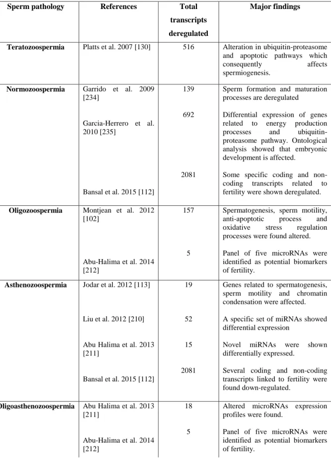
2.1. Review in literature data ... 33
2.1.1. Data collection ... 33
2.1.2. Data mining results ... 34
2.1.2.1. Teratozoospermia ...34
2.1.2.2. Normozoospermia ...34
2.1.2.3. Asthenozoospermia ...36
2.1.2.4. Oligoasthenozospermia ...39
2.1.2.5. Oligozoospermia...40
2.2. New emerging technology approaches: Sperm transcriptome profiling using Next generation sequencing... 42
viii
CHAPTER III ... 49
MATERIALS AND METHODS ... 49
3.1. Optimization of sperm RNA isolation ... 49
3.1.1. Materials and methods ... 51
3.1.1.1 Isolation of sperm cells ...51
3.1.1.2. Cell lysis ...51
3.1.1.3. Testing the effectiveness of lysis ...51
3.1.1.4. RNA isolation ...52
3.1.1.5. DNA isolation ...52
3.1.1.6. Nucleic acids QC metrics ...52
3.1.1.7. RNA library preparation and sequencing ...52
3.1.1.8. Global 5-methylcytosine (5-mC) measurement ...53
3.1.1.9. Statistical analysis ...53
3.1.2. Results and discussion ... 54
3.2. The ABCs of RNA-seq: An explanatory review on the basics of experimental design, laboratory performance and computational analysis in RNA-seq experiments ... 61
3.2.1. Design considerations ... 63
3.2.1.1. Library type ...63
3.2.1.2. Replicates ...63
3.2.1.3. Sample pairing ...64
3.2.1.4. Pooling ...64
3.2.1.5. Normalization of RNA-seq data ...64
3.2.1.6. Sequencing decisions...65
3.2.2. Sequencing, at a glance (Illumina)... 65
3.2.3. RNA-seq data analysis workflow ... 67
3.2.3.1. Quality control of raw data ...67
3.2.3.2. Preprocessing ...68
3.2.3.3. Alignment (mapping) ...69
3.2.3.4. Quality control for aligned reads ...72
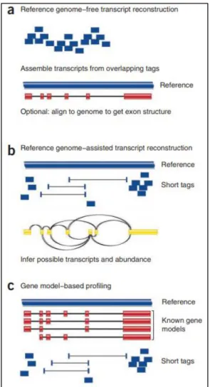
3.2.3.5. Reads assembly and quantification ...74
3.2.3.6. Differential expression analysis: normalizing and transforming read counts ...77
3.2.3.7. Variability within the experiment: data exploration ...78
3.2.3.8. Differential gene expression analysis ...80
3.2.3.9. Exploratory plots following DGE analysis ...80
3.3. Experimental work for sperm transcriptomic analysis ... 81
3.3.1 Patient population ... 81
ix
3.3.3. Sperm purification ... 82
3.3.4. RNA preparation ... 82
3.3.5. RNA-Seq library preparation ... 83
3.3.6. Sequencing Libraries ... 83
3.3.7. Bioinformatics... 83
3.4. Experimental work for sperm epigenetic analysis ... 84
3.4.1. Study participants... 84
3.4.2. Sperm damage assessment ... 84
3.4.2.1. Measurement of reactive oxygen species production ...84
3.4.2.2. Sperm Chromatin dispersion (SCD) test ...85
3.4.3. Sperm chromatin abnormalities assessment ... 85
3.4.3.1. Sperm chromatin structure assessment ...85
3.4.3.2. Histone retention assessment ...85
3.4.4.1. DNA extraction ...86
3.4.4.2. Global 5-mC, 5-hmC and 5-fC measurement ...86
3.4.5. Statistical analysis ... 86
CHAPTER IV ... 90
RESULTS ... 90
4.1. Comprehensive transcriptomic analysis of human spermatozoa in male infertility ... 90
4.1.1. Sperm transcriptome in oligozoospermia ... 90
4.1.1.1. Total number of transcripts and their expression level ... 90
4.1.1.2. Nature of spermatozoa transcripts ... 90
4.1.1.3. Functions of spermatozoal transcripts ... 91
4.1.1.3.1. Down-regulated genes ...91
4.1.1.3.2. Up-regulated genes ...96
4.1.1.3.3. Long non-coding RNAs ...97
4.1.2. Sperm transcriptome in asthenozoospermia ... 104
4.1.2.1. Total number of transcripts and their expression level ...104
4.1.2.2. Nature of spermatozoa transcripts ...104
4.1.2.3. Enrichment analysis of protein-coding genes for identification of biological and functional differences ...104
4.1.2.3.1. Down-regulated genes... 104
4.1.2.3.2. Up-regulated genes ... 109
4.1.2.3.3. Long non-coding RNAs ... 109
x
4.1.3.1. Total number of transcripts and their expression level ... 116
4.1.3.2. Nature of spermatozoa transcripts ... 116
4.1.3.3. Functions of spermatozoal transcripts ... 116
4.1.3.3.1. Complete genes differentially expressed... 116
4.1.3.3.2. Down-regulated genes... 119
4.1.3.3.3. Up-regulated genes ... 123
4.1.3.3.4. Long non-coding RNAs ... 123
4.1.4. Dissection of genes expression signatures associated with abnormal sperm phenotypes in infertile men by RNA seq analysis ... 128
4.1.4.1. Repertoire of DEG’s in sperm from infertile men as determined by Next Generation Sequencing of cDNA libraries...128
4.1.4.2. Data analysis strategy ...128
4.1.4.3. Molecular characterization of sperm pathologies types of infertile men...129
4.1.4.4. Identification of the best set of genes discriminating the different phenotypes of sperm pathologies of infertile men ...130
4.1.4.5. Transcriptional landscape of long non-coding RNAs in sperm from infertile men 132 4.2. Characterization of the sperm global epigenetic landscape associated with oxidative stress in infertile men ... 136
Conclusion ... 143
CHAPTER V ... 145
GENERAL DISCUSSION ... 145
5.1. Differential analysis of spermatozoal transcripts from infertile men with different sperm pathologies using RNA-seq ... 145
5.2. Assessment of global epigenetic changes and oxidative damage in sperm from infertile men ... 151
CONCLUSION AND FUTURE DIRECTIONS ... 155
REFERENCES ... 161
xi
LIST OF TABLES
Table 1. Male infertility-associated epigenetic modifications: DNA methylome alterations. ... 23 Table 2. Male infertility-associated epigenetic modifications: Aberrations in the spermatozoal chromatin proteome.
... 26
Table 3. Male infertility-associated epigenetic modifications: Altered sperm non-coding RNAs. ... 29 Table 4. Schematic representation of the microarray-based transcriptome studies evaluated in the present review.41 Table 5. The percentage of spermatozoa with intact cell membranes, after three physical treatments (means±SEM).
... 55
Table 6. Factors and Levels Assayed in Fractional Factorial Design for the optimisation of sperm RNA extraction:
Factors Screening. ... 58
Table 7. Summary of the experiments to find optimal conditions of sperm RNA isolation. ... 58 Table 8. Functional enrichments of the protein-protein interaction (PPI) network for the mouse orthologs
associated with male infertility phenotype. ... 92
Table 9. The spermatozoal enriched transcripts from oligozoospermic men associated with the oligozoospermia
mouse phenotype obtained from functional annotation analysis (Toppgene) based on their abundancy. ... 93
Table 10. The spermatozoal enriched transcripts from oligozoospermic men associated with the abnormal sperm
number mouse phenotype obtained from functional annotation analysis (Toppgene) based on their abundancy. .... 94
Table 11. Functional enrichments of the protein-protein interaction (PPI) network for the mouse orthologs
associated with abnormal sperm number. ... 96
Table 12. List of long non-coding RNAs associated with oligozoospermia... 100 Table 13. The molecular functions, biological processes, cellular localization, and pathways implicated of
spermatozoal transcripts in asthenozoospermia obtained from functional annotation analysis (Toppgene) of the abundantly down-regulated set of genes significantly differentially expressed.. ... 105
Table 14. The spermatozoal enriched transcripts from asthenozoospermic men associated with the abnormal sperm
motility mouse phenotype obtained from functional annotation analysis (Toppgene) based on their abundancy. .. 107
Table 15. Functional enrichments of the protein-protein interaction (PPI) network for the mouse orthologs
associated with abnormal sperm motility phenotype. ... 107
Table 16. The molecular functions, biological processes involved, cellular localization, and pathways implicated of
spermatozoal transcripts in teratozoospermia obtained from functional annotation analysis (Toppgene) of the complete set of genes significantly differentially expressed.. ... 118
Table 17. Functional enrichments of the protein-protein interaction (PPI) network for the mouse orthologs
associated with teratozoospermia phenotype. ... 119
Table 18. The spermatozoal enriched down-regulated transcripts from teratozoospermic men associated with the
teratozoospermia mouse phenotype obtained from functional annotation analysis (Toppgene) based on their abundancy... 121
Table 19. Biological pathway analysis of the set of common signature genes differentially expressed orthologs
related to “male infertility” phenotype identified by RNA seq. ... 131
xii
LIST OF FIGURES
Figure 1. Key molecular events for PGCs specification and migration. ... 5
Figure 2. Stages and molecular events associated with human spermatogenesis. Sequence of the stages spanning spermatogonial stem cells differentiation during embryogenesis and the release of mature spermatozoa. ... 11
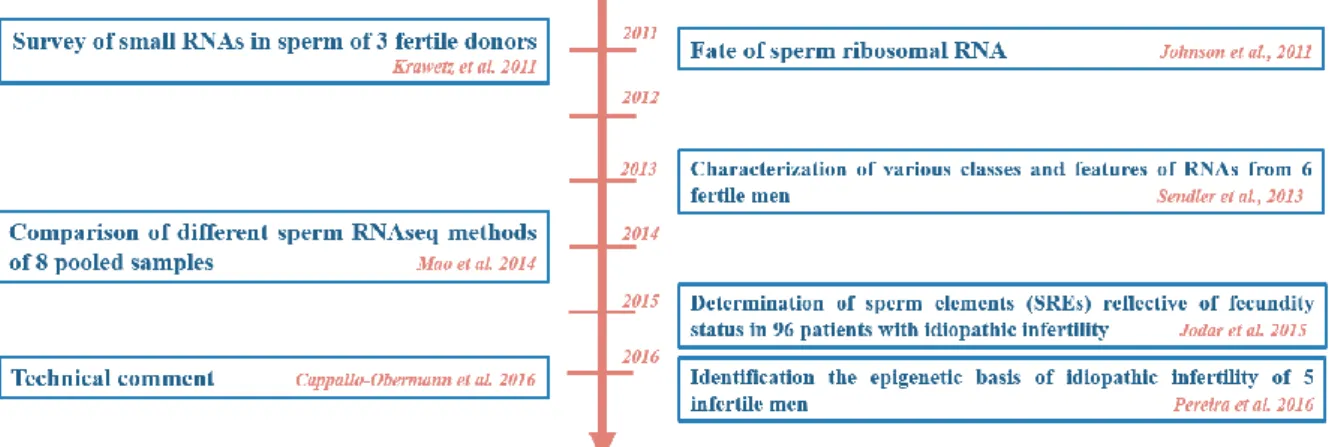
Figure 3. Timeline of studies involving transcriptomic profiling using Next generation sequencing from human spermatozoa. ... 46
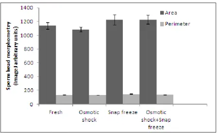
Figure 4. Sperm head morphometry analysis (mean ± SEM) after three physical treatments (osmotic choc, snap freeze, osmotic choc followed by snap freezing) compared with spermatozoa in fresh semen. ... 56
Figure 5. Histogram of relative fluorescence intensities (DAPI), reported as arbitrary fluorescence units (a.f.u.), after adhering to sperm nucleus under different physical treatment conditions. ... 56
Figure 6. Electropherograms (in a 1% agarose gel run for 40 minutes at 150 Volts) of DNA from sperm cells. ... 61
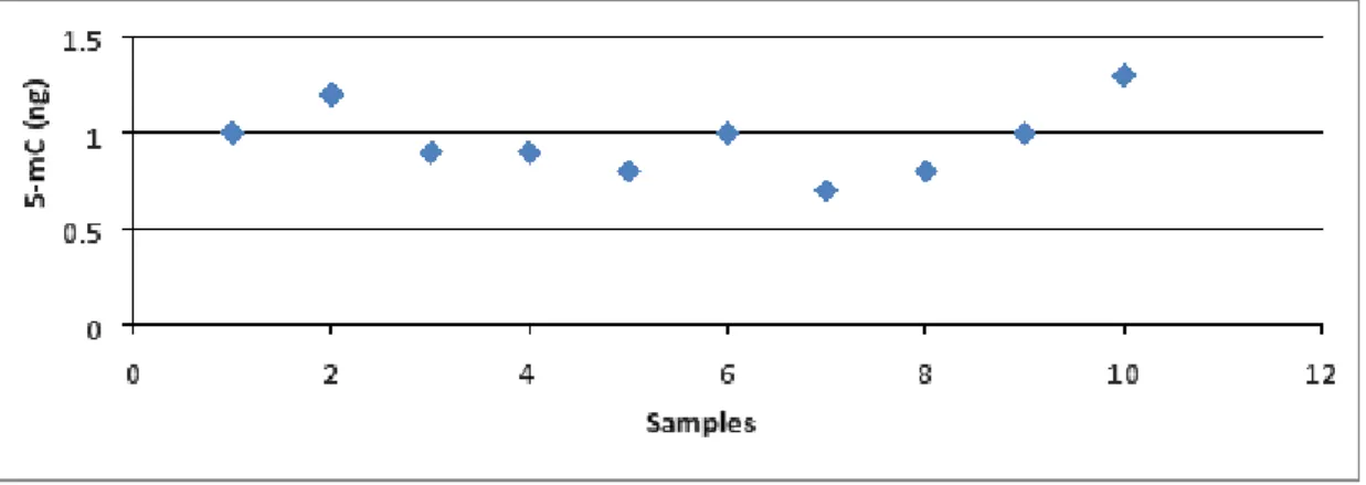
Figure 7. The levels of methylated DNA (5-mC) in total sperm DNA. ... 61
Figure 8. Illumina sequencing workflow using the sequencing by synthesis method adapted from Illumina. ... 66
Figure 9. FASTQC format and reports. ... 67
Figure 10. Quality control checks a: before quality trimming and b: after quality trimming ... 68
Figure 11. Three RNA mapping strategies: a: De novo assembly; b: Align to reference genome; c: Align to transcriptome. ... 69
Figure 12. An overview of cufflinks workflow ... 71
Figure 13. a: Tuxedo software components; b: Tuxedo protocol for differential gene expression ... 72
Figure 14. Post alignment quality control representations. ... 73
Figure 15. a: HTseq different modes for reads quantification; b: Gene transfer format (GTF) features ... 75
Figure 16. Overview of Cufflinks; b: Scheme of a simple deBruijn graph-based transcript assembly ... 76
Figure 17. Options for differential gene expression (DGE) analysis. ... 77
Figure 18. Illustration of variability within the experiment. ... 79
Figure 19. Exploratory plots following DGE analysis ... 81
Figure 20. Protein interaction map. Map was prepared using the STRING web tool (https://string-db.org) for the proteins encoded by the set of genes associated with the male infertility phenotype.. ... 92
Figure 21. Protein interaction map. Map was prepared using the STRING web tool (https://string-db.org) for the proteins encoded by the set of genes associated with the abnormal sperm number phenotype.. ... 96
Figure 22. (a) RNA–protein interaction network map (v1.0) prepared using the RAIN web tool (https://rth.dk/resources/rain/) for the long non-coding RNAs (lncRNAs) associated with oligozoospermia. (b) lncRNAs- top ranked candidate genes encoding proteins interaction map associated with oligozoospermia.. ... 98
Figure 23. Protein interaction map. Map was prepared using the STRING web tool (https://string-db.org) for the proteins encoded by the set of orthologous genes associated with abnormal sperm motility phenotype.. ... 106
Figure 24. (a) RNA–protein interaction network map (v1.0) prepared using the RAIN web tool (https://rth.dk/resources/rain/) for the long non-coding RNAs (lncRNAs) associated with asthenozoospermia. (b) lncRNAs- top ranked candidate genes encoding proteins interaction map associated with asthenozoospermia. .... 110
Figure 25. Pathway analysis of the ribosome (Kegg pathway) using the Pathview server (https://pathview.uncc.edu/). Genes significantly differentially expressed (DE) in asthenozoospermia ... 112
Figure 26. Pathway analysis of oxidative phosphorylation (a) and glycolysis (b) (Kegg pathway) using the Pathview server (https://pathview.uncc.edu/). Genes significantly differentially expressed (DE) in asthenozoospermia. ... 113
Figure 27. Pathway analysis of the calcium signaling pathway (Kegg pathway) using the Pathview server (https://pathview.uncc.edu/). Genes significantly differentially expressed (DE) in asthenozoospermia. ... 114
Figure 28. Protein interaction map. Map was prepared using the STRING web tool (https://string-db.org) for the proteins encoded by the set of mouse orthologous genes associated with teratozoospermia phenotype. Colored lines denote interactions and network nodes represent proteins. ... 117
Figure 29. Pathway analysis of Proteasome and Ubiquitin-mediated proteolysis (Kegg pathway) using the Pathview server (https://pathview.uncc.edu/). Genes significantly differentially expressed (DE) in teratozoospermia. ... 121
Figure 30. (a) RNA–protein interaction network map (v1.0) prepared using the RAIN web tool (https://rth.dk/resources/rain/) for the long non-coding RNAs (lncRNAs) associated with teratozoospermia. These candidate proteins were further prioritized based on topological features in protein-protein interaction network. (b) lncRNAs- top ranked candidate genes encoding proteins interaction map associated with asthenozoospermia.. ... 124
xiii
Figure 32. Transcriptional landscape of human sperm RNAs in ejaculates of infertile men with different sperm
abnormalities types. ... 130
Figure 33. Identification of the best set of genes discriminating the different phenotypes of sperm pathologies of
infertile men. ... 131
Figure 34. Relationship between sperm global DNA methylation with sperm histone retention (abnormal chromatin
packaging) a) by Aniline blue test and sperm chromatin alteration b) by Toluidine blue assay. ... 139
Figure 35. Relationship between sperm global DNA methylation with sperm ROS a) by NBT test and sperm DNA
fragmentation (SDF) b) by SCD test... 139
Figure 36. Relationship between sperm global DNA hydroxymethylation with sperm ROS by NBT test (a) and sperm
xv
LIST OF ABBREVIATIONS
5-fC: 5-formylcytosine
5-hmC: 5-hydroxymethylcytosine 5meC: 5th carbon of cytosine 8-OHdG: oxidative DNA adduct Ad: adult dark
aES: apical ectoplasmic specialization BAM: binary Alignment/Map
BH: benjamini-hochberg BTB: blood testis barrier CaM: calcium/calmodulin CAR: chromatin-associated RNA
CIGAR: Concise Idiosyncratic Gapped Alignment Report CSF1: colony stimulating factor 1
DAPI: 4′,6-diamidino-2-phenylindole DGE: differential gene expression DM-CpG: differentially methylated CpG DNA: deoxyribonucleic acid
DNMT: de novo methyltransferases DTT: dithiothreitol
ECM: extracellular matrix EG: exclusive gene
ELISA: enzyme-linked immunosorbent assay Enc: Encore NGS Multiplex System I
ERO: espèces réactives de l’oxygène FC: fold change
FDR (false discovery rate)
FFPES: Formalin Fixed Paraffin Embedded System
FPKM: fragments per kilobase of exons per million mapped GDE: genes differentially expressed
gDNA: genomic DNA
GnRH: gonadotropin releasing hormone GO: Gene ontology
GTF: Gene transfer format HAT: histone aceltyltransferase HDAC: histone deacetylase
HMR: hypomethylated DNA region ICSI: intracytoplasmic sperm injection IFT: intraflagellar transport
IMT: intra-manchette transport IQR: Inter-Qartile Range IVF: in vitro fertilization
KEGG: Kyoto Encyclopedia of Genes and Genomes lincRNA: long intergenic noncoding RNA
lncRNA: long non-coding RNA MBP: methyl-CpG binding protein MDS: multidimentional scaling miRNA: microRNA
MS: mass spectrometry
MSI: meiotic sex chromosome inactivation MTHFR: methylenetetrahydrofolate reductase NBT: Nitro Blue Tetrazolium
xvi ncRNA: non-coding RNA
NGS: next generation sequencing NMD: Nonsense-Mediated Decay Nt: nucleotides
ODF : outer dense fiber
OR: Ovation® RNA-Seq System V2 PCA: Principle Component Analysis PCR: polymerase chain reaction PE: Paired-end
PGC : primordial germ cells piRNA: Piwi-interacting RNA PPI: protein–protein interaction PRM : protamine
QC: quality control
qPCR: quantitative polymerase chain reaction RA: retinoic acid
RAIN: RNA-protein Association and Interaction Network RBP: RNA-binding protein
RIN: RNA integrity number RNA: ribonucleic acid RNA-seq : RNA sequencing RNP: ribonucleoprotein particle ROS: reactive oxygen species
RPKM: reads per kilobase of exons per million mapped rRNA: ribosomal RNA
RT-PCR: real time polymerase chain reaction SAC: spindle assembly checkpoint
SAM: Sequence Alignment/Map SCD: Sperm Chromatin dispersion SCF: stem cell factor
SEM:
SEP: sperm expression profiles Sf1: Steroidogenic factor 1 SP: SeqPlex RNA Amplification SR: Single read
SREs: sperm RNA elements
sSMC : small supernumerary marker chromosome
STRING : Search Tool for the Retrieval of Interacting Genes/Proteins SVM : support vector machine
TB: toluidine blue
TBC : tubulobulbar complex
TBDC: toluidine blue dark violet cell TBLC: toluidine blue light blue cell TET : ten-eleven-translocation TMM: Trimmed Mean of M-values TP : transition protein
UL: Ovation Ultralow Library Systems UPP: ubiquitin-proteasomal pathway VDAC : voltage-dependent anion channels WHO: world health organization
1
INTRODUCTION
Spermatozoa transfers transcripts as well as epigenetic information to the oocyte during fertilization. In this thesis I present two studies. In the first one, I applied an emerging technology next generation sequencing technology (NGS)) to systematically study human sperm Ribonucleic Acids (RNAs) in infertile men with different sperm pathologies and to gain insights into genes associated with male infertility. In this manner, paternal RNAs can serve as a biomarker to diagnose patients with subfertility or predict pregnancy outcomes [1]. In the second part, I characterized the sperm global epigenetic landscape in infertile men. as anyn changes in the epigenome may carry a high risk to be transmitted to the offspring, thus this examination of the epigenetic facet of sperm is valuable in part for characterizing the mechanisms of sperm pathogenesis, and in determining the risks of using a pathological sperm in assisted reproductive technologies (ART).
Outline of the thesis
The work presented in this thesis is divided into four major parts: (1) (Chapters 1 & 2) Introduction including extensive reviews of the literature and updated evidence regarding: the molecular basis of spermatogenesis, the sperm epigenetic layers and players in infertile men and the transcriptomic studies of male infertility; (2) (Chapter 3) Materials and methods including descriptions of the population, and the experimental methodology; (3) Results of the transcriptomic analysis of the sperm from infertile patients presenting different semen analysis abnormalities (count, motility, morphology) (Chapter 4 ) and assessment of the global epigenetic landscape in sperm from infertile men (Chapter 4) ; (4) A general conclusion of the main findings followed by the future directions (Chapter 5).
Chapters 1 & 2 present the molecular basis of spermatogenesis, the sperm epigenetic layers
and players in infertile men and the transcriptomic studies of male infertility.
In chapter 3, we describe the laboratory and computational methods used in this work. The aim is to provide an overview of the protocol of RNA extraction refinement and optimization for expression profiling, and how genomic data of sperm compiled in this work has been generated and analyzed. Therefore, we first describe methods used for sperm RNA
2
purification which are based on the experimental work performed in the framework of this thesis, followed by a description of the basics of RNA sequencing (RNA-seq) analysis. Finally, we will present the experimental work methodologies used for sperm transcriptomic and epigenetic analysis.
In chapter 4, we present results from a genome-wide expression analysis of sperm from infertile men at different pathologic conditions (oligozoospermia, asthenozoospermia, teratozoospermia) using RNA-Seq. Additionally, in the second section of this chapter, we characterized the global epigenetic landscape in sperm from infertile men associated with oxidative stress.
In Chapter 5, we discussed the rationale for appreciating the use of NGS for transcriptome analysis. Next, we presented our empirical evidence about the aberrant transcriptome in sperm for each sperm pathology.
In Chapter 6 we discussed the association between oxidative stress and an aberrant sperm epigenome in infertile men.
Finally, a conclusion highlights the significant strengths of this research that have been covered in this thesis as well as future directions for expanding this field of research, including some suggested future experiments.
In the same context we highlight the publication of two scientific publications listed below that tackled the sperm Deoxyribonucleic Acid (DNA) fragmentation in infertile men and antioxidants régime for sperm DNA damage. They are posted in supplememntary materials at the end of this manuscript.
Choucair, Fadi B., Eliane G. Rachkidi, Georges C. Raad, Elias M. Saliba, Nina S. Zeidan, Rania A. Jounblat, Imad F. Abou Jaoude, and Mira M. Hazzouri. "High level of DNA fragmentation in sperm of Lebanese infertile men using Sperm Chromatin Dispersion test." Middle East Fertility
Society Journal 21, no. 4 (2016): 269-276.
Choucair, Fadi, Elias Saliba, Imad Abou Jaoude, and Mira Hazzouri. "Antioxidants modulation of sperm genome and epigenome damage: Fact or fad? Converging evidence from animal and human studies." Middle East Fertility Society Journal 23, no. 2 (2018): 85-90.
3
CHAPTER I
MOLECULAR CONTROL O F SPERMATOGENESIS AND
POTENTIAL CAUSES OF MALE INFERTILITY
In this section, we will review in-depth the molecular control mechanisms of spermatogenesis. Spermatogenesis is governed by timely genetic and epigenetic events that we will try to piece together into a coherent complete biological picture.
An ever-expanding number of molecules have been implicated in the regulation of epigenetic processes during spermatogenesis. The second section will provide an extensive review of aberrant epigenetic regulation/modification and their association with fertility.
1.1. “Drawing” the molecular portrait of human spermatogenesis - a review of
the past familiar faces and a glimpse into the new features
1.1.1. Molecular control of spermatogenesis 1.1.1.1 Introduction
Spermatogenesis is a highly conserved process among many organisms consisting of
three key phases: a mitotic amplification phase, a meiotic phase, and a post-meiotic phase also known as spermiogenesis. Almost all the events during this process are orchestrated by complex molecular pathways, which are endowed by multiple genetic and/or epigenetic factors. A systematic understanding of the critical molecular events involved in spermatogenesis may help to pave the way for the diagnosis of sperm pathologies. In this paper, we will retrospectively review the dominant events operating during the initiation and progression of spermatogenesis in humans. We will provide an update on the orchestrated genetic and epigenetic networks in male germ cells during the mitotic phase, meiotic phase and spermiogenesis. We have considered genetic players based on human gene expression, and point mutations, data and mouse genetic and molecular aspects. Defining these broad mechanisms is a necessary prelude to determine the timing of aberrant events in the context of infertility.
4
1.1.1.2. Male germ cells development during embryogenesis
Lessons gleaned from mouse models revealed that primordial germ cells (PGCs) originate from epiblast cells in the yolk sac in response to the adjacent endoderm and extra-embryonic ectoderm signals. Several transcriptional regulator factors are required for the acquisition of the germ cell fate including intrinsic factors, such as Prdm1 (Positive Regulatory Domain I-Binding Factor 1) also known as Blimp1, a known transcriptional repressor which has a critical role in the foundation of the germ cell lineage [2], and extrinsic factors, such as BMPs (bone morphogenetic proteins) embryonic-derived signals [3].
In humans, it was suggested that BMP4- and WNT3A (expressed by embryonic epiblast cells) -mediated upregulation of PRDM [4]. Worth noting, WNT3A was suggested to be required for the responsiveness of epiblast cells to BMP4. PRDM1 in turns modulate the divergence of neural or germline fates through repression of SOX2 (SRY box-2). SOX2 is a transcription factor that is essential for maintaining self-renewal, or pluripotency, of undifferentiated embryonic stem cells [5]. In addition, SOX17 may also act with other factors, such as OCT4, in the generation of human germ cells. Following BMP4 signaling, epiblast cells begin to express Stella, which permits the specification of PGCs [4]. Thereafter, PGCs migrate through the hindgut to the genital ridges. Both migration and proliferation/apoptosis of PGCs during migration are directed by interactions between receptors, expressed by PGCs such as c-kit, and ligands, secreted by the surrounding somatic cells namely SCF (stem cell factor).
The migration process occurs within a rigidly controlled time frame aimed to a specific site. If PGCs end up in the wrong place at the wrong time they underwent apoptosis. The PGCs express the tyrosine kinase receptor c-KIT and the surrounding cells express its ligand the stem cell factor (SCF) [6]. In addition, another receptor the G-protein coupled receptor CXCR4 interacted with the chemokine CXCL12 (SDF1) is responsible for the maintenance of adult stem cell niches [7]. Moreover, several germ cell progenitor and pluripotency-associated genes are expressed for the maintenance of PGCs during migration such as Oct-4 or Nanos3 [8-9]. At this level, PGCs begin to express the migratory germ cell marker VASA which is the hallmark of postmigrating germ cells [10]. The above-mentioned events are summarized in Figure 1.
5 Figure 1. Key molecular events for PGCs specification and migration.
1.1.1.3. Male germ cells during the mitotic phase
PGCs now colonize the genital ridge, surrounded by somatic cells. They are now referred to as “gonocytes”. A genetically controlled masculinizing signal imposes the testicular differentiation, and promotes male germ cells fate through the secretome of the somatic surrounding compartment. Several genes are implicated in the male path sexual development including DHH, FGF9, M33, DMRT1, AMH, SRY and SOX9[11] which are regulatory genes known to be expressed in the mammalian genital ridge prior to sexual differentiation [12]. SRY and SOX9 are the 2 key genes involved in testis development. In fact, Sry expression regulates its downstream target SOX9, which in turn promotes male development and prevents ovarian pathways. Sry is expressed by pre-Sertoli cells and promotes their differentiation. GATA4, WT1 and SF1 are transcriptional factors involved in sex determination, have been shown to play a major role in controlling the gonadal specific Sry promoter [13-14]. Expression of Sry in the gonadal primordium causes the somatic supporting cell precursors of the gonad to differentiate as Sertoli cells. The expression of Steroidogenic factor 1 (Sf1) labels the cells of the mesonephros as Leydig precursors [15].
Gonocytes represent the fetal reservoir of stem cells. They are maintained in a quiescent undifferentiated state owing to the NOTCH1 signaling in Sertoli cells [16-17]. NOTCH is a negative regulator of GDNF and CYP26B1, which balances the effects of FGF2 (fibroblast growth factor 2) - a Sertoli cells growth factor activating GDNF- and SOX9/SF1 activator of CYP26B1 thus keeping GDNF and CYP26B1 at basal levels for gonocytes maintenance. Normally, CYP26B1 in Sertoli and Leydig cells regulates the concentration of retinoic acid which subsequently controls germ cell progression toward meiosis. However, the presence of CYP26B1 in the perinatal testis prevents meiotic entry. More so, GDNF induces the
6
activation of RET signaling which is required for gonocyte proliferation and maintenance. Thus, down-regulation of GDNF may contribute to the entry of gonocytes into mitotic arrest. The transformation of gonocytes into adult spermatogonia is usually completed postnatally by the age of 6 months. This conversion of gonocytes into adult dark (Ad) spermatogonia is driven by a significant increase in gonadotropin releasing hormone (GnRH) secretion which induces gonadotropin and testosterone production. Ad spermatogonia appear at three months of age, and have a characteristic dark nucleus. This period is termed mini puberty. Several factors are found to be involved in the gonocyte-to-Ad spermatogonia transition: transcription factors such as DMRTC2, EGR2; growth factor like proteins such as NRG1, NRG3; and
RNA binding and Y chromosome encoded genes such
as RBMY1B, RBMY1E, RBMY1J and TSPY4 [18]. It has been suggested that the NOTCH signaling in Sertoli cells is also involved in the transition of quiescent prospermatogonia into differentiated spermatogonia. In this manner, overactivation of NOTCH1 signaling would induce in part the up-regulation of the KIT receptor, and suppresses the expression of GDNF and CYP26B1.
This induces direct differentiation into KIT-expressing proliferating spermatogonia. It is worth noting that CYP26B1 is down-regulated and represses meiosis until puberty [16]. Add to that, the chromatin-modifying protein Swi-independent 3a (SIN3A) [19] and several members of the transforming growth factor beta (TGF-β) superfamily, such as the activins, inhibins, and bone morphogenetic proteins (BMPs) [20] play a role in the regulation of mitotic re-entry. During the first 2–3 years in boys, gonocytes transition into reserve active adult dark and adult pale spermatogonia [21]. It is important to distinguish between these two subpopulations, one responsive to Gdnf which adopts a self-renewing, stem cell phenotype, while the other advances to a differentiated, Kit-positive cell type, incapable of self-renewal [22]. Adark are considered the reserve stem cells whereas the Apale were the renewing stem cells [23]. The niche factor GDNF is secreted by Sertoli cells and promotes SSC (spermatogonial stem cells) renewal and maintenance.
GDNF binds to the GDNF-receptor α1 (GFRα1) and catalyses the activation of the c-RET receptor, which activates several signalling cascades, such as PI3K/AKT, MEK and SCR kinases [24]. This influence transcription factors such as BCL6B, ETV5, ID4 [23], and NANOS2 [25-26].Other genes not regulated by GDNF (e.g., Zbtb16, Taf4b, and Lin28), are likely controlled by different signals, promote SSC self-renewal and block differentiation [23]. Sertoli cell also secrete CXCL12 signals and via the CXCR4 receptor present on SSCs are able to regulate self-renewal through an undefined intercellular signaling pathway [27]. In
7
addition, Sertoli cells secreted-FGF2 activates MAP2K1and subsequently upregulates of the
Etv5, Bcl6b, and Lhx1 genes expressed in spermatogonial stem cells [24, 28]. Furthermore, a
new set of proteins have been found involved in spermatogonial self-renewal are the PIWI proteins (PIWIL2 and TDRD1) [3, 29].
In parallel, other extrinsic factors , such as colony stimulating factor 1 (CSF1) which is produced by Leydig cells and some peritubular myoid cells also drive SSC maintenance and self-renewal [30]. However, formation of the male germinal epithelium and SSC homing are achieved mainly through adhesion molecules such as integrins which mediate the binding of cells to the extracellular matrix or surrounding cells namely integrin beta 1 (ITGB1) [31]. It was proposed that two waves of spermatogonial proliferation occur: one between 4 and 8 years that leads to a modest increase of total spermatogonia numbers and another one around puberty which sharply expands the spermatogonia pool [13]. By 5 years of age, the adult dark and adult pale spermatogonia begin to differentiate into type B spermatogonia which by the age of 10 represent only 10% of all spermatogonia [16]. Type B spermatogonia undergo mitotic proliferation until puberty. In GDNF-responsive Ap spermatogonia, GDNF works synergistically with Neuregulin-1 (NRG1) induce spermatogonial differentiation to regulate the balance between SSC self renewal and differentiation [32]. Likewise, the inhibition of FOXO transcription factors through the activation of the PI3K/AKT pathway in the presence of extrinsic factors, results in down-regulation of the GDNF receptor c-Ret and commitment to spermatogenic differentiation [1, 28]. However, in Kit-positive Ap spermatogonia binding of Kit ligand also known as SCF (stem cell factor) secreted by Sertoli cells to the Kit receptor is critical for spermatogonial differentiation [3]. Additionally, Sohlh1 (spermatogenesis and oogenesis helix-loop-helix 1) and Sohlh2 are transcription factors that play a pivotal role in the differentiation of spermatogonia [33] by controlling Kit expression.
1.1.1.4. Male germ cells during the meoitic phase
Meiotic differentiation of spermatogonia is mediated by retinoic acid (RA) secreted by Sertoli cells [34]. In fact, RA signaling activates the PI3K/AKT/mTOR signaling pathway to induce the translation of Kit mRNAs [35]. Moreover, RA induces Stra8 expression which was suggested to play roles in premeiotic DNA synthesis and in meiotic progression [35-36] . Expression of DAZ1, DAZL and BOULE [37] is required for meiotic entry, where type B spermatogonia are divided into pre-leptotene spermatocytes. DAZ proteins can regulate the expression, transport and localization of target mRNAs of proteins. Such targets in mouse are genes related to: cell cycle progression such as Cdc25A; germ cell adhesion to Sertoli cells
8
such as Tpx-1, cytoskeleton assembly such as Cappβ1, protein degradation such as proteosome α7/C8 subunit, Pα7/c8, and microtubule-associated proteins such as Tpx2.
The meiotic prophase I further progresses into leptotene, zygotene, pachytene and diplotene spermatocytes. In leptotene spermatocytes, chromatin condensation is initiated and elements necessary for synapsis are recruited such as axial elements of the synaptonemal complex SYP3 [38]. Initiation of synapsis and meiotic recombination occur in zygotene spermatocytes.
During pachynema, we have full chromosomes synapsis and meiotic recombination. Interestingly, X and Y chromosomes lack homology and remain unsynapsed, thus forming the XY-body. Several proteins namely BRCA1, γ-H2AX and ATR silence the sex chromosomes by a mechanism called meiotic sex chromosome inactivation (MSI) [3]. Briefly, it was proposed that BRCA1 is involved in recruiting ATR and phosphorylate the histone H2AX, and that it is this phosphorylation that triggers chromatin condensation and transcriptional repression [39]. Finally, in diplotene spermatocytes the synaptonemal complex is dissociated. Recently, it was proposed that several checkpoints and surveillance mechanism exist, to eliminate chromosomally aberrant spermatocytes or silence unsynapsed chromosomes, in cases of improper synapsis or lower expression of cell cycle regulating genes [40]. During subsequent meiotic divisions a spindle assembly checkpoint (SAC) monitors the chromosomal alignment. After the second meiotic division, haploid round spermatids are formed.
1.1.1.5. Post-meoitic phase/ spermiogenesis
When meiosis is completed, round spermatids undergo complex morphogenesis processes through which they differentiate into elongated spermatids and eventually spermatozoa. Several cytological and morphological changes take place namely: acrosome and manchette biogenesis, expression of late spermatid genes, flagella formation, acquisition of signaling pathways related to motility and energy production, chromatin remodeling, cytoplasmic removal, and formation of the spermatid-Sertoli cell junction.
1.1.1.5.1. Acrosome biogenesis
It involves consecutive phases: the Golgi phase, also called the granule phase, where many pro-acrosomic granules are formed in trans-Golgi stacks followed by a fusion into a single, large acrosomic granule that associates on the top of the nuclear envelope by an F-actin structure the acroplaxome [41]. It starts to develop in round spermatids. Several genes have been involved in vesicle-to-vesicle fusion, transport and sorting of the acrosomal granules, namely Gopc, Agfg1 (previously called Hrb), CSNK2A2 [41-42] and SPATA16
9
[43]. Add to that, acrosome condensation following acrosomal vesicle formation, particularly
ZPBP1 is required for sperm acrosome formation, compaction and sperm:oocyte binding
[44].
1.1.1.5.2. Manchette biogenesis
Manchette is a transient microtubular structure that plays a role in shaping the nucleus and development of the sperm head. HOOK1 protein encoded by this gene implicated in the interaction between the Golgi apparatus to microtubules, connects the manchette to the nucleus and stabilizes this structure [45-46]. RIM-BP3, a manchette associated protein, interacts with HOOK1 and may be involved in linking manchette-bound HOOK1 to the nucleus [47]. The intra-manchette transport (IMT) process is involved in sperm head development, which encompasses several molecular motors (e.g. Kinesin II, dynein, myosin Va) associating with cargoes that are walking along the cystoskeletal elements, just like cytoskeletal rail-tracks for intracellular transport. IMT consists of both microtubule based and F-actin based transport [48], particularly LRGUK-1 is implicated in manchette formation and movement by binding to HOOK2 [49].
1.1.1.5.3. Axoneme development
Centrioles are microtubule-based cylindrical organelles that nucleate flagellum in round spermatids. It is composed of a microtubular structure, the flagellar axoneme that drives sperm motility. Tektins are cytoskeletal proteins that are associated with the axonemal outer doublet microtubules which are thought to play a fundamental role in ciliary movement [50]. In particular, tektin B1or tektin 2 [51], tektin 3[52], and tektin 1 [53] are localized in flagella of mature sperm in humans. In addition, SPAG6 is a protein associated with the central axonemal apparatus [54].
Ccdc181 is a linker protein between microtubules of the axoneme [55]. In order to maintain sperm motility, several proteins are localized on the sperm tail and are found involved in metabolite transport and signal transduction such as VDAC-1 and VDAC-2 (Voltage-dependent anion channels (VDAC), also known as mitochondrial porins) [56]. Recently VDAC3 has been detected in the sperm outer dense fiber, a structure surrounding the microtubular structure of the sperm tail [57]. Hence, it was speculated that VDACs might have an alternative structural organization and different functions in outer dense fiber (ODF) than in mitochondrial bioenergetics. AKAPs are scaffold proteins for kinases and phosphatases which can are involved in sperm motility signal transduction. AKAP3 and AKAP4 are abundantly expressed in the fibrous sheath of developing spermatids which encases the axoneme in the principle piece of the sperm. Intraflagellar transport (IFT) is
10
involved in sperm tail development using molecular motors associating with cargoes [48]. It was proposed that LRGUK-1 plays a role in the initiation of the axoneme growth to form the core of the sperm tail [49].
1.1.1.5.4. Cytoplasmic removal
Excess of cytoplasm is removed to ensure the development of a compact structure as well as to remove ribosomal RNAs to prevent spurious protein synthesis. It is noteworthy to mention that expulsion through the cytoplasmic droplet is not complete, and thus remnants of rRNA are cleaved to ensure complete translational cessation in sperm at fertilization. Accordingly, it was proposed that this mechanism may confer protection of some paternal RNAs required for early development [58]. Most of the excess of cytoplasm from spermatids is removed by the tubulobulbar complexes (TBCs) which are actin-filament-related structures that form at adhesion junctions between Sertoli cells and the maturing spermatids in the seminiferous epithelium [59]. In fact, CAPZA3 protein which interacts with F-actin in the TBC, plays a role in cytoplasmic removal and acrosomal biogenesis [60-61]. In addition, SPEM1 protein is implicated in residual cytoplasm removal from spermatids [62]. It was proposed that SPEM1may serve as a scaffold protein that can introduce ubiquitinated unwanted/defective proteins in the excess of cytoplasm to UBQLN1, which then delivers these proteins to the proteasome for degradation present in the manchette [63].
1.1.1.5.5. Chromatin remodeling
Chromatin remodeling is a major event that occurs during spermiogenesis leading to the formation of a mature spermatozoa with a compacted nucleus. Briefly, somatic histones are replaced by transition proteins (TP1 and TP2) which are subsequently replaced by protamines (PRM1 and PRM2) [64].. Several factors are implicated in the correct incorporation of protamines into sperm chromatin such asthe post-translational modifications of protamines.For example, PRM2 requires phosphorylation of its mature protein by a Ca2+/calmodulin-dependent protein kinase IV (Camk4) before it can be incorporated [65].
1.1.1.5.6. The expression of late spermatid genes
During late spermiogenesis several genes need to be expressed. This switch for expression of these genes is achieved through transcriptional regulators. Spermiogenes is governed by multiple independent mechanisms. The main modes for gene expression regulation target the two transcription factors, CREM (cAMP-responsive element modulator) and TBPL1 (TATA-binding protein like-1)[41].CREMT modulates the transcription of many important postmeiotic genes, such as Prm1, Prm2, Tnp1 and Tnp2 [37, 64-65].
11
TATA box binding protein-like 1 (TBPL1) protein is involved in transcription initiation, and many late spermiogenic genes are governed by TBPL1 [37, 66]. Additionally, PAPOLB (also called TPAP) is a testis-specific, cytoplasmic polyadenylate (polyA) polymerase which modulates specific transcription factors at posttranscriptional and posttranslational levels by extending the poly(A) tail of specific transcription factors mRNAs in round spermatids and, hence functioning in switching late spermiogenic genes [41, 66]. A recent study reported that MIWI proteins interact with piRNA and hence post-transcriptionally regulating gene expression of late spermatids [67].
1.1.1.5.7. Expression of cellular elements for energy production and motility
During spermiogenesis numerous genes essential for energy fuelling of sperm motility from glycolysis such as GAPDH2 glyceraldehyde 3-phosphate dehydrogenase 2 [68] are expressed. In addition many genes coding for signaling enzymes pivotal for sperm motility are expressed, namely Catsper calcium channels [69]. The aforementioned findings are summarized in Figure 2.
Figure 2. Stages and molecular events associated with human spermatogenesis. Sequence of the stages spanning spermatogonial stem cells differentiation during embryogenesis and the release of
mature spermatozoa.
1.1.1.6. Spermiation
The apical ectoplasmic specialization (aES) is implicated in spermiation, and is composed of various cell adhesion molecules and confers cell adhesion between Sertoli cells and spermatids. It is dissembled to allow the release of fully developed elongated spermatids
12
(i.e., spermatozoa) into the tubule lumen [41]. It was proposed that c-Yes regulates the phosphorylation status of the adhesion proteins which mediates their endocytosis, thereby destabilizing this junction at spermiation. This key regulator (c-Yes) has a specific spatial and temporal expression pattern during the epithelial cycle of spermatogenesis to mediate apical ES and blood testis barrier (BTB) dynamics [70]. It is noteworthy that c-Yes is present in human spermatozoa [71].
1.1.1.7. Gene expression pattern during spermatogenesis
Transcription is inactive and blocked during 2 stages of spermatogenesis: during meiotic recombination in lepto/zygotene spermatocytes and during the late steps of spermiogenesis in elongated spermatids owing to their tightly compacted DNA structure [72]. During this transcriptional repression the transcriptionally competent pachytene spermatocytes and round spermatids mRNAs are stored before being translated, through their association with RNA-binding proteins (RBPs) to form ribonucleoprotein particles (RNP) complexes [73]. Indeed, spermiogenesis-relevant mRNAs required for post-meiotic spermatid differentiation are synthesized in round spermatids, and they are subsequently translationally repressed. During spermiogenesis, translationally repressed mRNAs relevant to spermatid differentiation are gradually released and synthesized [74].
Many RNA-binding proteins are required during spermatogenesis to mediate post-transcriptional regulation at different cellular levels such as splicing, polyAdenylation, nuclear export, translational regulation (activation or repression) by targeting the 3’UTR of target mRNAs or the ribosome [72]. In human germ cells, PUM2/NANOS2 [75], DAZL, DAZ, BOULE [76], SAM68 [77-78], HIWI [79], RBMY [80] are different RBPs involved in post-transcriptional regulation.
1.2 Molecular and epigenetic causes of infertility
1.2.1. Molecular insights into the causes of male infertilityInfertility is a multifactorial and genetically complex disorder [81]. Several factors influence the risk for the development of male infertility and the course of this alteration. These factors may be genetic, lifestyle (e.g. aspects of nutrition, BMI), environment (e.g. occupational exposure), clinical (e.g. varicocele), and medications [82]. Clinically, the sperm pathologies spectrum ranges between different phenotypes including oligozoospermia, asthenozoospermia and teratozoospermia depending on the sperm conventional parameters abnormalities [83]
13
Oligozoospermia is one of the most common types of male infertility. Generally, it is associated with a markedly reduced sperm count. A value of 15 million/ml was determined to be the cut-off between fertile and oligozoospermia [83]. It is very heterogeneous, thus can be stratified and varies between mild/moderate to severe oligozoospermia. Several factors have been postulated as underlying cause of this pathology. Oligozoospermia represents a typical multicausal multifactorial “disease” with an intense crosstalk of the genetic background with the environment, including lifestyle habits, environment and diet. Certain genetic disorders can cause oligozoospermia such as Klinefelter's Syndrome, Kallmann's syndrome, and Testicular Malposition (cryptorchidism) [84]. Notably, oligozoospermia has also been associated with a number of genetic risk factors [85], namely specific microdeletions in the chromosome Y and polymorphisms [86]. Along this perspective, an elevated risk for chromosome abnormalities in their sperm was noticed, particularly sex chromosome abnormalities [87]. Likewise, many lines of evidence have suggested that oligozoospermia is related to various risk factors such as varicocele [88], obesity [89], oxidative stress [90-91], alcohol [92] and tobacco [93].
However, the vast majority of studies have been concerned with the cytogenetic analysis and Y chromosome microdeletions screening of sperm chiefly from severe oligozoospermic men in different ethnic groups. These studies highlight the genetic homogeneity of severe oligozoospermia susceptibility among different ethnic populations [94-97]. To further characterize oligozoospermia, some spermatozoa transcripts were evaluated in infertile subjects using Real-Time Polymerase Chain Reaction (RT-PCR) approach. The ubiquitin-conjugating enzyme 2B (UBE2B) RNA was shown altered in severe oligozoospermia [98]. Similarly, the expression of some heat shock proteins and heat shock factors [88], VASA gene [99] and apoptosis-related genes [100] in oligozoospermic infertile sperm was also deregulated.
Over the past decade, RNA microarrays have been invaluable tools for the deconvolution of complex biological mechanisms, resulting in a new understanding of male infertility pathogenesis and providing a foundation for the generation of new biomarkers for diagnosis, prognosis and the prediction of therapeutic responses. Few microarray-based studies have contributed significantly to our understanding of the development of oligozoospermia. In this manner, several dysregulated genes involved in oligozoospermia at the mRNA expression level were identified [101-103]. In regards to asthenozoospermia, this condition arises from damage to sperm motility [83]. A value of <40% of sperm progressive motility was
14
determined by the World Health Organisation (WHO) in 2010 to be the cut-off between fertile and asthenozoospermic individuals [83].
In the vast majority of cases, the cause of disease is not completely known, namely considered idiopathic. Despite a strong genetic component owing to various flagellar pathologies [104], mitochondrial and nuclear genomic alterations [105], the identification of genes important in “disease” susceptibility has progressed slowly, resulting in a limited understanding of the molecular basis of asthenozoospermia. Numerous environmental risk factors including, oxidative stress [105], lifestyle habits [106], food [107], and heavy metals [108] may contribute to asthenozoospermia. This slow progress can be attributed, in part, to the involvement of multiple of causes. In spite of this scanty information several attempts have been made to treat asthenozoospermia [109-111], treatment strategies has barely been improved.
Currently, several global approaches including array-based expression profiling based on RNA microarrays [112-114], proteomics based on 2D electrophoresis (2DG) and/or mass spectrometry (MS) [115-118], DNA methylation arrays [116, 119] have been used to detect the changes at different molecular levels (DNA, RNA, or protein) in asthenozoospermic men. In particular, while proteomics-based approaches the most commonly used method for studies on asthenozoospermia, microarray-based gene profiling has been rarely used. Nevertheless, it was demonstrated that asthenozoospermia bears a specific gene-expression signature [112, 114]. However, these studies have focused on comparative aspects of gene expression of specific spermatozoa transcripts by RT-PCR namely related to sperm function and structure [120-127]. As for teratozoospermia, it refers to abnormal sperm morphology. The WHO established a cut-off value of sperm normal forms of 4%, below this value individuals can be labeled teratozoospermic infertile [83]. Alterations may vary widely across the sperm structure affecting the head, mid-piece, and/or the tail [83]. Teratozoospermia, once regarded as multifactorial [128], can have both genetic and acquired causes, with interaction of these factors in many cases. Familial aggregations were early indicators of a genetic component in teratozoospermia [129]. Several mutations were pointed out to be associated with distinct sperm pathological phenotypes, such as globozoospermia, large headed spermatozoa, etc [129]. Accordingly, it may also involve a strong genetic susceptibility associated with unfavorable environmental conditions. The genetic component itself is complex and heterogeneous and thus requires a sensitive wide approach to recapitulate the genetic milieu of teratozoospermia.
![Figure 9. FASTQC format and reports; a: FASTQ read format [295]; b,c: Examples of FastQCreports [296]](https://thumb-eu.123doks.com/thumbv2/123doknet/12971721.377714/91.892.139.754.709.1092/figure-fastqc-format-reports-fastq-format-examples-fastqcreports.webp)
![Figure 10. Quality control checks a: before quality trimming and b: after quality trimming [298]](https://thumb-eu.123doks.com/thumbv2/123doknet/12971721.377714/92.892.113.822.547.842/figure-quality-control-checks-quality-trimming-quality-trimming.webp)
![Figure 15. a: HTseq different modes for reads quantification [317]; b: Gene transfer format (GTF) features [318].](https://thumb-eu.123doks.com/thumbv2/123doknet/12971721.377714/99.892.147.755.515.949/figure-htseq-different-modes-quantification-transfer-format-features.webp)
![Figure 16. Overview of Cufflinks [304]; b: Scheme of a simple deBruijn graph-based transcript assembly [320]](https://thumb-eu.123doks.com/thumbv2/123doknet/12971721.377714/100.892.112.785.577.1098/figure-overview-cufflinks-scheme-simple-debruijn-transcript-assembly.webp)
![Figure 18. Illustration of variability within the experiment; a: principle component analysis (PCA) (a: [324], b: [325]); c: a multidimentional scaling (MDS) [325]; d: a cluster dendogram [323]; e:](https://thumb-eu.123doks.com/thumbv2/123doknet/12971721.377714/103.892.130.761.674.1058/illustration-variability-experiment-principle-component-analysis-multidimentional-dendogram.webp)
