HAL Id: tel-02682436
https://tel.archives-ouvertes.fr/tel-02682436
Submitted on 1 Jun 2020
HAL is a multi-disciplinary open access archive for the deposit and dissemination of sci-entific research documents, whether they are pub-lished or not. The documents may come from teaching and research institutions in France or abroad, or from public or private research centers.
L’archive ouverte pluridisciplinaire HAL, est destinée au dépôt et à la diffusion de documents scientifiques de niveau recherche, publiés ou non, émanant des établissements d’enseignement et de recherche français ou étrangers, des laboratoires publics ou privés.
Carotenoid translocation and protein evolution in
cyanobacterial photoprotection
Fernando Muzzopappa
To cite this version:
Fernando Muzzopappa. Carotenoid translocation and protein evolution in cyanobacterial photopro-tection. Bacteriology. Université Paris Saclay (COmUE), 2019. English. �NNT : 2019SACLS493�. �tel-02682436�
Carotenoid translocation and protein
evolution in cyanobacterial
photoprotection
Thèse de doctorat de l'Université Paris-Saclay Préparée à l’Université Paris-Sud
École doctorale n°567 - Sciences du végétal : du gène à l'écosystème (SDV)
Spécialité de doctorat : Biologie
Thèse présentée et soutenue à Saclay, le 2/12/2019, par
Fernando Muzzopappa
Composition du Jury :
Philippe Minard
Professeur, Université Paris Sud et I2BC (UMR 9198 CEA/CNRS/UPS) Président Xenie Johnson
Directeur de recherche, BIAM (UMR 7265, CEA-Caradache/CNRS/AMU) Rapporteur Eric Maréchal
Directeur de recherche, LPCV (UMR 5168 CEA-Grenoble/CNRS/UGA) Rapporteur Jacques-Philippe Colletier
Directeur de recherche, IBS (UMR 5075 CEA-Grenoble/CNRS/UGA) Examinateur Francesca Zito
Directeur de recherche, IBPC (UMR 7099 CNRS/UPD) Examinateur Diana Kirilovsky
Directeur de recherche, I2BC (UMR 9198 CEA/CNRS/UPS) Directeur de thèse
NN T: 201 9 S AC L S 493
Preface
The work described in this PhD thesis was developed in the framework of the SE2B Marie Curie ITN. This training program consisted in a scientific Network involving 14 laboratories spread all over Europe focus on photosynthesis research. This Network provided an academic environment to 16 students, including myself, to carry out the PhD thesis. In this context, this thesis aims to study the involvement of carotenoid proteins in cyanobacterial photoprotective mechanisms.
In 2001, Diana Kirilovsky proposed the existence of a novel photoprotective mechanism involving the phycobilisome, the cyanobacterial antenna, decreasing the energy arriving at the reaction centers due to an increase of energy thermal dissipation. In 2006, our group confirmed the existence of such mechanism and discovered that a water-soluble carotenoid protein, the Orange Carotenoid Protein (OCP), is essential for it. Several scientific articles and thesis have been devoted to the elucidation of the molecular mechanism behind the role of the OCP. Recently, our group, in collaboration with an American laboratory, showed the first characterization of another family of carotenoid proteins, the Helical Carotenoid Proteins (HCP) which are homologs of one OCP domain.
The starting point of this thesis was the characterization of the homolog of the other domain of the OCP, the CTDH. This characterization and the analysis of the interaction between CTDH and HCP led to discovery of a novel carotenoid translocation mechanism among carotenoid proteins. That mechanism is deeply explored in this thesis, from a molecular point of view. Finally, the comparison of the OCP system with the CTDH/HCP system raised many questions about the evolution of the OCP. I attempted to shine some light on this problem by studying the role of the linker connecting the OCP domains in the different subclades of modern OCPs. This allowed as to trace the evolutionary history of the OCP.
II
Acknoledgment
During these three years I had the opportunity to learn a lot, about cyanobacteria and photosynthesis as well as how is the life in research. I would like to thank here all the people who were part of this process and also to all those, who contributed to the research described here. However not everything is science in life, moving to a different country leaving friends and family behind is a big challenge. This was only possible thanks to the support of family, old friends and those who I met here and now I am happy to call them friends.
First of all, I would like to thank Dr. Diana Kirilovsky for giving me the opportunity of working in this fascinating topic. Thank you for teaching me, not only how to perform an experiment but also how to develop a scientific career. For transmitting your passion for science and for all the advices given. Thank you for taking care of me and all the people of the lab.
Secondly, I want to acknowledge my family. I wouldn’t be here now without all you did for me. Ma, Pa and Lea; gracias por todo. To my grandparents, especially Roberto, Orlando and Marta who passed away when I was abroad. To Licha, for taking care of me and all the family. To Vir, thanks for everything… I do not attempt to express the inexpressible.
I also want to acknowledge Dr. Pierre Sétif and Dr. Dr. Anja Krieger-Liszkay, for their assistance and for being always available to share your knowledge with me.
To all my co-workers, Pablito, Adjélé, Léa, Kathleen, Jiao, Shira, Sandrine, Desire, Hortence, Ginga, Elodie, Marine and Melanie: thank you very much for the good mood, the laughs, the support and the patient. It was great to share the lab with you. I also want to thank all the ESRs, for all the good moments we shared in the SE2B events.
I want to acknowledge also our collaborators, Cédric Montigny, Dvir Harris, Noam Adir, Nikolai Sluchanko and Eugene Maksimov for all the fruitful collaborations we have had. I hope than in the future, the contribution between our labs will keep generating nice results.
Finally, I would like to thank all my friends who supported me. To all my friends in Argentina who showed me that some friendships are endless regardless where we are. To my new friends, the homeless, who showed me different cultures and enriched mine.
Contents
Preface I
Acknoledgment II
List of abreviations V
Part I: Introduction
1
The world of Cyanobacteria 2
Photosynthesis: A general overview 4
Photosynthetic components: pigments 7
Photosynthetic components: Antenna complex 9
Photosynthetic components: Photosystem II 13
Photosynthetic components: cytochrome b6f 14
Photosynthetic components: Photosystem I 16
Photosynthetic electron transport: linear versus cyclic 17
Photoprotection: introduction to oxidative stress 18
ROS scavengers and alternative electron pathways 20
Photoprotection: State transitions 20
Photoprotection: OCP-related NPQ 22
The Orange Carotenoid Protein: Structure 26
The Orange Carotenoid Protein: photoactivation 28
The Orange Carotenoid Protein activity 32
The Fluorescence Recovery Protein 35
The paralogs of the OCP domains 37
Aim of the thesis 40
List of publications 41
Bibliography 43
Part II: Results
51
Chapter I: The paralog of the C-terminal domain of the OCP: a novel carotenoid carrier. 51
Resume of the article 52
Introduction 55
Results 59
Discussion 82
Materials and Methods 90
Bibliography 96
Appendix: supplementary information 100
Chapter II: Analysis of a multidirectional carotenoid transfer
mechanism. 109
Resume of the article 110
Introduction 113
Results 116
Discussion 129
Appendix: supplementary information 133
IV
Bibliography 135
Chapter III: The CTDH structure: unraveling the role of the CTT in
carotenoid translocation. 143
Resume of the article 144
Introduction 147
Results (First part) 149
Results (Second part) 162
Discussion (Second part) 170
Materials and Methods 171
Bibliography 178
Appendix: supplementary information 184
Chapter IV: Molecular evolution of the Orange Carotenoid Protein. 193
Resume of the article 194
Introduction 197
Results 199
Discussion 210
Materials and Methods 212
Bibliography 216
Appendix: supplementary information 223
Part III: Conclusions and Perspectives
233
General discussion 234Perspectives 243
Conclusion 248
References 248
List of abbreviations
Ana: (Prefix) protein from Anabaena (Nostoc) PCC 7120. APC: Allophycocyanin.
Apo: (prefix) indicate absence of cofactor in a determined protein. ApcC: Linker protein of phycobilisome core.
ApcE: Terminal emitter of phycobilisome and linker of its core. ApcD, ApcF: Terminal emitter of phycobilisome.
ATP: Adenosine triphosphate. CAN: Canthaxanthin.
CrtO: β-carotene ketolase (required for echinenone and
3’hydroxy-echinenone synthesis).
CrtR: β-carotene hydroxylase (required for 3’hydroxy-echinenone and
zeaxanthin synthesis).
CTD: C-terminal domain of the OCP, isolated from the OCP. CTDH: paralog of the CTD.
Cyt b6f: cytochrome b6f Cys: cysteine.
CTT: C-terminal tail of the OCP-CTD or CTDH DNA: Deoxyribonucleic acid.
ECN: Echinenone. Fd: Ferredoxin.
Flv: Flavodiiron protein.
FRP: Fluorescence Recovery Protein. hECN: 3’hydroxy-echinenone.
HCP: Helical Carotenoid Protein, paralog of the NTD.
Holo: (prefix or suffix) indicate the presence of cofactor in a determined
protein.
VI
M: molar
MD: molecular dynamics.
NADP: Nicotinamide Adenine Dinucleotide Phosphate. NTD: N-terminal domain of the OCP, isolated.
NTE: N-terminal extension or arm of the OCP. NTF2: Nuclear Transporter Factor 2.
OCP: Orange Carotenoid Protein. OCPR: photoactivated OCP (red).
OCPO: inactive form of the OCP (orange).
OCP-CTD: C-terminal domain of the OCP, in the OCP. OCP-NTD: C-terminal domain of the OCP, in the OCP.
P680/P700: Special-pair chlorophyll of photosystem II/special-pair chlorophyll
of photosystem I
PAGE: polyacrylamide gel electrophoresis. PAM: pulse amplitude modulation.
PBS: phycobilisome. PC: phycocyanin.
PCC: Pasteur Culture Collection. PDB: Protein Data Bank
PSI/PSII: photosystem I / photosystem II. PQ/PQH2: plastoquinone / plastoquinol. RNA: Ribonucleic acid.
SEC: size exclusion chromatography.
Te: (prefix) protein from Thermosynechococcus elongatus. Tyr: tyrosine
Trp: tryptophan. WT: Wild-type.
“This Abstract, which I now publish, must necessarily be imperfect.” -Charles Darwin.
Part I
The world of Cyanobacteria
2
The world of Cyanobacteria
Cyanobacteria, formerly known as blue-green algae, are a diverse group of gram-negative prokaryotes. Among the vast group of bacteria, cyanobacteria are the only phylum able to perform oxygenic photosynthesis, a carbon fixation powered by light-driven water reduction. The cyanobacterial phylum is among the most ancient evolutionary linages, microfossils and phylogenetic analysis set the origin of this taxa at the mid Archean Eon (around 3 billion years ago)1. It is commonly accepted that oxygen produced by cyanobacterial photosynthesis had led to the Great Oxygenation Event (GEO), a rapid accumulation of oxygen in the Earth atmosphere 2.3 billion years ago2,3.
They are ubiquitous throughout the world; they inhabit a broad range of environments. Cyanobacteria are found in both marine water including coral reef4 and fresh water (lakes, reservoirs, slow flowing rivers and natural ponds). Cyanobacteria are also present in soils. They can be found in extreme habitats such as Antarctic ice, arid desert5 and hot springs.
The common cell structure of cyanobacteria is more elaborated than other bacteria. Although they have a broad range of sizes, on average it is higher than most of the prokaryotes6. Beside the external peptidoglycan layer of 10 nm and the
cytoplasmic membrane7,8, cyanobacteria possess, in the cytoplasm, an internal
thylakoid membrane system where the photosynthetic apparatus is located (with exception of Gloeobacter, a cyanobacterium without thylakoids and a photosynthetic apparatus in the cytoplasmic membrane9). Additionally, other compartments or inclusions can be found in cyanobacteria: polyphosphate granules, glycogen granules, cyanophycin (an aspartic acid backbone that works as nitrogen reservoir) and carboxysomes (a protein shell which concentrates the carbon dioxide and contains the ribulose-1,5-bisphosphate carboxylase/oxygenase (RuBisCo) and anhydrases).
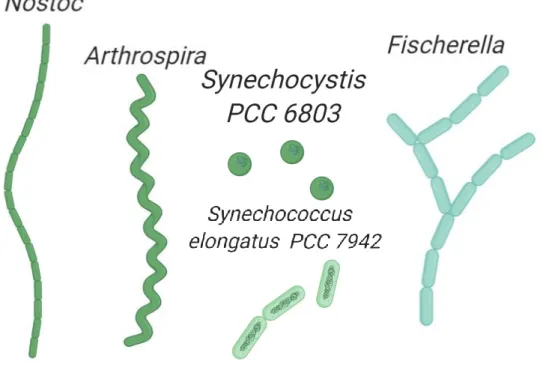
Their morphology can vary from unicellular to filamentous depending on the strain. While strains like Synechocystis PCC 6803 (hereafter Synechocystis) are spherical-shaped unicellular, other strains like Synechococcus elongatus PCC 7942 (hereafter Synechococcus) and Thermosynechococcus elongatus are rod-shaped unicellular and could be found in the environment as isolated, paired, linearly connected (2 to 4 cells), or in small clusters. Finally, other strains such as Nostoc punctiforme PCC 7120 (hereafter Nostoc) or Scytonema Hofmannii PCC 7110 (hereafter Scytonema) can form long filamentous structures. The latest allows cell differentiation among different cells belonging to a filamentous, in the case of Nostoc four types of cells are possible: vegetative cells (photosynthetic cells, it is the most abundant type under favorable growth conditions), heterocyst
(containing-nitrogenase enzyme cells involved in nitrogen fixation), akinetes (highly resistant spores formed under stress conditions) and hormogonia (gliding filamentous able to disperse in short distances, this cell type is involved in plant symbiosis)10. Based on these different properties, cyanobacteria have been classified into five different subsections. Subsections I and II are unicellular coccoids, the first divided by binary fission and the latest can additionally divide by multiple fission. Subsections III-V are filamentous cyanobacteria, subsection III filaments are formed by only vegetative cells, whereas subsection IV and V can undergo differentiation. In addition, subsection V can form branch patterns11.
This big diversity of phenotypes also is observed on the impact of cyanobacteria in other organisms, both negative and positive interaction have been reported. A good example of positive interaction is the symbiosis between cyanobacteria, usually belonging to the genus Nostoc, and another type of eukaryotic organisms. Generally, cyanobacterial symbionts supply fixed nitrogen to their host. The hosts can vary from fungi or algae to higher plants. On the other hand, cyanobacteria are also capable of form massive, high-dense blooms usually known as "harmful algal blooms" (HAB)12. HABs have considerable negative effects on the ecosystem. Cyanobacterial overgrowing depletes oxygen levels and decreases the sunlight in lakes and rivers, killing the other organisms. In addition, many bloom-forming cyanobacteria are toxin producers. Microcystin, produced as secondary metabolite, is one of the most common cyanotoxins. It is a potent inhibitor of protein phosphatases that poisons other organisms including mammalians. Most important, the occurrence of HABs is rising due to climate change (temperature increasing at the surface of water reservoirs) and increase of nutrients levels (especially phosphate) in lakes/rivers by human activity.
Symbiosis interaction involving cyanobacteria could have been extremely important in the first stages of eukaryotic evolution. It was proposed in 190513 that chloroplast, in eukaryotic cells, were derived from an ancestral cyanobacterium engulfed by another prokaryote. This so-called Endosymbiotic theory is supported by many experimental observations such as the similar way to replicate (binary fission) of organelles and prokaryotes and the presence of circular DNA and double membranes in organelles.
Photosynthesis: A general overview
4
Figure 1 | Diversity of cell morphology in Cyanobacteria. Schematic representations of
filamentous cyanobacteria Nostoc (subsection IV), Arthrospira (subsection III) and Fischerella (subsection V) are shown. In addition, unicellular cyanobacteria representations of Synechocystis and Synechococcus, both belonging to the subsection I, are presented.
Photosynthesis: A general overview
One of the most important biological processes on earth is the utilization of sun light to produce biologically compatible energy. This process, called photosynthesis, is a light-induced redox (reduction-oxidation) chemical reaction that can be formulated in a general way as:
𝐶𝑂2+ 2𝐻2𝐴 → (𝐶𝐻2𝑂) + 2𝐴 + 𝐻2𝑂 (eq. 1)
In the equation 1, H2A is the electron donor of the redox reaction and 2A is the
oxidized sub product. The oxygenic photosynthesis is a special case of photosynthesis where H2A is H2O and 2A is O2. Other types of anoxygenic
photosynthesis are possible, for example by using another electron donor such as H2S. This oxidation reaction is coupled to the reduction of CO2 into organic
compounds suitable for biological usage.
Photosynthetic organisms can be classified according to the bi-product generated from photosynthesis (‘2A’ in equation 1). If the bi-product is Oxygen, the photosynthetic organism is considered oxygenic and it performs oxygen-evolving
bacteria phyla are capable of photosynthesis. But only cyanobacteria, together with plants and eukaryotic algae, carry out oxygen-evolving photosynthesis.
Since the first experimental observations14, it was suggested that two different process are involved in oxygenic photosynthesis: a “light-reaction” and a “dark-reaction”. Now it is clear that the so called dark reactions are also light dependent, and many enzymes involved in this phase require light-activation15. A better
explanation of the photosynthetic mechanisms can be achieved by dividing it into four different phases16. The first phase involved the light harvesting by the antenna
complex and the following energy transfer to the photochemical centers. All the
photosynthetic organisms contain pigments (see the ‘Photosynthetic components: pigments’ section) which absorb light photons that ultimately are converted into chemical energy by photosynthesis. It was proved long time ago that not all the pigments are capable of photochemistry17. In fact, a large amount of pigments are part of antenna systems that transfer the energy18 to special pigments to carry out
photochemistry. The second phase is the charge separation and primary electron
transfer in reaction centers. In this phase, the excitation energy absorbed by the
antenna arrives into the reaction centers, a pigment-protein subunit capable of photochemistry. Combined, the antenna system and the reaction center form the photosynthetic unit (photosystem). In this transmembrane multiprotein complex, a special dimer of chlorophylls (green pigment) is promoted to an excited electronic state in which it became a strongly reducer species. This species rapidly transfers an electron to an electron acceptor (EA) generating an ion-pair state (P+EA-). The
electron could return to the chlorophyll-center in a recombination reaction, but this is generally avoided by the third phase, the stabilization by secondary reactions. After the charge separation is generated, very fast secondary reactions promote an electron transfer cascade that separate the negative and positive charges in the photosynthetic apparatus, avoiding recombination. This electron transport is coupled to the proton transmembrane movement and generates an electrochemical potential gradient. Afterwards, this gradient is used to generate ATP. The final phase is the synthesis of energetic products. In this phase, by using the ATP and the reduced compound (NADPH2) generated in the first three phases; the CO2 is
fixed into organic molecules. This last part of the photosynthesis is beyond of the scope of this work and it will not be explained in detail.
The first three phases (the light reactions) take place in the photosynthetic apparatus, a combination of pigment-binding protein complexes embedded in the thylakoid membrane. In plants, thylakoids membranes are an inner membrane system in the chloroplasts. In cyanobacteria, like in other prokaryotes, organelles (for instance, chloroplasts) are absents. Instead, a membrane system which is different of the cell membrane, harbors both photosynthetic and respiratory electron
Photosynthesis: A general overview
6
transport chains. This is one of the biggest differences between plant and cyanobacterial photosynthesis. Due to the fact that both electron transports are located in the same membrane system, in cyanobacteria some components are shared between photosynthetic and respiratory apparatus19. Electron carriers such as plastoquinol and plastocyanin (or cyt c6), as well as the cytochrome b6f, are
involved in electron transport in both apparatuses.
Figure 2 | Photosynthetic apparatus. Diagram of the composition of the photosynthetic membrane
in cyanobacteria (A) and plants (B). While in Plants the light-harvesting complex II (LHCII) in the membrane is the main antenna of photosystem II (PSII), in Cyanobacteria the soluble phycobilisome (PBS) plays that role. In both cases, the energy is transferred to the PSII. This energy is used by the PSII to reduce plastoquinol (PQ). PQ oxidization by the cytochrome b6f drive reduction of
plastocyanin (PC) (or cytochrome c6, in cyanobacteria).
A schematic representation of the oxygen-evolving photosynthetic apparatus from cyanobacteria and plants are shown in the figure 2. The basic components are the antenna complex (pigment-protein complex that harvest the light), photosystem II (PSII), plastoquinone, cytochrome b6f, plastocyanin and photosystem I (PSI). The
self-evident conclusion that can be drawn is that the main structural difference between both apparatuses is the antenna complex. In fact, the antenna complex is
the most diverse component among all photosynthetic organisms. It has been “re-invented” several times during evolution.
Photosynthetic components: pigments
The key players in harvesting the light energy are the pigments. These molecules play a functional and structural role in the photosynthetic apparatus. In this section, the structural and spectral properties of the main pigments involved in photosynthesis are shortly reviewed.
Chlorophyll. The main pigment in the reaction centers is the chlorophyll (Chl),
a planar organic molecule derived from a pyrrole. Diverse type of chlorophylls can be found in nature as a result of minor enzymatic modifications20. The most distributed chlorophyll in oxygenic photosynthetic organisms is Chl a, it occurs in reaction centers, in which it plays a major role during charge separation, and in light-harvesting complexes21. Oxidation of the methyl group at C-7 in Chl a into a formyl group result in a blue-shifted chlorophyll (Chl b) synthesis22. Chl b is found
in the accessory light-harvesting complexes in eukaryotic organisms. The only cyanobacteria containing this chlorophyll are the prochlorophytes23 which lack phycobiliproteins. Chl c which lacks the isoprenoid tail is commonly found in brown algae involved in the light-harvesting complex24. Chl d is found only in cyanobacteria belonging to the cyanobacterial genus Acaryochloris, representing the 95-99% of the total chlorophylls in this strain. Chl d absorbance is red-shifted compared with Chl a, and it is involved in light harvesting and reaction centers in these strains25. The recently discovered Chl f is the most red-shifted chlorophyll26.
It can be found as a minor component in some cyanobacteria produced under far-red growth light and it was shown that can occur in reaction centers performing photochemistry27. The structure of chlorophyll a, shown in the figure 3A, is composed of five cyclic substructures (rings) with a Magnesium atom coordinated to four Nitrogen atoms which are part of the inner rings. Chl a presents two mayor absorption bands, one in the blue region (around 430 nm) called soret band and other two in the red region (around 650 nm) called QX and QY. Light absorption in
the soret band lead to a second excited state (S2) that rapidly (in femtoseconds) decay to the lowest excited state (S1). This state is involved in all energy transfer process in photosynthesis.
Carotenoids. Among the most distributed pigments in the nature, carotenoids
are contained in almost all organisms despite the fact that are only produced in plants, algae and some bacteria (for instance, cyanobacteria) and fungi. There are hundreds of different carotenoids that play different roles in nature28, from photosynthesis to nutrition and disease prevention. In this work I will focus only on
Photosynthetic components: pigments
8
photosynthetic relevant carotenoids and their function on this process. They are elongated molecules formed by single and double carbon bounds containing one ring at each end. Examples of the carotenoid structure of Hydroxy-Echinenone, Echinenone, β-Carotene and Canthaxanthin are provided in the figure 3B. The conjugated π-electron system exhibits a strong absorption band in the range between 400 nm and 500 nm. This electronic transition is from the ground state (S0)
to the second excited state (S2)29. Transitions to the lowest excited state (S1) are
forbidden because of the symmetric of the carotenoid molecule30. Excited S2 relax
very fast (femtoseconds) to the S1 and from there to the ground state in a
non-radiative way31,32. In addition, ultrafast spectroscopy demonstrated a strong effect
of the solvent polarity on the S1 lifetime33. This was attributed to an additional excited state lying below S1 or coupled to S134, in which the electron density is shifted to the terminal ring of the polyene chain, designatede as Intramolecular Charge Transfer (ICT)32,35. Nevertheless, both excited states (S2/S1) play an
important role in photosynthesis. It is well documented the role of carotenoid in light harvesting. It was shown long time ago, that carotenoid can absorb and transfer energy to chlorophylls36 in the major antenna complex of plants and green algae (Light Harvesting Complex of Photosystem II, LHCII). In this case, excitation from carotenoid is transfer to QX or QY. Similar phenomenon had been observed in
Photosystem I from cyanobacteria37. Maybe the most investigated function of carotenoids in photosynthesis is the quenching of singlet-oxygen species (1O2)38.
This toxic molecule is generated in stress conditions and carotenoids can both decrease its concentration and prevent its formation (see the ‘Photoprotection: introduction to light stress’ section).
Phycobilins. In cyanobacteria and red algae, the antenna complex contains different
pigments than in plants, open tetrapyrroles that absorb light between 550 nm and 650 nm. The spectral properties of the phycobillins allow these organisms to benefit of a broader light spectrum. There are four kinds of phycobillins present in the nature: phycocyanobillin (PBP), phycoerythrobilin (PEB), phycoerythrocyanin (PEC) and phycoviolobilin (PVB). The structure of phycocyanobilin is shown in the figure 3C.
Figure 3 | Pigment Structures. Structure of Chlorophyll a is shown in (A). Representative
structures of carotenoid are shown in (B): Hydroxy-Echinenone, Echinenone, β-carotene and Canthaxanthin. Phycocyanobilin structure is shown in (C).
The spectral properties of the pigments described above are finely tuned by the protein environment in which they are embedded. These protein complexes, located in the thylakoid membrane, have evolved to bind and utilize the pigments to harvest and convert the light energy. In the following sections, the main protein complexes involved in oxygen-evolving photosynthesis in cyanobacteria are described and the main differences with plants are pointed out.
Photosynthetic components: Antenna complex
As it was mentioned before, a large part of the photosynthetic pigments that only collect and transfer the energy are in the antenna. Two big groups can be distinguished: integral-membrane antennas and extrinsic antennas. Another variant of antenna system are the inner membrane pigment-protein complexes (CP43 and CP47) that are closely associated with the reaction centers and are present in all oxygenic photosynthetic organisms. These antenna systems will be described together with the reactions center in the description of the photosystems.
Photosynthetic components: Antenna complex
10
A well-known example of membrane-bound antenna is the Light Harvesting Complex II (LHCII), present in plant but not in cyanobacteria. It exists as trimer and each monomer consist of a polypeptide of about 25 kilodaltons (kDa) binding several pigments: 8 chlorophylls a, 6 chlorophylls b and 4 carotenoids39. The chlorophylls are separated by approximated 10 Å, this close distance enable fast energy transfer between them (exciton equilibration in the LHCII trimer takes place in a few picoseconds) and effective energy transfer to the photosystem II. LHCII is part of a protein family called lhc. Other lhc genes, lhc1-4, are involved in the LCHI, the outer antenna complex associated with photosystem I40. The overall structure is well conserved in the lhc family as well as the pigment binding sites41,
but slight differences in the protein environment tune the photochemistry of each antenna component. In addition, between the core of PSII and the trimeric LHCII, there are minor monomeric antennas from the same gene family, namely CP24, CP26 and CP2942.
The best example and the most relevant for this work of extrinsic antenna is the
phycobilisome (PBS), the cyanobacterial antenna (Figure 4). The phycobilisome is
a large water-soluble pigment-protein complex that carries out light harvesting in cyanobacteria and red algae. The pigments of the phycobilisome are the phycobillins that are covalently attached to proteins, called phycobiliproteins. Depending on the number and type of phycobilin that is bound, the phycobiliproteins are divided in 4 kinds16: allophycocyanin (APC, λmax = 650 nm),
phycocyanin (PC, λmax = 620 nm), phycoerythrin (PE, λmax = 560 nm) and, the less
frequent, phycoerithrocyanin (PEC, λmax = 575 nm). Specific lyases catalyze the
covalent attachment of the bilin to phycobiliprotein cysteines, for example APC and PC contain phycocyanobilin. The different protein environments of the phycobilin in the different proteins give rise to different spectral properties which allow a very efficient energy transfer within the phycobilisome.
Figure 4 | Phycobilisome structure. (A) Side view of a Synechocystis phycobilisome. It is
composed of 6 rods formed by 3 phycocyanin trimers radiating from the core. The core (zoom view of bottom cylinders in B) is composed of APC trimer (shown in sky blue), APC trimer containing ApcD (shown in violet), the APC trimer containing ApcF and ApcE (shown in pink) and the linker protein, ApcC (shown in green). The rods are formed PC (dark blue). (C) The APC trimer structure is comprised of APC-α (green) and APC-β (orange), both bind billins (shown in blue). The APC structure used in the schema and in (C) correspond to PDB: 4PO5. Adapted from Muzzopappa and Kirilovsky 2019.
Despite the fact that different variants can be found in the nature, the most common architecture of the cyanobacterial phycobilisome (called hemidiscoidal PBS) is a fan-like structure with PE/PC rods that radiate from an APC core. All the phycobiliprotein can be found as heterodimer formed by two polypeptides, defined as chain α or β. Both α and β, are globular proteins (around 20 kDa) composed of eight helical elements. Evolutionary analysis of these proteins suggested that they evolved from the same ancestor and followed the same evolutionary pathway, giving rise to the same structural patterns (APC, PC, PE and PEC). This equal evolution of the α and β polypeptides (into APC, PC, PE and PEC) indicate that a phenomenon of co-evolution took place, guided by a strong interaction between α
Photosynthetic components: Antenna complex
12
and β. The basic subunit of the phycobilisome is formed by a heterodimer α /β that are assembled into ring-shaped trimers (α/β)3 and further into hexamers (α/β3)2.
Four trimers of APC heterodimers are associated forming cylinders in the phycobilisome core. The number of cylinders in the core depends on the strain: for example, Synechocystis have three cylinders arranged in a pyramid-like fashion, Nostoc has five and Synechococcus elongatus has two. The cylinders are hold together due to the binding of specific linker proteins. The basal cylinders which are in contact with the thylakoid membrane have a different composition than the upper cylinder. Two trimers are made up of pure APC α/β subunits. In the third trimer, one β subunit is replaced by ApcF and in the other subunit, an α subunit is replaced by ApcE. In the last trimer, in one subunit, the α polypeptide is replaced by ApcD. ApcF, ApcD and ApcE are modified APC proteins, with a red shifted spectrum compared with the bulk of APCs, ApcE also contain a linker domain that maintain the PBS core stability. The linker protein ApcC also stabilize the APC cylinders. In a similar way, the PBS rods are composed of two or three PC hexamers connected by linker proteins. Most of the marine cyanobacteria have PE and/or PEC hexamers at the end of the rods. These phycobiliproteins are blue-shifted compared with PC and expand the usable light range.
The overall arrangement of chromophores in the PBS facilitates the downhill energy transfer from the rods to the core and from there to the photosystems. Due to the intermediary distance between chromophore, around 20-40 Å, the model proposed is the Foster resonance energy transfer that required weak dipole coupling between chromophores and overlapping of emission spectrum of the donor and absorption spectrum of acceptor. Therefore, excitation of PC (Absorption λmax =
620 nm, Emission λmax = 645 nm) is transferred to the APC in the core (Absorption
λmax = 650 nm, Emission λmax = 660 nm). Then the energy is transferred towards
the terminal emitters (ApcD, ApcF and ApcE, Absorption λmax = 670 nm, Emission
λmax = 680 nm) and from there to the chlorophylls in the photosystems.
The PBS interact with the stromal side of the thylakoid membrane possibly through ApcE43. PBS diffusion on the membrane surface allow energy supply to both photosystems43. Phycobilisome are thought to be the main antenna of PS II, albeit they can also interact and transfer energy to PS I. It seems that the fraction of energy delivery to PSI or PSII depends on the strain, in Synechocystis44 is 50% to each photosystem but in Arthrospira45 80% is transferred to PSI. Depending on the strain, the different terminal emitters can interact distinctively with the photosystems, creating different energy pathway towards PSII and PSI. In Synechococcus elongatus sp. PCC 7002 deletion of ApcD render in a decrease of energy transfer to PSI and deletion of ApcF decreases energy transfer to PSII46. On the other hand, in Synechocystis ApcF is involved in energy transfer to both
photosystems and deletion of ApcD has a minor role in energy transfer47. Recently, it was shown that ApcD was involved in energy transfer to PSI and ApcF had a minor role in energy transfer in Synechococcus elongatus PCC 794248 (see “appendix 1: Additional publications”). In some strains, these proteins have a role on the regulation of the energy arriving to the photosystems (see below ‘Photoprotection: cyanobacterial state transition’).
Photosynthetic components: Photosystem II
In organism performing oxygen-evolving photosynthesis, such as plants, green algae and cyanobacteria, there are two photosynthetic units: Photosystem I (PSI) and Photosystem II (PSII). PSII is very unique biological complex due to the fact that is capable of carrying out a very thermodynamically demanding reaction, the water oxidation. A great effort was dedicated to isolate and study the photosystem II, leading to the elucidation of the structure of several PSII complexes from different organisms49–51, showing high degree of similarity between them.
The photosystem II is a membrane-integral complex composed of two identical monomers, each one of them formed by 19 protein subunits. Each monomer contains 36 chlorophyll a, and 7 carotenoid molecules. A schematic figure50,52 of cyanobacterial PSII is shown in figure 5A. The core of the reaction center is formed by two homologue proteins, D1 (encoded by the PsbA gene) and D2 (encoded by the PsbD gene), forming a symmetric heterodimer. Each monomer comprises of five α-helices exposing the N-termini to the stromal side of the thylakoid membrane. A chlorophyll dimer in the D1/D2, designated as PD1 and PD2, are
weakly coupled. The presence of other pigments in each subunit, another chlorophyll a (ChlD1 or ChlD2), one pheophytin (PhD1 or PhD2) and one plastoquinol
(QA and QB) creates two branches for the electron transfer chain (Figure 5B).
Interestingly, only one branch is active. After the excited state of the special pair PD1/PD2 is reached, the energy is enough to promote charge separation and the state
PD1+PhD1- is formed, via the intermediary ChlD153. Due to its spectral properties,
PD1+ is usually named as P680+. The radical pair is quickly stabilized (in about 400
ps) by electron transfer to QA, which is strongly bound to the protein matrix and it
is not exchangeable. The QA role is to receive a single electron and mediated the
double reduction of QB, which after being doubled reduced (by two successive
charge separations) can be replaced by a new plastoquinone from the plastoquinone pool. Attached to the lumenal side of D1/D2 heterodimer, there is an inorganic complex, Mn4CaO5, called Oxygen Evolving Complex (OEC)51. This complex
catalyzes the oxidation and splitting of water to restore the electron donated by P680+. Photo-oxidized P680+ drags one electron from the Mn atoms of OEC
Photosynthetic components: cytochrome b6f
14
through the Tyrosine 161 of D1 (Tyr Z). Four sequential oxidations of the OEC allows this complex to oxidize two water molecules and produce O2 and protons.
Specific amino acids of D1 (and one from CP43) coordinate the magnesium cluster and others protein (PsbO, PsbU, PsbV and extrinsic loops of D1, D2, CP43 and CP47) are required to stabilize it54. In cyanobacteria, there are several isoforms of D1. These isoforms have different tolerance to oxidative damage due to slight difference in the amino acid sequence. Therefore, the isoforms are differentially expressed depending on the light conditions.
Formed by two subunits (α and β) and one heme group, the cytochrome b559 (Cyt
b559)is located near the D2. The Cyt b559 is involved in PSII assembly55 and it is
essential for its photochemistry, since the minimum PSII active complex is composed of D1/D2/Cyt b55956. The role of Cyt b559 is still enigmatic but several
photoprotective mechanisms involving this component have been proposed57. To increase the light absorption, two antenna subunits are located surrounding the D1/D2 core. CP43 is composed by five helices and it contain 14 chlorophylls while CP47 is composed by six helices and three β-sheets containing 16 chlorophylls. Two small subunits, PsbI and PsbX stabilize the two peripheral chlorophylls (ChlZD1 and ChlZD2, respectively). In the lumenal side, PsbV, PsbU and PsbO
enclose the OEC.
Photosynthetic components: cytochrome b
6f
Another essential player of the electron flow in cyanobacterial membranes is the cytochrome b6f. This protein complex is a dimer with eight subunits per monomer.
Among these subunits, cytochromes (proteins that covalently bind one or more heme groups) are found58. Cytochrome f is formed by a single transmembrane helix that anchors the soluble domain, which harbor a heme group, in the luminal side of the membrane. The subunit cytochrome b6 possess four transmembrane helices and
coordinates two heme groups (bn and bp) and, an additional heme group, cn that is
electronically coupled with bn. An important component of the cytochrome b6f is the Rieske Fe-S protein. This subunit consists of two domains, a transmembrane N-terminal domain and a soluble domain which coordinate a Fe-S cofactor. Another four small subunits (PetG, PetL, PetM, and PetN), with structural role, and the subunit IV complete the complex monomer. In addition, one carotenoid and one chlorophyll, of unknown function, are embedded on the complex.
Figure 5 | Photosystem II structure. (A) Schematic side view of dimeric PSII complex. The
schematic representations are based on the PSII structure of Thermosynechococcus from Kamiya et al. 2003 and Umena et al. 2011. (B) Electron pathway in the reaction center of PSII, adapted from Cardona et al. 2012. The first charge separation originated the radical pair ChlD1+ PhD1- that rapidly
is converted into the more stable P680+ PhD1- (1). Then the Ph- can donate one electron to the
plastoquinone in QA (2). P680+ is reduced by the OEC via the TyrZ (3 and 4). Finally, the doubled
reduced QA can reduce the plastoquinone in QB (5).(C) Electron pathway in the reaction center of
PSI, adapted from Makita et al. 2017. Excitation of the reaction center of PSI lead to charge separation of P700+ A0-, that can reduce A1 and then Fe-S cluster FX. The electron moves quickly from
FX to FB through FA.
The electron transfer within the complex is well known and defined as “Q cycle”59. One reduced plastoquinone (PQH
2) binds to the Q0 site at cytochrome b6,
its oxidation lead to one electron transfer to the external plastocyanin, a soluble small copper-binding protein, through the Fe-S cluster and one electron transfer to one oxidized plastoquinone (PQ) bound to the Qi site, through the heme groups bn
Photosynthetic components: Photosystem I
16
and bp. In this first half cycle two protons are released to the lumenal side and one
proton from the stromal side was consumed during reduction of PQ into PQH in the Qi site. During the second half cycle, another PQH2 is fully oxidized, releasing the
2 protons in the lumenal side. One electron is transported to another plastocyanin and the other is used to reduce the PQ- in the Qi site to PQH2, consuming another
proton from the stromal side. The full cycle drives the oxidation of one PQH2,
reduction of two plastocyanin, the transport of two protons and the accumulation of two extra protons in the lumenal side.
Photosynthetic components: Photosystem I
The second photosynthetic unit in the oxygen-evolving photosynthetic apparatus is the photosystem I (PSI). PSI, like PSII, is a transmembrane pigment-protein complex. From a structural point of view, PSI from plants and cyanobacteria present some important differences. Maybe, the most remarkable difference is that cyanobacterial PSI, depending on the growth conditions can be found as trimer (in vivo60 and in vitro61) or monomer62. Plant PSI was found to be monomeric. This evolutionary loss of trimeric formation of PSI in plants may be explained by the fact that plant PSI incorporates antenna complex LHCI and the trimeric architecture of PSI does not allow such antenna binding63. According to this, LHCI is not found
in cyanobacteria64. Monomers of cyanobacterial PSI are formed by 12 subunits with
a molecular weight around 80 kDa. The core of the reaction center I is a heterodimer PsaA-PsaB which binds a special pair of chlorophylls (defined as P700), four additional chlorophylls (pair A and pair A0) and two phylloquinones molecules. An
important difference between PSII and PSI is that PSI core also contains one iron-sulfur cluster (Fe-S), FX. Another two Fe-S clusters (FA and FB) are bound to PsaC
protein, located next to FX in the stromal side. These Fe-S clusters play an important
role in the primary electron transfer. Additional pigments surrounding P700 form the intrinsic antenna of PSI, increasing the effective absorption area of PSI. Around 79 chlorophylls are bound to PsaA-PsaB and another 11 chlorophylls and 22 carotenoids are coordinated to the small subunits PsaJ, PsaK, PsaL, PsaM, and PsaX.
When the excitation reaches the PSI core a primary charge separation takes place leading to the oxidation of P700 into P700+ and the reduction the chlorophyll A0 (Figure 5C). Then, the electron moves quickly to FX through the phylloquinone A1.
The Fe-S cluster FX transfers the electron to FA, and from here it is transferred to
FB65. Ultimately the electron is used to reduce a soluble protein called Ferredoxin.
Due to the symmetry of PsaA-PsaB two branches for the electron transport are present. Interestingly, while in PSII only one branch is active, in PSI both branches
are active. The reduced state of P700+ is restored by electron transfer from the plastocyanin.
Photosynthetic electron transport: linear versus cyclic
As it was mentioned before, the light harvested by the antenna complexes induces a redox reaction in the core of the photosynthetic units PSII and PSI. This triggers an electron transport across the photosynthetic membrane with two important consequences: the generation of reducing power and the generation of a transmembrane potential by coupling the electron transport to proton accumulation in one side of the thylakoid membrane.
The canonical photosynthetic electron transport follows a linear fashion. Upon light absorption by the phycobilisomes (or LHCII), energy is transferred to PSII. This leads to the reduction of the plastoquinol pool consuming 2 H+ from the stromal side to generate PQH2, as it was described before. Water splitting by the
OEC produces 4 H+ in the lumenal side. The electrons flow from the plastoquinol
pool to the cytochrome b6f oxidizing PQH2 into PQ and accumulating four H+ in
the lumenal side, as it was detailed before. Plastocyanin transport electrons, one by one, from cytochrome b6f to PSI. These electrons are used to re-reduce the
photo-oxidize P700+ of PSI that was previously oxidized by the Ferredoxin. Ferredoxin is a soluble protein carrying a Fe-S cluster which is capable of reducing another protein, the Ferredoxin-NADP Reductase (FNR). FNR is a two domains protein. One domain binds Flavin Adenine Dinucleotide (FAD) and its function is to receive one-by-one electrons from Ferredoxin. The other domain binds NADP+ and
catalyzes the two-electron reduction to NADPH2. The transmembrane proton
gradient generated is ultimately used to catalyze phosphorylation of adenosine diphosphate (ADP) into adenosine triphosphate (ATP) by the ATPase. Both ATP and NADPH are used to fix CO2 into biologically usable molecules via the
Benson-Calvin cycle.
An alternative electron flow can take place if electrons are re-injected in the photosynthetic chain to circulate around PSI66. This mechanism leads to the
decrease of NADPH2 formation and an increase of proton accumulation on the
lumenal side of the membrane, leading to a greater ATP production and less reductive power. In cyanobacteria, two main pathways are involved in cyclic electron transport. The NDH-1 complex, a transmembrane protein complex that resembles the Complex-I of the respiratory chain in mitochondria67, is capable to receive electrons from Ferredoxin68 and transfer them to PSI via the PQ pool. This complex can bind Ferredoxin69 and NDH-1:PSI supercomplex has been detected70. In plants, the second pathway of electron recycling is through a
Photoprotection: introduction to oxidative stress
18
Ferredoxin/plastoquinone oxidoreductase named PGR564. PGR5 is a transmembrane protein which directly catalyzes the reduction of PQ and the oxidation of Ferredoxin. A second pathways exist on cyanobacteria, PGR5 homologues were suggested for this71 but the exact identity of the mechanism remains unknown.
Photoprotection: introduction to oxidative stress
In photosynthetic organisms, detrimental effects are triggered due to the over-reduction of the photosynthetic electron transport chain. This over-over-reduction of the chain can be generated by exposure to high lights or other conditions such as nutrient starvation and CO2 limitation. Under these conditions light-induced
reactions produce Reactive Oxygen Species (ROS) in the cell. ROS, that can be hydroxyl radical (•OH), hydrogen peroxide (H2O2), superoxide radical (•O−2) and
singlet oxygen (1O2), are highly reactive molecules that can damage DNA/RNA
molecules, oxidize lipids, proteins and cofactors. The long-lived excited state of chlorophyll, required for energy transfer during photosynthesis, plays a role in 1O2
production. In this long-lived state, the spins of the electrons of excited chlorophyll can rephase and populate the lowest energy-excited state, the chlorophyll triplet state (3Chl). 3Chl can react with 3O2 (the most common form of molecular oxygen)
producing singlet oxygen. The vast majority of chlorophylls are present in the antenna, in which the presence of nearby carotenoids decreases significantly the
1O
2 production73. The close distance between pigments in these complexes, allow
orbital overlap and spin-exchange reaction that quench the 3Chl by populating the carotenoid triplet state. This state is safely dissipated as heat. Nevertheless, at high light intensity both singlet oxygen production and protein degradation have been detected at the level of the PSII antenna74.
Most of the 1O2 is generated in PSII (reviewed in75). In high light conditions, the
plastoquinol pool is completely reduced and the photosynthetic electron transport is blocked. Charge recombination between the primary quinone acceptor (QA-) and
P680+ takes place. Two possible pathways are possible, the first one involves the
direct recombination into P680 and QA. The second possible pathway of charge
recombination goes through the repopulation of the first radical pair (P680+Phe-).
This radical pair can also undergo triplet radical formation, 3[P
680+Ph-], that
ultimately lead to the formation of 3P
680, which can react with 3O2 and produce
singlet oxygen. In the reaction center, the carotenoid molecule is located too far from the chlorophylls to quench the 3Chl state and singlet oxygen is efficiently
generated. The 1O2 can diffuse in the reaction center and damage the D1 subunit
leading to its degradation in a process called photoinhibition. This process take place even at low light intensity, but since the repair of PSII is very efficient in vivo only at high light intensities a net loss of PSII activity is observed76. It is important to note that other physiological conditions such as low temperatures or CO2
depletion can favor the limitation of the acceptor side of PSII and increase the probability of charge recombination. Additionally, the midpoint of QA can favor the
different recombination pathways. In cyanobacteria, an alternative isoform of D1, encoded by psbA377, replace D1 (psbA1) at high light conditions. This isoform shifts the redox potential of pheophytin, favoring the non-radiative recombination pathway without 1O2 production.
The cytochrome b6f and PSI also harbor chlorophylls and are theoretically
capable of 1O2 production. However, it has been proved that the contribution from
these complexes is minor compared with PSII. In cytochrome b6f, the protein
environment shorts the lifetime of the excited chlorophyll decreasing the chances of populating the triplet state78. Another potential source of 1O2 generation is during
chlorophyll synthesis, accumulation of precursors can also increase the singlet oxygen formation. In the electron acceptor side of PSI, the excess the photo-excitation can lead to reduction of O2 into O2- 79,80 (this reaction is also called
Mehler reaction) by the primary electron acceptors, which is further reduced to H2O2 and OH- 81. These ROSs can damage and inhibit PSI. Since the PSI turnover
time is about days82, photosynthetic organisms have mechanisms to keep P700
oxidized and avoid ROS production (see “ROS scavengers and alternative electron pathways.”). In agreement with this, PSI photoinhibition hardly takes place in vivo82.
In order to cope with the photo-oxidative stress, plants and cyanobacteria had developed many photoprotective mechanisms. Some of these mechanisms are collectively denominated as Non-Photochemical quenching (NPQ) since they decrease the chlorophyll fluorescence by increasing the thermal dissipation and, as consequence, reducing the energy arriving to the photosystems. They can be sorted by the molecular level at which they take place: for instance, preventive mechanisms dissipating the excess of energy at the antenna level or other mechanisms directly deal with already generated ROS. They also can be sorted by the time range when they take place: we can distinguish short-term mechanisms and long-term mechanisms. In the following sections, the principal photoprotective mechanisms of cyanobacteria will be introduced with special focus on the OCP-related Non-Photochemical-Quenching.
ROS scavengers and alternative electron pathways.
20
ROS scavengers and alternative electron pathways.
Cyanobacteria, as well as many organisms, contain enzymes to detoxify reactive oxygen species. Synechocystis uses an Iron Superoxide Dismutase (Fe-SOD) in order to catalyze the dismutation of 2 -O2 into H2O2 and O2. However, some marine
Synechococcus strains use copper/zinc SODs83. H2O2 is detoxified by different
enzymes: catalases, peroxidase and peroxiredoxins (reviewed in84). Beside the enzymatic reactions that decrease the ROS concentration presented above, cyanobacteria also contain small molecules capable of ROS scavenging. An already described example are the carotenoids. In addition, tocopherol can decrease the 1O2
concentration in the reaction center of PSII85. Collectively, these enzyme and
molecules deal with the ROS already produced in the photosynthetic machinery. In order to prevent the ROS production, other photoprotective mechanisms constitute an alternative pathway for electrons acting like a valve that releases the pressure from the electron transport chain. An example of this is the Plastid Terminal Oxidase (PTOX), that is present in some cyanobacteria86,87, receives electron from the PQ pool and reduce O2. This protect the PQ from over-reduction
and as a consequence avoid PSII photoinhibition.
Flavodiiron proteins (Flv) are a family of enzymes composed of two domains: the N-terminal metallo-β-lactamase-like domain harboring a non-heme diiron center capable of O2 and/or NO reduction and the C-terminal flavodoxin-like
domain, containing a flavin mononucleotide (FMN). The Flv found in photosynthetic organism belong to the Flv subgroup C that contains an additional domain that allows NADPH to be used as electron donor (reviewed by Allahverdiyeva et al.88). Cyanobacterial Flv can be clustered into four different groups, Flv 1 to Flv4. Flv1-Flv3 form a heterodimer that catalyze a “Mehler-like” reaction in the donor side of PSI. In this reaction, Flv1-Flv3 oxidize NAD(P)H and reduce O2 to H2O in a four-electron reaction without ROS production. This
photoprotective role is very important under fluctuating light89. Some cyanobacteria, for instance Synechocystis also contain a second pair of Flavodiiron proteins forming another heterodimer, Flv2-Flv4. Although its mechanism is not clear yet, Flv2-Flv4 are proposed to mediate the electron transfer from PSII (or PQ pool) to an unknown electron sink88,90 however this model is still under debate.
Photoprotection: State transitions
The light intensity is not the only issue for photosynthetic organisms, the quality the light (the portion of the electromagnetic spectrum irradiated, measured in wavelength) also can affect the photosynthetic capacity. Since in cyanobacteria PSI
contains more chlorophylls than PSII, illumination with blue (absorbed mostly by chlorophylls) or far-red light (absorbed mainly by PSI) will stimulate more PSI activity compared with PSII activity leading to oxidation of the components between the photosystem, such as the plastoquinol (PQ) pool. Since the main antenna of PSII is the PBS, orange illumination (mostly absorbed by PBS) leads to the induction of PSII activity and the reduction of the electron chain (and the PQ pool). This imbalance on the activity of the photosystem can promote the ROS production or/and decrease the photosynthetic capacity.
Plants, algae and cyanobacteria have developed a mechanism to re-balance the energy distribution between the photosystems called “state transition”. The imbalance is sensed by the redox-state of the PQ pool91. In darkness, cyanobacterial cells perform respiration (in the same thylakoid membrane) which lead to reduction of PQ pool. Under this condition cells are in State II, this state is characterized by a low PSII to PSI fluorescence ratio at 77K and a low PSII chlorophyll fluorescence yield at room temperature. If cells are illuminated with low intensities of blue or far-red light, PSI became more active and the PQ pool more oxidized inducing transition to State I. In this state, PSII Chl fluorescence yield increases at room temperature as well as the ratio PSII to PSI fluorescence at 77K. Illumination with orange light will activate PSII and reduce the PQ pool, inducing transition to State
II.
While it is accepted that in plants the redox state of the PQ pool is sensed by the cytochrome b6f which activate a specific kinase that phosphorylate LCHII92 and
induce the movement of antenna from PSII to PSI93, in cyanobacteria the
mechanism is not clear. The involvement of phosphorylation and cytochrome b6f
was controversial, but recently it was proved that none of them are involved94 in the mechanism of state transitions. Regarding the mechanism, based on diverse experimental observation several models were proposed: 1) Movement of PBS between the phycobilisomes. This model was proposed based on FRAP experiments that showed a high mobility of PBS compared with photosystems95,96.
According to this model, in state I PBS are bound to PSII and upon transition to state II they move to PSI. In addition, chemicals that alter the PBS-membrane interaction, such as phosphate, sucrose or betaine, inhibit the state transitions97. However, it was recently showed that these chemicals also affect processes in the membrane94. 2) Spillover between PSII and PSI98. This model requires a variable coupling between PSII and PSI. In State I this coupling is weak and the energy harvest by the PBS is transferred to the PSII while in state II strong coupling between PSII and PSI allows further transfer of this energy form PSII to PSI. This photosystem movement on the membrane is in agreement with the strong temperature dependence of the state transitions99 and electron microscopy
Photoprotection: OCP-related NPQ
22
observations100. Supporting this hypothesis, Liu and coworkers101 isolated supercomplexes involving both photosystems and PBS by using crosslinkers. 3) Detachment of PBS from PSII and Quenching in PSII. In this model is proposed that in state I the increase on room temperature fluorescence is due to PBS detachment from PSII. In addition, a recent work in Synechococcus elongatus PCC 7942 showed that in state II the PSII is quenched and not involved in spillover102.
The PBS are involved in almost all the models, as major antenna complexes, however the specific role of the antenna and the different terminal emitter is not so clear. As it was described before (see “Photosynthetic components: Antenna complex”) depending on the strain, the PBS terminal emitters play different role in the energy transfer to photosystems. In Synechocystis47, Synechococcus elongatus
PCC 700246 and Anabaena103, it was shown that deletion of apcD impaired state transitions. It was proposed that this was due to a permanent association between PBS and PSII, since ApcD was thought to be involved in energy transfer to PSI. On the other hand, deletion of apcF impaired state transition in Synechocystis but not in Synechococcus elongatus PCC 7002. Recently, a work48 carried out in our group, in which I collaborate by designing and constructing Synechococcus elongatus PCC 7942 mutants, showed that the role of these terminal emitters and their involvement on state transitions depend on the strain. In Synechococcus elongatus sp. PCC 7942, ApcD is involved in energy transfer to PSI but deletion of ApcF has no effect on energy transfer from PBS to any photosystem. By contrast, in Synechocystis ApcF is involved in energy transfer to both photosystems and deletion of ApcD does not affect the energy transfer47. In addition, while deletion of apcD in Synechocystis completely inhibit state transition and deletion of apcF also showed a strong alteration on this process, neither deletion of apcD nor apcF affects state transitions in Synechococcus elongatus PCC 7942. It is clear that the function of the terminal emitters is not the same in the different strains implying that their sequence or/and the overall architecture of the PBS dictate its role. Their role on state transition is not universal for all the cyanobacteria. Probably state transitions is the result of a series of concerted mechanisms and their relative contribution depends on the specific strain and the light regime that the strain experience.
Photoprotection: OCP-related NPQ
During evolution photosynthetic organisms have found their way to reduce the amount of photo-oxidative damage by just decreasing the energy arriving to PSII, the main source of ROS production. An elegant and efficient approach to achieve this relies on the thermal energy dissipation at the level of the antenna complex. This phenomenon can be followed by measuring how the chlorophyll fluorescence
yield decreases (due to a decrease in the antenna size of PSII) under high light illumination. This component of the (short-term) NPQ, named qE in plants and OCP-related NPQ in cyanobacteria, takes place in the time range of second and minutes. Another important feature of this mechanism is the reversibility of the process under normal light conditions.
A key factor in these mechanisms is the molecular sensing of the stressful conditions. In plants and green algae this is accomplished by sensing the ∆pH across the thylakoid membrane. Due to the molecular diversity of antenna among the photosynthetic organisms, the short-term NPQ mechanism is different in plants and cyanobacteria. In plants, it is known that protonation of PsbS subunit of LCHII, the xanthophyll cycle activation (conversion of Zeaxanthin to Violaxanthin) and aggregation of the antenna are required for the energy dissipation (reviewed in104). Besides that, the exact mechanism is still controversial.
In cyanobacteria, the absence of xanthophyll cycle and the lack of effect of ionic uncouplers on chlorophyll fluorescence (indicating that a ∆pH does not trigger any NPQ mechanism) prompted the researchers to hypothesize105 that there is not a short-term NPQ mechanism similar to plant qE in this phylum. Instead, the chlorophyll fluorescence changes were attributed to another NPQ mechanisms, state transitions (see above “photoprotection: state transition”). This view was challenged by El Bissati and co-workers in 2000 when they provided the first experimental evidence of the existence of this kind of mechanism99. They were able
to show the existence of a different process which takes place upon strong light illumination and it is independent on the temperature and membrane state. On the other hand, chlorophyll fluorescence changes during state transitions are dependent on the temperature and the membrane physical state. In addition, the authors were able to show that this quenching of the chlorophyll fluorescence was not related to photoinhibition and it can take place at low temperatures which inhibit state transitions. These experimental data pointed towards a novel photoprotective mechanism taking place at the level of the phycobilisome. The next step for describing this new mechanism was provided by Rakhimberdieva and co-workers, by measuring the action spectra of quenched cells (by inducing NPQ at high blue light illumination) they were able to proposed that the quenching was induced by a carotenoid molecule106.
The decisive breakthrough to decipher the NPQ mechanism on cyanobacteria was achieved by Wilson107 and co-workers in 2006. They could show that a previously described carotenoid protein, the Orange Carotenoid Protein (hereafter OCP) was directly involved in the cyanobacterial NPQ. The ΔOCP Synechocystis mutant showed a lack of blue-light induced NPQ. In addition, 77K fluorescence spectra of quenched and unquenched WT obtained by excitation of Chl or PBS,