HAL Id: in2p3-00348478
http://hal.in2p3.fr/in2p3-00348478
Submitted on 6 Jan 2009
HAL is a multi-disciplinary open access
archive for the deposit and dissemination of
sci-entific research documents, whether they are
pub-lished or not. The documents may come from
teaching and research institutions in France or
abroad, or from public or private research centers.
L’archive ouverte pluridisciplinaire HAL, est
destinée au dépôt et à la diffusion de documents
scientifiques de niveau recherche, publiés ou non,
émanant des établissements d’enseignement et de
recherche français ou étrangers, des laboratoires
publics ou privés.
Absolute cross-sections from X - γ coincidence
measurements
A. Lemasson, A. Shrivastava, A. Navin, M. Rejmund, V. Nanal, S.
Bhattacharyya, Avhishek Chatterjee, S. Kailas, K. Mahata, V.V. Parkar, et al.
To cite this version:
A. Lemasson, A. Shrivastava, A. Navin, M. Rejmund, V. Nanal, et al.. Absolute cross-sections from
X - γ coincidence measurements. Nuclear Instruments and Methods in Physics Research Section A:
Accelerators, Spectrometers, Detectors and Associated Equipment, Elsevier, 2009, 598, pp.445-449.
�10.1016/j.nima.2008.09.032�. �in2p3-00348478�
Absolute cross sections from X - γ coincidence measurements
A. Lemasson
a, A. Shrivastava
b, A. Navin
a, M. Rejmund
a, V. Nanal
c, S. Bhattacharyya
d,
A. Chatterjee
b, S. Kailas
b, K. Mahata
b, V.V. Parkar
b, R.G. Pillay
c, K. Ramachandran
b,
P.C. Rout
ba
GANIL, CEA/DSM - CNRS/IN2P3, Bd Henri Becquerel, BP 55027, F-14076 Caen Cedex 5, France
b
Nuclear Physics Division, Bhabha Atomic Research Centre, Mumbai 400085, India
c
Dept. of Nuclear and Atomic Physics, Tata Institute of Fundamental Research, Mumbai 400005, India
d
Variable Energy Cyclotron Centre, 1/AF Bidhan Nagar, Kolkata 700064, India.
Abstract
An activation technique using coincidences between characteristic X-rays and γ-rays to obtain absolute cross sections is described. This method is particularly useful in the case of nuclei that decay by electron capture. In addition to the reduction of possible contamination, an improved detection sensitivity is achieved as compared to inclusive mea-surements, thereby allowing the extraction of absolute fusion cross sections in the nano-barn range. Results of this technique for6
Li + 198
Pt system, at energies around the Coulomb barrier are described. Future applications with low intensity radioactive ion beams are also discussed.
Key words: X-γ coincidences, Activation, Fusion reactions, Absolute cross sections
1. Introduction
In general, it is necessary to reach the measure-ment limits to constrain the existence of new phe-nomena in any field of physics. The measurements of low cross sections for various nuclear processes similarly allow a detailed test of model assumptions. The recent evidence for a faster fall of the measured fusion cross section as compared to theoretical pre-dictions at energies much lower than the Coulomb barrier and its astrophysical implications have been discussed in Ref. [1]. The availability of low
inten-sity (∼ 105
pps) radioactive ion beams provides the opportunity to study the effect of exotic structures on the reaction mechanism. Both these studies re-quire making sensitive measurements and employ-ing techniques which are able to extract a weak sig-nal in the presence of a relatively large background. The fusion cross section is extracted from direct or indirect measurements of evaporation residues. The direct detection of evaporation residues can be per-formed using a recoil mass separator [1]. In the case of light and low incident energy beams, the small re-coil energies and large charge of the residue make these measurements difficult. Measurements of
in-beam characteristicγ-decay of excited evaporation
residues is another way of measuring the evapora-tion residues cross secevapora-tions [2]. It is thus possible to measure the individual evaporation residue cross sections and hence the (sum) total fusion cross sec-tion. If the evaporation residues are unstable, the decay to the daughter nucleus can be measured. In cases where some of the evaporation residues are stable, corrections need to be made to obtain the to-tal fusion cross section thus restricting the applica-tion of this technique.
Activation techniques when applicable, offer both a unique identification of the nucleus (obtained from
the knowledge of the energy and half-life of theγ/α
decay) and also a relatively lower background.
In-beam and off-In-beamγ-ray spectroscopy methods [3]
and measurements ofα decay [4,5] have been used
to obtain fusion cross sections. Moreover, if the evap-oration residues are decaying by electron capture, the observation of delayed X-rays offers a further advantage. During the electron capture, an atomic electron is captured by the nucleus, leaving a hole in an atomic level. X-rays or Auger electrons will be emitted in the subsequent deexcitation of the atom. The description of this technique, its application and
limitations to obtain absolute fusion cross section have been discussed in Ref. [6]. These two off-beam techniques can be combined, using coincidences
be-tween the characteristic X-rays andγ-rays from the
decay of an evaporation residue. The coincidences
between X-rays and γ-rays increases the detection
sensitivity by minimizing the background. Such a coincidence technique has been applied for nuclear spectroscopic studies of fission fragments to obtain an additional selectivity in building the relevant level
schemes [7]. X -γ - ray coincidences were also used
to obtain the average X-ray multiplicity and subse-quently the evaporation residue cross section [8].
In the following sections, the application of the X-γ
coincidence method to the 6
Li + 198
Pt system at
en-ergies around the Coulomb barrier VC(∼ 30 MeV) is
discussed. Results of this technique are compared
with an inclusive measurement at Elab = 35 MeV.
The sensitivity of the method is exemplified from the measurements of low cross sections at energies well below the Coulomb barrier. The applicability of this method to measurements with low intensity radioac-tive ion beams is also discussed.
2. Description of the method
Evaporation residues, produced in the fusion of an energetic projectile bombarding a target, are stopped in the target and catcher assembly. After removal from the irradiation setup, the target+catcher (sam-ple) is placed in an offline counting system. In the present case, the sample was placed in between two HpGe detectors and its activity was measured over a period of a few half-lives. If the evaporation residues are decaying by electron capture, the observation of
delayed X-rays in coincidence withγ-ray decay
char-acterize the nucleus of interest and lead to a
reduc-tion of the background. As the emission of X and
γ-ray are independent processes, there are no angular
correlation effects unlike in the case of γ-γ
coinci-dences.
From simple principles of radioactive decay, the evaporation residue cross section can be obtained for a one step decay [3]:
σER =
Ncλ
εγfγεXfXNtI [1 − exp(−λτirr)]
× [exp(−λt1) − exp(−λt2)]−1
(1)
In the above equation (that can be generalized) Nc
is the number of X-γ coincidences detected
be-tween times t1 and t2 after the irradiation of the
sample,εγ (εX) the absolute photo peak efficiency for
a givenγ (X)-ray, fγ (fX) are the absolute intensities
of the γ (X)-ray. λ is the radioactive decay constant
of the evaporation residue, τirr is the irradiation
1.0⋅102 1.0⋅103 1.0⋅104 1.0⋅105 1.0⋅106 0 200 400 600 800 1000 1200 Counts/0.25 keV Eγ (keV) a) ■201 Tl ❍200Tl ◆198,199Au ◆ ■ ◆ ❍ ◆ ❍ ❍ ❍ ❍ ❍ 5.0⋅104 1.5⋅105 2.5⋅105 3.5⋅105 60 65 70 75 80 85 90 Counts/0.25 keV Eγ (keV) b) Pt - Kα Pt - Kβ Hg - Kα Hg - Kβ Au - Kα Au - Kβ Pb - Kα Pb - K β
Fig. 1. a) Inclusive activation γ-ray spectra for the 6
Li + 198
Pt system at Elab of 35 MeV. Data were taken for 5.7 hours, 3.7
hours after a 3-hour irradiation. The dominant γ-rays arising from fusion and incomplete fusion reactions are labeled. b) An expanded view of the X-ray region shown in a).
time,Ntis the number of target atoms andI is the
number of incident beam particles.
The coincidence yields are extracted by demanding
an X-ray and obtaining the yield of the coincident
γ-ray, or vice versa. It may be noted that the character-istic X-rays can also arise from internal conversion
of the γ-rays in the daughter nucleus. In the next
section, measurements at a few energies in the6
Li
+198
Pt system are presented to highlight various as-pects of the method. The fusion excitation function measured over a wide energy range will be discussed elsewhere [9].
3. Experimental procedure and analysis
The measurements were carried out using beams
of 6
Li provided by the BARC-TIFR 14UD Pelletron accelerator at Mumbai. These beams, with labora-tory energies between 20 and 45 MeV, were used to
irradiate 95.7% enriched 198
Pt targets having a
typ-ical thickness of ∼ 1.3 mg/cm2
. A 2 cm × 2 cm sheet 2
was weighed by a precision balance to determine the foil thickness before making four targets. The recoil-ing heavy residues were stopped in the target and
an Al catcher foil (∼ 1 mg/cm2
). The intensity of the beam during the irradiation, was typically between 5 to 25 pnA and was monitored at one minute inter-vals. A commercially available current integrator was calibrated using a standard high precision current source. The uncertainty in the current measurement was less than 3%. The beam spot size was restricted to less than 3 mm diameter using a collimator close to the target. Following irradiations of duration vary-ing from 3 to 54 hours, the target and catcher foil were placed in between two HpGe coaxial detectors,
having Be windows, positioned 180◦ to each other.
HpGe detectors had a diameter of ∼ 55mm and a length of ∼ 75 mm. The coincidence measurements were made in two geometries with detectors placed at opposite side of the sample : a) front faces of both the detectors at a distance of 10 cm from the sample b) in a close geometry where the front faces of both the detectors were at a distance of 1.5 mm from the sample. The minimum time between the end of the irradiation and the beginning of the counting was ∼10 min. The measurements were performed within a 10 cm thick Pb shield to reduce background from cosmogenic and natural radioactivity. Further the HpGe detectors were wrapped with thin sheets of Cu-Cd to cut down the Pb X-rays. The energy signals from both the detectors and the signal from a Time to Amplitude Converter between the detectors were
recorded (1µs range) on an event by event basis. The
trigger to the acquisition system [10] was obtained from an OR condition of the constant fraction signals of the detectors. The electronic dead time was mea-sured independently for each detector using a 10 Hz pulser and was found to be typically less than 1%.
The energy resolution of the HpGe detectors was ∼ 1 keV at 80 keV, which allowed the separation of the
Kα1andKα2X-rays in the region of interest. The
ab-solute efficiencies of the detectors were determined
using a set of calibrated radioactive sources (57
Co,
133
Ba, 152
Eu and241
Am) mounted in the same geome-try and absorption conditions as the target. The over-all error in the absolute efficiency was estimated to be less than 5%. In order to increase the detection efficiency for measurements at lower beam energies, the sample was placed between the two detectors in a close geometry (1.5 mm from each detector face). Coincidence summing effects, corresponding to the
simultaneous detection of two or more γ-rays in a
detector, have to be taken into account to obtain the cross sections in such a geometry [11,12]. These ef-fects were verified to be negligible at 10 cm. These corrections are only dependent on the decay scheme of the relevant nuclei. In the present case due to sim-plicity of the decay schemes of the nuclei, the
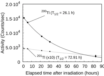
correc-0 5.0⋅103 1.0⋅104 1.5⋅104 2.0⋅104 0 10 20 30 40 50 60 70 80 90 Activity (Counts/sec)
Elapsed time after irradiation (hours)
200Tl (T
1/2 = 26.1 h)
201
Tl (x10) (T1/2 = 72.91 h)
Fig. 2. Activity as a function of the elapsed time after irradiation for the 367.9 keV (200
Tl) and 167.5 keV (201
Tl) transitions at Elab= 35MeV. The full and dotted lines are the fitted activities
incorporating the known half-life of the 200
Tland 201
Tl evapo-ration residues respectively.
tion could be easily accounted for. The nucleus de-pendent correction factor was determined from the measurements in both the geometries at three dif-ferent beam energies.
The compound nucleus, 204
Tl, decays
predomi-nantly to the199−202Tl isotopes by neutron
evapora-tion at the energies studied here. These evaporaevapora-tion residues are unstable and decay by electron
cap-ture to 199−202Hg respectively. Additionally, due to
the weak binding of the projectile (1.45 MeV), large cross sections for residues arising from breakup-fusion and/or transfer reactions were also observed. Some of these Tl isotopes have medical applications, hence their half-lives and the branching ratios for γ-emission from the daughter nuclei have been mea-sured recently to a large accuracy [13,14]. Figure 1a
shows a typical inclusiveγ-ray spectrum measured
atElab= 35 MeV. The relevant γ-rays corresponding
to the decay of the Tl, Au and Pt isotopes arising from fusion, incomplete fusion and neutron transfer are identified. In addition to their energy, the origin of
theγ-rays was also identified from the measurement
of their half-lives. The measuredγ-ray activities for
the 200
Tl (26.1 (1) h) and 201
Tl (72.91 (4) h) evapo-ration residues as a function of time at 35 MeV are shown in Fig. 2. As can be seen from the figure, the data are consistent with the known half lives.
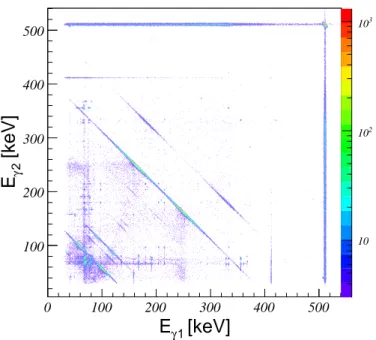
A typical γ − γ coincidence matrix measured at
Elab = 25 MeV (an energy lower than the Coulomb
barrier) is shown in Fig. 3. The back to back geome-try of the detectors maximizes the collection of nearly
backscattered events where aγ-ray deposits part of
its energy in one detector and then scatters to the other detector. These events correspond to the diag-onal lines, in the figure, having fixed energies such
[keV]
1 γE
0 100 200 300 400 500[keV]
2 γE
100 200 300 400 500 10 2 10 3 10Fig. 3. (color online) A part of the two dimensional coincidence matrix at Elab = 25MeV with the sample at 1.5 mm from the
face of the detectors. The data correspond to an irradiation of 14 hours and counted for 12 hours, 136 hours after the end of the irradiation.
X-rays can be clearly seen. A projection of such a co-incidence matrix is used to obtain the relevant spec-tra to exspec-tract the cross sections. Figure 4 shows both
the X-ray and γ-ray coincident spectra at Elab = 35
MeV where the measured coincident yields are large. Figure 4a shows the X-rays that are in prompt
co-incidence with the 367.9 keVγ-ray in 200
Hg.
Char-acteristics Kα1,2 and Kβ′1,2 Hg X-rays arising from
the electron capture can be seen. It should be noted
that the contamination of the Kα2Hg X-ray with Kα1
Au X-ray shown in Fig 1b. is removed as a
conse-quence of the γ-coincidence condition. A
compari-son between the coincidence (Fig. 4) and the related
inclusive (Fig. 1)γ-ray spectra shows that the
coin-cidence condition removes the uncorrelated transi-tions leading to a substantial reduction of the level of the background. For example, the peak to
back-ground ratio for the 367.9 keVγ-ray was found to be
around two orders of magnitude larger in the coinci-dence spectrum than in the inclusive one. The X-γ-ray coincidence yields can be extracted from the four
X-ray yields (Kα1, Kα2,Kβ′1 and Kβ′2) shown in Fig.
4a. Their well determined intensity ratios act as a consistency check of these yields. The X-γ-ray
coin-cidence yields can also be extracted fromγ-ray yields
obtained by gating on the Hg X-rays (Fig. 4b). The
rel-evantγ-rays corresponding to the decay of the
evapo-ration residues (199,200,201
Tl) can be seen. The198,199
Tl
residue can also arise from interactions with 196
Pt (2.56% impurity) in the target. The corrections for these channels are suitably taken into account to ob-tain the total fusion cross sections. In the remaining
100 500 900 1300 1700 60 65 70 75 80 85 90 Counts/0.5 keV Eγ (keV) a) Kα2 Kα1 Kβ’1 Kβ’2 5.0⋅104 2.0⋅105 3.5⋅105 360 365 370 100 500 900 1300 1700 2100 0 200 400 600 800 1000 1200 Counts/0.5 keV Eγ (keV) b) ★201Tl ❐200Tl ◆198Tl ✧199Tl ✧ ★ ✧ ❐ ◆ ❐ ❐ ❐ ❐ 5.0⋅104 2.5⋅105 4.5⋅105 65 75 85
Fig. 4. a) X-ray spectrum in coincidence with the 367.9 keV γ-ray transition (corresponding to the decay of200
Tl) at Elab= 35
MeV. The inset shows the γ-ray coincidence condition. b) γ-ray spectrum in coincidence with Kα X-rays of Hg at Elab = 35
MeV and the X-ray coincidence condition (inset). These exclusive spectra are to be compared with the inclusive measurements shown in Fig. 1.
part of the paper the cross sections have been pre-dominantly obtained using the method illustrated in Fig. 4b. A similar analysis was performed at other energies.
4. Results and Discussion
The evaporation residue cross sections extracted
using both inclusive γ and X-γ exclusive
measure-ments for the 6
Li +198
Pt system at 35 MeV are com-pared in Table 1. Residue cross sections were
ex-tracted using Eq. 1 and absolute γ and X-rays
in-tensities were taken from [13,14]. The errors were estimated from the variance-covariance matrix, con-sidering the peak height and background parame-ters as variables [10]. As can be seen from the Ta-ble 1 there is a good agreement between inclusive and coincidence methods showing the reliability of the present technique to obtain the cross section. 4
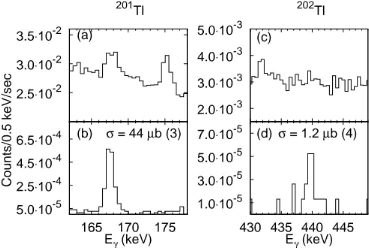
2.5⋅10-2 3.0⋅10-2 3.5⋅10-2 Counts/0.5 keV/sec 201 Tl (a) 5.0⋅10-5 2.5⋅10-4 4.5⋅10-4 6.5⋅10-4 165 170 175 Eγ (keV) (b) σ = 44 µb (3) 2.0⋅10-3 3.0⋅10-3 4.0⋅10-3 5.0⋅10-3 202 Tl (c) 1.0⋅10-5 3.0⋅10-5 5.0⋅10-5 7.0⋅10-5 430 435 440 445 Eγ (keV) σ = 1.2 µb (4) (d)
Fig. 5. γ-ray spectra at Elab= 23.6MeV. Expansion of the
rele-vant regions of interest corresponding to the evaporation residue
201
Tland 202
Tl, characterized by the 167.5 keV and 439.6 keV transitions respectively. (a) and (c) are the inclusive spectra and (b) and (d) correspond to X(Kα)-γ coincidence spectra .
Table 1
Evaporation residue cross sections for 200,201Tlat E
lab= 35MeV
obtained using different detector to sample distances (see text). Inclusive Coincidence Coincidence
(10 cm) (10 cm) (1.5 mm) Channel (mb) (mb) (mb) 201 Tl 25.7 (0.9) - 24.8 (1.1) 200 Tl 308 (2) 314 (8) 309 (2)
The non-observation of the201
Tl residue is consistent with the low coincidence efficiency for a detector to sample distance of 10 cm.
The sensitivity of the measurements to obtain low cross sections at energies well below the barrier are discussed below. Figure 5 shows the region of
inter-est of the inclusive and coincident γ-spectra (gated
by the Kα X-ray of Hg) for the
201,202
Tl nuclei.
Fig-ure 5a shows that the 167.5 keVγ-ray (201
Hg) is rid-ing on a large background, arisrid-ing from Compton
events of higher energyγ-rays. It can be seen from
the coincidence spectrum shown in Fig. 5b, that the background level is drastically reduced, leading to a peak to background ratio ∼ 70 times larger than in the case of the inclusive spectrum. Such an im-provement leads to a more accurate and reliable
de-termination of the γ yields, necessary to obtain the
fusion cross section. Figs. 5 c-d re-emphasize this
for the 439.6 keVγ-ray (202
Hg) where a much smaller
cross section is involved. In the inclusiveγ-ray
spec-trum (Fig. 5c), the yield of the 439.6 keV γ-ray is
not visible over the background. On the contrary, the
439.6 keVγ-ray can be clearly observed in the
coin-cidence spectrum (Fig. 5d). The extracted evapora-tion residue cross-secevapora-tions for the two channels are also indicated in figures 5 and 6.
Figure 6 shows the γ-ray spectra measured at
1⋅10-3 2⋅10-3 3⋅10-3 Counts/0.5 keV/sec (a) 6⋅10-3 7⋅10-3 8⋅10-3 (b) 5⋅10-6 1⋅10-5 165 170 175 Eγ (keV) (c)σ = 156nb (122) 1⋅10-3 2⋅10-3 3⋅10-3 (d) 500 1000 1500 Counts/0.5keV 1⋅10-3 2⋅10-3 3⋅10-3 (e) 500 1000 1500 5⋅10-6 1⋅10-5 430 435 440 445 Eγ (keV) (f) σ = 19nb (14) 0 2 4 6
Fig. 6. γ-ray spectra at Elab= 20.6MeV. Expansion of the
rele-vant regions of interest corresponding to the evaporation residue
201
Tland 202
Tl, characterized by the 167.5 keV and 439.6 keV transitions respectively. (a) and (d) correspond to the background spectra collected without a sample. (b) and (e) correspond to the inclusive spectra, (c) and (f) correspond to the X-γ coincidence spectra.
20.6 MeV corresponding to the data at the lowest
measured energy (E/VB ∼ 0.66). The data shown
was collected for 54 hours after an irradiation of 56 hours. At this energy the comparison between the inclusive (Figs. 6b and 6e) and coincidence (Figs. 6c and 6f) spectra clearly shows the significant increase of detection sensitivity resulting from the present coincidence technique. Poisson statistics and the maximum likelihood method [15] using CERN soft-ware package ROOT were used to obtain the quoted errors. This measurement of the absolute cross section down to the nanobarn level is a significant achievement.
In the remaining part of this section, limits arising from background on the extracted cross section are discussed. A comparison between the background spectra collected without a sample (Figs. 6a and 6d) and inclusive spectra (Figs. 6b and 6e) shows that the lower limit of the sensitivity of the measurement is presently not limited by the room background. The
cross sections for198,199Au arising fromd capture is
measured to be ∼ 4 orders of magnitude larger. The
Compton scattered γ-rays from their daughter
nu-clei (198,199
Hg) thus contribute to background in the X-rays region. In addition to the above, in the case of the 439.6 keV + 70.8 keV X-γ coincident events,
the Compton scattered events from the 511 keV
interest. This can be seen from the two dimensional spectra shown in Fig. 3 where the intersection of the
diagonal line (Eγ = Eγ1+ Eγ2= 511 keV) and the
gat-ing X-ray transition is overlappgat-ing with theγ-ray of
interest limiting the measured peak to background. Thus the sensitivity of the present measurement can be further improved by minimizing products formed in unwanted reactions leading to the emission of 511
keVγ-rays.
5. Applications to Radioactive Ion Beams
As described above, the lowest cross sections mea-sured was a few nb with a typical beam intensity of
1011
pps. Using the delayed X-rays method
measure-ments with 6
He + 64
Zn (106
pps) the authors of [16] were able to measure a total cross section as low as 50 mb. The application of the delayed X-rays tech-nique for the extraction of the fusion cross sections in the case of high Z evaporation residues is relatively more difficult due to the dominance of neutron evap-oration channels (thus characterized by the same
X-rays). Using off beamα decay measurements,
Pe-nionzhkevich et al. [5] were able to measure cross
sections of 1 mb for the6
He+206
Pb system.
GANIL is presently the only facility in the world to deliver low energy beams of the doubly Borromean
nucleus8
He. The only fusion cross section
measure-ments with8
He beams were performed using the
in-clusive in-beamγ-ray technique [17]. It was not
pos-sible to measure low cross sections at energies be-low the Coulomb barrier due to the competition with room background. With the technique and improved sensitivity discussed in this article it will be possible to measure sub-barrier fusion cross sections with a
weakly bound8
He beam and address the fundamen-tal questions of multidimensional tunneling includ-ing couplinclud-ing to the continuum.
6. Conclusion
The agreement between the inclusive and coin-cidence methods shows that the X-γ coincoin-cidence method suggested here can be reliably applied to obtain absolute cross section. This technique thus leads to a greater precision and accuracy of the mea-sured cross sections down to the nanobarn level and is ideally suited for measurements with low intensity radioactive ion beams.
7. Acknowledgments
We would like to thank P. Patale and M.S. Pose for their help during the experiment and the Pel-letron group for a smooth operation. We also thank
Y. Blumenfeld for providing the198
Pt targets. One of
us (A.L.) was partly supported by the Region Basse-Normandie (France).
References
[1] C. L. Jiang, K. E. Rehm, H. Esbensen, R. V. F. Janssens, B. B. Back, C. N. Davids, J. P. Greene, D. J. Henderson, C. J. Lister, R. C. Pardo, T. Pennington, D. Peterson, D. Seweryniak, B. Shumard, S. Sinha, X. D. Tang, I. Tanihata, S. Zhu, P. Collon, S. Kurtz, and M. Paul, Phys. Rev.C 71, (2005) 044613. [2] A. Navin, V. Tripathi, Y. Blumenfeld, V. Nanal, C. Simenel,
J. M. Casandjian, G. de France, R. Raabe, D. Bazin, A. Chatterjee, M. Dasgupta, S. Kailas, R. C. Lemmon, K. Mahata, R. G. Pillay, E. C. Pollacco, K. Ramachandran, M. Rejmund, A. Shrivastava, J. L. Sida, and E. Tryggestad, Phys. Rev. C 70, (2004) 044601.
[3] P. R. S. Gomes, T. J. P. Penna, R. Liguori Neto, J. C. Acquadro, C. Tenreiro, P. A. B. Freitas, E. Crema, N. Carlin Filho and, M. M. Coimbra, Nucl. Instr. and Meth. A 280 (1989) 395. [4] M. Dasgupta, D. J. Hinde, R. D. Butt, R. M. Anjos, A. C.
Berriman, N. Carlin, P. R. S. Gomes, C. R. Morton, J. O. Newton, A. Szanto de Toledo, and K. Hagino Phys. Rev. Lett. 82, 1395 (1999)
[5] Yu. E. Penionzhkevich, V. I. Zagrebaev, S. M. Lukyanov, and R. Kalpakchieva, Phys. Rev. Lett. 96, 162701 (2006). [6] R.G. Stokstad, Y Eisen, S. Kaplanis, D. Pelte, U Smilansky
and I. Tserruya, Phys. Rev. C 21 (1980) 2427.
[7] K. Butler-Moore, R. Aryaeinejad, J.D. Cole, Y. Dardenne, R.C. Greenwod, H.M. Winston, Nucl. Intr. and Meth. A 361 (1995) 245.
[8] H. J. Karwowski, S. E. Vigdor, W. W. Jacobs, S. Kailas, P. P. Singh, F. Soga, T. G. Throwe, T. E. Ward, D.L. Wark, J. Wiggins, Phys. Rev. C 25 (1982) 1355.
[9] A. Shrivsatava et al., (to be published).
[10] Data Acquisition and Analysis Package LAMPS. hhttp://www.tifr.res.in/˜pell/lamps.htmli
[11] K. Debertin and U. Schotzig, Nucl. Instr. and Meth. 158 (1979) 471.
[12] K. Debertin, R. G. Helmer, Gamma- and X-ray Spectrometry with Semiconductor Detectors. Elsevier Science Publishers, Amsterdam, The Netherlands (1988)
[13] Nucleide, Nuclear and Atomic Decay Data (2005), hhttp://www.nucleide.org/DDEP_WG/DDEPdata.htmi; Evaluated Nuclear Structure Data File (ENSDF) : hhttp://www.nndc.bnl.gov/ensdf/i
[14] Karla C. de Souza, Mnica L. da Silva, Jos U. Delgado, Roberto Poledna, Ricardo T. Lopes, Carlos J. da Silva, Appl. Radiat. Isot 60 (2004) 307.
[15] W.-M. Yao et al., J. Phys. G 33 (2006) 1.
[16] A. Di Pietro, P. Figuera, F. Amorini, C. Angulo, G. Cardella, S. Cherubini, T. Davinson, D. Leanza, J. Lu, H. Mahmud, M. Milin, A. Musumarra, A. Ninane, M. Papa, M. G. Pellegriti, R. Raabe, F. Rizzo, C. Ruiz, A. C. Shotter, N. Soic, S. Tudisco, and L. Weissman, Phys. Rev. C 69, 044613 (2004).
[17] A. Lemasson et al., (to be published).