HAL Id: inserm-00511205
https://www.hal.inserm.fr/inserm-00511205
Submitted on 24 Aug 2010HAL is a multi-disciplinary open access archive for the deposit and dissemination of sci-entific research documents, whether they are pub-lished or not. The documents may come from teaching and research institutions in France or abroad, or from public or private research centers.
L’archive ouverte pluridisciplinaire HAL, est destinée au dépôt et à la diffusion de documents scientifiques de niveau recherche, publiés ou non, émanant des établissements d’enseignement et de recherche français ou étrangers, des laboratoires publics ou privés.
Post-transcriptional controls - adding a new layer of
regulation to clock gene expression.
Marie Cibois, Carole Gautier-Courteille, Vincent Legagneux, Luc Paillard
To cite this version:
Marie Cibois, Carole Gautier-Courteille, Vincent Legagneux, Luc Paillard. Post-transcriptional con-trols - adding a new layer of regulation to clock gene expression.. Trends in Cell Biology, Elsevier, 2010, 20 (9), pp.533-41. �10.1016/j.tcb.2010.06.004�. �inserm-00511205�
Posttranscriptional controls - adding a new layer of control to clock gene
1
expression
2 3
Marie Cibois1, 2, 3, Carole Gautier-Courteille1, 2, Vincent Legagneux1, 2, Luc Paillard1, 2 4
5 6 7
1. Université de Rennes 1, Université Européenne de Bretagne, Institut Fédératif de 8
Recherche 140, Rennes, France 9
2. CNRS UMR6061 Institut de Génétique et Développement de Rennes, France 10
3. Present address Institut de biologie du développement de Marseille, UMR6216, CNRS-11
Université de la Méditerranée, Case 907, 13288 Marseille Luminy Cedex 09 France. 12
13 14
Corresponding author Paillard, L ([email protected]) 15
16 17
Manuscript length: 3387 words 18
Living organisms undergo biochemical, physiological and behavioural cycles with 19
periods ranging from seconds to years. The cycles with intermediate periods rely on 20
endogenous clocks that consist of oscillating gene expression. Our goal is to illustrate the 21
modalities and specific functions of posttranscriptional controls of gene expression 22
(exerted on pre-mRNAs and mRNAs) in biological clocks through two examples: the 23
circadian clock and the vertebrate somitic segmentation clock, an embryonic clock with 24
a period far below a day. We conclude that both uniformly and cyclically exerted 25
posttranscriptional controls underpin the set-up of clock functions. 26
27
Rhythmic gene expression in oscillators 28
Living organisms are submitted to periodic oscillations of biochemical, physiological 29
and behavioural parameters that are named biological rhythms. For a given process, the 30
periods of the cycles range from less than one second to several years (Box 1). The 31
biorhythms are subdivided into circadian (period approximately equal to 24 hours), ultradian 32
and infradian (respectively shorter and longer periods, See Glossary) [1]. 33
The present review will focus on essentially two rhythms, the ultradian rhythm that 34
underpins vertebrate somitic segmentation and the circadian rhythm. During vertebrate 35
embryo elongation, somites (presumptive muscles and bones) periodically bud off the non-36
segmented, posterior mesoderm (presomitic mesoderm). This results in a repetitive 37
organization all along the antero-posterior axis, which is referred to as somitic segmentation. 38
The periodic emergence of somites relies on an autonomous ‘clock’ within the non-segmented 39
mesoderm that oscillates with a period ranging from 30 minutes in zebrafish to 2 hours in 40
mice [2]. 41
In circadian rhythms, there also exists an internal clock that is able to free-run with a 42
period of approximately 24 hours. This clock exists in multicellular organisms, but also in 43
yeasts [3]. This autonomous clock is temporally ‘entrained’ by light–dark or temperature 44
cycles [4-6]. In mammals, it is located in the suprachiasmatic nucleus (SCN), a group of 45
hypothalamic neurons. Neuronal connections between the retina and the SCN explain the 46
entrainment by light-dark cycles, which is evidenced among others by the resetting of the 47
clock when light–dark cycles are shifted by some hours (in experimental conditions or 48
following long-distance travels in humans) [4,5]. 49
The mammalian circadian clock relies on eight proteins that are cyclically expressed in 50
the SCN (Figure 1A): Clock [7], Bmal1 (Mop3) [8], Per1, Per2 and Per3 [9], Cry1 and Cry2 51
[10], and Rev-Erbα [11]. The Clock–Bmal1 complex controls the expression of several genes 52
at the transcription level, among which Period (Per1 to Per3), Cryptochrome (Cry1 and 53
Cry2), and Rev-Erb
α
, through its association with E-box elements. The Per–Cry protein 54complexes interact with and inhibit Clock–Bmal1, and Rev-Erbα inhibits the transcription of 55
Bmal1. These two transcriptional feedback loops are responsible for the oscillations of Clock– 56
Bmal1 activity that themselves account for the circadian expression of the clock outputs 57
(Figure 1A) [4,5]. Several additional factors that modulate the mammalian circadian clock 58
were recently identified by RNAi or proteomic approaches [12,13]. The circadian and the 59
segmentation (Box 2) clocks both are set-up by transcriptional negative-feedback loops [2,14-60
18]. 61
In addition to transcriptional loops, the control of the degradation of the proteins 62
encoded by the clock genes determines their amounts in both clocks [17,19-21]. Several 63
posttranslational modifications determine the activity and the stability of clock proteins [19]. 64
Together, they represent a second layer of gene regulation in clock functions. A third layer of 65
gene regulation must now be considered when investigating biological rhythms (Figure 1B). 66
This layer, collectively referred to as posttranscriptional controls, encompasses all the 67
regulations that are exerted at the RNA level (Box 3). They are mediated by ribonucleoproteic 68
particles that include RNA-binding proteins (RNA-BPs) and non-coding RNAs, especially 69
microRNAs (miRNAs) [22-24]. Their contributions in essential clock functions are an 70
emerging and important field of study. 71
72
Circadian rhythms as a paradigm for dynamic posttranscriptional controls 73
The first evidence for posttranscriptional controls in circadian rhythms came from 74
pioneering work in the fruitfly Drosophila [25]. Since, oscillating mRNA stability during the 75
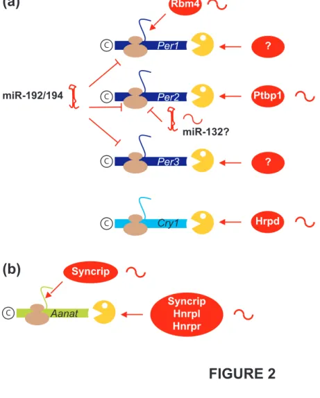
circadian cycle was also demonstrated in the mammalian core pacemaker (Figure 2). The 76
stabilities of Per2 and Cry1 mRNAs vary during the cycle in mice, and, together with 77
oscillating transcription, this results in rhythmic expression [26,27]. Woo and colleagues 78
found that the RNA-BPs Ptbp1 and Hnrpd are able to bind to the 3' untranslated regions of 79
Per2 and Cry1 mRNAs, respectively, and cause their rapid degradation [26,27]. Furthermore, 80
the levels of cytoplasmic Ptbp1 and Hnrpd oscillate during the circadian clock and are 81
correlated with target mRNA decay rates. In synchronized cultured cells, the oscillations of 82
Per2 and Cry1 mRNAs were affected when the levels of Ptbp1 and Hrpd were reduced by 83
RNAi. Together, these results suggest that oscillating amounts of cytoplasmic RNA-BPs may 84
be responsible for the oscillating stability of target mRNAs that in turn determines their 85
oscillating expression [26,27]. 86
Rhythmic translation is another strategy to achieve cyclic expression of clock genes in 87
the SCN, as demonstrated for Per1 mRNA (Figure 2). The RNA-BP Rbm4 is cyclically 88
expressed in-phase with Per1. It is able to bind to Per1 mRNA and to stimulate its translation. 89
Hence, translational stimulation by Rbm4 synergizes with transcriptional controls to amplify 90
the level of Per1 oscillations [28]. Interestingly, only Rbm4 protein, but not Rbm4 mRNA, is 91
cyclically expressed, indicating that Rbm4 expression is itself controlled at a translational or 92
posttranslational (protein degradation) level [28]. It is not known whether Rbm4 is required 93
for circadian rhythms in whole mammalian organisms, but manipulating its level in cultured 94
mammalian cells or in Drosophila affects circadian oscillations [28,29]. 95
In addition to RNA-BPs, microRNAs (miRNAs) also control several mRNAs within 96
the circadian pacemaker (Figure 2). miRNAs affect both mRNA stability and translation [22]. 97
In animals, the interactions between miRNAs and target mRNAs are mediated by limited 98
sequence conservation. A miRNA can have several mRNA targets that are difficult to 99
identify, although considering preferential evolutionary conservation improved the capacity to 100
predict miRNA-mRNA interactions in silico [30]. Cheng and colleagues [31] showed that the 101
miRNAs miR-219 and miR-132 have a circadian expression in the SCN, and they identified 102
several potential mRNA targets. Per2 protein is overexpressed upon treatment with an 103
antisense (antagomir) oligonucleotide against miR-132, which is consistent with miR-132 104
downregulating the translation of Per2 mRNA. Furthermore, circadian period length and 105
light-dependent clock resetting are altered in the absence of miR-219 and miR-132 106
respectively [31]. 107
The SCN emits circadian signals to other regions of the brain, including the pineal 108
gland. This gland synthesizes melatonin during the night and this circulating hormone relays 109
the circadian rhythm to the peripheral organs. Arylalkylamine N-acetyltransferase (Aanat) is 110
cyclically expressed in the pineal gland and is the rate-limiting enzyme in melatonin 111
synthesis. Its expression is controlled at several levels, including mRNA stability and 112
translation (Figure 2). The 3' untranslated region of Aanat mRNA contains a destabilizing 113
element, and three rhythmically expressed RNA-BPs (Hnrnpr, Hnrnpl, Syncrip) are able to 114
bind to this element and may play a role in the rhythmic degradation of Aanat mRNA [32]. In 115
addition, Aanat mRNA is translated through an IRES (internal ribosome entry site), and 116
Syncrip is able to bind to that IRES and stimulate Aanat mRNA translation. The oscillations 117
of Syncrip protein during circadian cycles result in in-phase oscillations of Aanat mRNA 118
translation, and manipulating the level of Syncrip impacts melatonin production in 119
pinealocytes [33]. It is probable that the oscillations of Hnrnpr, Hnrnpl and Syncrip are 120
themselves controlled by circadian cues sent by the SCN, but how this is achieved is unknown 121
(Figure 2). 122
In addition to brain, most mammalian organs contain autonomous clocks that are 123
entrained by cues emitted by the master clock [34], and posttranscriptional controls might 124
operate in these peripheral clocks too. A comprehensive microarray experiment revealed 125
ultradian rhythmic expression of several genes in mouse liver [35]. This might indicate some 126
ultradian clock, but an alternative cause could be mRNA degradation. If genes are transcribed 127
following circadian rhythms and the corresponding mRNAs are degraded following a 128
circadian, out-of-phase, rhythm, the mRNA levels might oscillate with a period of 12 hours 129
[35]. 130
A function for oscillating mRNA stability in circadian rhythm has also been described 131
in plants. A microarray screening in Arabidopsis thaliana identified two mRNAs whose 132
stabilities oscillate with a period of 24 hours. Disruption of the pathway responsible for the 133
rapid degradation of these mRNAs in the afternoon alters the oscillations of these mRNAs in 134
correlation with an altered circadian rhythm at the whole-plant level, indicating a link 135
between circadian rhythms in plants and specific mRNA decay [36]. 136
137
Clues for the importance of posttranscriptional controls in biological rhythms 138
How widespread are posttranscriptional controls of gene expression in biological 139
rhythms? A rough estimate is provided by identifying factors that control gene expression at 140
the posttranscriptional level and that display a rhythmic circadian expression. This is the case 141
for several miRNAs in the plant Arabidopsis thaliana [37], fly heads [38] and mouse retinas 142
[39]. 143
Several examples of oscillating RNA-BPs have also been reported, in addition to the 144
factors described in the previous section. In the green alga Chlamydomonas reinhardtii, the 145
capacity of the RNA-binding complex CHLAMY1 to bind to target mRNAs follows a 146
circadian rhythm [40]. CHLAMY1 comprises two subunits that both are RNA-BPs. 147
Experimentally manipulating the level of either of these two subunits strongly interferes with 148
the circadian rhythm, suggesting that these two proteins are at the heart of the circadian clock 149
in this species [41]. The Chlamydomonas clock is entrained by temperature cycles, and both 150
subunits of CHLAMY1 are involved in temperature integration [42]. In rats, the RNA-BP 151
Mbnl2 (Muscleblind 2) that is involved in alternative splicing of pre-mRNA has an oscillatory 152
expression in the pineal gland [43]. Finally, Nocturnin, a poly(A) ribonuclease (that causes 153
mRNA decay and translational repression by removing the poly(A) tails, see Box 3), is 154
cyclically expressed in the retina [44]. Surprisingly, mice in which the Nocturnin gene has 155
been inactivated display normal circadian rhythms and expression of clock genes (but altered 156
lipid metabolism or uptake) [45]. Hence, factors that control mRNA fate and display a 157
rhythmic expression pattern can be divided into two groups: those that directly influence the 158
clock, and those, like Nocturnin, that represent its readouts. 159
An additional clue to estimate the extent of translational controls in biological rhythms 160
is to compare the levels of cycling proteins with their corresponding mRNAs. Systematic 161
comparison of the transcriptome and the proteome of mouse liver showed that only half of the 162
genes that exhibit rhythmic protein expression also exhibit rhythmic mRNA expression [46]. 163
Interestingly, circadian variations in protein isoforms were also reported by these authors, 164
which are consistent with circadian modifications of alternative splicing [46]. The strong 165
discrepancies between transcriptome and proteome data suggest prevalent translational and/or 166
posttranslational (protein degradation) controls of cyclic gene expression in the circadian 167
clock. 168
169
One step forward: how are cyclic posttranscriptional controls generated? 170
As seen above, cyclical posttranscriptional controls are exerted on several mRNAs and 171
in several physiological systems. In some already discussed cases, the factors involved in 172
RNA regulations are uniformly expressed, but their activity or subcellular localisations 173
oscillate [26,27,41]. The mechanisms underlying these oscillations are unknown. 174
The factors controlling mRNA fate may also themselves be cyclically expressed, 175
owing to a cyclical transcriptional regulation, as demonstrated for miR-219 (see Figure 2) 176
[31], but also owing to posttranscriptional negative-feedback loops. In Neurospora crassa, 177
FRQ and FRH proteins form the FFC complex, which is able to recruit the RNA exosome (a 178
multi-subunit complex involved in mRNA degradation [47]) to frq mRNA, and to thereby 179
cause its degradation. Together with the capacity of FFC to repress the transcription of frq 180
gene, this posttranscriptional negative-feedback loop achieves circadian oscillations in N. 181
crassa [48]. In Arabidopsis thaliana, AtGRP7 and AtGRP8 are two RNA-BPs with a 182
circadian expression. AtGRP7 overexpression ablates circadian expression of Atgrp7 and 183
Atgrp8 mRNAs [49]. Both proteins are able to bind to their own pre-mRNAs and direct their 184
splicing pathways towards mRNA isoforms that contain a premature termination codon. 185
These isoforms are rapidly degraded by the non-sense-mediated mRNA decay (NMD) 186
pathway (see Box 3). Consequently, AtGRP7 and AtGRP8 negatively auto-regulate and cross-187
regulate their synthesis [50,51]. This mechanism very probably ensures a cyclical stability of 188
the mRNAs encoding AtGRP7 and AtGRP8, which contributes to their circadian oscillations. 189
In mammals, the RNA-BPs Rbm4 and Syncrip display oscillating expressions [28,33]. 190
It is tempting to speculate that these oscillations result from negative auto-regulations similar 191
to plant AtGRP7 and AtGRP8 or N. crassa FRQ. Indeed, several mammalian RNA-BPs 192
negatively regulate their own synthesis. PTBP1 and PTBP2 regulate the splicing of their own 193
respective pre-mRNAs and promote the skipping of an exon that results in an NMD sensitive 194
transcript [52,53]. They also cross-regulate each other through this splicing event [54,55]. 195
Similarly, the RNA-BP Celf2 negatively autoregulates its synthesis by inhibiting the splicing 196
of its own pre-mRNA [56]. Whether these negative auto-regulations of RNA-BPs generate 197
oscillations, and how these putative posttranscriptional negative-feedback loops are 198
interconnected with the master transcriptional loop, have not been tested in mammals. 199
200
Posttranscriptional controls do not need to be cyclically exerted to play a role in 201
biological rhythms. 202
Transcriptional negative-feedback loops result in successive activations and 203
repressions of gene promoters. When transcription is shut off, mRNAs decay following 204
exponential kinetics. If the decay of a given mRNA is sufficiently rapid (short half-life) 205
relative to the period of transcriptional oscillations, then almost complete removal of the 206
mRNA will occur before transcription resumes. This situation produces oscillations of mRNA 207
of maximum amplitude. However, if the transcription resumes before the mRNA is 208
completely degraded, then the amplitudes of the mRNA oscillations are reduced or the 209
oscillations are damped and, at the extreme of very stable mRNAs, completely disappear. 210
Therefore, rapid mRNA degradation is required to convert switches between active and 211
inactive transcription into oscillatory amounts of the corresponding mRNAs. One could 212
predict therefore that rapid and uniform mRNA decay is instrumental in the generation of 213
short-period (ultradian) biorhythms, and this prediction has at least been partially confirmed 214
in the case of vertebrate somitic segmentation clock. 215
The period of the somitic segmentation clock is comprised between 30 minutes and 2 216
hours [2]. Within one period, the amounts of several tens of mRNAs oscillate [57]. It takes no 217
more than a few minutes to have a cyclic mRNA completely degraded, indicating very short 218
half-lives. The data demonstrating the occurrence of posttranscriptional controls in somitic 219
segmentation are summarized in Table 1. 220
The expression pattern of Lunatic Fringe (Lfng, a modulator of Notch signalling, one 221
of the pathways required for segmentation) has been described in mice. In situ hybridizations 222
were made with both an exonic probe to reveal the mRNA and an intronic probe to reveal 223
sites of active transcription. The staining patterns with these two probes were very similar, 224
demonstrating that Lfng mRNA is degraded virtually as rapidly as the Lfng introns [58]. Since 225
splicing occurs co-transcriptionally, and excised introns are very rapidly degraded, these data 226
demonstrate the remarkable instability of Lfng mRNA. 227
Reporter genes also showed that mRNA degradation is required to achieve the 228
dynamic expression pattern of the clock genes. In Zebrafish, a GFP reporter controlled by the 229
Her1 promoter (an oscillating component of the core clock) accumulates in the presomitic 230
mesoderm owing to its high stability, suggesting a contrario the rapid decay of the 231
endogenous mRNA [59]. In Xenopus transgenic embryos, a characteristic striped expression 232
pattern of Hairy2a and Bowline, two genes downstream of the clock, is recapitulated by 233
reporter mRNAs only if they contain a destabilizing element in their 3' untranslated regions 234
(3'UTR) [60,61]. Taking as evidence for rapid mRNA degradation the capacity of a 3'UTR to 235
confer upon a reporter GFP gene a striped pattern of expression, several chick or mouse clock 236
mRNAs can be considered as unstable (Table 1 [60]). More recently, an approach combining 237
in ovo electroporation and an inducible promoter showed that chick Lfng mRNA is 238
destabilized by means of its 3'UTR [62]. 239
What happens to segmentation if the rapid degradation of the cyclic mRNAs is 240
impaired? Computational models of the zebrafish segmentation clock predict that the 241
oscillations of the core clock genes are sustained only if the corresponding mRNA and 242
proteins are unstable [18,63], but this was not experimentally tested at the mRNA level. In 243
Zebrafish, the ‘tortuga’ mutant shows an altered pattern of expression of Her1 with impaired 244
oscillations that is consistent with mRNA stabilisation [64]. The corresponding wild-type 245
gene product may therefore be responsible for the rapid decay of Her1 mRNA. This gene has 246
not been identified. In Xenopus, the RNA-BPs Celf1 and Fxr1p regulate the stability and/or 247
the translation of bound mRNAs, and knock-down of these proteins causes segmentation 248
defects [65,66]. This suggests that these proteins have to bind and control a subset of mRNAs 249
for correct segmentation to occur. The mRNA encoding Su(H), that is involved in Notch 250
signalling in the segmentation clock, was identified as a target of Celf1. Specifically, a 251
functional interaction between Celf1 and Su(H) mRNA is required for both the degradation of 252
this mRNA and somitic segmentation [67]. Together, these data show that uniform mRNA 253
regulation plays a key role in oscillations of the segmentation clock. 254
Continuous posttranscriptional controls were also described in the circadian clock. The 255
expression of the microRNAs 192 and mi-R194 in cultured mammalian cells [68], miR-256
122 in mouse liver [69] or bantam in fly heads [70] apparently does not follow a circadian 257
cycle (although miR-122 is cyclically transcribed but remains at approximately constant 258
levels due to a long high-life [69]). All these miRNAs continuously downregulate identified 259
target mRNAs encoding proteins involved in the circadian clock, and manipulating their 260
levels modifies the period and/or amplitude of circadian oscillations [68-70]. Other examples 261
are given by Per1 and Per3 mRNAs that are uniformly unstable in NIH3T3 cells and 262
transgenic mice, respectively [71,72]. The circadian oscillations of Per3 mRNA are strongly 263
modified when its mRNA degradation element is deleted [71]. Hence, constant 264
posttranscriptional repression may be required in some instances to achieve optimal circadian 265
oscillations in addition to cyclical posttranscriptional controls of gene expression. 266
267
Concluding remarks and future directions 268
The comparison of the segmentation and circadian clocks paves the way for future 269
researches (Box 4). Both mRNA degradation and translation, mediated by RNA-BPs and 270
miRNAs, have recognized functions in the circadian clock. In several instances, translational 271
efficiency and mRNA degradation oscillate in the circadian clock, and these oscillations fully 272
contribute to the clock. By contrast, the only known mode of posttranscriptional control in the 273
segmentation clock is constant mRNA degradation. In fact, we might simply lack data 274
concerning the different modes of posttranscriptional controls in the segmentation clock. 275
Using the circadian clock as a paradigm for posttranscriptional controls in clocks, we 276
recommend that the various modes of oscillating posttranscriptional controls should be 277
carefully investigated in the segmentation clock. Furthermore, most but not all known modes 278
of posttranscriptional controls were described in the circadian clock. Specifically, we know 279
nothing about the subcellular localization and the putative localized translation of the mRNAs 280
encoding factors of the clock. It might be of interest to investigate these points in the 281
regulation of mammalian circadian clock considering their recognized importance in neurons 282
[73]. 283
Another question is whether there exist human diseases caused by posttranscriptional 284
defects in clocks. Congenital vertebral malformations are often of genetic origin. Some of 285
them were associated with mutations affecting genes of the segmentation clock, but the 286
aetiology of most of them is unknown [74]. Factors involved in posttranscriptional regulations 287
in somitic segmentation, most of which were not identified, will be potential candidates for 288
causing these syndromes. Also several human troubles arise from defects in the circadian 289
clock, such as sleep disorders. Interestingly, fragile X patients suffer from sleep disorders 290
[75]. This syndrome is a consequence of impaired expression of the RNA-BP FMR1, and 291
Fmr1 KO mice display an altered circadian rhythm [76]. Fragile X syndrome provides 292
therefore a link between posttranscriptional controls, human pathology and the circadian 293
clock, and it can be anticipated that this will not remain an isolated example. 294
A last issue is the extent of posttranscriptional controls in clocks. Several inactivations 295
of gene encoding RNA-BPs were reported in mice. Some of them may be at the origin of 296
circadian troubles that remained unnoticed up to now, and this would merit careful 297
reinvestigation. For the RNA-BPs whose inactivations lead to clock troubles, the arising 298
question will be the identity of the mRNAs that are normally associated with that protein and 299
are deregulated upon its inactivation (and whose deregulation is responsible of the observed 300
troubles). Recent technological breakthroughs allow some optimism concerning our capacity 301
to ask that question. "CLIP" (Cross-linking and immunoprecipitation) allows the co-302
immunoprecipitation of RNA-BPs and associated RNAs [77]. Combined with next-generation 303
sequencing, it permits the genome-wide identification of the RNAs bound by a protein 304
(‘CLIPseq’) [78-80]. Maps of the interactions between miRNAs and mRNAs were drawn 305
from Argonaute CLIPseq [81,82]. Together, these recent technologies will provide us with a 306
genome-wide characterization of the network of posttranscriptional controls in virtually any 307
cell type, including those subject to clock oscillations, and will allow us fully appreciating the 308
extent of posttranscriptional controls in clocks. 309
310
Acknowledgments
311
We thank S. Hardy and H.B. Osborne for critical evaluation of the manuscript. V.L. is 312
a staff member of the INSERM (Institut National de la Santé et de la Recherche Médicale). 313
Work in the authors' laboratory is supported by a grant from the Agence Nationale de la 314 Recherche (ANR-07-JCJC-0097-01). 315 316
References
3171 Schibler, U. and Naef, F. (2005) Cellular oscillators: rhythmic gene expression and 318
metabolism. Curr Opin Cell Biol 17 (2), 223-229 319
2 Pourquie, O. (2003) The segmentation clock: converting embryonic time into spatial 320
pattern. Science 301 (5631), 328-330 321
3 Eelderink-Chen, Z. et al. (2010) A circadian clock in Saccharomyces cerevisiae. Proc 322
Natl Acad Sci U S A 107 (5), 2043-2047 323
4 Takahashi, J.S. et al. (2008) The genetics of mammalian circadian order and disorder: 324
implications for physiology and disease. Nat Rev Genet 9 (10), 764-775 325
5 Gery, S. and Koeffler, H.P. (2010) Circadian rhythms and cancer. Cell Cycle 9 (6) 326
6 Rensing, L. and Ruoff, P. (2002) Temperature effect on entrainment, phase shifting, 327
and amplitude of circadian clocks and its molecular bases. Chronobiol Int 19 (5), 807-328
864 329
7 Vitaterna, M.H. et al. (1994) Mutagenesis and mapping of a mouse gene, Clock, 330
essential for circadian behavior. Science 264 (5159), 719-725 331
8 Bunger, M.K. et al. (2000) Mop3 is an essential component of the master circadian 332
pacemaker in mammals. Cell 103 (7), 1009-1017 333
9 Zheng, B. et al. (2001) Nonredundant roles of the mPer1 and mPer2 genes in the 334
mammalian circadian clock. Cell 105 (5), 683-694 335
10 Kume, K. et al. (1999) mCRY1 and mCRY2 are essential components of the negative 336
limb of the circadian clock feedback loop. Cell 98 (2), 193-205 337
11 Preitner, N. et al. (2002) The orphan nuclear receptor REV-ERBalpha controls 338
circadian transcription within the positive limb of the mammalian circadian oscillator. 339
Cell 110 (2), 251-260 340
12 Zhang, E.E. et al. (2009) A genome-wide RNAi screen for modifiers of the circadian 341
clock in human cells. Cell 139 (1), 199-210 342
13 Robles, M.S. et al. (2010) Identification of RACK1 and protein kinase Calpha as 343
integral components of the mammalian circadian clock. Science 327 (5964), 463-466 344
14 Hardin, P.E. et al. (1990) Feedback of the Drosophila period gene product on 345
circadian cycling of its messenger RNA levels. Nature 343 (6258), 536-540 346
15 Dunlap, J.C. and Loros, J.J. (2006) How fungi keep time: circadian system in 347
Neurospora and other fungi. Curr Opin Microbiol 9 (6), 579-587 348
16 Hardin, P.E. (2005) The circadian timekeeping system of Drosophila. Curr Biol 15 349
(17), R714-722 350
17 Ko, C.H. and Takahashi, J.S. (2006) Molecular components of the mammalian 351
circadian clock. Hum Mol Genet 15 Spec No 2, R271-277 352
18 Giudicelli, F. et al. (2007) Setting the tempo in development: an investigation of the 353
zebrafish somite clock mechanism. PLoS Biol 5 (6), e150 354
19 Mehra, A. et al. (2009) Post-translational modifications in circadian rhythms. Trends 355
Biochem Sci 34 (10), 483-490 356
20 Lee, H. et al. (2009) Essential roles of CKIdelta and CKIepsilon in the mammalian 357
circadian clock. Proc Natl Acad Sci U S A 106 (50), 21359-21364 358
21 Hirata, H. et al. (2004) Instability of Hes7 protein is crucial for the somite 359
segmentation clock. Nat Genet 36 (7), 750-754 360
22 Filipowicz, W. et al. (2008) Mechanisms of post-transcriptional regulation by 361
microRNAs: are the answers in sight? Nat Rev Genet 9 (2), 102-114 362
23 Glisovic, T. et al. (2008) RNA-binding proteins and post-transcriptional gene 363
regulation. FEBS Lett 582 (14), 1977-1986 364
24 Keene, J.D. (2010) Minireview: global regulation and dynamics of ribonucleic Acid. 365
Endocrinology 151 (4), 1391-1397 366
25 So, W.V. and Rosbash, M. (1997) Post-transcriptional regulation contributes to 367
Drosophila clock gene mRNA cycling. Embo J 16 (23), 7146-7155 368
26 Woo, K.C. et al. (2009) Mouse period 2 mRNA circadian oscillation is modulated by 369
PTB-mediated rhythmic mRNA degradation. Nucleic Acids Res 37 (1), 26-37 370
27 Woo, K.C. et al. (2010) Circadian amplitude of cryptochrome 1 is modulated by 371
mRNA stability regulation via cytoplasmic hnRNP D oscillation. Mol Cell Biol 30 (1), 372
197-205 373
28 Kojima, S. et al. (2007) LARK activates posttranscriptional expression of an essential 374
mammalian clock protein, PERIOD1. Proc Natl Acad Sci U S A 104 (6), 1859-1864 375
29 Huang, Y. et al. (2009) Altered LARK expression perturbs development and 376
physiology of the Drosophila PDF clock neurons. Mol Cell Neurosci 41 (2), 196-205 377
30 Bartel, D.P. (2009) MicroRNAs: target recognition and regulatory functions. Cell 136 378
(2), 215-233 379
31 Cheng, H.Y. et al. (2007) microRNA modulation of circadian-clock period and 380
entrainment. Neuron 54 (5), 813-829 381
32 Kim, T.D. et al. (2005) Rhythmic serotonin N-acetyltransferase mRNA degradation is 382
essential for the maintenance of its circadian oscillation. Mol Cell Biol 25 (8), 3232-383
3246 384
33 Kim, T.D. et al. (2007) Rhythmic control of AANAT translation by hnRNP Q in 385
circadian melatonin production. Genes Dev 21 (7), 797-810 386
34 Kornmann, B. et al. (2007) System-driven and oscillator-dependent circadian 387
transcription in mice with a conditionally active liver clock. PLoS Biol 5 (2), e34 388
35 Hughes, M.E. et al. (2009) Harmonics of circadian gene transcription in mammals. 389
PLoS Genet 5 (4), e1000442 390
36 Lidder, P. et al. (2005) Circadian control of messenger RNA stability. Association 391
with a sequence-specific messenger RNA decay pathway. Plant Physiol 138 (4), 2374-392
2385 393
37 Sire, C. et al. (2009) Diurnal oscillation in the accumulation of Arabidopsis 394
microRNAs, miR167, miR168, miR171 and miR398. FEBS Lett 583 (6), 1039-1044 395
38 Yang, M. et al. (2008) Circadian regulation of a limited set of conserved microRNAs 396
in Drosophila. BMC Genomics 9, 83 397
39 Xu, S. et al. (2007) MicroRNA (miRNA) transcriptome of mouse retina and 398
identification of a sensory organ-specific miRNA cluster. J Biol Chem 282 (34), 399
25053-25066 400
40 Mittag, M. et al. (1994) Circadian expression of the luciferin-binding protein 401
correlates with the binding of a protein to the 3' untranslated region of its mRNA. Proc 402
Natl Acad Sci U S A 91 (12), 5257-5261 403
41 Iliev, D. et al. (2006) A heteromeric RNA-binding protein is involved in maintaining 404
acrophase and period of the circadian clock. Plant Physiol 142 (2), 797-806 405
42 Voytsekh, O. et al. (2008) Both subunits of the circadian RNA-binding protein 406
CHLAMY1 can integrate temperature information. Plant Physiol 147 (4), 2179-2193 407
43 Kim, J.S. et al. (2009) Muscleblind-like 2: circadian expression in the mammalian 408
pineal gland is controlled by an adrenergic-cAMP mechanism. J Neurochem 110 (2), 409
756-764 410
44 Baggs, J. and Green, C. (2003) Nocturnin, a Deadenylase in Xenopus laevis Retina. A 411
Mechanism for Posttranscriptional Control of Circadian-Related mRNA. Curr Biol 13 412
(3), 189-198 413
45 Green, C.B. et al. (2007) Loss of Nocturnin, a circadian deadenylase, confers 414
resistance to hepatic steatosis and diet-induced obesity. Proc Natl Acad Sci U S A 104 415
(23), 9888-9893 416
46 Reddy, A.B. et al. (2006) Circadian orchestration of the hepatic proteome. Curr Biol 417
16 (11), 1107-1115 418
47 Houseley, J. et al. (2006) RNA-quality control by the exosome. Nat Rev Mol Cell Biol 419
7 (7), 529-539 420
48 Guo, J. et al. (2009) The exosome regulates circadian gene expression in a 421
posttranscriptional negative feedback loop. Cell 138 (6), 1236-1246 422
49 Heintzen, C. et al. (1997) AtGRP7, a nuclear RNA-binding protein as a component of 423
a circadian-regulated negative feedback loop in Arabidopsis thaliana. Proc Natl Acad 424
Sci U S A 94 (16), 8515-8520 425
50 Schoning, J.C. et al. (2007) Auto-regulation of the circadian slave oscillator 426
component AtGRP7 and regulation of its targets is impaired by a single RNA 427
recognition motif point mutation. Plant J 52 (6), 1119-1130 428
51 Schoning, J.C. et al. (2008) Reciprocal regulation of glycine-rich RNA-binding 429
proteins via an interlocked feedback loop coupling alternative splicing to nonsense-430
mediated decay in Arabidopsis. Nucleic Acids Res 36 (22), 6977-6987 431
52 Wollerton, M.C. et al. (2004) Autoregulation of polypyrimidine tract binding protein 432
by alternative splicing leading to nonsense-mediated decay. Mol Cell 13 (1), 91-100 433
53 Rahman, L. et al. (2002) Alternative splicing of brain-specific PTB defines a tissue-434
specific isoform pattern that predicts distinct functional roles. Genomics 80 (3), 245-435
249 436
54 Spellman, R. et al. (2007) Crossregulation and functional redundancy between the 437
splicing regulator PTB and its paralogs nPTB and ROD1. Mol Cell 27 (3), 420-434 438
55 Boutz, P.L. et al. (2007) A post-transcriptional regulatory switch in polypyrimidine 439
tract-binding proteins reprograms alternative splicing in developing neurons. Genes 440
Dev 21 (13), 1636-1652 441
56 Dembowski, J.A. and Grabowski, P.J. (2009) The CUGBP2 splicing factor regulates 442
an ensemble of branchpoints from perimeter binding sites with implications for 443
autoregulation. PLoS Genet 5 (8), e1000595 444
57 Dequeant, M.L. et al. (2006) A complex oscillating network of signaling genes 445
underlies the mouse segmentation clock. Science 314 (5805), 1595-1598 446
58 Morales, A. et al. (2002) Periodic Lunatic fringe expression is controlled during 447
segmentation by a cyclic transcriptional enhancer responsive to notch signaling. Dev 448
Cell 3, 63-74 449
59 Gajewski, M. et al. (2003) Anterior and posterior waves of cyclic her1 gene 450
expression are differentially regulated in the presomitic mesoderm of zebrafish. 451
Development 130 (18), 4269-4278 452
60 Davis, R. et al. (2001) Molecular targets of vertebrate segmentation: two mechanisms 453
control segmental expression of Xenopus Hairy2 during somite formation. Dev Cell 1, 454
553-565 455
61 Hitachi, K. et al. (2008) Tbx6, Thylacine1, and E47 synergistically activate bowline 456
expression in Xenopus somitogenesis. Dev Biol 313 (2), 816-828 457
62 Hilgers, V. et al. (2005) In vivo analysis of mRNA stability using the Tet-Off system 458
in the chicken embryo. Dev Biol 284 (2), 292-300 459
63 Lewis, J. (2003) Autoinhibition with transcriptional delay: a simple mechanism for the 460
zebrafish somitogenesis oscillator. Curr Biol 13 (16), 1398-1408 461
64 Dill, K.K. and Amacher, S.L. (2005) tortuga refines Notch pathway gene expression in 462
the zebrafish presomitic mesoderm at the post-transcriptional level. Dev Biol 287 (2), 463
225-236 464
65 Gautier-Courteille, C. et al. (2004) EDEN-BP-dependent post-transcriptional 465
regulation of gene expression in Xenopus somitic segmentation. Development 131 466
(24), 6107-6117 467
66 Huot, M.E. et al. (2005) The RNA-binding protein fragile X-related 1 regulates somite 468
formation in Xenopus laevis. Mol Biol Cell 16 (9), 4350-4361 469
67 Cibois, M. et al. (2010) A strategy to analyze the phenotypic consequences of 470
inhibiting the association of an RNA-binding protein with a specific RNA. Rna 16 (1), 471
10-15 472
68 Nagel, R. et al. (2009) The miRNA-192/194 cluster regulates the Period gene family 473
and the circadian clock. Febs J 276 (19), 5447-5455 474
69 Gatfield, D. et al. (2009) Integration of microRNA miR-122 in hepatic circadian gene 475
expression. Genes Dev 23 (11), 1313-1326 476
70 Kadener, S. et al. (2009) A role for microRNAs in the Drosophila circadian clock. 477
Genes Dev 23 (18), 2179-2191 478
71 Kwak, E. et al. (2006) Essential role of 3'-untranslated region-mediated mRNA decay 479
in circadian oscillations of mouse Period3 mRNA. J Biol Chem 281 (28), 19100-480
19106 481
72 Wilsbacher, L.D. et al. (2002) Photic and circadian expression of luciferase in 482
mPeriod1-luc transgenic mice invivo. Proc Natl Acad Sci U S A 99 (1), 489-494 483
73 Wang, D.O. et al. (2010) Spatially restricting gene expression by local translation at 484
synapses. Trends Neurosci 33 (4), 173-182 485
74 Giampietro, P.F. et al. (2009) Progress in the understanding of the genetic etiology of 486
vertebral segmentation disorders in humans. Ann N Y Acad Sci 1151, 38-67 487
75 Gould, E.L. et al. (2000) Melatonin profiles and sleep characteristics in boys with 488
fragile X syndrome: a preliminary study. Am J Med Genet 95 (4), 307-315 489
76 Zhang, J. et al. (2008) Fragile X-related proteins regulate mammalian circadian 490
behavioral rhythms. Am J Hum Genet 83 (1), 43-52 491
77 Ule, J. et al. (2003) CLIP identifies Nova-regulated RNA networks in the brain. 492
Science 302 (5648), 1212-1215 493
78 Licatalosi, D.D. et al. (2008) HITS-CLIP yields genome-wide insights into brain 494
alternative RNA processing. Nature 456 (7221), 464-469 495
79 Sanford, J.R. et al. (2009) Splicing factor SFRS1 recognizes a functionally diverse 496
landscape of RNA transcripts. Genome Res 19 (3), 381-394 497
80 Yeo, G.W. et al. (2009) An RNA code for the FOX2 splicing regulator revealed by 498
mapping RNA-protein interactions in stem cells. Nat Struct Mol Biol 16 (2), 130-137 499
81 Chi, S.W. et al. (2009) Argonaute HITS-CLIP decodes microRNA-mRNA interaction 500
maps. Nature 460 (7254), 479-486 501
82 Zisoulis, D.G. et al. (2010) Comprehensive discovery of endogenous Argonaute 502
binding sites in Caenorhabditis elegans. Nat Struct Mol Biol 17 (2), 173-179 503
83 Robertson, J.B. et al. (2008) Real-time luminescence monitoring of cell-cycle and 504
respiratory oscillations in yeast. Proc Natl Acad Sci U S A 105 (46), 17988-17993 505
84 Veldhuis, J.D. et al. (1986) Spectrum of the pulsatile characteristics of LH release in 506
normal men. J Androl 7 (2), 83-92 507
85 Knight, J.E. et al. (2000) mRNA stability and polysome loss in hibernating Arctic 508
ground squirrels (Spermophilus parryii). Mol Cell Biol 20 (17), 6374-6379. 509
86 Hoppensteadt, F.C. and Keller, J.B. (1976) Synchronization of periodical cicada 510
emergences. Science 194 (4262), 335-337 511
87 Kawamura, A. et al. (2005) Zebrafish hairy/enhancer of split protein links FGF 512
signaling to cyclic gene expression in the periodic segmentation of somites. Genes 513
Dev 19 (10), 1156-1161 514
88 Kuersten, S. and Goodwin, E.B. (2003) The power of the 3' UTR: translational control 515
and development. Nat Rev Genet 4 (8), 626-637 516
89 Mauxion, F. et al. (2009) BTG/TOB factors impact deadenylases. Trends Biochem Sci 517
34 (12), 640-647 518
90 Garceau, N.Y. et al. (1997) Alternative initiation of translation and time-specific 519
phosphorylation yield multiple forms of the essential clock protein FREQUENCY. 520
Cell 89 (3), 469-476 521
91 Le Hir, H. and Seraphin, B. (2008) EJCs at the heart of translational control. Cell 133 522
(2), 213-216 523
92 Kamath, R. et al. (2003) Systematic functional analysis of the Caenorhabditis elegans 524
genome using RNAi. Nature 421, 231-237 525
93 Dietzl, G. et al. (2007) A genome-wide transgenic RNAi library for conditional gene 526
inactivation in Drosophila. Nature 448 (7150), 151-156 527
Glossary box
528
3' Untranslated Region (3'UTR): Region of the mRNA 3' to the translation stop codon. 529
Alternative splicing: Various ways to skip introns and splice exons. This mechanism 530
generates a large diversity of mRNA molecules from a single gene. Alternative splicing 531
includes mutually exclusive exons (where splicing leads to the inclusion of either of two 532
exons), exon skipping, intron retention, alternative 5' or 3' splice sites (leading to the retention 533
of all or only part of an exon) and alternative terminal exons. 534
Circadian rhythm: A cycle one day long (Latin circa, about, and dies, day). The period of a 535
circadian rhythm is 24 h when the organism is grown under a light-dark cycle (12h light, 12h 536
darkness), and about 24 h when the organism is released into free-running condition. Several 537
parameters cycle in circadian rhythms, the most obvious one in mammals being sleep and 538
wake. 539
Free-running rhythm: Circadian rhythm in the absence of external cues (like constant 540
darkness and temperature). 541
Half-life. Time required in the absence of synthesis to achieve degradation of half the initial 542
amount of a molecule (like an mRNA). 543
Infradian rhythm: A cycle of length above 24h. 544
Melatonin: Circulating hormone secreted by the pineal gland during the night in mammals. It 545
relays the circadian rhythm imposed by the central nervous system to the peripheral organs. 546
miRNA (micro RNA): Short double-stranded RNA, encoded by the genome, that controls 547
gene expression at several levels. In vertebrates, a prevalent feature of miRNAs is their 548
capacity to specifically repress the translation of target mRNAs by (limited) sequence 549
complementarity. 550
Period: Time interval between two reference points (two peaks for example). Inverse of 551
frequency. 552
Presomitic mesoderm: Posterior, non-segmented mesoderm, in which the segmentation 553
clock is active and from which segmented somites periodically bud off. 554
Somites: Transient embryonic repeated mesodermal structures. They are the origin of adult 555
skeletal muscles, bones and derm. 556
Somitic segmentation: Organisation of the somites as repeated units along the embryonic 557
antero-posterior axis. 558
Suprachiasmatic nucleus (SCN): A region of the hypothalamus. The master circadian clock 559
is located within the SCN. 560
Ultradian rhythm: A cycle of length shorter than 24h (e.g. the segmentation clock). 561
Box 1. Some examples of biological rhythms
562 563
Depending on the period, biorhythms are classified as ultradian (period T<24h), infradian 564
(T>24h) and circadian (T~24h). Ultradian rhythms include heart beating (T=fractions of 565
seconds to seconds), sleep episodes (T=tens of minutes), respiratory oscillations in yeasts 566
(T=1–5h [83]), somitic segmentation in vertebrates (T=30 minutes in Zebrafish, 2h in mice 567
[2]), or pulses of LH secretion by the pituitary gland (T~3h in men [84]). Infradian rhythms 568
include successions of torpor and arousal during the hibernation of small mammals 569
(T=several days [85]), female menstrual cycles (T=several days to several months), annual 570
rhythms (flowering of most plants), and even pluri-annual rhythms such as the emergence of 571
Cicada [86]. 572
Box 2. The vertebrate segmentation clock.
573
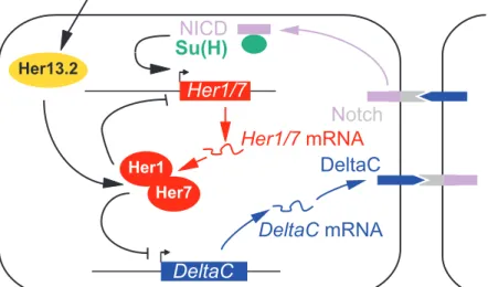
Please refer to the accompanying figure. 574
Title of the figure "The zebrafish core segmentation clock" 575
576
In zebrafish, the core segmentation clock consists of Her1 and Her7 proteins (see Figure). 577
Homodimers or heterodimers of these proteins bind to their own promoters and repress their 578
transcription. Taking into account transcriptional and translational delays, this results in 579
oscillating levels of these proteins. Furthermore, Her1/7 duplexes repress the transcription of 580
Delta-C, a transmembrane Notch ligand. When bound by its ligand, the Notch transmembrane 581
receptor undergoes a limited proteolysis that releases the Notch intracellular domain (NICD) 582
in the cytoplasm. NICD is then translocated to the nucleus. Together with Su(H) protein, the 583
NICD stimulates the transcription of target genes including Her1 and Her7. The stimulation 584
of Her1/7 transcription by Delta-C expressed in adjacent cells, and the ensuing repression of 585
Delta-C gene by Her1/7 achieves coordinated oscillations in neighbouring cells [18,63]. 586
Her13.2 reinforces the transcriptional inhibition mediated by Her1/7, and it is controlled by 587
the FGF pathway. This links the Notch and FGF signalling pathways [87]. Several other 588
genes are downstream of Her1/7 and are involved in somitic segmentation. In amniotes 589
(chick, mouse), the segmentation clock is more complex. It requires oscillations of the Notch 590
modulator Lunatic fringe, and of tens of mRNAs that encode proteins belonging to the FGF 591
and Wnt signalling pathways in addition to Notch [57]. 592
Box 3. Different levels of posttranscriptional controls of gene expression.
593 594
Please refer to the accompanying figure. 595
Title of the figure "pre-mRNA and mRNA fate in eukaryotic cells" 596
597
The posttranscriptional controls are exerted on RNA molecules and are indicated in red on the 598
figure. Concomitantly with nuclear transcription, pre-mRNAs are matured to mRNAs. Pre-599
mRNA maturation refers to three events: 5' capping, 3' cleavage and polyadenylation, and 600
intron excision coupled with exon splicing. Most pre-mRNAs can be cleaved and 601
polyadenylated at several sites (alternative cleavage/polyadenylation) and/or undergo several 602
splicing patterns (alternative splicing. In the figure, the second exon is either skipped or 603
spliced). Due to alternative cleavage/polyadenylation and alternative splicing, a large variety 604
of mRNAs can be obtained from a given pre-mRNA. 605
After nucleo-cytoplasmic export, mRNA translation and decay are controlled, and the 3' 606
poly(A) tail is a major site for these controls. Polyadenylated mRNAs are much more actively 607
translated than deadenylated mRNAs. The initiation factor eIF4G, that recruits the small 608
ribosomal subunit, is able to interact simultaneously with the 5' cap-binding protein eIF4E and 609
the 3' Poly(A) binding protein. The connection between mRNA 5' (cap) and 3' (poly(A) tail) 610
ends strongly stimulates translation [88]. In addition, polyadenylated mRNAs are much more 611
stable than deadenylated mRNAs. For most mRNAs, deadenylation is the rate-limiting step of 612
mRNA decay, and several factors that control mRNA stability do so by regulating the 613
deadenylation rate. In higher eukaryotes, the major pathway for mRNA decay is poly(A) tail 614
removal (deadenylation) followed by RNA exosome-mediated 3' to 5' exonucleolytic 615
degradation. [89]. The 5'-most AUG codon is generally the translation initiation codon, but 616
more distal initiation codons can also be used (alternative initiation of translation), resulting in 617
alternative protein isoforms. This mechanism was described for instance for the mRNA that 618
encodes FRQ, a component of the N. Crassa circadian clock [90]. 619
Nuclear and cytoplasmic controls are tightly coupled. A complex (EJC, exon junction 620
complex) is assembled during splicing immediately upstream of exon junctions, and remains 621
associated with the mRNA during nucleocytoplasmic export. This hallmark of a nuclear event 622
then influences cytoplasmic mRNA translation and degradation [91]. For example, the EJC is 623
involved in the recognition and rapid degradation of mRNAs containing a premature stop 624
codon by the ‘nonsense-mediated mRNA decay’ (NMD) pathway [91]. In addition, 625
alternative splicing can lead to mature transcripts that contain alternative 3' untranslated 626
regions (3'UTR), that are instrumental in mRNA stability and translation [88]. Consequently, 627
alternative cleavage/polyadenylation or splicing impacts mRNA half-life or translation. 628
Box 4. Future questions
629
- Uniform mRNA instability is the only mode of posttranscriptional controls demonstrated in 630
the segmentation clock. Do oscillating mRNA stability and/or oscillating mRNA translation 631
also play a role? 632
- In the circadian clock, the described mechanisms relate to most posttranscriptional controls 633
found to be governing the expression of other non-clock-related gene programs, but mRNA 634
intracellular traffic and local translation were not reported. Since they are prevalent 635
mechanisms in neurons [73], one could ask if they have a function in the circadian clock. 636
- A posttranscriptional feedback loop was demonstrated in N. crassa circadian clock [48], and 637
the levels of some RNA-BPs oscillate in mammalian circadian clocks [28,33]. Are there 638
posttranscriptional feedback loops in vertebrate clocks that could account for the oscillations 639
of these RNA-BPs? 640
- Systematic gene inactivations were reported in lower metazoans [92,93], and several genes 641
were disrupted by homologous recombination in mice. Some of them encode RNA-BPs or 642
miRNAs. Which inactivations lead to clock troubles, demonstrating an involvement of the 643
corresponding gene products in clock setting or robustness? 644
- What are the posttranscriptional networks in clocks? For the RNA-BPs and the miRNAs that 645
are involved in clocks, what are the associated mRNAs? 646
- Are deregulations of posttranscriptional networks in clocks at the origin of human diseases? 647
Figure legends
648
Figure 1. The mammalian circadian clock and its three layers of control 649
(a) Master circadian pacemaker in the suprachiasmatic nucleus (SCN). The Clock–Bmal1 650
complex directly stimulates the transcription of Per, Cry, Rev-Erb
α
, and of output clock-651controlled genes (CCGs) via binding to the E-box. Oscillatory activity of the Clock–Bmal1 652
complex is achieved by two negative feedback loops: the Per–Cry complex inhibits Clock– 653
Bmal1, and Bmal1 transcription is repressed by binding of Rev-Erbα to the RRE (ROR 654
response element). (b) Relationships between transcriptional, posttranscriptional and 655
posttranslational layers in the control of Per genes expression. Since Per proteins contribute to 656
the control of the Clock–Bmal1 complex, fine-tuning their levels is required to obtain 657
oscillations of clock genes. The levels of Per proteins are regulated at a transcriptional level 658
(yellow layer) by the Clock–Bmal1 complex (see Figure 1a). They are regulated at a 659
posttranslational level too (green layer), among others as Casein-kinase1-δ and -ε mediate Per 660
phosphorylation that targets them to ubiquitin/proteasome degradation [19,20]. Recent results 661
demonstrate that a third layer (posttranscriptional controls, red) should be added to complete 662
the picture. The oscillating controls (transcription, mRNA translation and degradation) are in 663
capital letters. 664
665
Figure 2. Posttranscriptional controls exerted on mRNAs encoding proteins involved in 666
circadian rhythms 667
Arrows and blunt-end lines towards ribosomes (brown) indicate stimulation and inhibition, 668
respectively, of mRNA translation. Arrows towards the exonucleolytic enzyme (yellow) 669
indicate stimulation of mRNA decay. The sinusoidal symbols on the right of the factors 670
involved in posttranscriptional controls indicate oscillating levels of these factors. (a) 671
Components of the master circadian clock in the SCN. (b) Aanat, a pineal, rate-limiting 672
enzyme in melatonin synthesis. 673
Gene Function in the clock Evidence for posttranscriptional controls
References
Llnfg in amniotes
Encodes modulator of Notch signalling
mRNA instability inferred from expression pattern in mice; 3'UTR of chick mRNA confers rapid degradation to a reporter mRNA
[58,62]
zebrafish Her1 Encodes component of the core clock
The expression pattern of a reporter mRNA controlled by Her1 promoter is different from that of endogenous Her1 due to increased mRNA stability. Expression pattern in the Tortuga mutant consistent with Tortuga gene product being responsible for Her1 mRNA instability [59,64] Xenopus Hairy 2a, Hairy 1, Esr5, Nrarp, Bowline, Chick Hairy 1, Mouse Hes1, human HES4
Mouse Hes1 and human HES4 may be components of segmentation clock. The other genes encode factors
downstream of the
segmentation clock. Some of them are involved in setting the antero-posterior polarity of forming somites
In Xenopus, the 3'UTR of Hairy 2a confers instability on a reporter mRNA. The expression pattern of Hairy 2a or Bowline was recapitulated in transgenic embryos with the appropriate promoter and a 3'UTR of one of these genes, but not with a 3'UTR of a stable mRNA.
[60,61]
Xenopus Su(H) (homologue of mammalian Rbpj)
Binds to Notch intracellular domain to stimulate expression of Notch target genes
mRNA instability is conferred by association with the RNA-BP Celf1. A specific impairment of the interaction between Celf1 and Su(H) mRNA causes segmentation defects.
[65,67]
674
Table 1. Posttranscriptional controls of gene expression in the segmentation clock. 675
FIGURE 1
(a)
CLOCK BAML1 Clock Bmal1 Per Cry Clock Per Cry CCG Boî Boîte EE-boxte E BoîBoîE-boxte Ete E Cry1, 2 Per1-3 Boî Boîte EE-boxte E Rev-Erb α Boî -BoîE-boxte Ete E -Bmal1 Boî Boîte ERREte E REα Bmal1 miR-219 CCG protein