doi: 10.3389/fonc.2020.01231 Edited by: Nils Cordes, Technische Universität Dresden, Germany Reviewed by: Elisa Giannoni, University of Florence, Italy Priyanka Gupta, University of Alabama at Birmingham, United States *Correspondence: François Kuonen francois.kuonen@chuv.ch
Specialty section: This article was submitted to Molecular and Cellular Oncology, a section of the journal Frontiers in Oncology Received: 10 January 2020 Accepted: 16 June 2020 Published: 22 July 2020 Citation: Lorusso G, Rüegg C and Kuonen F (2020) Targeting the Extra-Cellular Matrix—Tumor Cell Crosstalk for Anti-Cancer Therapy: Emerging Alternatives to Integrin Inhibitors. Front. Oncol. 10:1231. doi: 10.3389/fonc.2020.01231
Targeting the Extra-Cellular
Matrix—Tumor Cell Crosstalk for
Anti-Cancer Therapy: Emerging
Alternatives to Integrin Inhibitors
Girieca Lorusso
1, Curzio Rüegg
1and François Kuonen
2*
1Experimental and Translational Oncology, Department of Oncology Microbiology and Immunology, Faculty of Science and
Medicine, University of Fribourg, Fribourg, Switzerland,2Department of Dermatology and Venereology, Hôpital de Beaumont,
Lausanne University Hospital Center, Lausanne, Switzerland
The extracellular matrix (ECM) is a complex network composed of a multitude of
different macromolecules. ECM components typically provide a supportive structure to
the tissue and engender positional information and crosstalk with neighboring cells in
a dynamic reciprocal manner, thereby regulating tissue development and homeostasis.
During tumor progression, tumor cells commonly modify and hijack the surrounding
ECM to sustain anchorage-dependent growth and survival, guide migration, store
pro-tumorigenic cell-derived molecules and present them to enhance receptor activation.
Thereby, ECM potentially supports tumor progression at various steps from initiation, to
local growth, invasion, and systemic dissemination and ECM-tumor cells interactions
have long been considered promising targets for cancer therapy. Integrins represent key
surface receptors for the tumor cell to sense and interact with the ECM. Yet, attempts to
therapeutically impinge on these interactions using integrin inhibitors have failed to deliver
anticipated results, and integrin inhibitors are still missing in the emerging arsenal of drugs
for targeted therapies. This paradox situation should urge the field to reconsider the role
of integrins in cancer and their targeting, but also to envisage alternative strategies. Here,
we review the therapeutic targets implicated in tumor cell adhesion to the ECM, whose
inhibitors are currently in clinical trials and may offer alternatives to integrin inhibition.
Keywords: extracellular matrix, tumor, progression, crosstalk, clinical perspectives
INTRODUCTION: TARGETING THE ECM-TUMOR CELL
CROSSTALK
The extra-cellular matrix (ECM) is a dynamic niche continuously undergoing quantitative
and qualitative remodeling by renewed synthesis and proteolytic modifications. During ECM
remodeling, changes to its physical structure and organization occur, leading to a dysregulation
in fiber composition, tissue architecture, and stiffness contributing to cancer progression and
fibrosis (
1
). The cell can sense the surrounding ECM fibers by transmembrane surface molecules,
such as integrins or other glycoproteins, acting as cellular mechano-chemical sensors. The
relevance of the finely tuned integration and crosstalk between the ECM molecules, the cellular
cytoskeleton, and the downstream signaling pathways, has been widely recognized and studied
(
2
,
3
). Their complex dynamic bi-directional interactions and mechano-transduction control have
been associated to fundamental physiological processes such
as branching tissues morphogenesis and angiogenesis during
development and homeostasis. These interactions are also
relevant to pathological conditions including cancer, from
initial malignant transformation to the disruption of tissue
polarity and promotion of invasiveness toward dissemination
and metastasis development (
4
,
5
). Integrins represent the
key cell surface receptors for the cell to sense the ECM,
triggering signaling pathways that determine cell fate and
evolution toward a malignant phenotype and resistance to
therapy (
6
,
7
). Numerous experimental and preclinical studies
conducted over the past decades highlighted the central role
of integrins in affecting different steps of tumorigenesis,
by controlling tumor cell adhesion, proliferation, migration,
invasion, and survival (
6
). This made integrins appealing
therapeutic targets leading to the development of integrin
inhibitors and their clinical testing in cancer trials. Unfortunately
and unexpectedly, integrin inhibitors failed to deliver any
tangible therapeutic benefits for cancer patients (
8
–
10
). This
failure may be due to the intrinsic complexity of integrin
signaling that we still do not fully understand. But they also
question the pharmacokinetic/pharmacodynamics properties of
the integrin inhibitors developed, the integrin subunit and the
associated biological process targeted, the preclinical models
used as well as the design of the clinical trials performed
(
7
,
8
). Addressing those yet unanswered questions is likely
to pave the road toward successful introduction of a novel
generation of integrin inhibitors in clinical practice. In the
meantime, long-ago discovered non-integrin ECM receptors
as well as intra-cellular downstream effectors of the
ECM-tumor cell crosstalk (signaling molecules) taking part in several
key aspects of tumor progression, were largely neglected.
Considering the clinical failure of integrin inhibitors, these
ECM-tumor crosstalk targets are potential candidates that may be
therapeutically exploited in alternative to integrin inhibitors.
Here we review those currently tested in anti-cancer clinical
trials, and portray their biology and activity in promoting
tumor evolution.
NON-INTEGRIN TUMOR CELL
RECEPTORS TO THE ECM
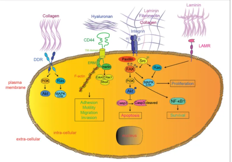
CD44
CD44 is a non-kinase transmembrane glycoprotein expressed in
various cancer types (
11
). CD44 extracellular domain contains
binding sites for various ECM proteins such as collagen, laminin,
and fibronectin (
12
,
13
), while hyaluronic acid (HA) produced
both by tumor cells and tumor stroma is the main and
most specific CD44 ligand (
14
,
15
) (Figure 1). CD44 functions
are modulated by both glycosylation and alternative splicing
(
16
–
18
). Unlike the standard CD44 (CD44s), variant CD44
isoforms (CD44v) contain exons with specific post-translational
modifications allowing binding of tumor-promoting cytokines
like osteopontin (OPN), hepatocyte growth factor (HGF),
vascular endothelial growth factor (VEGF), and basic fibroblast
growth factor (bFGF) (
19
–
23
). Upon HA binding, CD44
proteins change conformation, oligomerize, and redistribute in
glycolipid-enriched domains (GEMs) at the cell membrane (
24
,
25
). There, activated CD44 preferentially interacts with activated
receptor tyrosine kinases (RTKs) (
26
), various adaptor proteins
such as ankyrin or the ERM (ezrin, radixin, and meosin),
ultimately leading to cytoskeletal changes (spectrin, F-actin)
(
27
,
28
), Src family kinases (SFK) members accumulation (
29
),
and activation of downstream pathways, such as Rho-GTPases
(
30
–
33
), PI3K/AKT, or Ras/MAPK (
34
,
35
) (Figure 1). Since
the seminal discovery of their role in metastasis (
36
), CD44s
and CD44v have been implicated in various steps of tumor
progression. In particular, HA-induced CD44 conformational
changes and subsequent cytoskeletal modifications promote
tumor cell migration, invasion, and epithelial-to-mesenchymal
transition (EMT) (
27
,
28
,
30
,
37
–
45
). In glioma cells,
HA-CD44 interactions were shown to occur specifically at the
leading edge of migrating cells upon regulation by activated
protein kinase C (PKC) (
46
). Upon HA binding, various
proteases cleave CD44 allowing dynamic cytoskeletal changes,
filopodia formation and ultimately CD44-mediated migration
(
47
–
50
). Recently, non-catalytic MMP-9–mediated activation of
CD44 was shown to promote tumor cell amoeboid migration
(
51
). Since mesenchymal migration is based on integrin—ECM
interactions, it is tempting to hypothesize that CD44 may
support migration plasticity and escape to integrin inhibition
(
52
–
54
). Further along tumor progression, circulating tumor cells
(CTC) need to extravasate at distant organs. CD44 expressed
on CTC was shown to interact with the HA coat produced
by endothelial cells and initiate the process of tumor cell
extravasation (
55
), particularly to the bone marrow, as shown
in various tumor models through in vitro studies (
56
,
57
).
Importantly, both Cathepsin K, a potent collagenase typically
expressed by osteoclasts during osteolysis, and MMP-9 were
reported to be induced upon HA-mediated CD44 activation
in prostate and breast cancer cells, suggesting their role in
the colonization of metastatic osteolytic prostate and/or breast
cancer cells (
58
–
60
). CD44 alternative splicing was reported to
promote lung colonization by metastatic cancer cells (
61
). Recent
studies implicated HA-CD44 interaction in tumor cell resistance
to chemotherapy, by inducing multi-drug resistance 1 gene
(MDR1) expression (
62
), ABC drug transporters (
63
),
ankyrin-induced drug fluxes (
62
), and tumor cell survival pathways
like ErbB2 signaling and PI3K/AKT pathway (
64
). Alternatively,
HA-CD44 interactions may provide chemo-resistance through
decreased apoptosis/cell death pathways by inducing
anti-apoptotic proteins like inhibitors of the apoptosis family
members (IAPs) (
65
–
68
), reducing pro-apoptotic proteins (
69
)
or modulating autophagy (
70
).
Altogether, CD44 is involved at multiple steps of tumor
progression and its inhibition appears as a promising alternative
for tumor-ECM targeting therapies. Low molecular mass HA,
soluble CD44, CD44 blocking antibodies, CD44 blocking
peptides/aptamers, CD44-targeting sh/siRNA or silibinin (a
plant-derived inhibitor of CD44 expression) have all been
used successfully to interfere with CD44 function in preclinical
FIGURE 1 | Extracellular matrix—tumor cell interactions. In addition to integrins, DDR, CD44, LAMRs, FAK, and SFK represent emerging therapeutic targets currently tested in clinical trials for solid tumors. Downstream effectors interactions were simplified for clarity reasons. DDR, discoidin domain receptor; LAMR, 36/67 kDa laminin receptors; FAK, focal adhesion kinase; PI3K, phosphoinositide-3-kinase; MAPK, mitogen-activated protein kinases; Casp3, caspase 3; NF-κB1, nuclear factor-kappa B1.
models of solid tumor progression (Table 1). The CD44-blocking
antibody RO5429083 was tested in a phase I, dose-escalation
clinical study in metastatic or locally advanced, CD44-positive
malignant solid tumors (NCT01358903) as well as in a phase
I clinical study, alone or in combination with cytarabine, for
acute myelogenous leukemia (NCT01641250). Alternatively,
CD44 targeting may serve to specifically deliver cytotoxic drugs
or radioisotopes to tumor cells. Bivatuzumab-mertansine, a
CD44v6-specific targeting antibody linked to the cytotoxic
drug mertansine, was tested in phase I dose-escalation
clinical studies for CD44v6-positive recurrent or metastatic
breast cancers (NCT02254031, NCT02254005) and advanced
squamous cell carcinoma of the head and neck (NCT02254044,
NCT02254018). The
186Re-labeled bivatuzumab was tested
in phase I biodistribution studies for non-small cell lung
cancers (NCT02204059) and adenocarcinoma of the breast
(NCT02204046). Although preliminary, these results encourage
further clinical assessment of CD44-targeting therapies, either
alone or in combination.
Discoidin Domain Receptors (DDR)
DDR1 and DDR2 belong to the family of the transmembrane
receptor tyrosine kinase (RTK) with an extracellular discoidin
domain binding to collagen in its native triple-helical
conformation (
227
,
228
) (Figure 1). DDR1 and DDR2 bind to
various collagen isoforms with different affinities. DDR1 typically
binds to collagens I-VI and VIII, while DDR2 preferentially
binds to collagens I-III and X (
228
–
231
). Upon collagen binding,
DDRs cluster and get activated through auto-phosphorylation at
multiple tyrosine residues within the cytosolic part of the protein
(
232
,
233
), leading to the recruitment of adaptor or signaling
proteins like ShcA, SHP-2, SFKs, the proline-rich tyrosine kinase
2 (Pyk2), and the non-muscle myosin heavy chain (NMHC) IIA
(
234
,
235
). In cancer cells, DDR activation was reported to induce
Ras/MAPK (
236
), PI3K/AKT (
236
), Notch (
237
), NF-κB (
238
),
PKCα/JAK/Stat (
239
), and p130CS/JNK pathways (
234
), thereby
participating in various steps of tumor progression (Figure 1).
Both DDR1 and DDR2 were shown to promote tumor cell
proliferation, survival (
236
,
238
,
240
,
241
), and migration
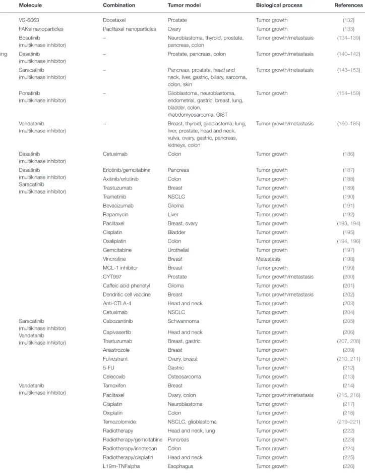
TABLE 1 | In vivo preclinical studies for solid tumors.
Molecule Combination Tumor model Biological process References
Targeting CD44
Low molecular mass HA – Ovary, peripheral nerve Tumor growth/metastasis (71–73)
soluble CD44 – Melanoma, breast Tumor growth (74–76)
CD44 blocking antibody – Breast, colon, pancreas, liver Tumor growth, metastasis (77–81)
CD44v6 blocking antibody – Pancreas Metastasis (80,82,83)
CD44 peptide – Melanoma, gastric Tumor growth/metastasis (81,83–85)
CD44v3 peptide – Glioblastoma Tumor growth (71–73,84,86)
CD44v6 si/shRNA – Colon, gastric Tumor growth (82)
CD44/Epcam aptamer Ovary Tumor growth (82,85)
Silibinin – Prostate Tumor growth (86)
Targeting DDR
DDR1 blocking antibody – Breast Tumor growth (87)
7rh (DDR1 inhibitor) – Gastric, pancreas Tumor growth (88,89)
WRG-28 (DDR2 inhibitor) – Breast Metastasis (90)
Dasatinib
(multikinase inhibitor)
– Lung Tumor growth (91)
Nilotinib
(multikinase inhibitor)
– Colon Metastasis (92)
7rh (DDR1 inhibitor) Dasatinib Nasopharyngeal carcinoma Tumor growth (93) 7rh (DDR1 inhibitor) LY-411575 (Notch
inhibitor)
Lung Tumor growth (91)
DDR1-IN1 (DDR1 inhibitor) Temzolomide/radiotherapy Glioblastoma Tumor growth (94) Dasatinib
(multikinase inhibitor)
JQ1 (BET inhibitor) Lung Tumor growth (95)
LAMR small molecule inhibitor – Breast Metastasis (96)
Targeting LAMR
LAMR37blocking antibody – Fibrosarcoma Metastasis (97)
OFA/iLRP-blocking antibody – Melanoma Metastasis (98–100)
OFA/iLRP-based immunotherapy – Fibrosarcoma, sarcoma Tumor growth/metastasis (99,100) FAK C-terminal domain – Fibroblasts, breast Tumor growth/metastasis (101,102) Targeting
FAK
TAE-226 – Glioma, ovary Tumor growth (103)
VS-6062 (FAK/Pyk2 inhibitor) – Prostate, pancreas, melanoma, basal cell carcinoma
Tumor growth/metastasis (104–107)
VS-4718 – Breast, ovary Tumor growth/metastasis (108,109)
VS-6063 – Ovary Tumor growth (110)
Compounds 14, Y15, Y11 – Breast, pancreas, colon Tumor growth (111–114) Compounds C4, INT2-31, M13,
R2 (FAK scaffold inhibitors)
– Breast, pancreas, neuroblastoma, melanoma, colon
Tumor growth (115–121)
BI853520 – Breast, mesothelioma Tumor growth (122,123)
NVP-TAE-226 – Ewing sarcoma Tumor growth/metastasis (124)
NVP-TAE-226 Docetaxel Ovary Tumor growth (125)
VS-6062 (FAK/Pyk2 inhibitor) Sunitinib Liver Tumor growth (126)
VS-6062 (FAK/Pyk2 inhibitor) Vemurafenib Colon Tumor growth (127)
Compound Y15 5-FU Colon Tumor growth (113)
Compound Y15 Gemcitabine Pancreas Tumor growth (112,128)
Compound C4 (FAK scaffold inhibitor)
Temzolomide Glioblastoma Tumor growth (128)
Doxorubicin Breast Tumor growth (115)
Compound R2 (FAK scaffold inhibitor)
Doxorubicin, 5-FU Colon Tumor growth (121)
PF5735228 WZ811 (CXCR4
inhibitor)
Lung Tumor growth (129)
VS-4718 HDAC inhibitors Lung, Esophagus Tumor growth (130)
VS-4718 PD-1 antagonist, T cell immunotherapy
Pancreas Tumor growth (131)
TABLE 1 | Continued
Molecule Combination Tumor model Biological process References
VS-6063 Docetaxel Prostate Tumor growth (132)
FAKsi nanoparticles Paclitaxel nanoparticles Ovary Tumor growth (133) Bosutinib
(multikinase inhibitor)
– Neuroblastoma, thyroid, prostate, pancreas, colon Tumor growth/metastasis (134–139) Targeting SFK Dasatinib (multikinase inhibitor)
– Prostate, pancreas, colon Tumor growth/metastasis (140–142)
Saracatinib (multikinase inhibitor)
– Pancreas, prostate, head and neck, liver, gastric, biliary, sarcoma, colon, skin
Tumor growth/metastasis (143–153)
Ponatinib
(multikinase inhibitor)
– Glioblastoma, neuroblastoma, endometrial, gastric, breast, lung, bladder, colon,
rhabdomyosarcoma, GIST
Tumor growth (154–159)
Vandetanib (multikinase inhibitor)
– Breast, thyroid, glioblastoma, lung, liver, prostate, head and neck, vulva, ovary, gastric, pancreas, kidneys, colon
Tumor growth/metastasis (160–185)
Dasatinib
(multikinase inhibitor)
Cetuximab Colon Tumor growth (186)
Dasatinib
(multikinase inhibitor) Saracatinib (multikinase inhibitor)
Erlotinib/gemcitabine Pancreas Tumor growth (187)
Axitinib/erlotinib Colon Tumor growth (188)
Trastuzumab Breast Tumor growth (189)
Trametinib NSCLC Tumor growth (190)
Bevacizumab Glioma Tumor growth (191)
Rapamycin Liver Tumor growth (192)
Paclitaxel Breast, ovary Tumor growth (193,194)
Cisplatin Bladder Tumor growth (195)
Oxaliplatin Colon Tumor growth (194,196)
Gemcitabine Urothelial Tumor growth (197)
Vincristine Breast Metastasis (198)
MCL-1 inhibitor Breast Tumor growth (199)
CYT997 Prostate Tumor growth/metastasis (200)
Caffeic acid phenetyl Glioma Tumor growth (201)
Dendritic cell vaccine Breast Tumor growth/metastasis (202)
Anti-CTLA-4 Head and neck Tumor growth (203)
Cetuximab NSCLC Tumor growth (204)
Saracatinib (multikinase inhibitor) Vandetanib (multikinase inhibitor)
Cabozantinib Schwannoma Tumor growth (205)
Capivasertib Head and neck Tumor growth (206)
Trastuzumab Breast, gastric Tumor growth (207,208)
Anastrozole Breast Tumor growth (209)
Fulvestrant Ovary, breast Tumor growth (210,211)
5-FU Gastric Tumor growth (212)
Celecoxib Osteosarcoma Tumor growth (213)
Vandetanib (multikinase inhibitor)
Tamoxifen Breast Tumor growth (214)
Paclitaxel Ovary, colon Tumor growth/metastasis (215,216)
Cisplatin Neuroblastoma Tumor growth (217)
Oxiplatin Colon Tumor growth (218)
Temozolomide NSCLC, glioblastoma Tumor growth (219–221) Radiotherapy Head and neck, lung Tumor growth (222) Radiotherapy/gemcitabine Pancreas Tumor growth (223)
Radiotherapy/irinotecan Colon Tumor growth (224)
Radiotherapy/cisplatin Head and neck Tumor growth (225)
(
242
–
245
). Interestingly, EMT was reported to rely on the switch
from DDR1 (epithelial) to DDR2 (mesenchymal) expression
(
246
), although various reports implicate both DDR1 and DDR2
in EMT-mediated tumor cell invasion (
234
,
247
). More recently,
DDRs were implicated in the late stages of metastatic tumor
progression (
244
,
248
). Typically, DDR1 drives site-specific
metastasis of lung cancer cells to bone (
248
). Additionally, the
collagen-dependent interaction between Transmembrane 4 L6
Family Member 1 (TM4SF1) and DDR1 regulates dormancy
vs. growth at the metastatic site (
239
). Finally, both DDR1
and DDR2 promote resistance to radio- and chemo-therapy
in various preclinical models (
94
,
236
–
238
,
249
). However,
despite these converging evidences implicating DDRs in tumor
progression, one should consider that DDR-mediated effects
are highly versatile and cell-dependent. For example, DDR1
was shown to either support or prevent integrin α2β1-mediated
cell migration in different experimental models (
234
,
250
,
251
).
Moreover, the dynamic regulation of DDR expression during
tumor progression will determine the consequences of DDR
inhibition (
231
). Thus, the complex regulation of DDR activity
in tumor cells may stand for the controversy concerning their
contribution to cancer progression (
243
,
248
,
252
–
254
) and
affect the potential efficacy of DDR targeting in cancer. Still, the
recent identification of activating mutations in the cytoplasmic
signaling portions of DDR affecting intracellular signaling
(
240
,
255
–
257
) opens new perspectives in the identification of
patients who might benefit the most from DDR inhibition.
DDR1 and DDR2 kinases are efficiently inhibited by
multikinase inhibitors like ponatinib, imatinib, dasatinib, and
nilotinib (
258
). Dasatinib, nilotinib, a DDR1 blocking antibody,
the selective DDR1 inhibitors 7rh and DDR1-IN-1 and the
selective allosteric DDR2 inhibitor WRG-28 were shown
to efficiently prevent DDR-mediated tumor progression in
preclinical models (Table 1). Driven by these encouraging results,
dasatinib was tested in a phase II clinical trial for patients
with advanced non-small cell lung cancers harboring a DDR2
mutation (NCT01514864). Unfortunately, it was abandoned
because of lack of efficacy and slow enrollment. Currently,
nilotinib is being assessed in a phase II clinical trial for malignant
locally advanced or metastatic solid neoplasms presenting
DDR1 or DDR2 mutations (NCT02029001). Importantly,
non-canonical activation of DDR1 was shown to promote metastasis
through tyrosine kinase-independent signaling in preclinical
models (
239
), warranting cautious assessment of RTK inhibitors
to target DDR. Further efforts should aim at the development
of specific DDR1 and DDR2 inhibitors targeting canonical and
non-canonical activation routes, the identification of the patients
who may benefit the most from DDR inhibition and their use in
combination therapies.
36/67 kDa Laminin Receptors (LAMR)
The 67 kDa (LAMR
67) laminin receptor was first identified as
a receptor for laminin 1 (
259
–
261
) (Figure 1). It is currently
hypothesized that LAMR
67arises from post-translational
modifications of the precursor 37 kDa laminin receptor
(LAMR
37), although the precise mechanisms (like sumoylation)
are still to be resolved (
262
–
264
). LAMRs harbor multiple cellular
localizations, as assessed by the wide range of cellular processes
they are implicated in: ribosomal biogenesis (
265
), protein
translation (
266
–
268
), pre-rRNA processing (
269
), cellular
adhesion and migration (
267
,
270
), invasion (
271
), cellular
proliferation (
272
,
273
), cytoskeletal modulation (
267
,
274
),
and chromatin and histone modifications (
275
). Both LAMR
37and LAMR
67were identified at the cell membrane where they
potentially bind to laminins, associate with integrins (
276
,
277
)
and get phosphorylated (
278
,
279
). Although the downstream
signaling mechanisms are still unelucidated, various authors
reported modifications of Ras/MAPK and JNK/p38 signaling
upon laminin-binding to LAMRs (
280
), possibly through
interactions with FAK and paxillin (
267
,
281
) (Figure 1).
Given their various implications in cellular regulation, it is
not surprising to find elevated LAMR expression in various
cancers (
282
–
288
) and their involvement in tumor cell growth,
migration, invasion, and aggressiveness (
266
,
282
,
289
).
Importantly, laminin 1—LAMR interaction was shown to be
implicated in tumor cell adhesion (
271
,
290
) and invasion
(
291
,
292
) and LAMR down-regulation was shown to promote
tumor cell apoptosis (
293
–
296
). Whether this is mediated by
laminin 1-dependent activation of LAMR remains unknown.
Recent data suggest that LAMR interaction with FAK may
depend on laminin 1—LAMR interaction and promote
Ras/MAPK and/or PI3K/AKT-mediated survival (
297
,
298
).
However, LAMR was found to promote tumor progression
through various laminin 1-independent manners, such as
regulation of telomerases (
299
), reviewed in (
300
).
Despite various emerging strategies aimed to target LAMR
(
300
), in vivo preclinical studies assessing the feasibility and
efficiency of targeting LAMR are still scant. Both a LAMR
37blocking antibody and a small molecule inhibitor preventing
laminin-LAMR interaction were shown to impede metastatic
progression (Table 1). The green tea-derived
epigallocatechin-3-gallate (EGCG) is a small molecule affecting a large number
of cellular targets, including LAMR
67(
301
) and LAMR
37(
302
). EGCG is currently assessed in a phase I study
for chemopreventive effect in patients with curative-intent
resections of colorectal cancer (NCT02891538). Interestingly,
the immunogenic LAMR tumor-associated antigen, referred as
oncofoetal antigen immature laminin receptor protein
(OFA-iLRP), has been successfully used as a tumor antigen for
vaccine-based therapies in preclinical studies (Table 1). Cellular
immunotherapy using autologous dendritic cell loaded with
OFA-iLRP was tested in a phase I-II clinical study for
metastatic breast cancers (NCT00879489). Altogether, LAMR
targeting appears promising for cancer therapy, although major
efforts should aim at the development of specific inhibitors
and acquisition of stronger preclinical data prior to further
clinical trial.
DOWNSTREAM EFFECTORS OF
INTEGRIN-MEDIATED TUMOR CELL
ADHESION TO THE ECM
Focal Adhesion Kinase (FAK)
Focal adhesion kinase (FAK) is a cytoplasmic non-receptor
protein tyrosine kinase. It is an important cell signaling
hub
highly
phosphorylated
upon
integrin
activation,
and has long been recognized as promoting cancer cell
migration, proliferation, and survival/chemoresistance through
downstream activation of Rho-GEF, talin, cortactin, SFKs,
PI3K/AKT, Ras/MAPK, or NF-κB pathways (
303
,
304
)
(Figure 1). More recent studies have described that besides
its classical localization at the plasma membrane of tumor
cells, FAK can also translocate to the nucleus and act as a
transcription factor driving the expression of cytokines and
chemokines favoring tumor immune evasion, independently of
integrin signaling (
305
). In pancreatic cancer, FAK inhibition
increases the immune infiltrate within the tumor environment,
thereby sensitizing tumors to immune-checkpoint blockade
(
306
). In addition, FAK inhibition also affect stromal cells. By
targeting carcinoma-associated endothelial cells, FAK inhibition
enhances vascular permeability, drug delivery, and overcomes
chemo-resistance to DNA-damaging agents (
307
). Altogether,
these data largely support the potential for therapeutic benefits
of FAK inhibitors, used alone or in combination therapies, in
the arsenal of anti-cancer strategies, illustrated by their success
in various preclinical models (Table 1). FAK inhibition mostly
relies on small molecule inhibitors working through various
mechanisms: ATP competitive kinase inhibition (TAE-226,
VS-4718, VS-6062, VS-6063, GSK-2256098, PF-573228), FAK
scaffold inhibition (compounds 14, Y11, Y15, C4, INT2-31,
M13, R2), or more recently ATP competitive non-kinase
inhibition (BI853520) (Table 1). In combination, FAK inhibition
was reported to improve the efficacy of chemotherapeutic
agents (docetaxel, paclitaxel, temzolomide, 5-FU, gemcitabine,
doxorubicin), targeted therapies (EGFR inhibitor, Src inhibitor,
sunitinib, BRAF inhibitor, CXCR4 inhibitor, HDAC inhibitor),
or immunotherapy (PD1 antagonists, T cell immunotherapy)
(Table 1). Acceptable safety profiles were obtained in phase I
clinical trials for VS-6062 (
104
,
308
), GSK-2256098 (
309
–
311
),
VS-6063 (
312
,
313
), VS-4718 and BI853520 (
314
–
316
), with
VS-6062, GSK-2256098, and VS-6063 showing stabilization of
disease in patients with various advanced solid tumors. Both
GSK-2256098, in combination with trametinib, and VS-6063,
however, failed to show efficacy in phase II clinical trials for
pancreatic adenocarcinoma and malignant mesothelioma,
respectively [NCT02428270, (
317
)]. This unexpected failure
may have been prevented by the stratification of the patients
based on FAK amplification/activity in order to select for
the best responders. VS-6063 is currently tested in multiple
clinical trials: (i) a phase II clinical trial in a pre-operative
setting for malignant mesothelioma (NCT02004028); (ii) a
phase II clinical trial in association with the PD-1 inhibitor
pembrolizumab for advanced solid tumors (NCT02758587,
NCT03727880); (iii) a phase I clinical trial in association with
the RAF/MEK inhibitor RO5126766 for advanced solid tumors
(NCT03875820); (iv) a phase I clinical trial in association with
the anti-PDL1 antibody avelumab for epithelial ovarian cancer
(NCT02943317); (v) a phase I clinical trial in association with
pembrolizumab and gemcitabine for advanced solid tumors
(NCT02546531). The results of these ongoing clinical trials will
be decisive to shape the future development of FAK inhibitors in
clinical practice.
Src Family Kinases (SFK)
The SFK, composed of c-Src, Fyn, Yes, Lck, Lyn, Hck, Fgr,
and Blk, are cytoplasmic non-receptor protein tyrosine
kinases. Their prominent functions are mediated by their
SH2 and SH3 domains interacting with various RTKs (such
as EGF-R, HER2, IGF-R, HGF-R, and PDGF-R), thereby
participating in integration and regulation of RTK signaling.
But SFK also participate in ECM-mediated signaling. Through
phosphorylation of FAK, SFK activation stabilizes focal adhesion
complexes enhancing cell adhesion to the ECM (
318
) (Figure 1).
Altogether, SFK are implicated in many steps of tumorigenesis,
including proliferation, migration, invasion, survival in the
circulation and at distant metastatic sites (
319
–
324
), achieved
through modulation of various downstream effectors as
PI3K/AKT, Ras/MAPK, or Stat3 (
325
,
326
). Additionally,
SFK activation confers therapeutic resistance to targeted RTK
therapies (e.g., Trastuzumab/Herceptin for HER2), to
hormone-receptor endocrine therapies (e.g., Tamoxifen for Estrogen
Receptor), as well as to traditional chemo- and radiotherapies
(
327
). Given their central role in tumor cell signaling and
pleiotropic functions in cancer, SFK represent a promising target
for anti-cancer therapies. SFK are currently most efficiently
targeted
using
non-specific
ATP-competitive
multikinase
inhibitors, such as dasatinib, bosutinib, saracatinib, ponatinib,
and vandetanib, targeting many different tyrosine kinases (such
as BCR-ABL, Kit, PDGFR, EGFR, RET, VEGFR) in addition to
SFK members (
328
). With the exception of vandetanib, approved
for the treatment of thyroid medullary carcinoma, dasatinib,
ponatinib, and bosetanib have been approved by the FDA
for hematological malignancies only, based on their BCR/Abl
inhibitory capacity (
328
). In vivo preclinical data, however,
suggest their potential efficacy in solid tumors as well, alone or in
combination, although not necessarily through SFK inhibition
(Table 1). Up to date, the results of phase II clinical trials with
SKF inhibitors in monotherapy have been disappointing, as
they showed only modest or no efficacy (
326
,
329
). Such failure
may be largely attributed to the current lack of biomarkers
for the identification patients with aberrant SFK, the lack of
specificity of SFK inhibitors, and the sometimes opposing
effects of SFK members at various steps of tumor progression
(
330
,
331
). The interpretation of the numerous ongoing clinical
trials (http://www.clinicaltrials.gov/) as well as the design of
future successful clinical trials testing SFK inhibitors for solid
tumors will largely depend on our capacity to overcome these
important issues.
CONCLUSION
Despite huge expectations based on preclinical studies, integrin
inhibitors failed to deliver anticipated results and have not
entered the clinical practice yet. Understanding and surmounting
the pitfalls of integrin inhibition will be crucial to further
sustain the targeting of tumor cell–ECM interactions as an
anticancer strategy. Yet, other long-time discovered molecules
at the interface between tumor cell and ECM as CD44,
DDR, LAMR, FAK, and SFK, are emerging as alternative
therapeutic targets in clinical trials. Alike integrin inhibitors,
their therapeutic relevance will depend on the specificity
and pharmacokinetic/dynamic properties of the inhibitors
developed, on the adequacy of the preclinical models used
for validation, on the biological process targeted, on the
biomarkers used for the identification of best responders
and on the combination strategies applied in clinical trials.
Importantly, our growing knowledge of the biology of ECM—
tumor cell interactions will be instrumental in overcoming these
important pitfalls and extend the arsenal of clinically valuable
inhibitors targeting the ECM—tumor cells crosstalk in the
near future.
AUTHOR CONTRIBUTIONS
GL wrote the review and edited the manuscript. CR edited the
manuscript. FK planned the outline, wrote the review, and edited
the manuscript. All authors read and approved the submitted
version of the manuscript.
FUNDING
Work in our laboratories was supported by the Swiss National
Science Foundation grants PZ00P3_185926 (to FK) and
31003A_179248 (to CR).
REFERENCES
1. Lu P, Weaver VM, Werb Z. The extracellular matrix: a dynamic
niche in cancer progression. J Cell Biol. (2012) 196:395–406.
doi: 10.1083/jcb.201102147
2. Hynes RO. The extracellular matrix: not just pretty fibrils. Science. (2009) 326:1216–9. doi: 10.1126/science.1176009
3. Hynes RO. Stretching the boundaries of extracellular matrix research. Nat Rev Mol Cell Biol. (2014) 15:761–3. doi: 10.1038/nrm3908
4. Bonnans C, Chou J, Werb Z. Remodelling the extracellular matrix in development and disease. Nat Rev Mol Cell Biol. (2014) 15:786–801. doi: 10.1038/nrm3904
5. Ringer P, Colo G, Fassler R, Grashoff C. Sensing the mechano-chemical properties of the extracellular matrix. Matrix Biol. (2017) 64:6–16. doi: 10.1016/j.matbio.2017.03.004
6. Cooper J, Giancotti FG. Integrin signaling in cancer: mechanotransduction, stemness, epithelial plasticity, and therapeutic resistance. Cancer Cell. (2019) 35:347–67. doi: 10.1016/j.ccell.2019.01.007
7. Hamidi H, Ivaska J. Every step of the way: integrins in cancer
progression and metastasis. Nat Rev Cancer. (2018) 18:533–48.
doi: 10.1038/s41568-018-0038-z
8. Alday-Parejo B, Stupp R, Ruegg C. Are integrins still practicable targets for anti-cancer therapy? Cancers. (2019) 11:978. doi: 10.3390/cancers11070978
9. Kapp TG, Rechenmacher F, Sobahi TR, Kessler H. Integrin
modulators: a patent review. Expert Opin Ther Pat. (2013) 23:1273–95. doi: 10.1517/13543776.2013.818133
10. Vicente-Manzanares M, Sanchez-Madrid F. Targeting the integrin interactome in human disease. Curr Opin Cell Biol. (2018) 55:17–23. doi: 10.1016/j.ceb.2018.05.010
11. Yin T, Wang G, He S, Liu Q, Sun J, Wang Y. Human cancer cells with stem cell-like phenotype exhibit enhanced sensitivity to the cytotoxicity of IL-2 and IL-15 activated natural killer cells. Cell Immunol. (2016) 300:41–5. doi: 10.1016/j.cellimm.2015.11.009
12. Ishii S, Ford R, Thomas P, Nachman A, Steele G Jr, Jessup JM. CD44 participates in the adhesion of human colorectal carcinoma cells to laminin and type IV collagen. Surg Oncol. (1993) 2:255–64. doi: 10.1016/0960-7404(93)90015-Q
13. Jalkanen S, Jalkanen M. Lymphocyte CD44 binds the COOH-terminal heparin-binding domain of fibronectin. J Cell Biol. (1992) 116:817–25. doi: 10.1083/jcb.116.3.817
14. Aruffo A, Stamenkovic I, Melnick M, Underhill CB, Seed B. CD44 is the principal cell surface receptor for hyaluronate. Cell. (1990) 61:1303–13. doi: 10.1016/0092-8674(90)90694-A
15. Banerji S, Wright AJ, Noble M, Mahoney DJ, Campbell ID, Day AJ, et al. Structures of the Cd44-hyaluronan complex provide insight into a fundamental carbohydrate-protein interaction. Nat Struct Mol Biol. (2007) 14:234–9. doi: 10.1038/nsmb1201
16. Stamenkovic I, Amiot M, Pesando JM, Seed B. A lymphocyte molecule implicated in lymph node homing is a member of the cartilage link protein family. Cell. (1989) 56:1057–62. doi: 10.1016/0092-8674(89)90638-7
17. Goldstein LA, Zhou DF, Picker LJ, Minty CN, Bargatze RF, Ding JF, et al. A human lymphocyte homing receptor, the hermes antigen, is related to cartilage proteoglycan core and link proteins. Cell. (1989) 56:1063–72. doi: 10.1016/0092-8674(89)90639-9
18. Idzerda RL, Carter WG, Nottenburg C, Wayner EA, Gallatin WM, St John T. Isolation and DNA sequence of a cDNA clone encoding a lymphocyte adhesion receptor for high endothelium. Proc Natl Acad Sci USA. (1989) 86:4659–63. doi: 10.1073/pnas.86.12.4659
19. Bennett KL, Jackson DG, Simon JC, Tanczos E, Peach R, Modrell B, et al. CD44 isoforms containing exon V3 are responsible for the presentation of heparin-binding growth factor. J Cell Biol. (1995) 128:687– 98. doi: 10.1083/jcb.128.4.687
20. Tremmel M, Matzke A, Albrecht I, Laib AM, Olaku V, Ballmer-Hofer K, et al. A CD44v6 peptide reveals a role of CD44 in VEGFR-2 signaling and angiogenesis. Blood. (2009) 114:5236–44. doi: 10.1182/blood-2009-04-219204
21. Todaro M, Gaggianesi M, Catalano V, Benfante A, Iovino F, Biffoni M, et al. CD44v6 is a marker of constitutive and reprogrammed cancer stem cells driving colon cancer metastasis. Cell Stem Cell. (2014) 14:342–56. doi: 10.1016/j.stem.2014.01.009
22. Megaptche AP, Erb U, Buchler MW, Zoller M. CD44v10, osteopontin and lymphoma growth retardation by a CD44v10-specific antibody. Immunol Cell Biol. (2014) 92:709–20. doi: 10.1038/icb.2014.47
23. Weber GF, Ashkar S, Glimcher MJ, Cantor H. Receptor-ligand interaction between CD44 and osteopontin (Eta-1). Science. (1996) 271:509–12. doi: 10.1126/science.271.5248.509
24. Lesley J, Hyman R, Kincade PW. CD44 and its interaction
with extracellular matrix. Adv Immunol. (1993) 54:271–335.
doi: 10.1016/S0065-2776(08)60537-4
25. Liu D, Sy MS. Phorbol myristate acetate stimulates the dimerization of CD44 involving a cysteine in the transmembrane domain. J Immunol. (1997) 159:2702–11.
26. Misra S, Toole BP, Ghatak S. Hyaluronan constitutively regulates activation of multiple receptor tyrosine kinases in epithelial and carcinoma cells. J Biol Chem. (2006) 281:34936–41. doi: 10.1074/jbc.C600138200
27. Fehon RG, McClatchey AI, Bretscher A. Organizing the cell cortex: the role of ERM proteins. Nat Rev Mol Cell Biol. (2010) 11:276–87. doi: 10.1038/nrm2866
28. Lokeshwar VB, Fregien N, Bourguignon LY. Ankyrin-binding domain of CD44(GP85) is required for the expression of hyaluronic
acid-mediated adhesion function. J Cell Biol. (1994) 126:1099–109.
doi: 10.1083/jcb.126.4.1099
29. Foger N, Marhaba R, Zoller M. Involvement of CD44 in cytoskeleton
rearrangement and raft reorganization in T cells. J Cell Sci.
(2001) 114:1169–78.
30. Bourguignon LY, Zhu H, Zhou B, Diedrich F, Singleton PA, Hung MC. Hyaluronan promotes CD44v3-Vav2 interaction with Grb2-p185(HER2) and induces Rac1 and Ras signaling during ovarian tumor cell migration and growth. J Biol Chem. (2001) 276:48679–92. doi: 10.1074/jbc.M1067 59200
31. Ponta H, Sherman L, Herrlich PA. CD44: from adhesion molecules to signalling regulators. Nat Rev Mol Cell Biol. (2003) 4:33–45. doi: 10.1038/nrm1004
32. Bourguignon LY. Hyaluronan-mediated CD44 activation of RhoGTPase signaling and cytoskeleton function promotes tumor progression. Semin Cancer Biol. (2008) 18:251–9. doi: 10.1016/j.semcancer.2008.03.007 33. Bourguignon LY, Singleton PA, Zhu H, Diedrich F. Hyaluronan-mediated
CD44 interaction with RhoGEF and Rho kinase promotes Grb2-associated binder-1 phosphorylation and phosphatidylinositol 3-kinase signaling leading to cytokine (macrophage-colony stimulating factor) production and breast tumor progression. J Biol Chem. (2003) 278:29420–34. doi: 10.1074/jbc.M301885200
34. Orian-Rousseau V, Morrison H, Matzke A, Kastilan T, Pace G, Herrlich P, et al. Hepatocyte growth factor-induced Ras activation requires ERM proteins linked to both CD44v6 and F-actin. Mol Biol Cell. (2007) 18:76–83. doi: 10.1091/mbc.e06-08-0674
35. Weber GF. Molecular mechanisms of metastasis. Cancer Lett. (2008) 270:181–90. doi: 10.1016/j.canlet.2008.04.030
36. Gunthert U, Hofmann M, Rudy W, Reber S, Zoller M, Haussmann I, et al. A new variant of glycoprotein CD44 confers metastatic potential to rat carcinoma cells. Cell. (1991) 65:13–24. doi: 10.1016/0092-8674(91)90403-L 37. Bourguignon LY, Singleton PA, Zhu H, Zhou B. Hyaluronan promotes
signaling interaction between CD44 and the transforming growth factor beta receptor I in metastatic breast tumor cells. J Biol Chem. (2002) 277:39703–12. doi: 10.1074/jbc.M204320200
38. Bourguignon LY, Wong G, Earle CA, Xia W. Interaction of low molecular weight hyaluronan with CD44 and toll-like receptors promotes the actin filament-associated protein 110-actin binding and MyD88-NFkappaB signaling leading to proinflammatory cytokine/chemokine production and breast tumor invasion. Cytoskeleton. (2011) 68:671–93. doi: 10.1002/cm.20544
39. Bourguignon LY, Wong G, Earle C, Krueger K, Spevak CC. Hyaluronan-CD44 interaction promotes c-Src-mediated twist signaling, microRNA-10b expression, and RhoA/RhoC up-regulation, leading to Rho-kinase-associated cytoskeleton activation and breast tumor cell invasion. J Biol Chem. (2010) 285:36721–35. doi: 10.1074/jbc.M110.162305
40. Zhao S, Chen C, Chang K, Karnad A, Jagirdar J, Kumar AP, et al. CD44 expression level and isoform contributes to pancreatic cancer cell plasticity, invasiveness, and response to therapy. Clin Cancer Res. (2016) 22:5592–604. doi: 10.1158/1078-0432.CCR-15-3115
41. Bellerby R, Smith C, Kyme S, Gee J, Gunthert U, Green A, et al. Overexpression of specific CD44 isoforms is associated with aggressive cell features in acquired endocrine resistance. Front Oncol. (2016) 6:145. doi: 10.3389/fonc.2016.00145
42. Cho SH, Park YS, Kim HJ, Kim CH, Lim SW, Huh JW, et al. CD44 enhances the epithelial-mesenchymal transition in association with colon cancer invasion. Int J Oncol. (2012) 41:211–8. doi: 10.3892/ijo.2012.1453 43. Brown RL, Reinke LM, Damerow MS, Perez D, Chodosh LA, Yang J, et al.
CD44 splice isoform switching in human and mouse epithelium is essential for epithelial-mesenchymal transition and breast cancer progression. J Clin Invest. (2011) 121:1064–74. doi: 10.1172/JCI44540
44. Ni J, Cozzi PJ, Hao JL, Beretov J, Chang L, Duan W, et al. CD44 variant 6 is associated with prostate cancer metastasis and chemo-/radioresistance. Prostate. (2014) 74:602–17. doi: 10.1002/pros.22775
45. Thomas L, Byers HR, Vink J, Stamenkovic I. CD44H regulates tumor cell migration on hyaluronate-coated substrate. J Cell Biol. (1992) 118:971–7. doi: 10.1083/jcb.118.4.971
46. Lamontagne CA, Grandbois M. PKC-induced stiffening of
hyaluronan/CD44 linkage; local force measurements on glioma cells. Exp Cell Res. (2008) 314:227–36. doi: 10.1016/j.yexcr.2007.07.013
47. Nagano O, Saya H. Mechanism and biological significance of CD44 cleavage. Cancer Sci. (2004) 95:930–5. doi: 10.1111/j.1349-7006.2004.tb03179.x 48. Nagano O, Murakami D, Hartmann D, De Strooper B, Saftig P, Iwatsubo
T, et al. Cell-matrix interaction via CD44 is independently regulated by different metalloproteinases activated in response to extracellular Ca(2+) influx and PKC activation. J Cell Biol. (2004) 165:893–902. doi: 10.1083/jcb.200310024
49. Nakamura H, Suenaga N, Taniwaki K, Matsuki H, Yonezawa K, Fujii M, et al. Constitutive and induced CD44 shedding by ADAM-like proteases and membrane-type 1 matrix metalloproteinase. Cancer Res. (2004) 64:876–82. doi: 10.1158/0008-5472.CAN-03-3502
50. Sugahara KN, Hirata T, Tanaka T, Ogino S, Takeda M, Terasawa H, et al. Chondroitin sulfate E fragments enhance CD44 cleavage and CD44-dependent motility in tumor cells. Cancer Res. (2008) 68:7191–9. doi: 10.1158/0008-5472.CAN-07-6198
51. Orgaz JL, Pandya P, Dalmeida R, Karagiannis P, Sanchez-Laorden B, Viros A, et al. Diverse matrix metalloproteinase functions regulate cancer amoeboid migration. Nat Commun. (2014) 5:4255. doi: 10.1038/ncomms5255 52. Friedl P, Alexander S. Cancer invasion and the microenvironment: plasticity
and reciprocity. Cell. (2011) 147:992–1009. doi: 10.1016/j.cell.2011.11.016 53. Te Boekhorst V, Preziosi L, Friedl P. Plasticity of cell migration
in vivo and in silico. Annu Rev Cell Dev Biol. (2016) 32:491–526. doi: 10.1146/annurev-cellbio-111315-125201
54. Schmidt S, Friedl P. Interstitial cell migration: integrin-dependent and alternative adhesion mechanisms. Cell Tissue Res. (2010) 339:83–92. doi: 10.1007/s00441-009-0892-9
55. Siegelman MH, Stanescu D, Estess P. The CD44-initiated pathway of T-cell extravasation uses VLA-4 but not LFA-1 for firm adhesion. J Clin Invest. (2000) 105:683–91. doi: 10.1172/JCI8692
56. Okado T, Hawley RG. Adhesion molecules involved in the binding of murine myeloma cells to bone marrow stromal elements. Int J Cancer. (1995) 63:823–30. doi: 10.1002/ijc.2910630613
57. Draffin JE, McFarlane S, Hill A, Johnston PG, Waugh DJ. CD44 potentiates the adherence of metastatic prostate and breast cancer cells to bone marrow endothelial cells. Cancer Res. (2004) 64:5702–11. doi: 10.1158/0008-5472.CAN-04-0389
58. Lark MW, Stroup GB, James IE, Dodds RA, Hwang SM, Blake SM, et al. A potent small molecule, nonpeptide inhibitor of cathepsin K (SB 331750) prevents bone matrix resorption in the ovariectomized rat. Bone. (2002) 30:746–53. doi: 10.1016/S8756-3282(02)00675-0
59. Corey E, Brown LG, Quinn JE, Poot M, Roudier MP, Higano CS, et al. Zoledronic acid exhibits inhibitory effects on osteoblastic and osteolytic metastases of prostate cancer. Clin Cancer Res. (2003) 9:295–306. 60. Littlewood-Evans AJ, Bilbe G, Bowler WB, Farley D, Wlodarski B, Kokubo T,
et al. The osteoclast-associated protease cathepsin K is expressed in human breast carcinoma. Cancer Res. (1997) 57:5386–90.
61. Yae T, Tsuchihashi K, Ishimoto T, Motohara T, Yoshikawa M, Yoshida GJ, et al. Alternative splicing of CD44 mRNA by ESRP1 enhances lung colonization of metastatic cancer cell. Nat Commun. (2012) 3:883. doi: 10.1038/ncomms1892
62. Bourguignon LY, Peyrollier K, Xia W, Gilad E. Hyaluronan-CD44 interaction activates stem cell marker Nanog, Stat-3-mediated MDR1 gene expression, and ankyrin-regulated multidrug efflux in breast and ovarian tumor cells. J Biol Chem. (2008) 283:17635–51. doi: 10.1074/jbc.M800109200
63. Ricciardelli C, Ween MP, Lokman NA, Tan IA, Pyragius CE, Oehler MK. Chemotherapy-induced hyaluronan production: a novel chemoresistance mechanism in ovarian cancer. BMC Cancer. (2013) 13:476. doi: 10.1186/1471-2407-13-476
64. Misra S, Ghatak S, Zoltan-Jones A, Toole BP. Regulation of multidrug resistance in cancer cells by hyaluronan. J Biol Chem. (2003) 278:25285–8. doi: 10.1074/jbc.C300173200
65. Bourguignon LY, Earle C, Wong G, Spevak CC, Krueger K. Stem cell marker (Nanog) and Stat-3 signaling promote MicroRNA-21 expression and chemoresistance in hyaluronan/CD44-activated head and neck squamous cell carcinoma cells. Oncogene. (2012) 31:149–60. doi: 10.1038/onc.2011.222 66. Chen L, Bourguignon LY. Hyaluronan-CD44 interaction promotes c-Jun signaling and miRNA21 expression leading to Bcl-2 expression and chemoresistance in breast cancer cells. Mol Cancer. (2014) 13:52. doi: 10.1186/1476-4598-13-52
67. Bourguignon LY, Wong G, Earle C, Chen L. Hyaluronan-CD44v3 interaction with Oct4-Sox2-Nanog promotes miR-302 expression leading to self-renewal, clonal formation, and cisplatin resistance in cancer stem cells from head and neck squamous cell carcinoma. J Biol Chem. (2012) 287:32800–24. doi: 10.1074/jbc.M111.308528
68. Fedorchenko O, Stiefelhagen M, Peer-Zada AA, Barthel R, Mayer P, Eckei L, et al. CD44 regulates the apoptotic response and promotes disease development in chronic lymphocytic leukemia. Blood. (2013) 121:4126–36. doi: 10.1182/blood-2012-11-466250
69. Park YS, Huh JW, Lee JH, Kim HR. shRNA against CD44 inhibits cell proliferation, invasion and migration, and promotes apoptosis of colon carcinoma cells. Oncol Rep. (2012) 27:339–46. doi: 10.3892/ijo.2016.3801 70. Lv L, Liu HG, Dong SY, Yang F, Wang QX, Guo GL, et al. Upregulation
of CD44v6 contributes to acquired chemoresistance via the modulation of autophagy in colon cancer SW480 cells. Tumour Biol. (2016) 37:8811–24. doi: 10.1007/s13277-015-4755-6
71. Slomiany MG, Dai L, Tolliver LB, Grass GD, Zeng Y, Toole BP. Inhibition of functional hyaluronan-CD44 interactions in CD133-positive primary human ovarian carcinoma cells by small hyaluronan oligosaccharides. Clin Cancer Res. (2009) 15:7593–601. doi: 10.1158/1078-0432.CCR-09-2317 72. Ween MP, Hummitzsch K, Rodgers RJ, Oehler MK, Ricciardelli C. Versican
induces a pro-metastatic ovarian cancer cell behavior which can be inhibited by small hyaluronan oligosaccharides. Clin Exp Metastasis. (2011) 28:113–25. doi: 10.1007/s10585-010-9363-7
73. Slomiany MG, Dai L, Bomar PA, Knackstedt TJ, Kranc DA, Tolliver L, et al. Abrogating drug resistance in malignant peripheral nerve
sheath tumors by disrupting hyaluronan-CD44 interactions with
small hyaluronan oligosaccharides. Cancer Res. (2009) 69:4992–8. doi: 10.1158/0008-5472.CAN-09-0143
74. Ahrens T, Sleeman JP, Schempp CM, Howells N, Hofmann M, Ponta H, et al. Soluble CD44 inhibits melanoma tumor growth by blocking cell surface CD44 binding to hyaluronic acid. Oncogene. (2001) 20:3399–408. doi: 10.1038/sj.onc.1204435
75. Xu XM, Chen Y, Chen J, Yang S, Gao F, Underhill CB, et al. A peptide with three hyaluronan binding motifs inhibits tumor growth and induces apoptosis. Cancer Res. (2003) 63:5685–90.
76. Yu Q, Toole BP, Stamenkovic I. Induction of apoptosis of metastatic mammary carcinoma cells in vivo by disruption of tumor cell surface CD44 function. J Exp Med. (1997) 186:1985–96. doi: 10.1084/jem.186.12.1985 77. Ghatak S, Misra S, Toole BP. Hyaluronan oligosaccharides inhibit
anchorage-independent growth of tumor cells by suppressing the phosphoinositide 3-kinase/Akt cell survival pathway. J Biol Chem. (2002) 277:38013–20. doi: 10.1074/jbc.M202404200
78. Li L, Hao X, Qin J, Tang W, He F, Smith A, et al. Antibody against CD44s inhibits pancreatic tumor initiation and postradiation recurrence in mice. Gastroenterology. (2014) 146:1108–18. doi: 10.1053/j.gastro.2013.12.035 79. Weigand S, Herting F, Maisel D, Nopora A, Voss E, Schaab C,
et al. Global quantitative phosphoproteome analysis of human tumor xenografts treated with a CD44 antagonist. Cancer Res. (2012) 72:4329–39. doi: 10.1158/0008-5472.CAN-12-0136
80. Seiter S, Arch R, Reber S, Komitowski D, Hofmann M, Ponta H, et al. Prevention of tumor metastasis formation by anti-variant CD44. J Exp Med. (1993) 177:443–55. doi: 10.1084/jem.177.2.443
81. Hibino S, Shibuya M, Engbring JA, Mochizuki M, Nomizu M, Kleinman HK. Identification of an active site on the laminin alpha5 chain globular domain that binds to CD44 and inhibits malignancy. Cancer Res. (2004) 64:4810–6. doi: 10.1158/0008-5472.CAN-04-0129
82. Misra S, Hascall VC, De Giovanni C, Markwald RR, Ghatak S. Delivery
of CD44 shRNA/nanoparticles within cancer cells: perturbation
of hyaluronan/CD44v6 interactions and reduction in adenoma
growth in Apc Min/+ MICE. J Biol Chem. (2009) 284:12432–46. doi: 10.1074/jbc.M806772200
83. Khurana SS, Riehl TE, Moore BD, Fassan M, Rugge M, Romero-Gallo J, et al. The hyaluronic acid receptor CD44 coordinates normal and metaplastic gastric epithelial progenitor cell proliferation. J Biol Chem. (2013) 288:16085–97. doi: 10.1074/jbc.M112.445551
84. Xu Y, Stamenkovic I, Yu Q. CD44 attenuates activation of the hippo signaling pathway and is a prime therapeutic target for glioblastoma. Cancer Res. (2010) 70:2455–64. doi: 10.1158/0008-5472.CAN-09-2505
85. Zheng J, Zhao S, Yu X, Huang S, Liu HY. Simultaneous targeting of CD44 and EpCAM with a bispecific aptamer effectively inhibits intraperitoneal ovarian cancer growth. Theranostics. (2017) 7:1373–88. doi: 10.7150/thno.17826
86. Nambiar D, Prajapati V, Agarwal R, Singh RP. In vitro and in vivo anticancer efficacy of silibinin against human pancreatic cancer BxPC-3 and PANC-1 cells. Cancer Lett. (2013) 334:109–17. doi: 10.1016/j.canlet.2012.09.004 87. Zhong X, Zhang W, Sun T. DDR1 promotes breast tumor growth
by suppressing antitumor immunity. Oncol Rep. (2019) 42:2844–54. doi: 10.3892/or.2019.7338
88. Jin H, Ham IH, Oh HJ, Bae CA, Lee D, Kim YB, et al. Inhibition of discoidin domain receptor 1 prevents stroma-induced peritoneal metastasis in gastric carcinoma. Mol Cancer Res. (2018) 16:1590–600. doi: 10.1158/1541-7786.MCR-17-0710
89. Aguilera KY, Huang H, Du W, Hagopian MM, Wang Z, Hinz S, et al. Inhibition of discoidin domain receptor 1 reduces collagen-mediated tumorigenicity in pancreatic ductal adenocarcinoma. Mol Cancer Ther. (2017) 16:2473–85. doi: 10.1158/1535-7163.MCT-16-0834
90. Grither WR, Longmore GD. Inhibition of tumor-microenvironment interaction and tumor invasion by small-molecule allosteric inhibitor of DDR2 extracellular domain. Proc Natl Acad Sci USA. (2018) 115:E7786–94. doi: 10.1073/pnas.1805020115
91. Ambrogio C, Gomez-Lopez G, Falcone M, Vidal A, Nadal E, Crosetto N, et al. Combined inhibition of DDR1 and notch signaling is a therapeutic strategy for KRAS-driven lung adenocarcinoma. Nat Med. (2016) 22:270–7. doi: 10.1038/nm.4041
92. Jeitany M, Leroy C, Tosti P, Lafitte M, Le Guet J, Simon V, et al. Inhibition of DDR1-BCR signalling by nilotinib as a new therapeutic strategy for metastatic colorectal cancer. EMBO Mol Med. (2018) 10:e7918. doi: 10.15252/emmm.201707918
93. Lu QP, Chen WD, Peng JR, Xu YD, Cai Q, Feng GK, et al. Antitumor activity of 7RH, a discoidin domain receptor 1 inhibitor, alone or in combination with dasatinib exhibits antitumor effects in nasopharyngeal carcinoma cells. Oncol Lett. (2016) 12:3598–608. doi: 10.3892/ol.2016.5088
94. Vehlow A, Klapproth E, Jin S, Hannen R, Hauswald M, Bartsch JW, et al. Interaction of discoidin domain receptor 1 with a 14-3-3-Beclin-1-akt1 complex modulates glioblastoma therapy sensitivity. Cell Rep. (2019) 26:3672–83.e7. doi: 10.1016/j.celrep.2019.02.096
95. Xu C, Buczkowski KA, Zhang Y, Asahina H, Beauchamp EM, Terai H, et al. NSCLC driven by DDR2 mutation is sensitive to dasatinib and JQ1 combination therapy. Mol Cancer Ther. (2015) 14:2382–9. doi: 10.1158/1535-7163.MCT-15-0077
96. Kim DG, Lee JY, Kwon NH, Fang P, Zhang Q, Wang J, et al. Chemical inhibition of prometastatic lysyl-tRNA synthetase-laminin receptor interaction. Nat Chem Biol. (2014) 10:29–34. doi: 10.1038/nchembio.1381 97. Narumi K, Inoue A, Tanaka M, Isemura M, Shimo-Oka T, Abe T, et al.
Inhibition of experimental metastasis of human fibrosarcoma cells by anti-recombinant 37-kDa laminin binding protein antibody. Jpn J Cancer Res. (1999) 90:425–31. doi: 10.1111/j.1349-7006.1999.tb00765.x
98. McClintock SD, Warner RL, Ali S, Chekuri A, Dame MK, Attili D, et al. Monoclonal antibodies specific for oncofetal antigen–immature laminin receptor protein: effects on tumor growth and spread in two murine models. Cancer Biol Ther. (2015) 16:724–32. doi: 10.1080/15384047.2015.1026484 99. Barsoum AL, Liu B, Rohrer JW, Coggin JH Jr, Tucker JA, Pannell LK,
et al. Production, safety and antitumor efficacy of recombinant oncofetal antigen/immature laminin receptor protein. Biomaterials. (2009) 30:3091–9. doi: 10.1016/j.biomaterials.2009.02.022
100. Rohrer JW, Barsoum AL, Coggin JH Jr. Identification of oncofetal antigen/immature laminin receptor protein epitopes that activate BALB/c mouse OFA/iLRP-specific effector and regulatory T cell clones. J Immunol. (2006). 176:2844–56. doi: 10.4049/jimmunol.176.5.2844
101. Hauck CR, Hsia DA, Puente XS, Cheresh DA, Schlaepfer DD. FRNK blocks v-Src-stimulated invasion and experimental metastases without effects on cell motility or growth. EMBO J. (2002) 21:6289–302. doi: 10.1093/emboj/cdf631
102. Mitra SK, Mikolon D, Molina JE, Hsia DA, Hanson DA, Chi A, et al. Intrinsic FAK activity and Y925 phosphorylation facilitate an angiogenic switch in tumors. Oncogene. (2006) 25:5969–84. doi: 10.1038/sj.onc.1209588 103. Liu TJ, LaFortune T, Honda T, Ohmori O, Hatakeyama S, Meyer T, et al.
Inhibition of both focal adhesion kinase and insulin-like growth factor-I receptor kinase suppresses glioma proliferation in vitro and in vivo. Mol Cancer Ther. (2007) 6:1357–67. doi: 10.1158/1535-7163.MCT-06-0476
104. Roberts WG, Ung E, Whalen P, Cooper B, Hulford C, Autry C, et al. Antitumor activity and pharmacology of a selective focal adhesion kinase inhibitor, PF-562,271. Cancer Res. (2008) 68:1935–44. doi: 10.1158/0008-5472.CAN-07-5155
105. Stokes JB, Adair SJ, Slack-Davis JK, Walters DM, Tilghman RW, Hershey ED, et al. Inhibition of focal adhesion kinase by PF-562,271 inhibits the growth and metastasis of pancreatic cancer concomitant with altering the tumor microenvironment. Mol Cancer Ther. (2011) 10:2135–45. doi: 10.1158/1535-7163.MCT-11-0261
106. Jeong K, Murphy JM, Rodriguez YAR, Kim JS, Ahn EE, Lim SS. FAK inhibition reduces metastasis of alpha4 integrin-expressing melanoma to lymph nodes by targeting lymphatic VCAM-1 expression. Biochem Biophys Res Commun. (2019) 509:1034–40. doi: 10.1016/j.bbrc.2019.01.050 107. Kuonen F, Surbeck I, Sarin KY, Dontenwill M, Ruegg C, Gilliet
M, et al. TGFbeta, fibronectin and integrin alpha5beta1 promote invasion in basal cell carcinoma. J Invest Dermatol. (2018) 138:2432–42. doi: 10.1016/j.jid.2018.04.029
108. Tanjoni I, Walsh C, Uryu S, Tomar A, Nam JO, Mielgo A, et al. PND-1186 FAK inhibitor selectively promotes tumor cell apoptosis in three-dimensional environments. Cancer Biol Ther. (2010) 9:764–77. doi: 10.4161/cbt.9.10.11434
109. Walsh C, Tanjoni I, Uryu S, Tomar A, Nam JO, Luo H, et al. Oral delivery of PND-1186 FAK inhibitor decreases tumor growth and spontaneous breast to lung metastasis in pre-clinical models. Cancer Biol Ther. (2010) 9:778–90. doi: 10.4161/cbt.9.10.11433
110. Kang Y, Hu W, Ivan C, Dalton HJ, Miyake T, Pecot CV, et al. Role of focal adhesion kinase in regulating YB-1-mediated paclitaxel resistance in ovarian cancer. J Natl Cancer Inst. (2013) 105:1485–95. doi: 10.1093/jnci/djt210 111. Golubovskaya VM, Nyberg C, Zheng M, Kweh F, Magis A, Ostrov D, et al.
A small molecule inhibitor, 1,2,4,5-benzenetetraamine tetrahydrochloride, targeting the y397 site of focal adhesion kinase decreases tumor growth. J Med Chem. (2008) 51:7405–16. doi: 10.1021/jm800483v
112. Hochwald SN, Nyberg C, Zheng M, Zheng D, Wood C, Massoll NA, et al. A novel small molecule inhibitor of FAK decreases growth of human pancreatic cancer. Cell Cycle. (2009) 8:2435–43. doi: 10.4161/cc.8.15.9145
113. Heffler M, Golubovskaya VM, Dunn KM, Cance W. Focal adhesion kinase autophosphorylation inhibition decreases colon cancer cell growth and enhances the efficacy of chemotherapy. Cancer Biol Ther. (2013) 14:761–72. doi: 10.4161/cbt.25185
114. Golubovskaya VM, Figel S, Ho BT, Johnson CP, Yemma M, Huang G, et al. A small molecule focal adhesion kinase (FAK) inhibitor, targeting Y397 site: 1-(2-hydroxyethyl)-3, 5, 7-triaza-1-azoniatricyclo [3.3.1.1(3,7)]decane; bromide effectively inhibits FAK autophosphorylation activity and decreases cancer cell viability, clonogenicity and tumor growth in vivo. Carcinogenesis. (2012) 33:1004–13. doi: 10.1093/carcin/bgs120
115. Kurenova EV, Hunt DL, He D, Magis AT, Ostrov DA, Cance WG. Small molecule chloropyramine hydrochloride (C4) targets the binding site of focal adhesion kinase and vascular endothelial growth factor receptor 3 and suppresses breast cancer growth in vivo. J Med Chem. (2009) 52:4716–24. doi: 10.1021/jm900159g
116. Kurenova E, Liao J, He DH, Hunt D, Yemma M, Bshara W, et al. The FAK scaffold inhibitor C4 disrupts FAK-VEGFR-3 signaling and inhibits pancreatic cancer growth. Oncotarget. (2013) 4:1632–46. doi: 10.18632/oncotarget.1365
117. Stewart JE, Ma X, Megison M, Nabers H, Cance WG, Kurenova EV, et al. Inhibition of FAK and VEGFR-3 binding decreases tumorigenicity in neuroblastoma. Mol Carcinog. (2015) 54:9–23. doi: 10.1002/mc.22070 118. Ucar DA, Kurenova E, Garrett TJ, Cance WG, Nyberg C, Cox A,
et al. Disruption of the protein interaction between FAK and IGF-1R inhibits melanoma tumor growth. Cell Cycle. (2012) 11:3250–9. doi: 10.4161/cc.21611
119. Ucar DA, Magis AT, He DH, Lawrence NJ, Sebti SM, Kurenova E, et al. Inhibiting the interaction of cMET and IGF-1R with FAK effectively reduces growth of pancreatic cancer cells in vitro and in vivo. Anticancer Agents Med Chem. (2013) 13:595–602. doi: 10.2174/1871520611313040009
120. Golubovskaya VM, Palma NL, Zheng M, Ho B, Magis A, Ostrov D, et al. A small-molecule inhibitor, 5’-O-tritylthymidine, targets FAK and Mdm-2
interaction, and blocks breast and colon tumorigenesis in vivo. Anticancer Agents Med Chem. (2013) 13:532–45. doi: 10.2174/1871520611313040002 121. Golubovskaya VM, Ho B, Zheng M, Magis A, Ostrov D, Morrison C, et al.
Disruption of focal adhesion kinase and p53 interaction with small molecule compound R2 reactivated p53 and blocked tumor growth. BMC Cancer. (2013) 13:342. doi: 10.1186/1471-2407-13-342
122. Tiede S, Meyer-Schaller N, Kalathur RKR, Ivanek R, Fagiani E, Schmassmann P, et al. The FAK inhibitor BI 853520 exerts anti-tumor effects in breast cancer. Oncogenesis. (2018) 7:73. doi: 10.1038/s41389-018-0083-1 123. Laszlo V, Valko Z, Ozsvar J, Kovacs I, Garay T, Hoda MA, et al. The
FAK inhibitor BI 853520 inhibits spheroid formation and orthotopic tumor growth in malignant pleural mesothelioma. J Mol Med. (2019) 97:231–42. doi: 10.1007/s00109-018-1725-7
124. Moritake H, Saito Y, Sawa D, Sameshima N, Yamada A, Kinoshita M, et al. TAE226, a dual inhibitor of focal adhesion kinase and insulin-like growth factor-I receptor, is effective for Ewing sarcoma. Cancer Med. (2019) 8:7809–21. doi: 10.1002/cam4.2647
125. Halder J, Lin YG, Merritt WM, Spannuth WA, Nick AM, Honda T, et al. Therapeutic efficacy of a novel focal adhesion kinase inhibitor TAE226 in ovarian carcinoma. Cancer Res. (2007) 67:10976–83. doi: 10.1158/0008-5472.CAN-07-2667
126. Bagi CM, Christensen J, Cohen DP, Roberts WG, Wilkie D, Swanson T, et al. Sunitinib and PF-562,271 (FAK/Pyk2 inhibitor) effectively block growth and recovery of human hepatocellular carcinoma in a rat xenograft model. Cancer Biol Ther. (2009) 8:856–65. doi: 10.4161/cbt.8.9.8246
127. Chen G, Gao C, Gao X, Zhang DH, Kuan SF, Burns TF, et al. Wnt/beta-Catenin pathway activation mediates adaptive resistance to BRAF inhibition in colorectal cancer. Mol Cancer Ther. (2018) 17:806–13. doi: 10.1158/1535-7163.MCT-17-0561
128. Golubovskaya VM, Huang G, Ho B, Yemma M, Morrison CD, Lee J, et al. Pharmacologic blockade of FAK autophosphorylation decreases human glioblastoma tumor growth and synergizes with temozolomide. Mol Cancer Ther. (2013) 12:162–72. doi: 10.1158/1535-7163.MCT-12-0701
129. Dragoj M, Bankovic J, Sereti E, Stojanov SJ, Dimas K, Pesic M, et al. Anti-invasive effects of CXCR4 and FAK inhibitors in non-small cell lung carcinomas with mutually inactivated p53 and PTEN tumor suppressors. Invest New Drugs. (2017) 35:718–32. doi: 10.1007/s10637-017-0494-4 130. Dawson JC, Serrels B, Byron A, Muir MT, Makda A, Garcia-Munoz A, et al. A
synergistic anti-cancer FAK and HDAC inhibitor combination discovered by a novel chemical-genetic high-content phenotypic screen. Mol Cancer Ther. (2019) 19:637–49. doi: 10.1101/590802
131. Jiang H, Hegde S, Knolhoff BL, Zhu Y, Herndon JM, Meyer MA, et al. Targeting focal adhesion kinase renders pancreatic cancers responsive to checkpoint immunotherapy. Nat Med. (2016) 22:851–60. doi: 10.1038/nm.4123
132. Lin HM, Lee BY, Castillo L, Spielman C, Grogan J, Yeung NK, et al. Effect of FAK inhibitor VS-6063 (defactinib) on docetaxel efficacy in prostate cancer. Prostate. (2018) 78:308–17. doi: 10.1002/pros.23476
133. Byeon Y, Lee JW, Choi WS, Won JE, Kim GH, Kim MG, et al. CD44-Targeting PLGA nanoparticles incorporating paclitaxel and FAK siRNA Overcome chemoresistance in epithelial ovarian cancer. Cancer Res. (2018) 78:6247–56. doi: 10.1158/0008-5472.CAN-17-3871
134. Bieerkehazhi S, Chen Z, Zhao Y, Yu Y, Zhang H, Vasudevan SA, et al. Novel Src/Abl tyrosine kinase inhibitor bosutinib suppresses neuroblastoma growth via inhibiting Src/Abl signaling. Oncotarget. (2017) 8:1469–80. doi: 10.18632/oncotarget.13643
135. Kim WG, Guigon CJ, Fozzatti L, Park JW, Lu C, Willingham MC, et al. SKI-606, an Src inhibitor, reduces tumor growth, invasion, and distant metastasis in a mouse model of thyroid cancer. Clin Cancer Res. (2012) 18:1281–90. doi: 10.1158/1078-0432.CCR-11-2892
136. Rabbani SA, Valentino ML, Arakelian A, Ali S, Boschelli F. SKI-606 (Bosutinib) blocks prostate cancer invasion, growth, and metastasis in vitro and in vivo through regulation of genes involved in cancer growth and skeletal metastasis. Mol Cancer Ther. (2010) 9:1147–57. doi: 10.1158/1535-7163.MCT-09-0962
137. Messersmith WA, Rajeshkumar NV, Tan AC, Wang XF, Diesl V, Choe SE, et al. Efficacy and pharmacodynamic effects of bosutinib (SKI-606), a