HAL Id: hal-01933810
https://hal.inria.fr/hal-01933810
Submitted on 3 Dec 2018HAL is a multi-disciplinary open access
archive for the deposit and dissemination of sci-entific research documents, whether they are pub-lished or not. The documents may come from teaching and research institutions in France or abroad, or from public or private research centers.
L’archive ouverte pluridisciplinaire HAL, est destinée au dépôt et à la diffusion de documents scientifiques de niveau recherche, publiés ou non, émanant des établissements d’enseignement et de recherche français ou étrangers, des laboratoires publics ou privés.
Depolarization versus repolarization abnormality
underlying inferolateral J wave syndromes – new
concepts in sudden cardiac death with apparently
normal hearts
Michel Haïssaguerre, Koonlawee Nademanee, Mélèze Hocini, Ghassen Cheniti,
Josselin Duchateau, Antonio Frontera, Frédéric Sacher, Nicolas Derval,
Arnaud Denis, Thomas Pambrun, et al.
To cite this version:
Michel Haïssaguerre, Koonlawee Nademanee, Mélèze Hocini, Ghassen Cheniti, Josselin Duchateau, et al.. Depolarization versus repolarization abnormality underlying inferolateral J wave syndromes – new concepts in sudden cardiac death with apparently normal hearts. Heart Rhythm, Elsevier, 2019, 16 (5), pp.781-790. �10.1016/j.hrthm.2018.10.040�. �hal-01933810�
Depolarization versus repolarization abnormality underlying
inferolateral J wave syndromes – new concepts in sudden cardiac
death with apparently normal hearts
Michel Haïssaguerre1,2,3, MD* , Koonlawee Nademanee4, MD*, Mélèze Hocini1,2,3, MD, Ghassen Cheniti1, MD, Josselin Duchateau1,2,3, MD, Antonio Frontera1, MD,
Frédéric Sacher1,2,3, MD, Nicolas Derval1,2,3, MD, Arnaud Denis1,2,3, MD,
Thomas Pambrun1,2,3, MD, Rémi Dubois2,3, PhD, Pierre Jaïs1,2,3, MD, David Benoist2,3, PhD, Richard D. Walton2,3, PhD, Akihiko Nogami5, MD, Ruben Coronel2, MD,
PhD, Mark Potse,2,6,7 PhD, Olivier Bernus2,3, PhD
1Bordeaux University Hospital, Bordeaux, France
2IHU LIRYC, Electrophysiology and Heart Modeling Institute, Bordeaux, France 3Univ Bordeaux, U1045, Bordeaux, France
4Bumrungrad Hospital, Bangkok, Thailand 5University of Tsukuba, Ibaraki, Japan
6CARMEN research team, Inria Bordeaux Sud-Ouest, Talence, France 7Univ Bordeaux, UMR5251, Bordeaux, France
*The two first authors have contributed equally Corresponding author:
Prof. Michel Haissaguerre
Hopital Cardiologique Haut-Leveque Avenue de Magellan
Bordeaux, France
[email protected] +33664067871
Running Title: New concepts in inferolateral J wave Syndromes
This work was supported by the National Research Agency (ANR-10-IAHU04-LIRYC), the European Research Council (FP7/2007-2013 grant agreement number 322886–SYMPHONY) and the Leducq-Foundation (RHYTHM network). The authors acknowledge a research grant from Medtronic.
Abstract:
Early repolarization indicates a distinct electrocardiographic phenotype affecting the junction between the QRS complex and the ST segment in inferolateral leads (inferolateral J-Wave Syndromes). It has been considered a benign electrocardiographic variant for decades, but recent clinical studies have demonstrated its arrhythmogenicity in a small subset, supported by experimental studies showing transmural dispersion of repolarization. Here, we review the current knowledge and the issues of risk stratification which limit clinical management. In addition we report on new mapping data of patients refractory to pharmacological treatment using high-density electrogram mapping at the time of inscription of J-wave. These data demonstrate that distinct substrates, delayed depolarization and abnormal early repolarization, underlie inferolateral J-wave syndromes, with significant implications. Finally, based on these data, we propose a new simplified mechanistic classification of sudden cardiac deaths without apparent structural heart disease.
Early repolarization indicates a distinct electrocardiographic phenotype affecting the junction (J-point or J-wave) between the QRS complex and the ST segment in inferolateral leads. It was initially described as a benign ECG finding or found in association with hypothermia1-4. Subsequently, many other conditions producing this phenotype have been described such as hypercalcemia, acute ischemia, brain injury, and others.
The link with an increased risk of arrhythmic death was later demonstrated in sporadic cases
6-7 and in case-control studies of unexplained sudden cardiac death (SCD), and finally in
association with various types of structural heart disease (SHD)8-12. This article will focus on J-wave syndromes affecting inferolateral leads and review the current knowledge and the limitations in risk stratification. In addition we report new clinical mapping data using high-density electrode mapping, which provide evidence that two distinct substrates, delayed depolarization and early repolarization abnormality, underlie inferolateral J-wave syndromes.
Diagnosis of Early Repolarization/inferolateral J-wave Syndrome
Expert consensus recommendations13, 14 distinguish an early-repolarization ECG pattern (ER) and an early-repolarization syndrome (ERS). The pattern is defined as the presence of a J-point elevation≥1 mm in ≥2 contiguous inferior and/or lateral leads of a standard 12-lead ECG. The syndrome is said to be present when the pattern is found in 1) a patient resuscitated from otherwise unexplained ventricular fibrillation (VF) or; 2) an SCD victim with a negative autopsy and medical chart review with a previous ECG. The J-point elevation may be a notch or a slur, with or without elevation of the ST segment.
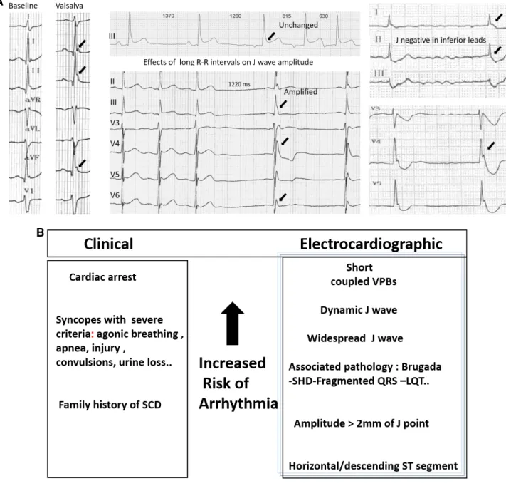
The definition of J-wave is sometimes ambiguous owing to the small amplitude and spontaneous changes of the signal. The J/ST elevation in inferior leads may be more easily missed as the pattern is less stereotypical than the Brugada syndrome (BrS). Bipolar limb leads (I,III,aVF) may be less sensitive than unipolar precordial leads in identifying discrete J/ST-segment abnormalities, and in assessing the spatial extent of the J-wave. Although unemphasized previously, a J-wave may also be negative and less apparent when it follows a negative QRS complex. Figure 1A illustrates these electrocardiographic variations.
Clinical Significance
The ECG pattern of early repolarization was first reported in 1936 as a normal variant1. In
segment in an accidentally hypothermic man2. In 1953, Osborn described a “current of injury” in experimental acidosis and hypothermia in dogs and which was often associated with the development of VF3.
The ER was known for decades to be an innocent ECG feature that was more common in young men and in athletes, with a prevalence varying between 1 to 24 % in the general population. However, case reports of unexpected SCD associated with an inferolateral J-wave abnormality were published as early as 1984, most of them concerning victims from Japan and Southeast Asia6,7,8,9. Then, the potential arrhythmogenicity of ER was shown in wedge preparations by Yan and Antzelevitch5. Finally, a definitive association between J-waves and
the pathogenesis of idiopathic VF was described in case-control studies8-12,15-19. Although these results have raised some concerns for the affected patients, the high prevalence of J-waves in the general population causes them to portend a low absolute arrhythmia risk20,21.
Risk in asymptomatic subjects
The prevalence of J-waves in athletes is reported to be as high as 44%22-25. In a study involving 704 athletes (14% harboring a J-wave), with a follow-up of 6 years there was no observation of arrhythmic events24. Similarly, no association with higher risk of death was
observed during long-term follow-up in young adults with J-waves25. Interestingly a correlation was found between J-point elevation and septal hypertrophy, as well as a possible link with exercise-induced hypertrophy23, 25-27. False tendons have also been related to the genesis of J-waves, potentially through altered depolarization in the Purkinje system due to localized stretching28. Although J-waves are common in African Americans, it is estimated that there is no increased risk in this subgroup whereas a higher risk may be present in Asian populations29-31. Noteworthy, the definition of ER in the prior literature varies in terms of wave or ST-elevation pattern; it may include individuals with only ST-elevation and no J-wave, which impacts on the reported prognostic value.
Risk in association with SHD
A large number of studies have suggested that J-waves associated with SHD increase the vulnerability to ventricular arrhythmias but not to non-arrhythmic cardiac events32-37. In a meta-analysis of 19 studies including 7268 SHD patients, a higher risk was associated with J waves specifically in inferior leads, with a notching configuration, and a horizontal or descending ST segment12. An increased arrhythmic risk has also been shown in the event of
artery occlusion were associated with 53% risk of VF34. J-waves, particularly in the inferior leads, were also associated with increased risk in patients with chronic coronary artery disease, or with dilated or hypertrophic cardiomyopathy35-37. Therefore the presence of inferior J-waves associated with SHD increases the incidence of arrhythmic events. J-waves its association with other ECG abnormalities (Brugada, Long QT, short QT) also increase the risk of arrhythmias38-41.
J waves and Idiopathic VF– risk stratification
Among patients with a history of idiopathic VF, several reports have provided clinical evidence of an increased prevalence of J-wave patterns. In the most recent study from Korea, J-waves were observed in 35 of 81 patients (43%) with idiopathic VF, and were associated with a higher risk of recurrences19. The incidence of idiopathic VF associated with inferolateral J-waves is estimated to be 3:100,000. This represents a low absolute risk by comparison with a prevalence of 1 % to 24 % in the population8,10,15-19. Therefore, the
majority of individuals with ER are at minimal risk for arrhythmic events so that asymptomatic patients with no family history of SCD should be reassured20-21.
In symptomatic patients several ECG markers have been assessed in relation with outcome. Though, the spontaneous variability of the J-ST pattern is a limiting factor. Clinical variables such as gender or ethnicity have provided conflicting results in terms of prognostic value. A family history of sudden death has been reported to be more common in association with SCD42 and it likely increases the individual risk. The prognostic value of QRS slurring versus notching, often coexisting or changing in time, is still unclear. The following variables have been shown to be associated with an increased risk of malignant arrhythmias: i) a horizontal or descending type of ST segment in the inferior leads, as opposed to an upward ST segment15-17,25,31; however the prevalence of this more risky ST pattern in controls may be up to 3%, reducing its specific value; ii) a higher magnitude of J-waves (>2.0 mm) in the inferior leads10; iii) an extensive ECG pattern involving anterior (leads V1–V3) and inferior leads8,18,41.
A consistent observation at the time of malignant arrhythmias is the amplification of inferolateral J-waves which then recedes after spontaneous or pharmacological arrhythmia termination7,8,43,44. A higher J-wave amplitude recorded soon after a syncope (compared with prior or later ECGs) suggest that a malignant arrhythmia may have occurred. The variability of J-waves after longer cycle lengths (post pause) is in our opinion an essential marker of
electrical vulnerability. Unfortunately there are no specific means to challenge the dynamicity of J-wave patterns in the vulnerable patients, like e.g. sodium blocker provocation in BrS. A Valsalva maneuver or Holter monitoring are however useful to appreciate the J-wave dynamics on the 12-lead ECG during cycle-length variations. Finally, VF induction maneuvers during an electrophysiology study are of little utility. In a multi-center study involving 81 patients followed up by ICD interrogations, VF inducibility could not predict the incidence of subsequent arrhythmias45.
In summary, from the different published reviews it appears that no clinically strong risk-stratification can be performed to identify the small subset of patients at high risk and that may facilitate primary prevention. Genetic variant analysis or specific pharmacological testing may hopefully become of prognostic importance in the future14. Currently the decision of therapeutic prevention in high-risk patients (by ICD or sometimes by Quinidine) or loop recorder implantation in intermediate-risk patients, is based on the severity of clinical variables, and electrocardiographic patterns: T-wave negativity, J-wave amplitude, the spatial extent of the J-wave pattern, and dynamic changes in the J-wave13,20-21. The presence of
short-coupled premature beats is not a risk factor as it indicates imminent threat of VF. Figure 1B summarizes our hierarchical view of risk factors.
The evidence for repolarization and depolarization abnormalities as distinct mechanisms
There is an ongoing controversy on whether the “J-wave syndromes” (Brugada and inferolateral J-waves) are due to repolarization or depolarization abnormalities. The repolarization mechanism is founded on studies in right or left ventricular wedge preparations, demonstrating that J-waves can be a consequence of a transmural repolarization gradient due to a differential distribution and function of the transient outward current9. The depolarization mechanism is founded on studies demonstrating that structural discontinuities can cause conduction disturbances by current-to-load mismatch and display the phenotype of J-waves46.In humans, however, the current data indicate heterogeneous mechanisms.
Brugada syndrome
In BrS the best evidence in support of a depolarization abnormality in humans came from Nademanee et al and other groups47-53 showing late fractionated electrograms on the
epicardial side of the right ventricular outflow tract. These electrograms were then correlated with the presence of interstitial fibrosis and reduced gap-junction expression in biopsies or autopsy studies from affected patients46,48,49. Such microstructural abnormalities were also reported in prior anatomical studies by Martini et al and Corrado et al54,55. Noteworthy, Antzelevitch's group has demonstrated that fractionated electrograms in animal wedge preparations can also be caused by repolarization disparities and concealed phase 2 reentry56. Our current clinical experience with high-resolution epi-endocardial mapping techniques (at the full organ level) demonstrates that the late epicardial fragmented potentials are continuous to the main depolarization front. This provides a strong argument in favor of a depolarization abnormality as a primary substrate of BrS in humans.
Inferolateral J wave syndrome
In inferolateral J-waves, repolarization abnormalities have been well established as the dominant substrate. However, a slurred end of the QRS complex is also a well-known marker of delayed activation in SHD (termed as ‘peri-infarction block’ or ‘epsilon wave’)57.The
J-wave here indicates “post-excited” myocardium in the same way as the delta J-wave in WPW syndrome indicates “pre-excited” myocardium.
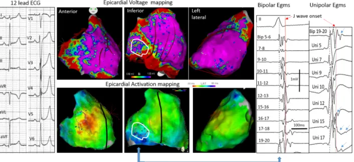
High density invasive mapping data have been collected from 38 patients with inferolateral J-wave syndrome in 3 centers58. These patients had no demonstrated SHD and most were referred for VF recurring despite antiarrhythmic drugs including Quinidine. Electro-anatomical mapping was performed during sinus rhythm to obtain endocardial and epicardial electrograms (2000-6000 recorded points) in unipolar and bipolar mode (unipolar filters of 0.05- 250/500 Hz-, and bipolar 30-250/500Hz). A 2mm interelectrode spacing was used to minimize the recording of far-field potentials and specific attention was paid to the electrograms coincident with the J-wave. Abnormal electrograms were defined as prolonged fragmented electrograms with more than 3 components and a local duration superior to 70 ms59; these criteria are similar to those defining structural alteration and fibrotic tissue in SHD. The electrograms occurring within (and possibly prolonging) the J-wave were considered as belonging to depolarization if they were sharp and in temporal and spatial continuity of the depolarization field mapped at the end of the QRS complex . They were considered as indicating ventricular repolarization if either they were not in continuity with the surrounding depolarization (presence of an electrical gap > 50ms), or displayed a slow pattern (hump) in unipolar mode that was not related to a near-field potential. Such slow
potentials have been reported previously during direct or indirect epicardial recordings, in patients with J-wave or hypothermia conditions.33,60-63.
Based on these definitions, the results58 indicate that the electrocardiographic J-wave can be the phenotypic expression of either delayed depolarization or early repolarization, in the inferior part of ventricles. In patients who had a concomitant BrS (spontaneous or provoked by a sodium channel blocker) inferolateral J-waves were consistently caused by delayed depolarization of inferior myocardium. In patients without BrS, inferolateral J-waves were caused by delayed depolarization in 24% of patients, whereas an ER was the cause of J-waves in 76%. Note that the true prevalence of depolarization versus repolarization abnormalities may be different, as the patients were often only referred after quinidine failure, which is likely to favor a higher proportion of delayed repolarization patients. Ajmaline testing was performed in all patients and resulted in J-wave amplification or ST elevation in the inferior leads in a few patients. The latter had a delayed depolarization whereas no patient with early repolarization had J/ST wave amplification on Ajmaline.
It is noteworthy that in a prior non-invasive mapping study, Zhang et al did not observe conduction abnormalities in inferolateral J-wave syndrome62 during sinus rhythm. A likely explanation is that the small depolarization fronts that are responsible for the late fractionated electrograms cannot be recognized as such by a mathematical inverse solution from body surface potentials.
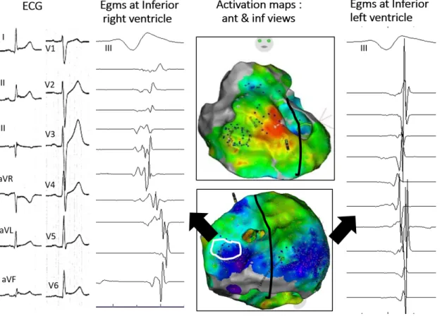
Examples of J-waves caused by late depolarization are shown in Figures 2 and 3. The fragmented electrograms occurred timely during the inscription of J-waves and were in continuity with the depolarization field. The great majority of abnormal electrograms were found on the epicardium (right ventricle and two cases in left ventricle) while two patients had late electrograms recorded endocardially and epicardially. They were recorded predominantly in the inferior right ventricle, at the sites of terminal activation as predicted by a modeling study64. In the patients with BrS and inferolateral J-wave syndromes the electrograms recorded in the inferior right ventricle were similar to those in the RVOT. The pathogenesis of inferolateral J-waves here is dominantly due to abnormal delayed conduction; either limited to the inferior myocardium (right or left – endo or epicardial), or combined with other locations (such as the anterior right ventricle). The affection responsible for electrogram fractionation (altered myocardial cells or their connections, fibrotic or fatty tissue infiltrations) is undetermined. In addition not only a heterogeneous delay of activation but
also a homogeneous delayed activation may lead to a J-wave64. Finally, there is probably a potential contribution of a repolarization disparity (secondary to the depolarization alterations) to explain a part of J/ST wave fluctuations or arrhythmogenesis, which requires additional studies46.
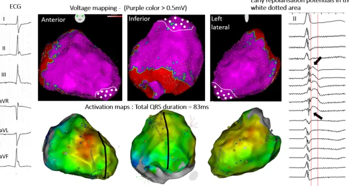
Examples of J-waves due to (true) early repolarization are illustrated in Figures 4,5. In these patients we could not find electrograms indicative of delayed depolarization coincident with J-waves, but low frequency (hump) potentials were present at the beginning of the ST segment in unipolar recordings. The spatial location and extent of potentials were epicardially dominant in the inferior septal projection and adjacent left ventricle60-63. Although other
hypotheses cannot be totally excluded46, the J-wave mechanism may represent a lower epicardial voltage across the ventricular wall as shown by Yan and Antzelevitch5,9,57. These authors have emphasized that a short-coupled ectopic beat was “a strong piece of evidence supporting an endo-epicardial myocardial gradient of repolarization leading to phase 2 reentry.”. Such arrhythmogenesis due to abbreviated action potentials (loss of the dome) is technically difficult to demonstrate in clinical conditions. However we observed that VF is commonly initiated from Purkinje triggers (also with short coupling interval) whereas initiation from myocardial triggers (potentially due to phase 2 reentry) is more particularly observed in patients with multifocal ectopy and widespread early repolarization8.
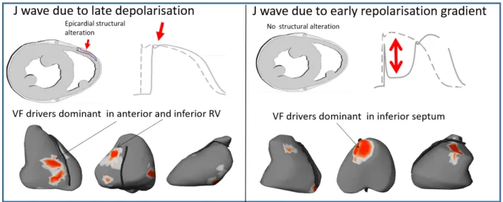
VF drivers in the 2 forms of inferolateral J wave syndromes
VF drivers were mapped using a non-invasive method59 and showed dominant drivers in the inferior part of myocardium during the initial stages of VF (Fig 6). VF in patients with J-waves indicating delayed depolarization was dominantly associated with drivers in the inferior and anterior right ventricle. VF in early repolarization was rather associated with drivers located in the inferior septum and adjacent regions. Note that this epicardial region overlying the inferior septum may be the breakthrough site of activity originating from the Purkinje posterior fascicle. Importantly, the cycle length of VF (measured at the 10th second after initiation) was significantly shorter in early repolarization than in delayed depolarization (n=16, 148±5ms vs 175±4ms, p=0.013) consistent with shorter ventricular refractory periods. Table S1 summarizes the differences between J-waves due to early repolarization versus late depolarization.
Implications of repolarization vs depolarization origins of inferolateral J wave –
The classification of inferolateral J-waves in two distinct substrates has implications in terms of terminology, pathogeny, genetics, and therapy.
While the terminology of early repolarization appears adequate in the cases of repolarization abnormality, it is erroneous when the J-wave is due to a late depolarization. The term inferolateral J-wave may thus be more adequate and generic. Further studies are needed to provide additional phenotypic features that may help distinguishing between depolarization and repolarization abnormality. It is likely that a stable J-wave pattern not influenced by long cycle lengths is caused by a depolarization abnormality, potentially exacerbated at short cycle lengths; and vice versa for early repolarization. In a prior study by Roten et al, the response of inferior J-wave patterns to isoproterenol varied individually. Inferior J-waves were persistent in 35% (rather suggestive of delayed depolarization) and decreasing or normalizing in 65% of patients (rather suggestive of early repolarization). Baseline QRS width was significantly larger in patients with persistent J-waves65.
In terms of genetic predisposition, mutations in genes coding for subunits of the IK-ATP channel or cardiac L-type calcium channel have been described, however without validation by functional expression studies in most. Loss–of-function mutations in Na-channel genes have also been reported, including a significant proportion of them associated with an ST elevation in inferior leads under Na channel blocker. We speculate that a late-depolarization J-wave will be more likely associated with gene variants in the Na channel, connexins, and structural proteins, while mutations in the ion channels carrying Ito, IK-ATP or ICa will be more associated with early repolarization.
The immediate benefit of distinguishing J-wave subtypes is its therapeutic potential. In patients with late-depolarization J-waves, substrate ablation targeting the delayed electrograms is feasible like in BrS or other structural heart diseases. In patients with ER J-waves, we do not know whether ablation of abnormal repolarizing tissue is applicable and safe, but trigger ablation is an effective option when anti-arrhythmic drugs and particularly quinidine have failed.
Mechanistic classification of VF associated with apparently normal hearts
The present review shows that similar ECG phenotypes may be caused by fundamentally different substrates. Inferolateral J-waves can be the expression of voltage gradients at the initial phase of repolarization (‘early-repolarization’) or the expression of delayed depolarized areas. Delayed depolarization is associated with electrogram fractionation indicating local structural alteration, whereas in the early repolarization group, there is likely no structural abnormality although VF triggers originating from the Purkinje system are often found. Similar findings have been reported recently in idiopathic VF, using high density electrogram characterization59. The study involved 24 patients with no electrocardiographic phenotype: J-waves, long or short QT syndromes were excluded; imaging and ajmaline testing were strictly negative. Localized areas of abnormal depolarization were identified in 62% of them. Most abnormal areas were epicardial, affecting dominantly the right ventricle, and of limited size (5% of the total surface). In the remaining subset of IVF devoid of myocardial alteration (38% of patients), Purkinje triggers were evidenced and appeared as the dominant mechanism. Therefore, the spectrum of arrhythmogenic diseases leading to SCD in apparently normal hearts, including the J-wave syndromes, appears to comprise an important emerging subgroup in which the dominant substrate are localized depolarization abnormalities that may or may not have an electrocardiographic expression. A simplified mechanistic classification based on the primary pathogenesis is proposed in figure 7.
Conclusion
Inferolateral J-waves are subtle ECG phenotypes that may be responsible of SCD in patients with no apparent structural heart disease. Their occurrence at the QRST junction can be the expression of distinct substrates, early repolarization or delayed activation abnormality or mixed forms. Distinguishing between these substrates could significantly improve genetic interpretation, risk stratification and the therapeutic approach. Further studies are also needed for quantitative assessment of the recorded signals and understanding of the pathogenesis of inferolateral J-waves.
References
1- Shilpey R, Hallaran W. The four lead electrogram in 200 normal men and women. Am Heart J 1936; 11:325-345.
2- Tomaszewski W. Changements electrocardiographiques observes chez un homme mort de froid. Arch Mal Cœur 1938 ; 31 : 525-528.
3- Osborn JJ. Experimental hypothermia: respiratory and blood pH changes in relation to cardiac function. Am J Phys 1953; 175:389-398..
4- Kambara H, Phillips J. Long-term evaluation of early repolarization syndrome (normal variant RS-T-segment elevation). Am J Cardiol 1976; 38:157-161.
5- Yan GX, Antzelevitch C. Cellular basis for the electrocardiographic J wave. Circulation. 1996; 93:372–379.
6- Otto CM, Tauxe RV, Cobb LA, Greene HL, Gross BW, Werner JA, Burroughs RW,
Samson WE, Weaver WD, Trobaugh GB. Ventricular fibrillation causes sudden death in southeast Asian immigrants. Ann Intern med 1984; 101: 45-47.
7- Aizawa Y, Tamura M, Chinushi M, Naitoh N, Uchiyama H, Kusano Y, Hosono H, Shibata A. Idiopathic ventricular fibrillation and bradycardia-dependent intraventricular block Am Heart J 1993 ;126 :1473-4.
8- Haissaguerre M, Derval N, Sacher F et al. Sudden cardiac arrest associated with early repolarization. N Engl J Med. 2008; 358: 2016–23. 1157-1164.
9- Antzelevitch C, Yan GX. J-wave syndromes: Brugada and early repolarization syndromes. Heart Rhythm. 2015; 12:1852–66.
10- Tikkanen JT, Anttonen O, Junttila MJ, Aro AL, Kerola T, Rissanen HA, Reunanen A, Huikuri HV. Long-term outcome associated with early repolarization on electrocardiography. N Engl J Med. 2009; 361: 2529–37.
11- Nam GB, Ko KH, Kim J, Park KM, Rhee KS, Choi KJ, Kim YH, Antzelevitch C. Mode of onset of ventricular fibrillation in patients with early repolarization pattern vs. Brugada syndrome. Eur Heart J. 2010; 31: 330–9.
and sudden cardiac death in patients with structural heart diseases. Heart Rhythm. 2017; 14: 1157-1164.
13- MacFarlane PW,Antzelevitch C, Haissaguerre M, Huikuri HV, Potse M, Rosso R, Sacher F, Tikkanen JT, Wellens H, Yan GX. The early repolarization pattern: a consensus paper. J Am CollCardiol. 2015; 66:470-7.
14- Antzelevitch C, Yan GX, Ackerman MJ, et al. J-Wave syndromes expert consensus conference report: Emerging concepts and gaps in knowledge. Heart Rhythm 2016. 32:315-339.
15- Rosso R, Kogan E, Belhassen B, Rozovski U, Scheinman MM, Zeltser D, Halkin A, Steinvil A, Heller K, Glikson M, Katz A, Viskin S. J-point elevation in survivors of primary ventricular fibrillation and matched control subjects: incidence and clinical significance. J Am CollCardiol. 2008; 52:1231–8.
16- Sinner MF, Reinhard W, Muller M et al. Association of early repolarization pattern on ECG with risk of cardiac and all-cause mortality: a population-based prospective cohort study. PLoS Med. 2010; 7 e1000314.
17- Derval N, Simpson CS, Birnie DH et al. Prevalence and characteristics of early repolarization in the CASPER registry: cardiac arrest survivors with preserved ejection fraction registry. J Am Coll Cardiol. 2011; 58:722–8.
18- Junttila MJ, Tikkanen J T, Kentta T et al. Early repolarization as a predictor of arrhythmic and non-arrhythmic cardiac events in middle-aged subjects. Heart Rhythm. 2014; 11:1701–6.
19- Seong CS, Gwag HB, Hwang JK, Park SJ, Park K-M, Kim JS. Clinical significance of fragmented QRS complexes or J waves in patients with idiopathic ventricular arrhythmias. PLoS ONE 2018; 13:e0194363.
20- Viskin S, Rosso R, Halkin A. Making sense of early repolarization. Heart Rhythm. 2012; 9:566–9.
21- Obeyesekere N, Krahn AD . Early Repolarization – What Should the Clinician Do? Arrhythm Electrophysiol Rev. 2015; 4:96-9.
22- Tikkanen JT, Junttila MJ, Anttonen O, Aro AL, Luttinen S, Kerola T, Sager SJ, Rissanen HA, Myerburg RJ, Reunanen A, Huikuri HV. Early repolarization:
electrocardiographic phenotypes associated with favorable long-term outcome. Circulation. 2011; 123:2666–73.
23- Biasco L, Cristoforetti Y, Castagno D, Giustetto C, Astegiano P, Ganzit G, Gribaudo CG, Gaita F. Clinical, electrocardiographic, echocardiographic characteristics and long term follow up of elite soccer players with J-point elevation. Circ Arrhythm Electrophysiol. 2013;6:1178–84.
24- Quattrini FM, Pelliccia A, Assorgi R, DiPaolo FM1, Squeo MR, Culasso F, Castelli V, Link MS, Maron BJ. Benign clinical significance of J-wave pattern (early repolarization) in highly trained athletes. Heart Rhythm. 2014; 11:1974–82.
25- Ilkhanoff L, Soliman EZ, Prineas RJ, Walsh JA 3rd, Ning H, Liu K, Carr JJ, Jacobs DR Jr, Lloyd-Jones DM. Clinical characteristics and outcomes associated with the natural history of early repolarization in a young, biracial cohort followed to middle age: the coronary artery risk development in young adults (CARDIA) study. Circ Arrhythm Electrophysiol. 2014;7:392.
26- Guillem JP, Haissaguerre M, Lemétayer P, Montserrat P, Le Hérissier A, Warin JF. Echocardiographic study of the early repolarization syndrome. Demonstration of dynamic obstruction with isoprenaline. Arch Mal Coeur Vaiss. 1988; 81:199-206.
27- Noseworthy PA, Weiner R, Kim J, Keelara V, Wang F, Berkstresser B, Wood MJ, Wang TJ, Picard MH, Hutter AM Jr, Newton-Cheh C, Baggish AL. Early repolarization pattern in competitive athletes: clinical correlates and the effects of exercise training. Circ Arrhthm Electrophysiol. 2011; 4:432–40.
28- Nakagawa M, Ezaki K, Miyazaki H, Wakisaka O, Shinohara T, Teshima Y, Yufu K, Takahashi N, Saikawa T. Electrocardiographic characteristics of patients with false tendon: possible association of false tendon with J waves. Heart Rhythm. 2012; 9:782–8.
29- Olson KA, Viera AJ, Soliman EZ, Crow RS, Rosamond WD. Long-term prognosis associated with J-point elevation in a large middle-aged biracial cohort: the ARIC study. Eur Heart J. 2011; 32:3098–106.
30- Perez MV, Uberoi A, Jain NA, Ashley E, Turakhia MP, Froelicher V. The prognostic value of early repolarization with ST-segment elevation in African Americans. Heart Rhythm. 2012; 9:558–65.
31- Haruta D, Matsuo K, Tsuneto A, Ichimaru S, Hida A, Sera N, Imaizumi M, Nakashima E, Maemura K, Akahoshi M. Incidence and prognostic value of early repolarization pattern in the 12-lead electrocardiogram. Circulation. 2011; 123:2931–7.
32- Tikkanen JT, Wichmann V, Junttila MJ, Rainio M, Hookana E, Lappi OP, Kortelainen ML, Anttonen O, Huikuri HV. Association of early repolarization and sudden cardiac death during an acute coronary event. Circ Arrhythm Electrophysiol. 2012; 5:714–8.
33- Ghosh S, Cooper DH, Vijayakumar R, Zhang J, Pollak S, Haïssaguerre M, Rudy Y. Early repolarization associated with sudden death: insights from noninvasive electrocardiographic imaging. Heart Rhythm 2010; 7, 534–537.
34- Demidova MM, Martin-Yebra A, van der Pals J, Koul S, Erlinge D, Laguna P, Martínez JP, Platonov PG. Transient and rapid QRS-widening associated with a J-wave pattern predicts impending ventricular fibrillation in experimental myocardial infarction. Heart Rhythm. 2014; 11:1195–2001.
35- Patel RB, Ng J, Reddy V et al. Early repolarization associated with ventricular arrhythmias in patients with chronic coronary artery disease. Circ Arrhythm Electrophysiol. 2010; 3:489–95.
36- Pei J, Li N, Gao Y, Wang Z, Li X, Zhang Y, Chen J, Zhang P, Cao K, Pu J. The J-wave and fragmented QRS complexes in inferior leads associated with sudden cardiac death in patients with chronic heart failure. Europace. 2012; 14:1180–7.
37- Tsuda T, Hayashi K, Konno T, Sakata K, Fujita T, Hodatsu A, Nagata Y, Teramoto R, Nomura A, Tanaka Y, Furusho H, Takamura M. J Waves for Predicting Cardiac Events in Hypertrophic Cardiomyopathy. JACC Clin Electrophysiol. 2017; 3:1136-1142.
38- Laksman ZW, Gula LJ, Saklani P, Cassagneau R1, Steinberg C, Conacher S, Yee R1, Skanes A, Leong-Sit P, Manlucu J, Klein GJ, Krahn AD. Early repolarization is associated with symptoms in patients with type 1 and type 2 long QT syndrome. Heart Rhythm. 2014; 11:1632–8.
39- Peters S, Selbig D. Early repolarization phenomenon in arrhythmogenic right ventricular dysplasia-cardiomyopathy and sudden cardiac arrest due to ventricular fibrillation. Europace. 2008;10:1447–9.
40- Letsas KP, Sacher F, Probst V et al. Prevalence of early repolarization pattern in inferolateral leads in patients with Brugada syndrome. Heart Rhythm. 2008;5:1685–9.
41- Kamakura T, Kawata H, Nakajima I et al. Significance of non-type 1 anterior early repolarization in patients with inferolateral early repolarization syndrome. J Am CollCardiol. 2013;62:1610–8.
42- Nunn LM, Bhar-Amato J, Lowe MD, Macfarlane PW, Rogers P, McKenna WJ, Elliott PM, Lambiase PD. Prevalence of J-point elevation in sudden arrhythmic death syndrome families. J Am Coll Cardiol 2011;58:286–9.
43- Aizawa Y, Sato A, Watanabe H, et al. Dynamicity of the J-wave in idiopathic ventricular fibrillation with a special reference to pause-dependent augmentation of the J-wave. J Am Coll Cardiol 2012; 59:1948-1953.
44 Mizumaki K, Nishida K, Iwamoto J, Nakatani Y, Yamaguchi Y, Sakamoto T, Tsuneda T, Kataoka N, Inoue H. Vagal activity modulates spontaneous augmentation of J-wave elevation in patients with idiopathic ventricular fibrillation. Heart Rhythm. 2012; 9:249–55. 45- Mahida S, Derval N, Sacher F et al. Role of electrophysiological studies in predicting risk of ventricular arrhythmia in early repolarization syndrome. J Am Coll Cardiol. 2015; 65:151–9.
46- Hoogendijk MG, Potse M, Linnenbank AC et al Mechanism of right precordial ST-segment elevation in structural heart disease: excitation failure by current-to-load mismatch. Heart Rhythm. 2010; 7: 238-48.
47- Nademanee K, Veerakul G, Chandanamattha P, Chaothawee L, Ariyachaipanich A, Jirasirirojanakorn K, Likittanasombat K, Bhuripanyo K, Ngarmukos T. Prevention of ventricular fibrillation episodes in Brugada syndrome by catheter ablation over the anterior right ventricular outflow tract epicardium. Circulation. 2011; 123:1270–1279.
48- Nademanee K, Raju H, de Noronha SV, et al. Fibrosis, Connexin-43, and Conduction abnormalities in the Brugada Syndrome. J Am Coll Cardiol 2015; 66:1976-1986.
49- Coronel R, Casini S, Koopmann TT, et al. Right ventricular fibrosis and conduction delay in a patient with clinical signs of Brugada syndrome: a combined electrophysiological, genetic, histopathologic, and computational study. Circulation. 2005; 112:2769–2777.
50- Sacher F, Jesel L, Jais P, Haissaguerre M. Insight into the mechanism of Brugada syndrome: epicardial substrate and modification during ajmaline testing. Heart Rhythm 4/1/2014 2014;11:732-734.
51- Brugada J, Pappone C, Berruezo A, Vicedomini G, Manguso F, Ciconte G, GiannelliL, Santinelli V. Brugada Syndrome Phenotype Elimination by Epicardial Substrate Ablation. Circ Arrhythm Electrophysiol 2015; 8:1373-81.
52- Chung FP, Raharjo SB, Lin YJ, et al A novel method to enhance phenotype, epicardial functional substrates, and ventricular tachyarrhythmias in Brugada syndrome. Heart Rhythm. 2017; 14:508-517.
53- Pappone C, Brugada J, Vicedomini G et al. Electrical Substrate Elimination in 135 Consecutive Patients With Brugada Syndrome. Circ Arrhythm Electrophysiol. 2017; 10:e005053.
54- Martini M, Nava A, Thiene G , Buja GF Canciani B, Scognamiglio R, Daliento L, Dalla Volta S. Ventricular fibrillation without apparent structural heart disease . Am Heart J 1989; 118: 1203-1209.
55- Corrado D, Basso C, Buja G, Nava A, Rossi L, Thiene G. Right bundle branch block, right precordial st-segment elevation, and sudden death in young people. Circulation. 2001; 103:710-7.
56- Patocskai B, Yoon N, Antzelevitch C. Mechanisms Underlying Epicardial Radiofrequency Ablation to Suppress Arrhythmogenesis in Experimental Models of Brugada Syndrome. JACC: Clinical Electrophysiology 2017; 3: 353.
57- Liu T, Zheng J, Yan GX. J Wave Syndromes: History and Current Controversies Korean Circ J. 2016;46(5):601-609.
58- Nademanee K, Hocini M, Nogami A, Veerakul G, Cheniti G, Bokan RM, Lou Q, Haissaguerre M. Mapping And Ablation Of Ventricular Fibrillation Associated With Early Repolarization Syndrome. Heart Rhythm Scientific Sessions 2018; B-AB11-03.
59- Haissaguerre M, Hocini M, Cheniti G et al Localized structural alterations underlying a subset of unexplained sudden cardiac death. Circ Arrhythm Electrophysiol. 2018; 11:e006120.
60- Emslie-Smith D, Sladden GE, Stirling GR. The significance of changes in the electrocardiogram in hypothermia. Br Heart J. 1959; 21: 343–351.
61- Boineau JP. The early repolarization variant—an electrocardiographic enigma with both QRS and J-STT anomalies J ElectroCardiology2007; 40: 3e1–e10.
62- Zhang J, Hocini M, Strom M, Cuculich PS, Cooper DH, Sacher F, Haïssaguerre M, Rudy Y. The Electrophysiological Substrate of Early Repolarization Syndrome: Noninvasive Mapping in Patients. JACC Clin Electrophysiol. 2017; 3:894-904.
63- Nakagawa K, Nagase S, Morita H, Ito H. Left ventricular epicardial electrogram recordings in idiopathic ventricular fibrillation with inferior and lateral early repolarization. Heart Rhythm 2014; 11:314-317.
64- Meijborg VMF, Potse M, Conrath CE, Belterman CNW, De Bakker JMT and Coronel
R: Reduced Sodium Current in the Lateral Ventricular Wall Induces Inferolateral J-Waves. Front. Physiol.2016; 7:365.
65- Roten L, Derval N, Sacher F et al. Heterogeneous response of J-wave syndromes to beta-adrenergic stimulation . Heart Rhythm. 2012 ; 9:1970-6.
A
B
Figure 1: (A) ECG variations in inferolateral J-Waves. Left: Valsalva or strong inspiration maneuver producing J-wave amplification. Middle: Cycle-length prolongation associated with either unchanged pattern or amplification. Right: negative J-waves in inferior leads and positive J-waves in lateral leads; note the majoration post pause. (B) Hierarchical view of electrocardiographic and clinical risk factors.
Figure 2: Inferolateral J-wave syndrome due to abnormal depolarization. Upper central maps shows bipolar electrogram voltage (purple indicating voltage>1 mV) with low voltage in inferior right ventricle. Lower maps show the activation mapping with blue indicating the latest activated regions, here in the inferobasal right ventricle. Right panel shows fragmented electro-grams preceding and coincident with the J-wave (white contour) in bipolar and unipolar (arrows) mode.
Figure 3: Inferolateral J-wave syndrome due to abnormal depolarization. The maps (center) show activation mapping with blue indicating the latest acti-vated regions; herein the inferobasal right and left ventricles. Low voltage fragmented electrograms coincident with J-wave are only present in inferior right ventricle (white contour) compared with electrograms in inferior left ven-tricle.
Figure 4: Inferolateral J-wave syndrome due to early repolarization. Upper maps show bipolar electrogram voltage without evidence of low voltage area. Lower maps shows the latest activated regions (blue) in the inferobasal and lat-erobasal right ventricle. There are no late depolarization electrograms coinci-dent to the J-wave but slow early-repolarization potentials (arrows); which are present in the apical region (white-dotted area). Note that J-wave is small on lead II (right) and underestimates the extent of early repolarization recorded by epicardial mapping.
Figure 5: Another case of early repolarization. 12-lead ECGs (left) shows a global J-wave pattern. There are no late depolarization bipolar electrograms coincident with the J-wave; but early repolarization potentials are recorded diffusely in the inferior left ventricle.
Figure 6: Typical location of VF driver regions. The locations of reentries are shown in red. They are predominantly located in the right ventricle in early repolarization (right) versus the inferior septum in late depolarization J-waves (left).
Figure 7: Spectrum of arrhythmogenic diseases leading to SCD in apparently normal hearts, and a mechanistic classification based on the primary patho-genesis.