HAL Id: tel-01264118
https://tel.archives-ouvertes.fr/tel-01264118
Submitted on 28 Jan 2016HAL is a multi-disciplinary open access archive for the deposit and dissemination of sci-entific research documents, whether they are pub-lished or not. The documents may come from teaching and research institutions in France or abroad, or from public or private research centers.
L’archive ouverte pluridisciplinaire HAL, est destinée au dépôt et à la diffusion de documents scientifiques de niveau recherche, publiés ou non, émanant des établissements d’enseignement et de recherche français ou étrangers, des laboratoires publics ou privés.
Investigating the role of human HAT (histone
acetyltransferase) containing complexes, ATAC and
SAGA, in living cells
Nikolaos Vosnakis
To cite this version:
Nikolaos Vosnakis. Investigating the role of human HAT (histone acetyltransferase) containing com-plexes, ATAC and SAGA, in living cells. Genomics [q-bio.GN]. Université de Strasbourg, 2014. En-glish. �NNT : 2014STRAJ119�. �tel-01264118�
UNIVERSITÉ DE STRASBOURG
École doctorale des Sciences de la Vie et de la Santé
IGBMC - CNRS UMR 7104 - Inserm U 964
THÈSE
présentée par :Nikolaos Vosnakis
soutenue le : 16 décembre 2014
pour obtenir le grade de :
!"#$%&'($')*%+,-$&.,#/'($'0#&1.2!%&3
Discipline/ Spécialité
: Aspects moléculaires et cellulaires de la biologie
Investigating the role of human HAT
(histone acetyltransferase) containing
complexes, ATAC and SAGA, in living cells
THÈSE dirigée par :
M. Laszlo TORA Directeur de recherche, université de Strasbourg
RAPPORTEURS :
M. Edouard BERTRAND Chargé de Recherches, Institut de Génétique Moléculaire de Montpellier M. Marc TIMMERS Professeur, University Medical center d !"#$%&"
AUTRES MEMBRES DU JURY :
2
Acknowledgements
First I would like to thank Dr Edouard Bertrand, Dr Evi Soutoglou and Prof Marc Timmers for accepting to be members of my jury. Thank you for your time and effort to evaluate my thesis. I thank Laszlo for giving me the opportunity to work in an excellent scientific environment. Thank you for your guidance and full support throughout these four years. Your trust and encouragement helped me to be motivated even when things did not go exactly as planned. I thank Didier for his bonne humeur!!" #$%" &'( helping me to manage scientific, social and administrative issues, especially in the year that Laszlo was in a different continent. I would also like to thank old and current members of the Tora lab. Particularly, I would like to thank Monica not only for teaching me how to work in the cell culture but also for her support during the first year of my thesis, Anne for always being kind and helpful but also for sharing food at the canteen, David for sharing precious reagents and a useful advice. Thank you Sarina for the nice chats during those years. Chen-Yi thanks for being such a good friend and for your help in the lab. Thanks Jacques for your scientific advice and the historical references. I want to thank Tiago, Federica, Ivanka and Sascha for bringing high quality fresh air in the lab.
My acknowledgements go to all the people with whom I worked with and provided their expertise in different aspects of this project. I thank Dr Pascal Didier (Yves Mely lab, Laboratoire de Biophotonique et Pharmacologie, UdS) for our excellent collaboration in the FCS experiments. All the people of the IGBMC imaging facility and particularly Marc Koch for his help and valuable advice on FRAP and FLIP experiments. I thank Eli Scheer for always being helpful in the lab and particularly for her major contribution in all the protein extraction and IP experiments; without her this part of the project would not be possible. Thanks to Matthieu Stierle for the generation of the anti-ADA2b antibody. I thank Virginie Chavant from the proteomic platform of the IGBMC, that worked on the analysis of most of our IPs. Many thanks to Marjorie Fournier for useful and extensive discussions on the proteomic part of the project and also for her patience in teaching me how to properly treat very long protein lists. I thank Amelie Weiss and Dr Laurent Brino from the HTS platform of IGBMC for their contribution in automated microscopy experiments. I would also like to thank Prof Marc Timmers, Dr Marc Vigneron and Dr Simon Trowitzsch (Imre Berger lab, EMBL, Grenoble) for kindly sharing reagents that were invaluable for the experiments of this study. Last but not least I thank Betty Heller and all the stuff of the IGBMC cell culture facility for always being helpful.
Moreover, I thank Dr Maria-Elena Torres-Padilla and Dr Patrick Heun, members of my mid-thesis committee for their useful comments and discussions.
3 I also thank the IGBMC PhD programme and the Fondation pour la Recherche Médicale (FRM) for the financial support of my thesis project.
A special thanks goes to all the good friends from Strasbourg that tolerate(d) me and made these four years special. It would be a long list to mention. Thank you for the fun, memorable moments and the multidimensional support.
4
ABSTRACT
SAGA and ATAC are multisubunit Histone Acetyl-Transferase (HAT) containing complexes that facilitate RNA polymerase II (Pol II) transcription by altering the chromatin state via post-translational modifications of histones. ATAC and SAGA share a set of subunits among which GCN5, that exhibits different catalytic properties in the context of each complex. Many studies have described the roles of the complexes in gene-specific transcription regulation and provided information about their genome wide distribution and function. However, little is known about the dynamic properties of the complexes in living cells. In addition, structural and functional studies suggest a modular organisation of the complexes. Nevertheless, there is little information on the regulation of their assembly and how the common subunits are differentially incorporated in the two complexes. The first goal of my Ph.D. project was to investigate the intranuclear dynamics of human ATAC and SAGA complexes in living cells and study how it can be related to transcription regulation. I used an experimental approach based on live-cell imaging methods such as FRAP (Fluorescence Recovery After Photobleaching) and FCS (Fluorescence Correlation Spectroscopy). I exploited these two techniques, to characterise the mobility of the two complexes and compare their dynamic properties with other important actors of Pol II transcription. I showed that all tested ATAC and SAGA subunits are highly mobile in the nucleus of living cells and they only exhibit very transient interactions with chromatin. Moreover, I investigated the existence of a possible connection between active transcription and the dynamics of the two complexes. Thus, the data collected from this set of experiments show, for the first time in living cells, that the very dynamic behaviour of the ATAC and SAGA is a key property of both complexes which explains certain aspects of their function as chromatin modifiers. During my Ph.D. studies, I also became interested in the mechanisms that regulate the dynamic properties of SAGA and ATAC subunits related to the assembly of the complexes. This question was investigated by a combination of imaging experiments (in fixed and living cells) and quantitative proteomics. FLIP (Fluorescence Loss In Photobleaching), showed that exogenously overexpressed ATAC- and SAGA-specific HAT-module subunits (ADA2a and ADA2b respectively) differ significantly in their subcellular localisation dynamics. In addition, I showed that the relative abundance of GCN5 and ADA2a affects the subcellular distribution of the latter protein. With the application of quantitative proteomic analysis, based on MudPIT (Multidimensional Protein Identification Technology), the findings of the imaging experiments were expanded on endogenous proteins and provided strong evidence that the cytoplasmic and nuclear assembly pathways of SAGA and ATAC complexes are different. Altogether, our data contributed to our understanding of the way ATAC and SAGA exhibit their functions and revealed novel protein-protein interaction related to the assembly of the complexes.
5
Table of contents
Table of contents ... 5 Table of Figures ... 9 Table of Tables ... 10 1 Introduction ... 111.1 Core promoter elements in Pol II transcription initiation ... 12
1.2 RNA pol II: a structure-function paradigm ... 13
1.3 General Transcription Factors and Pre-Initiation Complex (PIC) formation ... 16
1.3.1 General Transcription Factors ... 16
1.3.2 Preinitiation complex assembly pathways from a deterministic perspective ... 21
1.4 Chromatin organisation ... 23
1.4.1 Histone PTMs and modulation of chromatin organisation and function .. 25
1.4.2 Posttranslational modification of histones ... 28
1.5 The transcriptional coactivators SAGA and ATAC ... 33
1.5.1 The SAGA complex in S. cerevisiae ... 34
1.5.2 The ADA complex in S. cerevisiae ... 35
1.5.3 The SAGA-LIKE (SLIK) complex of in S. cerevisiae ... 35
1.5.4 The SAGA complex in metazoans ... 36
1.5.5 The modular organisation of SAGA reflects its multiple functions. ... 38
1.5.6 Regulation of SAGA recruitment ... 41
1.5.7 ATAC, the second GCN5 containing complex present in metazoans ... 44
1.5.8 Structural organisation of ADA2a and ADA2b and their role in GCN5 activity regulation ... 47
1.5.9 Evidence for distinct functional roles of ATAC and SAGA in gene specific transcription regulation ... 49
6
1.6 Investigating mechanisms of action of transcription factors in living
cells 54
1.6.1 Live-cell fluorescence microscopy techniques reveal transcription factors dynamics !55
1.6.2 Functional dynamics of transcription factors revealed by FRAP and FCS..58
1.7 Paradigms and implications of transcription-associated complexes assembly regulation ... 62
2 Materials and methods ... 68
2.1 Cell culture ... 68
2.2 eGFP constructs and cDNAs ... 68
2.3 Transfection ... 69
2.4 Transcription inhibition conditions ... 69
2.5 Antibodies ... 69
2.6 Preparation of Nuclear and Cytoplasmic extracts ... 70
2.7 Immunoprecipitations ... 70
2.8 Western blot ... 71
2.9 MudPIT mass spectrometry analyses ... 71
2.10 Microscopy ... 72
2.10.1 FRAP and FLIP ... 72
2.10.2 Fluorescence Correlation Spectroscopy ... 74
2.10.3 High content analysis ... 75
3 "#$%&'()*+, )$, *(-*#(#+).$, /0, 112324, #$5, 6272, '.#'+)8#+.(, '.9*:-;-&, #(-, highly dynamic in human cell nuclei and their mobility is unaffected by +(#$&'()*+).$,)$<)=)+).$!> ... 76
3.1 Results (I) ... 76
3.1.1 FRAP based approach to investigate human SAGA and ATAC dynamics ... 76
3.1.2 Semi-quantitative FRAP indicates that SAGA and ATAC are highly mobile complexes !!!78
7 3.1.3 The nuclear behaviour of SAGA and ATAC shows that they belong in a class very dynamic transcription factors ... 80 3.1.4 Characterization of the dynamics of SAGA and ATAC complexes after transcription inhibition ... 83 3.1.5 Investigation of SAGA and ATAC dynamics by FCS ... 92 3.1.6 Unbiased fitting of FCS data reveals two distinct diffusing species for all studied transcription factors ... 93 3.1.7 The highly dynamic behaviour of SAGA and ATAC complex is an inherent property of the complexes ... 95 3.1.8 Effects of transcription inhibition on SAGA and ATAC subunits analysed by FCS 98
3.2 DISCUSSION (I) ... 101
3.2.1 Human ATAC and SAGA function involves transient interactions with chromatin 101 3.2.2 Dynamics of SAGA and ATAC are not linked to active transcription ... 103 3.2.3 Approaches to clarify the interpretation of FCS data ... 105 3.2.4 FRAP experiments to study the effect of SAGA and ATAC on Pol II recruitment in living human cells ... 105 3.2.5 FRAP/FCS experiments to investigate SAGA and ATAC recruitment in living cells 106 3.2.6 Alternative systems to investigate site specific recruitment of ATAC and SAGA in living cells ... 107
4 "#$%&'()*+,)$,*(-*#(#+).$,//0,11?@$#9)'&,.A,)$+(#'-::%:#(,5)&+()=%+).$,.A,2324, #$5,6272,'.9*.$-$+&!BB ... 109
4.1 RESULTS (II) ... 109
4.1.1 Exogenously overexpressed ADA2a and ADA2b localize to different cellular compartments in fixed human U2OS cells ... 109 4.1.2 ADA2a and ADA2b have different intracellular dynamics as revealed by live- cell imaging ... 112 4.1.3 Intranuclear mobility of exogenous ADA2a is significantly affected by the co-expression of GCN5 ... 116
8 4.1.4 The replacement of the ADA2a SWIRM domain with the corresponding
domain of ADA2b retains ADA2a in the nucleus ... 118
4.1.5 The cytoplasmic and nuclear assembly pathways of endogenous ATAC and SAGA complexes are different ... 124
4.2 Discussion (II) ... 130
4.2.1 Evidence for differential subcellular compartmentalization of human ATAC and SAGA complex assembly ... 130
4.2.2 Evaluating changes in localisation of ADA2a relative to ADA2b and GCN5 abundance !!!133
4.2.3 Implications for the role of SWIRM domain in intracellular dynamics and assembly of human SAGA and ATAC ... 134
4.2.4 Alternative imaging approaches that allow direct monitoring of nucleocytoplasmic shuttling ... 135
4.2.5 Identifying domains responsible for ADA2a-GCN5 interaction in living cells !!136
5 Supplementary Data ... 138
Résumé de thèse ... 139
9
Table of Figures
Figure 1.1. RNA polymerase II structure and the localisation of its four mobile modules. ... 14
Figure 1.2. 3D structure of the TATA-binding protein (TBP) core domain ... 17
Figure 1.3. Functional domains on yeast TFIIB sequence. ... 20
Figure 1.4. Preinitiation complex assembly pathways. ... 22
Figure 1.5.Chromatin organisation. ... 23
Figure 1.6. The atomic structure of core histones and the nucleosome core particle. ... 25
Figure 1.7. Domain organization of the GCN5 and PCAF enzymes in human, Drosophila and yeast. ... 36
Figure 1.8. Proposed models of yeast SAGA structure and subunits interaction. ... 39
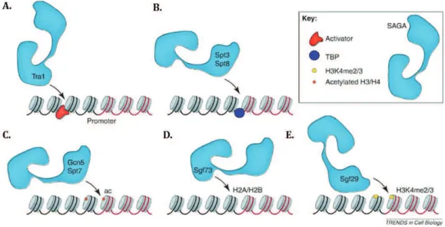
Figure 1.9. Different modes of SAGA recruitment require distinct subunits. ... 42
Figure 1.10. Domain organisation and sequence relationships of human ADA2a and ADA2b. ... 47
Figure 1.11. Schematic summary of ADA2b domain interactions with GCN5 and ADA3. ... 48
Figure 1.12. Schematic representation of a FRAP and FLIP experiment. ... 56
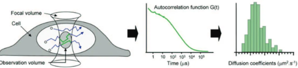
Figure 1.13. Principle of fluorescence correlation spectroscopy ... 57
Figure 1.14. FRAP studies reveal a broad range of dynamics for nuclear proteins. ... 59
Figure 1.15. Model for nuclear import of RNA pol II. ... 63
Figure 1.16. Model for holo-TFIID assembly. ... 66
Figure 3.1. FRAP curves of SAGA and ATAC subunits. ... 78
Figure 3.2. Comparison of half times of recovery between human SAGA and ATAC subunits. ... 80
Figure 3.3 Qualitative comparison between FRAP curves of GCN5 and other GTFs. ... 82
Figure 3.4. Actinomycin D treatment efficiently inhibits transcription globally. ... 84
Figure 3.5. Actinomycin D treatment alters the FRAP curves of all tested factors. ... 85
Figure 3.6. Actinomycin D affects the mobile fraction of most tested SAGA and ATAC subunits. . 86
Figure 3.7. Actinomycin D affects the t1/2 of several factors, including that of GFP. ... 87
Figure 3.8. DRB treatment efficiently inhibits transcription globally. ... 88
Figure 3.9. DRB has mild effect on the FRAP curves of most tested factors. ... 89
Figure 3.10. DRB has no significant effect on the mobile fraction of most tested factors. ... 90
Figure 3.11. DRB effects on the t1/2 of tested SAGA/ATAC subunits and GTFs. ... 91
Figure 3.12. Distribution of diffusion constants of eGFP-tagged human transcription factors... 94
Figure 3.13. Comparison of DeGFP with Dfast and Dslow of the eGFP-tagged factors ... 96
Figure 3.14. Comparison of Ds between eGFP and the eGFP-tagged factors. ... 99
Figure 4.1. Overexpressed ADA2a and ADA2b have different localisation patterns in U2OS cells. ... 110
10 Figure 4.3. Exogenously overexpressed ADA2a-eGFP is shuttling between the nucleus and the
cytoplasm. ... 113
Figure 4.4. Differential intracellular dynamics of ADA2a and ADA2b revealed by FLIP. ... 115
Figure 4.5. FRAP reveals the effect of GCN5 on nuclear ADA2a mobility. ... 117
Figure 4.6. Swapping domains between ADA2a and ADA2b affects the localisation of ADA2a. . 119
Figure 4.7. ADA2a SWAP domain mutants have different intracellular dynamics. ... 120
Figure 4.8. ADA2s SWAP mutants have different half time of loss than ADA2a. ... 122
Figure 4.9. anti-ADA2a and )ADA2b antibodies efficiently IP ATAC and SAGA complexes respectively. ... 124
Figure 4.10. Purity of nuclear and cytoplasmic extracts. ... 125
Figure 4.11. Endogenous ADA2a associates with ATAC components only in the nucleus. ... 126
Figure 4.12. ZZZ3 interacts with ATAC components only the nucleus. ... 128
Figure 4.13. ADA3 interacts with ATAC components only in the nucleus... 129
Figure 5.1. Examples of nonlinear fitting of average autocorrelation curves of free eGFP and eGFP-tagged factors. ... 138
Table of Tables
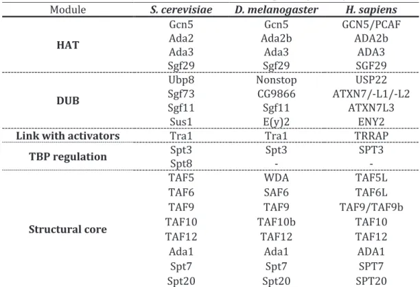
Table 1.1. Subunits and functional modules of SAGA complex in yeast, fly and human... 37Table 1.2. Subunit composition of ATAC and SAGA complexes in H. sapiens ... 45
11
1 Introduction
The availability of the right type of proteins at the right amounts is a fundamental prerequisite of homeostasis for all living organisms. In eukaryotes, and particularly in metazoans, protein abundance mainly depends on the tight regulation of mRNA production. Although some genes are constitutively expressed, the expression level of others has to be adjusted depending on the differentiation stage of a cell and the conditions of the environment. The amount of mRNA and the corresponding protein produced from a gene can be also changed by regulatory pathways related to the processing, the stability or the transport of the transcript in the cytoplasm. In addition, there are cellular mechanisms that regulate translation efficiency and protein stability. However, the most common strategy for gene expression regulation involves changes of the transcription rate of a gene.
Transcription of most protein encoding genes in eukaryotes is performed by RNA polymerase II (Pol II). Pol II function must be highly regulated so that it transcribes genes in a very specific spatiotemporal manner dictated by cell cycle, metabolic or tissue specific needs of a cell. Moreover, the molecular pathways regulating the function of Pol II must be sensitive enough to respond rapidly in changes and signals of the extra or intra-cellular environment. Such type of regulation requires the interaction between sequence-specific DNA-binding factors, general transcription factors (GTFs), co-activators, the epigenetic state of target sequences and Pol II. In the beginning of this chapter, some of the main steps of the above interactions will be summarised to provide the general functional insights on the role of factors that are particularly relevant to this study. Second, a more focused selection of information regarding the role of the SAGA co-activator complex in transcription regulation will be described. In addition, I will summarize some of the information available for the ATAC co-activator complex in response to stress and other pathways. Moreover, I will discuss how Fluorescence Recovery After Photobleaching (FRAP) and Fluorescence Correlation Spectroscopy (FCS) have been applied to investigate the dynamics of transcription factors in living cells and summarise important findings and implications of such studies. Last but not least, as many of the transcription related complexes are composed by multiple subunits, I will also discuss the findings and the implications of studies investigating different aspects of complex assembly regulation in living cells.
12
1.1 Core promoter elements in Pol II transcription initiation
Pol II transcription can be divided in three phases: i) initiation, that involves the recruitment of Pol II at the promoter of a target gene by a set of proteins, called general (or basal) transcription factors (GTFs) as well as the Mediator complex; ii) elongation, during which the RNA transcript is synthesized by the polymerase, and iii) termination, that results in the release of both Pol II and the RNA product from the transcribed DNA template.
Initiation is considered to be the liming step of eukaryotic transcription. Particularly, it is the formation of a productive preinitiation complex (PIC), consisting of Pol II and the general transcription factors allowing basal levels of transcription, that can be rate limiting. It has been shown that both the sequence and the chromatin environment of the core promoter of a gene both serve as a platform for the recruitment of GTFs and the nucleation of the PIC (Thomas & Chiang, 2006; Muller & Tora, 2014).
One could assume that since the efficiency of PIC formation seems to be a universal mechanism of Pol II transcription initiation regulation, the core promoter sequences that serve as PIC assembly sites would also be conserved. Indeed, Pol II core promoters may contain characteristic, highly conserved DNA motifs. Highly conserved core promoter elements are the TATA box, that is recognised by TBP (TATA-binding protein) a component of the TFIID (Transcription Factor II D) complex and BRE, recognised by TFIIB (Transcription Factor II B)(Deng & Roberts, 2007; Fuda et al, 2009). In total, seven distinct types of core promoter elements have been identified in eukaryotes. It must be noted that these core promoter elements are not present in every core promoter. There are core promoters that lack any of the identified conserved motifs indicating that various other sequences and chromatin features are involved Pol II initiation regulation (Thomas & Chiang, 2006; Sandelin et al, 2007; Juven-Gershon et al, 2008; Fuda et al, 2009).
Multiple core promoter elements can be present upstream or downstream of the TSS (Transcription Start Site) of a transcription unit. The combinatorial effect of these sequences, exhibited by the recruitment of different sets of GTFs, in combination with interaction with distant regulatory sites (i.e. enhancers) results in the very accurate but also diverse transcription initiation regulation (Thomas & Chiang, 2006; Fuda et al, 2009).
13
1.2 RNA pol II: a structure-function paradigm
RNA polymerase II (RNA pol II/ Pol II) is the multisubunit complex that is responsible for the transcription of most eukaryotic protein-coding genes. It interacts with the general transcription factors the template DNA and multiple other factors that orchestrate its function during transcription initiation, elongation and termination. The characterisation of Pol II structure has provided a better understanding of the way those interactions are achieved.
Yeast and human Pol II are composed of 12 highly conserved subunits (in human RPB1 to RPB12) (Edwards et al, 1991; Young, 1991). Among those, RPB5, 6, 8, 10 and 12 are shared between the other two classes of eukaryotic DNA dependent RNA polymerases (RNA pol I and RNA pol III). On the other hand, RPB4, 7, 9 and the unstructured Carboxy-Terminal Domain (CTD) extension of RPB1 are specific to RNA pol II (Woychik et al, 1990; Carles et al, 1991; Young, 1991; Hampsey, 1998; Wild & Cramer, 2012). It is RPB1 together with RPB2 that form the active site of the enzyme and bear the catalytic activity of the complex (Lee & Young, 2000).
X-ray structure analysis of the S. cerevisiae Pol II holocomplex has shown that Rpb1 and Rpb2 are at the centre of the complex forming the active site. Rpb5, Rpb6 and Rpb8 bind directly to Rpb1 whereas Rpb9 binds to Rbp2. Rpb7 links Rpb4 to Rpb6. The remaining subunits (Rpb3, Rpb10, Rpb11, and Rpb12) create a bridge that links Rpb1 to Rpb2 (Figure 1.1, A). Four mobile modules termed core, clamp, shelf and jaw-lobe, have been described at the central part of the complex (Figure 1.1, B). The active site is found at the base of a cleft at the centre of the complex. The clamp module has a broad range of movement that can swing over the active centre region and holds the DNA-RNA hybrid in place. It is has been suggested that after interactions of promoter DNA with GTFs (especially TFIIB), only the template strand of DNA can slip in the cleft to be scanned for a transcription start site (Cramer, 2000; Cramer et al, 2001; Kostrewa et al, 2009).
14 Figure 1.1. RNA polymerase II structure and the localisation of its four mobile modules.
A. Front (left) and top (right) view of the complex shown as a ribbon diagram. The 12 subunits Rpb1-Rpb12 are coloured according to the key between the views. Dashed lines represent disordered loops. B. Backbone traces of the four mobile modules of the Pol II structure core. Jaw-lobe, clamp, and shelf modules structure shown in grey, blue, yellow and pink, respectively. Modified from (Cramer et al, 2001) and (Armache et al, 2005).
Pol II exhibits a broad range of interactions with different sets of factors at each phase of transcription (initiation, elongation, termination). For efficient PIC formation, Pol II has to directly or indirectly interact with the GTFs. However, transcription initiation and start of RNA synthesis require disengaging of Pol II from the promoter (promoter clearance) and association with different sets of factors that regulate transcription elongation and termination. Functional and structural studies have shown that the CTD of RPB1, the largest subunit of Pol II, is particularly important for the regulation of these interactions and pre-mRNA processing.
As mentioned above the CTD of RPB1 is an unstructured sequence that in human cells contains 52 tandem repeats of the consensus heptapeptide Tyr-Ser-Pro-Thr-Ser-Pro-Ser (YSPTSPS). The CTD is a dynamic structure that functions as a scaffold for the interaction with regulators of Pol II function. Two functionally distinct forms of Pol II can be distinguished depending on the level of CTD phosphorylation. Hypophosphorylated CTD is characteristic of the IIA form of Pol II which is related to early steps of transcription initiation and Pol II recruitment to the PIC (Lu et al, 1991; Serizawa et al, 1993). During transcription elongation and termination, a cycle of phosphorylation and dephosphorylation of S2 and S5 result in a hyperphosphorylated CTD that characterises the IIO form of Pol II. Specifically, phosphorylation of S5 by cyclin-dependent kinase 7 (CDK7), associated with the GTF TFIIH, is a signal for PIC destabilisation and is a key step for the transition from initiation to elongation (Serizawa et al, 1993). Subsequently, CDK9 present in positive transcription elongation factor b (P-TEFb) phosphorylates S2, required for productive elongation and recruitment of mRNA 3!-end processing factors (Komarnitsky et al,
15 2000; Ahn et al, 2004). Apart from directly targeting CTD serine 2, CDK9 in the context of P-TEFb has another important role that regulates productive elongation. It has been shown that at many metazoan genes, right after initiation and the synthesis of a short (20-65 nt) RNA molecule, Pol II is poised at promoter proximal positions (Muse et al, 2007; Core et al, 2008; Nechaev et al, 2010; Rahl et al, 2010). This paused Pol II form is promoted by the interaction of DRB sensitivity inducing factor (DSIF) and Negative Elongation Factor (NELF) that inhibit further elongation. Phosphorylation of these two factors by P-TEFb results in Pol II release and allows productive elongation (Peterlin & Price, 2006; Cheng & Price, 2007).
The level of S2 and S5 phosphorylation is also regulated by protein phosphatases, some of which have been shown to interact with GTFs. For example, TFIIF-associated CTD phosphatase 1 (FCP1) interacts with TFIIB (Chambers et al, 1995), RPB4 (Kimura et al, 2002b), and TFIIF subunit RAP74 (Chambers et al, 1995; Kobor et al, 2000), and dephosphorylates S2. Interestingly, the interaction TFIIF with FCP1 has been shown to enhance S2 dephosphorylation suggesting that this stimulation may regulate the transition of Pol II from the IIO (elongating) to the IIA (initiating) form (Chambers et al, 1995). Interestingly, Ssu72 integral component of the CPF mRNA *+-end processing complex, has been shown to interact with general transcription factors and affects Ser5-P and Ser7-P depending on the orientation relative to the backbone polarity of the CTD (Rosado-Lugo & Hampsey, 2014). The so called CTD phospho-code has been expanded with studies showing that Tyr1-P, Thr4-P, and Ser7-P are also phosphorylated at distinct steps of RNA pol II transcription underlining the regulatory plasticity of this structure (Heidemann et al, 2013).
Apart from phosphorylation, the CTD can be extensively modified by glycosylation. Glycosylation occurs mainly on the fourth position of the heptapeptide with the glycosylated residues being mainly a threonine but also a serine (in non-consensus sequences) (Kelly et al, 1993). Since glycosylation has been found to be mutually exclusive to phosphorylation (Comer & Hart, 2001; Iyer et al, 2003) it has been suggested that it could prevent kinases from phosphorylating the CTD during early initiation (Thomas & Chiang, 2006). Lastly, extensive Pol II ubiquitination has been linked to DNA damage signalling pathways (Woudstra et al, 2002; Bucheli & Sweder, 2004; Gillette et al, 2004). Arrested Pol II at sites of DNA lesions gets polyubiquitinated and among other functions it may act as a signal for the recruitment of nucleotide excision repair (NER) factors (Bregman et al, 1996).
16
1.3 General Transcription Factors and Pre-Initiation Complex (PIC)
formation
Accurate, site-specific recruitment and engagement of RNA pol II in transcription necessitates the presence of a specific set of factors, which are collectively called general transcription factors (GTFs). These factors are Transcription Factor (TF) IIA, TFIIB, TFIID, TFIIE, TFIIF, and TFIIH. They were initially identified as enzymatically active fractions of subcellular extracts under the presence of which, purified Pol II could transcribe in vitro a DNA template (Weil et al, 1979). Among the GTFs there are two large multisubunit complexes: TFIID that composed of TBP plus 13 TBP Associated Factors (TAFs) and TFIIH that has10 subunits. TFIIB is the only GTFs that is a single polypeptide (Thomas & Chiang, 2006).
1.3.1 General Transcription Factors
TFIID
TFIID is a multisubunit complex that was identified as a factor required for RNA pol II transcription initiation. It is composed of TBP plus 13 TAFs. TFIID mediates promoter recognition via interactions with elements around the TSS (Fire et al, 1984; Young, 1991). Key component of the complex is TBP which in the sequential assembly model is the factor that in the context of TFIID recognises the promoter to start nucleation of the PIC. TBP is highly conserved in eukaryotes with a size of 339 aa in human. The importance of this factor for the regulation of eukaryotic transcription is also apparent from the fact that apart from TFIID, TBP is also present in the core-promoter binding factors of RNA pol I and RNA pol III, (SL1 and TFIIIB respectively). However, although there is a single type of TBP in yeast, one or two TBP related (TRF1 and TRF2, also called TLF) are present in metazoans. In addition, vertebrates contain a third TRF, called TRF3 or TBP2 (Gazdag et al, 2007). Interestingly, in mammals TRF2 and TRF3/TBP2 are involved in the regulation of gonad-specific genes, without the simultaneous presence of TBP (Davidson, 2003; Müller et al, 2010)(Gazdag et al, 2007). TBP has a bipartite structure which is highly conserved particularly at the carboxy-terminal part, which interacts directly with promoter sequences inducing a 90° bending of DNA. This, results in the formation
of an asymmetric platform that facilitates PIC assembly (Kim et al, 1993; Kim & Burley, 1994; Nikolov et al, 1996).
17 Figure 1.2. 3D structure of the TATA-binding protein (TBP) core domain
Ribbon representation of the 3D structure of the TATA-binding protein (TBP) core domain determined by X-ray crystallography. The regions of TBP contacted by transcription factor TFIIA, TFIIB and the DNA are indicated. Adapted from (Davidson, 2003)
DNA-TBP interactions can be highly regulated in a positive or negative way. Negative regulation is required as it has been suggested that non-specific binding to A/T rich regions may lead to the formation of non-productive transcription complexes (Coleman & Pugh, 1995). Several protein-protein interactions have been involved in this type of regulation which inhibits promoter recognition and/or TBP interactions with TFIIA and TFIIB. Association of TBP with the amino-terminal region of BTAF1 in the B-TFIID complex blocks TBP-promoter binding (Pereira et al, 2001). In addition, the carboxy-terminal region of BTAF1 disrupts TBP-DNA complex formation in an ATP dependent manner (Auble et al, 1997; Chicca et al, 1998; Pereira et al, 2001; Andrau et al, 2002). It has to be noted that apart from their inhibitory role on TBP activity, these functions of BTAF1 facilitate the release of TBP from non-specific sites, thus allowing its redistribution to actual promoter regions (Collart, 1996; Li et al, 1999; Geisberg et al, 2002). Another mechanism of negative regulation is based on the affinity of specific domains of TAF1 (the largest TFIID subunit) with functional regions of TBP. Interaction of the two factors can block TBP-DNA complex formation but can also compete for TFIIA binding (Kokubo et al, 1993; Kokubo et al, 1998; Liu et al, 1998). Similarly, negative cofactor 2 (NC2) can inhibit the interaction of TBP with TFIIA and TFIIB by blocking the respective interaction surfaces of TBP. The complexity of TBP activity regulation is elevated from the interaction between NC2 and BTAF1. Particularly, the NC2, but bot NC2- component of human NC2, was found associated with BTAF1. Interestingly, it was shown that this interaction promotes BTAF1 association with TBP (Klejman et al, 2005). The interplay between these factors, together with the potential of TBP to form homodimers unable to bind DNA (Coleman et al, 1995; Jackson-Fisher et al, 1999), illustrates the complex network of protein interactions that regulate TBP/TFIID mediated promoter recognition. Two GTFs that promote TBP-DNA complex formation are TFIIA and TFIIB. There are three major mechanisms by which TFIIA enhances TBP activity. The first include the dissociation of TBP dimers that increase the availability of monomeric TBP (Coleman et al, 1999). Second, TFIIA competes with the negative regulator TAF1 for their common binding regions on TBP (Kokubo
18 et al, 1998). Third, its association with TBP-promoter complex prevents BTAF1 from exhibiting its destabilising role (Auble & Hahn, 1993). TFIIB has also similar effects as it enhances and stabilizes the formation of TBP-DNA complex (Imbalzano et al, 1994a; Zhao & Herr, 2002). TBP association with TAFs forms the TFIID complex that recognises a broader range of sequences. This binding spectrum is particularly important since a recognisable TATA element is absent from the majority of vertebrate promoters. Different TAFs mediate the recognition of distinct promoter elements expanding possible TFIID-DNA contacts. Thus the presence of TAFs is essential for transcription from these promoters that lack canonical TATA-box (Martinez et al, 1994; Huisinga & Pugh, 2004). Moreover the dynamic association of TAFs in TFIID has been suggested to provide increased potential for cell type specific transcription initiation regulation. This is particularly important for metazoans and the regulation of gene expression at different developmental stages (Müller et al, 2010).
Additionally, the fact that TAFs have been found to interact with transcriptional activators suggests that TFIID has a co-activator function. Interestingly, it has been demonstrated that different domains of the same activator can interact with different TAFs or that the same TAF can interact with many activators (Gill et al, 1994; Chiang & Roeder, 1995; Verrijzer & Tjian, 1996; Rojo-Niersbach et al, 1999; Asahara et al, 2001). Thus, activator mediated TFIID recruitment to TATA-less promoter elements can regulate productive PIC assembly at these sites. TAFs were also found to interact with several GTFs like TFIIA (Yokomori et al, 1993), TFIIE (Ruppert & Tjian, 1995), TFIIB (Goodrich et al, 1993), but also with the catalytic subunits of Pol II (Wu & Chiang, 2001). The extensive network of protein contacts that TAFs are involved could play a key role in facilitating the nucleation of PIC in different cellular background.
Apart from the interactions with activators, TAFs can direct the recruitment of TFIID to promoters via interactions with covalently modified histones. It is well established that H3K4m3 and H3K9ac are enriched at active promoters (Wang et al, 2008b). Certain TAFs contain structural domains which interact with histone modifications. Particularly, TAF1 has a double bromodomain that specifically recognises H4K5/12 of H4K8/16 but also H4K16 with a lower affinity (Jacobson et al, 2000). As these marks are found in nucleosomes within actively transcribed chromatin regions, TAF1 could act as reader that recruits TFIID at active promoter sites. In addition, a chromatin interacting domain that has been found at the C-terminal region of TAF3 is the plant homeodomain (PHD). Proteomic screening based on stable isotope labelling by amino acids in cell culture (SILAC) showed that TAF3 binds specifically, with high affinity to H3K4m3 via the PHD domain. Interestingly, the simultaneous presence of acetylated lysines
19 H3K9 and H3K14 further enhanced the observed crosstalk between H3K4m3 and TFIID (Vermeulen et al, 2007).
Overall the broad range of TAF mediated interactions summarised above, allow the regulation of recruitment and stabilisation of TFIID at active promoter without the requirement of a canonical TATA element.
TFIIA
Human TFIIA contains three subunits (,, - and .). Early studies showed that TFIIA is essential for transcription (Reinberg & Roeder, 1987), while later, it was shown that basal transcription does not necessitate its presence (Van Dyke et al, 1988; Wu & Chiang, 1998). These contradictions on the role of TFIIA probably result from the differences of complexity in the systems by which transcription efficiency was studied (partially or highly purified system). As mentioned above, it is believed that TFIIA acts as an antirepressor of inhibitors, that are present for example in a partially purified system, by facilitating the interaction of TFIID, TBP and DNA (Imbalzano et al, 1994b; Kang et al, 1995). There are two particularly important functions of TFIIA related with PIC formation. First, it promotes the dissociation of TBP dimmers increasing the efficiency of promoter elements recognition (Coleman et al, 1999). Second, it competes with TAF1/BTAF1 inhibition, stabilising the formation of the PIC (Auble & Hahn, 1993; Kokubo et al, 1998). In addition, the interactions of TFIIA with several components of TFIID (i.e. TAF1, TAF4, TAF11, TBP), activators, and GTFs (e.g. TFIIE) indicate that it could also have coactivator function (Kobayashi et al, 1995; Langelier et al, 2001; Solow et al, 2001; Dion & Coulombe, 2003).
TFIIB
TFIIB is composed of 316 aminoacids in human (345 in yeast) with 5 functional domains along its sequence. It is a key factor for the recruitment of Pol II to the preinitiation complex. The interaction between its amino-terminal zinc ribbon domain (B-ribbon) is involved in the recruitment of RNA pol II to the promoter and its carboxy-terminal domain (B-core) binds TBP and DNA at the core promoter (Nikolov et al, 1995; Kostrewa et al, 2009) (Figure 1.3). Its B-linker domain plays a role in TSS selection whereas the B-reader domain contributes to the opening of DNA at the promoter. In addition TFIIB interacts directly with RAP30 and RAP74 (subunits of TFIIF) and TAF9 (Ha et al, 1993; Fang & Burton, 1996; Deng & Roberts, 2007).
20 Figure 1.3. Functional domains on yeast TFIIB sequence.
Organization of the distinct functional domains of TFIIB along its sequence. Known interactions and transcription initiation steps related to each domain are mentioned above the scheme. Adapted from (Kostrewa et al, 2009).
According to the proposed model for the role of TFIIB in initiation-elongation transition, it is the B-ribbon that recruits promoter DNA to Pol II and positions the active site above the cleft of the polymerase, resulting in the closed complex conformation (Kostrewa et al, 2009). Subsequently, the B-linker facilitates the melting of 20 bp downstream of the TATA box and the template strand can slip in the cleft and be positioned in the template tunnel. The role of B-reader is important at this point to stabilise the generated bubble near the active centre. The transition to the open complex formation is completed by the loading of the DNA duplex into the downstream cleft. Next, DNA template strand is scanned for a start site (Inr motif), a process in which again the B-reader is involved. The RNA chain synthesis starts with the first phosphodiester bond formation when the first two nucleotides are opposite the Inr motif. The following phase, termed abortive transcription, includes the generation of short DNA-RNA hybrids that are not stably bound, resulting in the ejection of short RNAs. In the final phase (promoter escape), once the synthesised RNA becomes longer than seven nucleotides TFIIB is released and the elongation complex is formed (Kostrewa et al, 2009).
TFIIF
TFIIF is formed by the heterodimerisation of its two subunits RAP30 and RAP74. It regulates PIC formation at multiple levels. It directly interacts with Pol II facilitating its association with promoter bound TFIID-TFIIB complex (Flores et al, 1991). It has been suggested that TFIIF induces topological changes in the promoter DNA that is wrapped around Pol II (Robert et al, 1998). Thus, the affinity of Pol II with the TBP-TFIIB complex is increased and the factors assembling into PIC are stabilising at the promoter region. As TFIIF is closely interacting with TFIIB and Pol II, it is thought that it affects the specificity of the selection of the transcription start site (Hampsey, 1998). An additional way by which TFIIF is increasing Pol II transcription specificity is by inhibiting and/or reversing the binding of Pol II at non-specific sites but also functioning as an elongation factor (Cojocaru et al, 2008). Lastly, the direct interaction between TFIIF and TFIIE is important for the next step of PIC assembly as it recruits TFIIE and TFIIH (Maxon et al, 1994).
21 TFIIE
In human, TFIIE is a heterooctamer of TFIIE, and TFIIE-. According to the sequential PIC assembly pathway, TFIIE and TFIIH are recruited at the promoter after the assembly of the TFIID-TFIIB-Pol II/ TFIIF complex. TFIIE plays a crucial role in promoter melting as it interacts directly with TFIIB, TFIIF, TFIIH, Pol II and promoter DNA. Particularly, it stimulates all the enzymatic activities of TFIIH (ATPase, CTD kinase, helicase) facilitating the transition from initiation to elongation (Ohkuma et al, 1991; Ohkuma & Roeder, 1994; Watanabe et al, 2003). TFIIH
As mentioned above, TFIIH is recruited to the promoter together with TFIIE. TFIIH is a complex of 10 subunits that via its multiple enzymatic activities plays an important role both in transcription initiation and in nucleotide excision repair (NER) DNA damage response (Drapkin et al, 1994). TFIIH consists of two functionally distinct subcomplexes. The core subcomplex of TFIIH contains XPB, p62, p52, p44, and p34. The DNA-dependent ATPase activity of this subcomplex is required for stable promoter opening and transcription initiation (Conaway & Conaway, 1989; Roy et al, 1994). Absence of TFIIH leads to promoter stalling of Pol II and abortive transcription. Moreover, the ATP-dependent helicase activity of XPB is required for promoter clearance of Pol II (Kumar et al, 1998). The cyclin activating kinase (CAK, composed by CDK7, Cyclin H, MAT1) subcomplex of TFIIH phosphorylates Pol II CTD (Lu et al, 1992). It has been demonstrated that Pol II associates with the pre-initiation complex in a hypophosphorylated form (II A) whereas it has to be hyperphoshorylated (IIO) to escape from the promoter and switch to the elongating form. TFIIH has a crucial role in this transition via its CDK7 subunit that phosphorylates serine 5 of Pol II CTD (Lu et al, 1991; Serizawa et al, 1993).
1.3.2 Preinitiation complex assembly pathways from a deterministic perspective
The discovery of GTFs initiated extensive investigations, using mainly in vitro approaches, to discover the way these factors interact with Pol II and the DNA template to form a stable pre-initiation complex (PIC). Two models derived from these studies:
The sequential assembly pathway: According to the model describing this pathway, the PIC is assembled at the promoter in a stepwise manner. Particularly, it was described that TFIID is the GTF that binds first at the promoter core element and that this interaction is further stabilised by the presence of TFIIA and TFIIB (Buratowski et al, 1989; Van Dyke et al, 1989). The next steps involve the recruitment of Pol II together with TFIIF that results in the formation of a
22 stable complex between TFIID-TFIIA-TFIIB-Pol II/TFIIF and the DNA template at the promoter site. Finally TFIIE are TFIIH are recruited to complete the formation of a stable transcription PIC (Figure 1.4, A).
The Pol II holoenzyme pathway: The second model came up as a consequence of the observation that Pol II could be purified as a holoenzyme complex containing not only some GTFs but also other factors that included the SWI/SNF chromatin remodelling complex, the acetyltransferase GCN5, and the co-activator mediator (Kim et al, 1994; Koleske & Young, 1994). The absence of TFID from these purifications was in favour of the notion that TFIID may act independently as a factor that binds to the core promoter, its binding is stabilised by TFIIA and then allows the recruitment of the Pol II holoenzyme (Thomas & Chiang, 2006) (Figure 1.4, B).
Figure 1.4. Preinitiation complex assembly pathways.
According to the two pathways suggested for the recruitment of GTFs in a highly ordered process, preinitiation complex (PIC) formation can occur: (A) by stepwise recruitment of the general transcription machinery (the sequential assembly pathway) or (B) by recruitment of preassembled RNA pol II holoenzyme and TFIID complexes (the two-component pathway).
There is no study providing conclusive evidence to support the validity of either model. However, although the models seem contradictory their co-occurrence in vivo cannot be excluded. Moreover, as it is discussed in chapter 1.6, the findings of studies based on live imaging techniques are challenging this static view of PIC assembly. Indeed the suggested models may represent the two extremes of a dynamic system under different conditions. In addition, #$"/01'(2#$2"#31452"'&"467#(8'2/5"94$'043":#3"&6(2:4(";5'01</5#24%="'6(">/4?"#@'62" the interactions of the GTFs and promoters, as well as other transcription related regulatory elements. The 5'01</5#2/'$=" @4/$9" 2:4" &#ct that these regulatory elements are not readily accessible due to the compaction of DNA into chromatin. Thus, the function of additional co-factors is needed to cross this additional barrier.
23
1.4 Chromatin organisation
The eukaryotic genome is tightly packed in the confined space of nucleus in the form of chromatin. Thus, it is chromatin that should be considered as the actual substrate of fundamental DNA dependent nuclear processes like transcription, replication and repair. Chromatin is not just a static DNA-protein complex but rather a macromolecular assembly with a broad range of conformational dynamics in vitro and in vivo (Figure 1.5).
Figure 1.5.Chromatin organisation.
Schematic illustrating the different levels of chromatin compaction. DNA is wrapped around a histone octamer to form nucleosomes that are connected by stretches of linker DNA in a @4#%3"'$"#"32(/$93!!"5'$&'(0#2/'$A"B65<4'3'04"&'<%/$9"(436<23"2'"2:4"&'(0#2/'$" of a fiber-like structure of about 30 nm in diameter. Further compaction of the 30 nm fibers into higher-order structures eventually results in the formation of chromosomes. As chromatin is a dynamic structure, those distinct levels of organisation may not be clearly distinguished in living cells. Adapted from (Jansen & Verstrepen, 2011).
The basic structural unit of the chromatin fiber is the nucleosome core, a particle that consists of 147 base pairs of DNA wrapped around a histone octamer (Kornberg, 1974; Arents & Moudrianakis, 1993; Richmond & Davey, 2003). Each histone octamer is composed of two copies of H2A, H2B, H3, and H4 (core histones) assembled in an H3/H4 tetramer and two H2A/H2B dimers (Figure 1.6). All four core histones have a histone fold domain at the carboxy-terminal end. These domains are unique, evolutionarily conserved dimerization motifs. They are particularly significant for the assembly of the nucleosome core since they organise the central 129 of 147 base pairs in left-handed superhelical turns. Consequently, these are the domains that mainly contribute to DNA-protein interactions (Baxevanis et al, 1995; Richmond & Davey,
24 2003). A direct consequence of the nucleosome structure is that it may act as an obstacle for transcription.
In addition, core histones form extensions of disordered tails at their amino-terminal ends. Histones H2A and H2B have a shorter carboxy-terminal tail domain too. Core histone tails protrude from the nucleosome and they are considered as determinants of chromatin fiber dynamics. They interact loosely with nucleosomal DNA hence they are the most accessible and mobile parts of the nucleosome.
A nucleosome is composed by the nucleosome core together with the DNA segments that connect it with the preceding and succeeding nucleosome cores. Those sequences called linker DNA, vary in length among species and cell types, with the average nucleosome length being 200 bp (Schalch et al, 2005). The combination of 11 nm nucleosome arrays results in a condense fiber of approximately 30 nm diameter. Note however, that several recent studies questioned the existence of the 30 nm chromatin fiber (Ausio, 2014; Razin & Gavrilov, 2014). The compaction of the nucleosome arrays into the higher order form is facilitated by a fifth histone called H1 (Thoma et al, 1979). H1 does not interact with the core histones but it can stabilise the interaction of 20 additional bp with the periphery of the nucleosome resulting in a chromatin particle of approximately 160 bp named chromatosome (Simpson, 1978; Song et al, 2014).
25 Figure 1.6. The atomic structure of core histones and the nucleosome core particle. A.: Core histones H3 (green) and H4 (yellow) form a tetramer. B.: A dimer is formed from H2A (red) and H2B (pink). C.: The DNA makes 1.7 turns around the histone octamer to form the nucleosome core particle that has a disk-like structure. Each strand of DNA is shown in different shade of blue. Histones are coloured as in (A) and (B). Adapted from (Khorasanizadeh, 2004).
Initially, formation of chromatin was considered just as way of packaging large genomes in the confined nuclear volume. However, increasing evidence shows that the formation of this highly packed yet dynamic structure may also result in the selective restriction of transcription factor movements and thus their accessibility to their regulatory sites.
1.4.1 Histone PTMs and modulation of chromatin organisation and function
Chromatin, from the basic building unit (nucleosome) to the complex conformation of chromatin fibers into higher order assemblies (chromosomes), has specific structural characteristics. However, there are several mechanisms that can alter chromatin architecture at different levels. Post-translational modifications (PTMs) of histones, chromatin remodelling complexes and exchange of canonical histones with histone variants have been extensively studied. Histone PTMs have an impact both on chromatin structure but also in transcription regulation. In the following sections the general ways that histone PTMs impact chromatin structure and consequently transcription efficiency are discussed and the functional output of some of the best-characterised histone PTMs is summarised.
26 Before describing the role of specific histone modification, I will summarise the two general ways in which these modifications affect the accessibility of chromatin-associated factors with their target sites. The first is by directly altering the electrostatic charge of histones and/or the affinity between different nucleosomes. The second, non-mutually exclusive way that chromatin modifications can affect transcription, is by defining the type of chromatin binding proteins that are recruited at regulatory regions.
1.4.1.2 Direct structural effects of histone modifications on nucleosomes and chromatin
The first evidence that histone PTMs may affect the properties of nucleosomes came from studies related to the effects of histone tails on chromatin conformation. It was shown that removing the amino-terminal histone tails from nucleosome cores had a negative effect on their further compaction into a 30 nm chromatin fiber (Fletcher & Hansen, 1995). Moreover, removing hyperacetylation from histone tails from nucleosomes was increasing the accessibility of sequence specific factors to their binding sites (Vettese-Dadey et al, 1996). On the other hand, removal of histones tails by trypsin digestion did not seem to affect the stability of the nucleosome core (Ausio et al, 1989). Thus, the role of histone tails seemed to be more related to the interaction between nucleosomes and the accessibility of transcription factors to their sites than with the stability of the nucleosome core. An example of how histone tail PTMs can affect chromatin structure is the acetylation of H4K16. Experiments in which nucleosomal arrays containing acetylated H4K16 were compared to arrays lacking this PTM or lacking the full H4 tail, showed that H4K16 acetylated arrays did not form the 30 nm fiber as it was the case for non-acetylated arrays (Shogren-Knaak et al, 2006).
Apart from the effect of modifications on sites found at the histone tails, nucleosome stability can be affected by modifications that alter histone/DNA interactions. PTMs at the histone fold domains or the additions of larger moieties like ubiquitin on histones are thought to have this type of effects. A relevant example is acetylation of lysine 56 of histone H3 (H3K56ac). It occurs at the region where DNA enters and exits the nucleosome suggesting that acetylation could affect DNA-histone interaction. This modification has been extensively studied in yeast and among others has been shown to play a role in genomic instability, repair and transcriptional initiation and elongation, by altering nucleosome stability (Celic et al, 2006; Haldar & Kamakaka, 2008; Williams et al, 2008; Värv et al, 2010). In human cells, the same modification was identified in a screen for histone PTMs responsive to DNA damage (Tjeertes et al, 2009) and has also been found to correlate positively with the binding of Nanog, Sox2, and Oct4 transcription factors at their target gene promoters (Tan et al, 2013). This fact indicates the H3K56ac is related to fundamental, highly conserved chromatin regulated processes.
27 On the other hand ubiquitination of K123 of H2B, although it involves the addition of bulky peptide like ubiquitin (~8kDa) is believed not to disrupt but actually increase nucleosome stability during transcription initiation and elongation (Chandrasekharan et al, 2009). However, these effects have to be further investigated. In addition, it has been shown to serve as a bridge for the recruitment of methylases of histone H3 (Lee et al, 2007).
1.4.1.3 Histone PTMs as docking sites for effector proteins
An alternative way, in which posttranslational modifications of histones can affect the structure of chromatin, is that they consist 0#(73"&'(":/32'$4"CDE" (4#%4(3!!"2:#2"#33'5/#24"?/2:"5:('0#2/$" >/#" %'0#/$3" 2:#2" (45'9$/34" 1#(2/56<#(" 0'%/&/5#2/'$3A" F$" 0#$8" 5#343G" 2:434" (4#%4(3!!" 5#$" @4" subunits of chromatin-remodelling or chromatin-modifying complexes. In contrast to histone modifiers, chromatin-remodelling complexes are ATP-dependent and their effect on chromatin has to do with repositioning/sliding of nucleosomes on DNA, removing histones, or exchanging core histones with histone variants (Lusser & Kadonaga, 2003; Clapier & Cairns, 2009). The activity of these complexes is required, among others, to maintain the compaction of chromatin, to attribute specialised characteristics in well-defined chromatin regions and to regulate the accessibility of DNA binding factors in the compact environment of chromatin. Particularly for transcription activation, the activity of chromatin modellers can expose DNA regulatory elements facilitating the binding of activators to upstream enhancers, or enable the association of the components of the basal transcriptional machinery with promoters (Clapier & Cairns, 2009; Voss & Hager, 2014).
The interaction of nucleosome chromatin-modifying or -remodelling complexes with DNA or modified histones is carried out via specific domains of their subunits. Well-studied examples of a motif common in this type of complexes are the bromodomains that recognise acetylation, the chromo-like domains of the Royal family and the PHD domains that recognise methylation, and the domain within 14-3-3 proteins that recognises phosphorylation (Dhalluin et al, 1999; Macdonald et al, 2005; Wysocka et al, 2006; Mujtaba et al, 2007). These types of interactions can tether a chromatin-remodelling or )modifying complex on chromatin regions enriched with histones that bare a particular modification. In this way, the abundance of a histone PTMs can target and/or regulate the activity of a remodeller or chromatin-modifying complex (Suganuma & Workman, 2011).
28
1.4.2 Posttranslational modification of histones
It is more than 50 years since the first evidence for a relationship between the posttranslational modifications of histones and gene regulation was found (Allfrey et al, 1964). Since then, several types of modifications have been found. Histone PTMs may involve the addition of small chemical moieties on histones (acetylation, methylation, phosphorylation etc.) or the attachment of large globular proteins like ubiquitin and SUMO (Small Ubiquitin-like Modifier). An important feature of these modifications, that amplifies the number and the complexity of potential molecular signals chromatin can receive and transmit, is the fact that they are reversible.
1.4.2.1 Histone Acetylation
Histone acetylation occurs with the transfer of an acetyl group from acetyl-CoA to the specific lysines on histones and is catalysed by enzymes called histone acetyltransferases (HATs). The reverse process, removal of the acetyl group is performed by histone deacetylases (HDACs). The opposite action of these two enzyme families renders histone acetylation a highly dynamic modification (Yang & Seto, 2007).
The addition of the acetyl group to lysine is neutralising the positive charge of this residue. Thus, acetylated lysines have lower affinity for DNA. When amplified along large chromatin regions this reduction of histone/DNA affinity results in chromatin decompaction of various levels. Hence, it is not surprising that histone acetylation has been positively correlated with active transcription with that corresponding marks found at the sites of active promoters and euchromatic regions (Katan-Khaykovich & Struhl, 2002; Pokholok et al, 2005; Roh et al, 2005). In general, acetylated histone residues are considered marks of active transcription. However, as for most histones PTMs, acetylation of specific residues has been found to affect different cellular pathways. Particularly, enrichment in H3K9ac and H3K14ac has been found in chromatin regions that promote gene expression (Pokholok et al, 2005; Li et al, 2007). H4K16ac can affect the global conformation of chromatin resulting in a less packed fiber that consequently becomes more accessible for nuclear proteins (Shogren-Knaak et al, 2006). Acetylation can also act as a signal for the assembly of chromatin as H4K5ac and H4K12ac have been described as marks for the deposition of newly synthetized histones (Sobel et al, 1995; Roth et al, 2001).
The enzymes that add the acetyl group to lysines are called histone acetyltransferases (HATs) Most nuclear HATs function in the context of larger complexes and exhibit different substrate
29 specificities (Carrozza et al, 2003). Although the first report of histone acetylation and speculation for its effects on transcription was already made 50 years ago (Allfrey et al, 1964), the first direct link between proteins that affect the levels of histone acetylation and transcriptional regulation resulted mainly from two studies 20 years later. In the first study, it was found that a HAT from Tetrahymena was homologous to Gcn5 (general control non-repressed 5)(Brownell et al, 1996), an already known coactivator of transcription in yeast (Georgakopoulos & Thireos, 1992). The second, showed that a histone deacetylase identified from in vivo screen was homologous to a yeast transcriptional regulator (Taunton et al, 1996). Soon after, it was shown that both the histone acetyltransferase and the deacetylase, were components of larger complexes, in the context of which the enzymes exhibit enhanced catalytic activity, and can be efficiently recruited to their target sites (Grant et al, 1997; Kadosh & Struhl, 1997; Roth et al, 2001).
1.4.2.2 Histone Phosphorylation
Histones can be phosphorylated on serine, threonine and tyrosine residues that mostly localise on their amino-terminal tails. The covalent attachment of a phosphate group, from ATP to the hydroxyl group of the target amino-acid side chain is catalysed by kinases whereas removal of the modification is carried out by phosphatases. The negative charge that is added to the target histone during phosphorylation, classifies this modification in the category of histone PTMs that also affect chromatin conformation. Kinases might be targeted to their chromatin sites either directly by the presence of DNA binding motifs in their sequence or indirectly by associating with chromatin interacting factors (Oki et al, 2007; Rossetto et al, 2012).
Phosphorylation of histones has been linked both with global changes of chromatin structure but also with specific signalling pathways. Particularly, in mammals, phosphorylation of H3 at Ser10, is mediated in mitosis by the Aurora-B kinase and it has been shown to be involved in the initiation of the chromosome condensation process. However, the presence of the mark is not necessary to maintain the compact chromatin state (Van Hooser et al, 1998; Crosio et al, 2002; Johansen & Johansen, 2006). A well-studied example of histone phosphorylation involved in signalling, is the case of the histone variant H2A.X (called .H2A.Z when phosphorylated) (Rogakou et al, 1998). Two serine residues of its carboxy-terminal are phosphorylated in quick response to DNA double strand break (DSB) damage. .H2A.Z is then recognised by DNA repair enzymes that are recruited on the damage site. It is also recruiting the kinases that catalyse the phosphorylation of H2A.X. In that way, a positive feed-back loop is generated that spreads .H2A.Z to large DNA regions (Kang et al, 2005).
30 1.4.2.3 Histone Methylation
As described above, acetylation and phosphorylation of histones is changing the charge of the modified protein. On the contrary, methylation has no such effect. It mainly occurs on the side chains of lysines and arginines of H3 and H4. Lysines can be mono-, di- or trimethylated whereas arginines can be monomethylated and symmetrically or asymmetrically dimethylated. This range of possible methylated states is indicative of the complexity underlying the effects of this mark (Ng et al, 2009).
The enzymes depositing methylation marks are collectively called histone methyltransferases (HMTs). Most HMTs have a highly conserved catalytic domain called SET, found in all studied eukaryotes. Many studies have focused on the study of HMTs especially on those that regulate developmental genes as components of larger protein complexes like Polycomb and Trithorax (Dillon et al, 2005; Steffen & Ringrose, 2014).
Methylation is a mark that can have diverse effects in chromatin structure and transcription regulation. These effects depend on the methylated residue, the level of methylation and the proteins that recognise the modification. H3K4me3, H3K36me3, or H3K79me3 are marks characterising transcriptionally active regions. Heterochromatic regions and regions that are transcriptionally repressed are found enriched in H3K9me3, H3K27me3, and H4K20me3. However an interesting finding that reveals the complexity but also the regulatory potential of this histone modification is the monomethylated H3K9, H3K27, and H4K20 which are predominantly found in euchromatic regions. Thus, it has been shown that even the degree of methylation of the residue can differentially regulate gene expression (Black et al, 2012).
1.4.2.4 Histone Ubiquitination
Modifications described above result in the addition of small chemical moieties on a histone. In contrast to those, ubiquitination entails the addition of a bulky polypeptide (~8 kDa). The addition of such a moiety is expected to have significant effects on chromatin structure and transcriptional regulation.
Three enzymes, that have been classified in three types, act sequentially to complete the covalent binding of ubiquitin on lysines of histone or non-histone proteins. First, an E1 enzyme activates ubiquitin in an ATP dependent step. Next, activated ubiquitin is transferred to the active site of a ubiquitin carrier protein enzyme, E2. The last step requires the action of a