A rhoptry antigen of Plasmodium falciparum is protective
in Saimiri monkeys
R. G.RIDLEY1, B. TAKACS2, H. ETLINGER2 and J. G. SCAIFE1* 1
Department of Molecular Biology, University of Edinburgh, King's Buildings, Mayfield Road, Edinburgh EH9 3JR, Scotland
2
Central Research Units, F Hoffman-La Roche Limited, CH-4002 Basel, Switzerland (Accepted 2 May 1990)
SUMMARY
A non-polymorphic antigen associated with the rhoptry organelles of Plasmodium falciparum has been purified by immuno-affinity chromatography. The antigen, RAP-1 (rhoptry associated protein-1), which is defined by monoclonal antibodies which inhibit parasite growth in vitro, is a multi-component antigen consisting of four major proteins of 80, 65, 42 and 40 kDa and two minor proteins of 77 and 70 kDa. These proteins were electro-eluted from preparative sodium dodecyl sulphate polyacrylamide gels and protected Saimiri sciureus monkeys from a lethal blood-stage infection of P. falciparum malaria. Sera from the protected animals recognized only proteins of the RAP-1 antigen when used to probe a Western blot of total parasite protein extract, confirming that RAP-1 is responsible for eliciting the protective immune response. Key words: Plasmodium falciparum, Saimiri sciureus monkeys, rhoptry antigen, protection.
INTRODUCTION
The ability of some species of new world monkeys to support the erythrocytic stage of malarial infection has allowed vaccination studies to be undertaken in these primates and early experiments showed that vaccination with merozoites or schizonts from
Plas-modium falciparum protected against a lethal
blood-stage infection (Mitchell et al. 1977; Siddiqui, 1977). Many merozoite and schizont antigens have since been identified and their genes cloned and sequenced. However, only two well-defined antigens have so far been purified to homogeneity and successfully used to protect monkeys from infection; the major merozoite surface antigens derived from the protein PMMSA (Hall et al. 1984; Perrin et al. 1985a; Siddiqui et al. 1987) and a 41 kDa rhoptry antigen (Perrin et al. 19856). In the case of one antigen, RESA, recombinant protein derived from a repeat region of the gene has offered some protection (Collins et al. 1986). Synthetic peptides have been used in some vaccination experiments. A synthetic peptide corresponding to the N-terminus of the PMMSA derived 83 kDa protein gave good pro-tection (Cheung et al. 1986) and a mixture of three peptides corresponding to the N-termini of the PMMSA derived 83 kDa protein and two proteins of 55 kDa and 35 kDa, which have not been well characterized, gave almost full protection (Pattaroyo
et al. 1987). These peptides also formed part of a
complex of peptides from a variety of malarial antigens which showed some promise in a human vaccine trial (Pattaroyo et al. 1988).
To our knowledge, only PMMSA has been * Reprint requests to Professor J. G. Scaife.
repeatedly shown to offer a high degree of protection in this type of experiment (Hall et al. 1984; Perrin
et al. 1985 a; Siddiqui et al. 1987; our unpublished
results). Because of the small number of candidate antigens for vaccine development which have been rigorously tested in model animal systems, we have sought other antigens which may have protective properties.
One source of such antigens may be the rhoptry organelles, which are located at the apex of the merozoite and are intimately involved in erythrocyte invasion (Aikawa, 1971; Bannister et al. 1986). In fact, early studies on the rodent malarial parasite
Plasmodium yoelii revealed a rhoptry antigen which
could partially protect mice against malaria (Holder & Freeman, 1981). We have previously reported two monoclonal antibodies 2.13 and 7.12, which define an antigen, RAP-1 (rhoptry associated protein-1) associated with the rhoptries (Hall et al. 1983). They both inhibit parasite growth in vitro (I. A. Hope and J. G. Scaife, unpublished observations) suggesting that their target antigen could induce protective immunity in an anti-malarial vaccine. T h e RAP-1 antigen, which also offers the advantage of being non-polymorphic (Clark et al. 1987) has recently been characterized and its gene cloned (Ridley et al. 1990). We report here the purification of this antigen and assess its ability to protect Saimiri sciureus monkevs from malarial infection.
MATERIALS AND METHODS Standard techniques
Indirect immunofluorescence assays were performed as described previously (Hall et al. 1983). Sodium Parasitology (1990), 101, 187-192 Printed in Great Britain
dodecyl sulphate polvacrvlamide gel electrophoresis (SDS-PAGE), using the method of Laemmli (1970), together with Western blotting analysis have also been previously described (Takacs & Staehli, 1987).
P. falciparum in vitro culture was maintained
according to the method of Trager & Jensen (1976), later modified by Zolg et al. (1982).
Affinity-purification of RAP-1 antigen
Purified mAb 2.13 (Hall et al. 1983) was coupled to cyanogen bromide-activated Sepharose 4B (Pharmacia Ltd, Milton Keynes, UK) as recom-mended by the manufacturer. Cell pellets of bulk cultures of a P. falciparum isolate, Kl from Thailand (Thaithong & Beale, 1981) were harvested as pre-viously described (Goman et al. 1982) and stored at — 70 °C until required. They were then extracted at 4 °C for 1 h with an extraction buffer ( 2 °0 (w/v))
sodium deoxycholate, 1 °0 (w/v) Nonidet P40,
50 m.M Tris-HCl (pH 8-(;), 5 m\i ethylene-diaminetetra-acetic acid (EDTA), 5 m.\i l,2-di(2-aminoethoxy)ethane-iV,iY,Ar',7V'-tetra-acetic acid (EGTA)) containing 5 mM-iodoacetamide, 0'2 m\i phenylmethylsulphonyl fluoride (PMSF) and 10/yg/ml each of the protease inhibitors pepstatin, chymostatin, antipain, leupeptin and soybean trypsin inhibitor (all from Sigma Chemical Co. Ltd, Poole, UK). Fresh P M S F was added to 0 2 m.M and the extract was centrifuged at 100000 £ for 3 h at 4 °C. The supernatant fraction had fresh P M S F added to 0'2 m\i and was passed through a 10 ml column of Sepharose 4B-bound mAb 2.13 equilibrated with the extraction buffer. The column was then washed extensively with wash buffer (0'5 °0 (w/v) sodium
deoxycholate, 1 °0 (w/v) Xonidet P40, 50 mM T r i s
-HCl, pH 80, 5 m.M EDTA, 5 m.M EGTA) fol-lowed by at least 5 column volumes of the wash buffer containing 015 M NaCl and was finally re-equilibrated in salt-free wash buffer before eluting with 50 m\i diethylamine in wash buffer at pH 115. The protein solution was neutralized with solid glycine and the cocktail of protease inhibitors at 10/<g/ml was added prior to storage at —70 °C.
Monkey immunization experiment
RAP-1 was purified from the Kl strain of P.
falciparum by affinity chromatography followed by
electro-elution of the protein bands from 15 °0
preparative SDS-polyacrylamide gels. Four Saimiri
sciureus monkeys each received, subcutaneously,
25 //g protein in complete Freund's adjuvant on day 1 and 25 //g protein in incomplete Freund's adjuvant on days 24 and 48. Control monkeys received the adjuvant in phosphate-buffered saline on the same days. Sera were taken on day 56. On day 60, monkeys received, intravenously, 3'5 x 10' P.
falci-parum (Uganda Palo Alto strain) infected
erythro-Fig. 1. Monoclonal antibody 2.13 punctate
immunofluorescence pattern obtained with the schizont stage of a Plasmodiutn falciparum-infected erythrocyte.
cytes from a donor monkey. Parasitaemias were determined by light microscopy of blood smears stained with Giemsa.
RESULTS
Localization of the antigen RAP-1 to the rhoptry organelles of the merozoite is shown in Fig. 1. The punctate fluorescence pattern produced by mAb 2.13 defines rhoptry organelles of parasites at the late schizont stage of the erythrocytic cycle. Each parasite within the schizont contains a pair of rhoptry organelles.
The RAP-1 antigen was purified from extracts of cultured parasites by affinity chromatography using mAb 2.13. It consists of four major bands (p80, p65, p42 and p40) with Mr = 80, 65, 42 and 40 kDa and two minor bands, Mr = 11 and 70 kDa which varied
in intensity depending on the preparation (Fig. 2). The high yield of p80 with respect to p70 was heavily dependent on the addition of protease inhibitors during extraction, purification and storage of the antigen, suggesting that p70 is a degradation product of p80.
The ability of human immune sera to recognize RAP-1 was tested (Fig. 3). The components p80, p77, p70, p65 and p40 were all well recognized by the sera; however, the signal for p42, though present, was very weak and is not visible in the figure. Interestingly, p80 showed much stronger immune recognition than any of the other bands, suggesting that it contains an immunodominant epitope lacking in the other RAP-1 components.
The protein bands observed by Coomassie stain-ing in Fig. 2 were further purified by electro-elution
p80 P77 p70 p65 \ _
r—
(kDa) — 200 — 97 — 69 (kDa) - 200 - 97 r •"» — 69 p42 p40 _ 46 - 46 — 30 - 30Fig. 2. Immuno-affinity purified rhoptry antigen RAP-1 was subjected to SDS-PAGE in the presence of 2-mercaptoethanol and stained with Coomassie Blue. The major fractions eluted from an immuno-affinity column are shown and the sizes of the major bands are
indicated.
1 2
Fig. 3. Recognition of RAP-1 by human immune sera. Ten samples of Nigerian human immune sera were pooled and used to probe a Western blot of total
Plasmodium falciparum protein (lane 1) and
affinity-purified RAP-1 (lane 2).
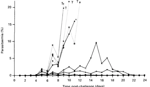
20
-8 10 12 14 16 18 20 22 24
Time post-challenge (days)
Fig. 4. Effect of inoculation with RAP-1 on parasitaemia in Saimiri monkeys. Control monkeys are
represented by dotted lines and experimental animals by full lines. Positions are marked to indicate when drug treatment was administered (T) or death occurred ( + ).
from preparative gels and used to inoculate Saimiri monkeys. Four animals were treated with the antigen while four control animals were injected with saline (Fig. 4). All eight animals were challenged with blood-stage parasites (Fig. 4). One of the four
experimental monkeys died prematurely with a ruptured spleen at a parasitaemia significantly lower than is normally tolerated in these animals. This monkey produced the lowest titre of antibody by indirect immunofluorescence. The other three
re-(kDa) — 200
— 94
— 46
- 30
band is likewise frequently observed in total malarial protein extract (lane 1) even though it is of low abundance in affinity-purified RAP-1 as a result of using protease inhibitors during the extraction and purification of antigen (lane 2) (see also Fig. 2).
Importantly, no proteins were recognized by the monkey sera other than those associated with the RAP-1 antigen. This confirms the purity of the immunizing antigen and strongly suggests that there are no cross-reactive epitopes on other malarial antigens. We conclude that the RAP-1 antigen elicits an immune response in Saimiri monkeys which confers protection against falciparum malaria.
- 20
1 2
Fig. 5. Recognition of RAP-1 by monkey antisera. Sera taken from the experimental monkeys prior to parasite challenge were pooled and used to probe a Western blot of total Plasmodium falciparum protein (lane 1) and affinity-purified RAP-1 (lane 2).
covered completely after developing relatively low parasitaemias. By contrast, all of the control animals showed a rapid rise in parasitaemia to 20 °0 by days
8-12 and were cured by chloroquine and Fansidar treatment. No correlation was observed between antibody titre and peak parasitaemia in the three protected animals.
Sera taken from the animals after immunization and four days prior to parasite challenge, were assayed for reactivity against RAP-1 and for re-activity against total parasite protein (Fig. 5). The sera, which stained rhoptries in indirect immuno-fluorescence (not shown), recognized purified RAP-1 in Western blots (lane 2) and detected only components of the RAP-1 antigen when used to probe a Western blot of total malarial protein extract (lane 1). The sera of the four control animals gave negative results in these tests.
The protein bands in the total protein extract recognized by the monkey sera in lane 1 correspond to the bands at 80, 70, 65 and 42 kDa observed in Fig. 2. We are satisfied that the lower band observed in Fig. 5 corresponds to p42 as co-migratory signals have been observed using rabbit antisera raised specifically against this protein. The protein detected above p80 at 83 kDa is frequently observed when blotting total malarial protein extract with both monoclonal and polyclonal antibodies against RAP-1 and most likely corresponds to a newly synthesized precursor protein prior to cleavage of its N-terminal signal sequence (Ridley et al. 1990). The 70 kDa
DISCUSSION
In this report, we show that the non-polymorphic rhoptry antigen RAP-1 (Clark et al. 1987) can be extracted from P. falciparum and can protect Saimiri monkeys against a lethal infection of falciparum malaria. It effectively inhibits early multiplication of the pathogen and permits complete recovery without further intervention. The sera from the immune animals recognized only RAP-1 antigen proteins when used to probe a Western blot of total parasite protein, confirming that the antigen is responsible for generating this protection. Furthermore, in keeping with the antigen's non-polymorphic nature, the response is not strain limited. The immunizing antigen, purified from a Thai isolate, K l , protected against an isolate of East African origin, Palo Alto Uganda.
It is not possible at this stage to specify how humoral and cellular mechanisms participate in the protection of the monkeys. However, some com-ments on the immunogenicity of this antigen are possible.
The RAP-1 antigen is obviously complex, con-sisting of up to six protein species. The recognition by monoclonal antibodies of all the higher molecular weight proteins (p80, p77, p70 and p65) and pulse-chase data (Clark et al. 1987) has led to the conclusion that these proteins are all derived from the same precursor protein (Ridley et al. 1990). In addition, evidence has also been presented that p42 and p40 may be derived from these higher molecular weight proteins (Ridley et al. 1990). In the light of these observations it is interesting to note that the 80 kDa protein is much more strongly recognized by human immune sera than the other proteins, suggesting that a strong immunogenic determinant is present on the 80 kDa protein which is lacking on the other proteins. In contrast, the 42 kDa component appears to be poorly immunogenic in man, though the denatured protein was immunogenic in monkeys.
It should be noted that sera from the experimental animals recognized a recombinant fusion peptide derived from the merozoite surface antigen, PMMSA (Gentz et al. 1988) (not shown). However,
the sera did not recognize parasite PMMSA or its products in a Western blot (Fig. 5), nor did they give the immunofluorescence pattern characteristic of this antigen (not shown). Perhaps the epitopes recognized in the fusion peptide (made in Escherichia
coli) are special to this construct.
It needs to be stressed that RAP-1 is different from another antigen of similar size and subunit composition, denned by a mAb Hb31cl3, previously used in monkey immunization experiments (Perrin
et al. 19856). Antibodies raised against the two
antigens do not cross-react (R. Ridley and U. Certa, unpublished observations). In addition, the 80 kDa component of the antigen defined by mAb Hb31cl 3 has been identified as a serine protease (Braun-Breton, Rosenberry & Pereira da Silva, 1988). The gene sequence encoding RAP-1 has been cloned and sequenced and does not contain any of the motifs characteristic of serine proteases (Ridley et al. 1990). In summary, a non-polymorphic rhoptry antigen, RAP-1, has been purified from P. falciparum malarial extracts using immuno-affinity chromato-graphy and electro-elution from polyacrylamide gels. This antigen was successfully used to protect Saimiri monkeys from a lethal infection of P. falciparum malaria. Further assessment of the antigen's value as a vaccine requires monkey immunization experi-ments using recombinant protein material derived from the gene sequence of RAP-1. Attempts to express the gene in bacteria are currently in progress. We thank Hugues Matile for indirect immunofluorescence analysis of monkey sera, Richard Pink and Ulli Certa for discussions, Anne McGowan and Inez Bolliger for out-standing technical support, Graham Brown for photogra-phy and Annie Wilson for preparing the manuscript. This work was supported by Hoffman-La Roche and the Medical Research Council.
REFERENCES
AIKAWA, M. (1971). Fine structure of malaria parasites.
Experimental Parasitology 30, 284—320.
BANNISTER, L. H., MITCHELL, G. H., BUTCHER, G. A. &
DENNIS, E. D. (1986). Lamellar membranes associated with rhoptries in erythrocytic merozoites of
Plasmodium knoivlesi; a clue to the mechanism of
invasion. Parasitology 92, 291—303.
BRAUN-BRETON, C , ROSENBERRY, T. L. & PEREIRA DA SILVA,
L. (1988). Induction of proteolytic activity of a membrane protein in Plasmodium falciparum by phosphatidylinositol specific phospholipase C. Nature,
London 322, 457-9.
CHEUNG, A., I.EBAN, J., SHAW, A. R., MERKLI, B., STOCKER, J., CHIZZOLINI, C , SANDER, C. & PERRIN, L. H. ( 1 9 8 6 ) .
Immunization with synthetic peptides of Plasmodium
falciparum surface antigen induces antimerozoite
antibodies., Proceedings of the National Academy of
Sciences, USA 83, 8328-32.
CLARK, J. T., ANAND, R., AKOGLU, T. & MCBRIDE, J. S.
(1987). Identification of proteins associated with the
rhoptry organelles of Plasmodium falciparum merozoites. Parasitology Research 73, 425-34.
COLLINS, W. E., ANDERS, R. F . , PAPPAIOANOU, M., CAMPBELL, G. H., BROWN, G. V., KEMP, D. ] . , COPPEL, R. L., SKINNER, J. C , ANDRYSIAK, P. M . , FAVOLORO, J. M . , CORCORAN, L. M., BROADERSON, J. R., MITCHELL, G. F. &
CAMPBELL, c. c. (1986). Immunization of Aotus monkeys with recombinant proteins of an erythrocyte surface antigen of Plasmodium falciparum. Nature,
London 323, 259-62.
GENTZ, R., CERTA, U., TAKACS, B., MATILE, H., DOBELI, H., PINK, R., MACKAY, M., BONE, N. & SCAIFE, J. G. ( 1 9 8 8 ) .
Major surface antigen pi 90 of Plasmodium falciparum : detection of common epitopes present in a variety of plasmodia isolates. EMBO Journal 7, 225-30.
GOMAN, M., LANGSLEY, G., HYDE, J. E., YANKOFSKY, N . K.,
ZOLG, J. w. & SCAIFE, j . G. (1982). The establishment of
genomic DNA libraries for the human malaria parasite P. falciparum and identification of individual clones by hybridisation. Molecular and Biochemical
Parasitology 5, 391^-00.
HALL, R., HYDE, J. E., GOMAN, M., SIMMONS, D. L., HOPE, I. A., MACKAY, M., SCAIFE, J., MERKLI, B., RICHLE, R. &
STOCKER, J. (1984). Major surface antigen of a human malaria parasite cloned and expressed in bacteria.
Nature, London 311, 379-82.
HALL, R., MCBRIDE, J., MORGAN, G., TAIT, A., ZOLG, J. W.,
WALLIKER, D. & SCAIFE, j . G. (1983) Antigens of the
erythrocytic stages of Plasmodium falciparum detected by monoclonal antibodies. Molecular and Biochemical
Parasitology 7, 247-65.
HOLDER, A. A. & FREEMAN, R. R. (1981). Immunization
against a blood-stage rodent malaria using purified parasite antigens. Nature, London 294, 361-4. LAEMMLI, u. K. (1970). Cleavage of structural proteins
during the assembly of the head of bacteriophage T 4 .
Nature, London 227, 680-5.
MITCHELL, G., RICHARDS, W. H. G., BUTCHER, G. A. & COHEN, s. (1977). Merozoite vaccination of douroucouli monkeys against falciparum malaria. Lancet i, 1335-8.
PATTAROYO, M. E., ROMERO, P . , TORRES, M. L., CLAVIJO, P . , MORENO, A., MARTINEZ, A., RODRIGUEZ, R., GUZMAN, F. &
CABBEZZAS, E. (1987). Induction of protective immunity against experimental infection with malaria using synthetic peptides. Nature, London 328, 629—32.
PATTAROYO, M. E., AMADOR, R., CLAVIJO, P . , MORENO, A., GUZMAN, F . , ROMERO, P . , TASCON, R., FRANCO, A., MURILLO, L. A., PONTON, G. & TRUJILLO, G. ( 1 9 8 8 ) . A
synthetic vaccine protects humans against challenge with asexual blood stages of Plasmodium falciparum malaria. Nature, London 332, 158-61.
PERRIN, L. H., MERKLI, B., LOCHE, M., CHIZZOLINI, C ,
SMART, J. & RICHLE, R. (1985 a). Antimalaria immunity
in Saimiri monkeys: immunization with surface components of asexual blood stages. Journal of
Experimental Medicine 160, 441-51.
PERRIN, L. H., MERKLI, B., GABRA, M. S., STOCKER, J. W.,
CHIZZOLINI, c. & RICHLE, R. (1985ft). Immunisation
with a Plasmodium falciparum merozoite surface antigen induces a partial immunity in monkeys.
Journal of Clinical Investigation 75, 1718-21. RIDLEY, R. G., TAKACS, B., LAHM, H . - W . , DELVES, C. J.,
GOMAN, M., CERTA, U., MATILE, H., WOOLLETT, G. R. &
protective rhoptry antigen from Plasmodium
falciparum. Molecular and Biochemical Parasitologv
41, 125-34.
SIDDIQUI, w. A. (1977). An effective immunization of experimental monkeys against a human malaria parasite, Plasmodium falciparum. Science 197, 388-9.
SIDDIQUI, W. A., TAM, L. Q., KRAMER, K. J., HUI, G. S. N . , CASE, S. E., YAMAGE, K. M., CHANG, S. P . , CHAN, E. B. T.
& KAN, s.-c. (1987). Merozoite surface coat precursor protein completely protects Aotus monkeys against
Plasmodium falciparum malaria. Proceedings of the National Academy of Sciences, USA 84, 3014-18.
TAKACS, B. & STAEHI.I, c. (1987). Activated macrophages and antibodies against the plant lectin, GF1—B4,
recognise the same tumour-associated structures
(TAS). Journal of Immunology 138, 1999-2007.
THAITHONG, s. & BEAI.E, G. (1981). Resistance of ten Thai isolates of Plasmodium falciparum to chloroquine and pyrimethamine by in vitro tests. Transactions of the
Royal Society of Tropical Medicine and Hygiene 75,
271-3.
TRAGER, w. & JENSEN, B. (1976). Human malaria parasites in continuous culture. Science 217', 254—7.
Z O L G , J . W . , M C L E O D , A. J . , D I C K S O N , I. H . & S C A I F E , J. G. (1982). Plasmodium falciparum: modification of the
in vitro culture conditions improving parasite yields. Journal of Parasitologv 68, 1072-80.