CA125 production by the peritoneum: in-vitro and in-vivo studies
5
0
0
Texte intégral
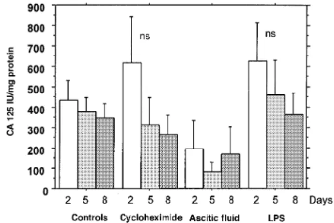
(2) M.Epiney et al.. Talbot et al. (Talbot et al., 1989) found a postoperative CA125 increase after laparotomy in the sera of patients with normal preoperative concentration, while a third study (Yedema et al., 1993) showed a significant CA125 increase in the serum after laparotomy for benign diseases and non-ovarian malignancies. The purpose of the present study was to investigate in vivo and in vitro if the peritoneum is a source of CA125 and if it contributes to circulating CA125. In practical terms, we wanted to see if the increase in CA125 was higher after a peritoneal surgery and if the proportion of patients having an increased CA125 concentration was higher in patients having undergone a peritoneal surgery compared with non-peritoneal surgery.. Materials and methods Tissue cultures Uterine peritoneum and myometrium were obtained from patients undergoing hysterectomy for reasons other than malignancy. Abdominal peritoneum was obtained from women undergoing elective laparotomy. After removal, the tissue pieces were minced with scissors, washed and placed in HBSS (Hanks’ balanced salt solution, Sigma, St Louis, MO, USA) containing 200 IU/ml penicillin (Hoechst, Darmstadt, Germany), 200 µg/ml streptomycin (Hoechst) and 2.5 µg/ml fungizone (Gibco, Basle, Switzerland). Explants of uterine and abdominal peritoneum, and myometrium were cultured in RPMI 1640 (Merck, Darmstaadt, Germany) containing 10% FCS (fetal calf serum) (Animed, Basel, Switzerland), 100 ng/ml streptomycine, 0.1 IU/ml penicillin and 2.5 ng/ml fungizone (referred to as complete RPMI hereafter). To mimic bacterial infection, complete RPMI was supplemented in some experiments by 100 µg/ml lipopolysaccharides (LPS; Sigma, Buchs, Switzerland). In other experiments, ascitic fluid (30% v/v) was added to complete RPMI to mimic cancer. Finally, complete RPMI was also supplemented sometimes with cycloheximide (10 µg/ml, Sigma) to inhibit protein synthesis. Media were collected on days 2, 5 and 8 and CA125 concentration determined in duplicate in the supernatants by an immunoradiometric assay kit (CIS Biointernational, Saclay, France) according to the instructions of the manufacturer. Total proteins were measured in each supernatant with the protein Biorad kit (Biorad, Munchen, Germany) using bovine serum albumin as the standard. The concentration of CA125 in media supplemented with ascitic fluid but in absence of tissue was subtracted from the CA125 concentrations found in the culture supernatants supplemented with ascitic fluid and in presence of tissue. Results were expressed as units of CA125 per mg total protein. Statistical analyses were performed on log transformed values by analysis of variance (ANOVA) and paired t-test (when appropriate) using the Statview program (Abacus, Berkeley, CA, USA). Prospective study In a prospective study we analysed a population of 40 patients admitted for surgery under general anaesthesia. The first group included nine men and 10 women undergoing abdominal surgery with peritoneum opening. Cases with endometriosis or ovarian cancer were excluded. The second group included 11 men and 10 women undergoing surgical intervention elsewhere. Data concerning age, sex, preoperative diagnosis, type of surgery, anatomicopathological diagnosis, length of the anaesthesia and the operation were recorded. A 5 ml blood specimen on heparin was obtained 24–48 h preoperat-. 1262. Figure 1. CA125 concentrations in uterine peritoneal explant cultures (n ⫽ 12) according to the duration of incubation. Controls were cultured in complete RPMI (see Materials and methods). Explants were also cultured in RPMI supplemented with 10 µg/ml cycloheximide (n ⫽ 12) with 30% v/v ascitic fluid (n ⫽ 9) and with 100 µg/ml lipopolysaccharides (LPS; n ⫽ 4). ively and 48 h postoperatively from each patient. After centrifugation (10 min at 1500 g), plasma samples were stored at –20°C until assayed. CA125 concentration was determined in duplicate by the same immunoradiometric assay as described above. The change between pre- and postoperative CA125 concentrations was calculated and log transformed. Statistical analyses were performed by Student’s t and χ2 tests.. Results Tissue cultures In the uterine peritoneal explant cultures (n ⫽ 12), we observed CA125 concentrations varying between 83.9 ⫾ 135.1 and 627.8 ⫾ 367.9 IU/mg protein (mean ⫾ SD) according to the days of culture and the treatments. Irrespective of the culture conditions, CA125 concentrations gradually decreased with the time of incubation. However, no viability test was done to exclude the possibility that this decrease was due to cell death. The release of CA125 in cultures of uterine peritoneal explants was not significantly changed by the presence of cycloheximide (a protein synthesis inhibitor) (n ⫽ 12; Figure 1). Ascitic fluid (n ⫽ 9) or lipopolysaccharides (n ⫽ 4) did not change significantly the CA125 concentrations as compared to complete RPMI alone (Figure 1). Cultures of abdominal peritoneal explants (n ⫽ 11), uterine peritoneal explants (n ⫽ 12) and myometrial explants (n ⫽ 9) released similar CA125 concentrations. The concentrations of CA125 were not significantly different between the tissues for the same incubation time (Figure 2). Similarly to uterine peritoneal explants, the presence of cycloheximide did not change significantly the CA125 concentrations released by myometrial explant cultures (n ⫽ 9, Figure 3). Prospective study The prospective study included two groups of patients: the first group (nine men and 10 women) having abdominal surgery with opening of the peritoneum and the second (11 men and 10 women) with extraperitoneal surgery. Details of the patients and the corresponding CA125 concentrations are given in.
(3) CA125 production by the peritoneum. Discussion. Figure 2. CA125 concentrations in cultures of abdominal (n ⫽ 4) and uterine (n ⫽ 12) peritoneal explants and myometrial explants (n ⫽ 9), according to the duration of incubation.. Figure 3. CA125 concentration in cultures of myometrial explants (n ⫽ 9) supplemented or not with 10 µg/ml cycloheximide.. Table I. Of the 40 patients, seven (three men and four women) had a preoperative CA125 concentration above 35 IU/ml and were excluded from further analysis as discussed below. Table II shows the characteristics of the remaining patients with a preoperative CA125 ⬍35 IU/ml. The groups are comparable in age. The mean postoperative value of CA125 in the group ‘peritoneal surgery – men’ was high (55.8 IU/ml ⫾ 63.2) with a high standard deviation. This is due to one case of gastrojejunal anastomosis cancer with a postoperative CA125 concentration of 167.11 IU/ml. The postoperative CA125 values were not significantly different from the preoperative ones when considering the different groups (peritoneal surgery or extraperitoneal surgery in men and/or women). Since the CA125 assay had an interassay coefficient of variation of 5.5%, we counted the number of patients who had a postoperative CA125 concentration above the preoperative concentration ⫹5.5% and considered this increase as significant. With this approach, 53.3% of patients had an increased postoperative CA125 in the group of peritoneal surgery whereas only 16.7% of the patients had such an increase in the group of extraperitoneal surgery. This was statistically significant (χ2 test, P ⬍ 0.03). Analysing the female subgroup for the same parameters, 33% of women with peritoneal surgery had a higher postoperative CA125 concentration against 0% in the extraperitoneal group (P ⬍ 0.03). In the male group, we found 83.3 against 27.3% (P ⬍ 0.05).. We could not demonstrate in-vitro de-novo synthesis of CA125 under our experimental conditions since an inhibitor of protein synthesis such as cycloheximide did not change the concentrations of CA125 found in the incubation medium. Addition of cycloheximide has been shown to decrease significantly the concentrations of CA125 secreted in the medium by explants of decidua (Bischof et al., 1986). Our results suggest that uterine and abdominal peritoneum, and myometrium are not a source of CA125 production. In the explant cultures we noticed that the CA125 concentrations decreased progressively with the time of incubation. This can be explained either by inhibition of de-novo production of the antigen in our culture conditions or by a release of stored CA125. It must be added, however, that the interpretation of these data is somewhat difficult because the half-life of CA125 in vitro is unknown and the viability of the explants in culture could not be assessed. Our observations seem to exclude the possibility that these tissues actively produce CA125 in vitro. Different inflammatory situations such as pelvic inflammatory disease, endometriosis (especially with adhesions), malignant pathologies with ascites and peritonitis are known to induce high circulating concentrations of CA125. In order to mimic cancer or an inflammation, peritoneal explant cultures were performed in medium supplemented with ascitic fluid or LPS, speculating that these two substances could stimulate potential CA125 production. Even under these conditions, we do not find a significant effect on the CA125 release. This observation is in contrast with previous results (Zeillemaker et al., 1994). These last authors cultured mesothelial cells for 6 h and observed that the secretion of CA125 by cells grown in medium with LPS, IL-1β or TNFα was statistically higher than in unstimulated cells. However, the kinetics of the two studies are different: we measured CA125 after 2–8 days. This could perhaps explain the different results. The difference in the culture medium and the use of antibiotics certainly does not explain our negative results. Among the 40 patients included in the prospective study, seven had a preoperative CA125 ⬎35 IU/ml associated with malignant (pancreas, lymphoma, gastric and breast) and benign (uterine fibroid, thyroid) conditions. This underlines the lack of specificity of this marker for ovarian cancer. We excluded these cases from further analysis in order to avoid a possible decrease in postoperative CA125 due to tumour removal, as shown previously (Yedema et al., 1993). Although our sample size was limited, we observed a significant difference in the proportion of patients who had a postoperative increase of CA125 when patients with abdominal and extra-abdominal surgery were compared. This suggests that, in vivo, the peritoneum might contribute to circulating CA125 concentrations. These results confirm earlier studies (Talbot et al., 1989; Yedema et al., 1993) in which a postoperative increase of CA125 after laparotomy was observed. In both studies, CA125 measurements were performed between 4 and 28 days after surgery and the maximal rise in CA125 was detected 2–4 weeks postoperatively (Talbot et al., 1989). Our postoperative 1263.
(4) M.Epiney et al.. Table I. Diagnosis, preoperative and postoperative CA125 concentrations in 40 men and women undergoing peritoneal or extraperitoneal surgery Men Diagnosis. Peritoneal surgery Pancreatic cancer Malignant lymphoma Gastric sarcoma Gastrojejunal anastomosis carcinoma Colon carcinoma Sigmoiditis Peritoneal pseudomyxoma Rupture of abdominal scar Abdominal arterial bypass. Women CA125 preop (IU/ml). CA125 postop (IU/ml). 333.4 99.1. 208 84.7. 66.3. 43.1. 27.4. 167.1. 16.2 15 14.8. 22.4 97.3 12. Adenomyosis Adenomyosis Colon carcinoma. 24 18 13.6. 19.8 20.8 25.2. 11.4. 17.8. Serous ovary cyst. 11. 9.5. 6. 18.4. Stomach carcinoma Myoma. Extraperitoneal surgery Prostatic 23.6 hyperplasia 23.2 15.8 13.6. 19.1 16.9 10. 11.6. 18.2. Spinal stenosis. 10.4. 6.9. Acromial syndrome Occlusive arterial disease Revision of sternum. 8 7.9. 6.5 7.3. 7.2. 6.8. Spinal stenosis Varicose veins. 5.2 4.9. 11.9 3.8. CA125 preop (IU/ml). Peritoneal surgery Myoma 117.6 Serous ovary 31.6 cystadenoma Colic 30.5 invagination Endometrial 30 carcinoma. 9.8 9.8. Extraperitoneal surgery Thyroid 69.5 hyperplasia nodular Breast cancer 43.8 Breast cancer 42.6 Femur and 30.8 wrist fracture Multinodulary 14.2 goitre Papillary thyroid 12.2 cancer Shoulder synovitis 9.6 Breast cancer 8.3. 21.4. Carotid stenosis Dysphagia Occlusive arterial disease Spinal stenosis. Diagnosis. Occlusive arterial disease Breast cancer. Ca125 postop (IU/ml). 43.6 27.5 43.8 19.2. 6 8.2 56.1 31.4 33.9 27.7 13 11.6 7 8.2. 8. 8.4. 7.9. 6.4. Preop ⫽ preoperatively; postop ⫽ postoperatively.. Table II. Characteristics of patients with preoperative CA125 concentration ⬍35 IU/ml Peritoneal surgery. Mean age (years, SD) Length of surgery (mean, SD) Length of anaesthesia (mean, SD) Preoperative CA125 (IU/ml, SD) Postoperative CA125 (IU/ml, SD) Postoperative–preoperative CA125 (IU/ml, SD). Extraperitoneal surgery. Men (n ⫽ 6). Women (n ⫽ 9). 63 ⫾ 16 179 ⫾ 150 216 ⫾ 154. 57.1 ⫾ 12 118 ⫾ 43 149 ⫾ 40. 59.6 ⫾ 11 165 ⫾ 136 205 ⫾ 150. 59.9 ⫾ 16 130 ⫾ 9.1 172 ⫾ 89. 15.1 ⫾ 7. 19.8 ⫾ 9.3. 12 ⫾ 6.5. 13 ⫾ 8.2. 11.7 ⫾ 6.4. 11.8 ⫾ 7.4. –0.27 ⫾ 3.7. –1.2 ⫾ 1.3. 55.8 ⫾ 63.2. 20 ⫾ 11.6. 40.8 ⫾ 57.6. 0.2 ⫾ 7.8. measurements were done after 48 h and an increased CA125 concentration, although not statistically significant, could already be observed. This marginal increase is taken as indirect evidence of the peritoneal contribution to circulating CA125 1264. Men (n ⫽ 11). Women (n ⫽ 7). concentration. It must be admitted, however, that in the five cases with abdominal tumours, the contribution of tumour manipulation to increased postoperative CA125 concentrations cannot be excluded..
(5) CA125 production by the peritoneum. Acknowledgements The skilful technical help of Claire Guffat is acknowledged.. References Abrao, M.S., Podgaec, S., Braz, M.F. et al. (1997) The use of biochemical markers in the diagnosis of pelvic endometriosis. Hum. Reprod., 12, 2523–2527. Barbati, A., Anceschi, M.M., Broccucci, L. et al. (1990) CA125 is released by human amnion cells in culture. Am. J. Obstet. Gynecol., 162, 266–268. Barbieri, R.L., Niloff, J.M., Bast, R.C. et al. (1986) Elevated serum concentrations of CA125 in patients with advanced endometriosis. Fertil. Steril., 45, 630–634. Bast, R.C., Feeney, M., Lazarus, H. et al. (1981) Reactivity of a monoclonal antibody with human ovarian carcinoma. J. Clin. Invest., 68, 1331–1337. Bast, R.C., Klug, T.L., St John, E. et al. (1983) A radioimmunoassay using a monoclonal antibody to monitor the course of epithelial ovarian cancer. N. Engl. J. Med., 309, 883–887. Bergmann, J.F., Bidart, J.M., George, M. et al. (1987) Elevation of CA125 in patients with benign and malignant ascites. Cancer, 59, 213–217. Bischof, P., Tseng, L., Brioschi, P.A. et al. (1986) Cancer antigen 125 is produced by human endometrial stromal cells. Hum. Reprod., 1, 423–426. Bischof, P., Mignot, T.M. and Ce´ dard, L. (1989) Are pregnancy-associated plasma protein-A (PAPP-A) and CA125 measurements after IVF-ET possible predictors of early pregnancy wastage? Hum. Reprod., 4, 843–847. Bischof, P., Galfetti, M.A., Seydoux, J. et al. (1992) Peripheral CA125 levels in patients with uterine fibroids. Hum. Reprod., 7, 35–38. Bon, G, Kenemans, P., Dekker, J. et al. (1999) Fluctuations in CA125 and CA 15–3 serum concentrations during spontaneous ovulatory cycles. Hum. Reprod., 14, 566–570. De Bruijn, H.W.A., Van Beeck Calkoen-Carpay, T., Jager, S. et al. (1986) The tumor marker CA125 is a common constituent of normal cervical mucus. Am. J. Obstet. Gynecol., 154, 1088–1091. Dietel, M., Arps, H., Klapdor, R. et al. (1986) Antigen detection by monoclonal antibodies CA 19–9 and CA125 in normal and tumor tissue and patients’ sera. J. Cancer Res. Clin. Oncol., 111, 257–265. Fedele, L., Vercellini, P., Arcaini, L. et al. (1988) CA125 in serum, peritoneal fluid, active lesions and endometrium of patients with endometriosis. Am. J. Obstet. Gynecol., 158, 166–170. Duk, J.M, Aalders, J.G., Fleuren, G.J. et al. (1988) CA125: a useful marker in endometrial carcinoma. Am. J. Obstet. Gynecol., 155, 1097–1101. Garzetti, G.G., Ciavattini, A., Tranquilli, A.L. et al. (1994) Serum CA125 concentration in endometriosis patients: role of pelvic and peritoneal irritation. Gynecol. Endocrinol., 8, 27–31. Haga, Y, Sakamoto, K., Egami, H. et al. (1986) Clinical significance of serum CA125 values in patients with cancers of the digestive system. Am. J. Med. Sci., 292, 30–34. Halila, H., Stenman, U.H. and Seppala, M. (1986) Ovarian cancer antigen CA125 levels in pelvic inflammatory disease and pregnancy. Cancer, 57, 1327–1329. Ho, H.N., Wu, M.Y., Chen, S.U. et al. (1997) Total antioxidant status and nitric oxide do not increase in peritoneal fluids from women with endometriosis. Hum. Reprod., 12, 2810–2815. Jacobs, I.J., Fay, T.N., Stabile, I. et al. (1988) The distribution of CA125 in the reproductive tract of pregnant and non-pregnant women. Br. J. Obstet. Gynecol., 95, 1190–1194. Johansson, J., Santala, M. and Kauppila, A. (1998) Explosive rise of serum CA125 following the rupture of ovarian endometrioma. Hum. Reprod., 12, 3503–3504. Kabawat, S.E., Bast, R.C., Bahn, A.K. et al. (1983) Tissue distribution of a coelomic-epithelium-related antigen recognized by the monoclonal antibody OC 125. Int. J. Gynecol. Pathol., 2, 275–285. Lehtovirta, P., Apter, D. and Stenman, U.H. (1990) Serum CA125 levels during the menstrual cycle. Br. J. Obstet. Gynecol., 97, 930–933. Meisser, A., Campana, A. and Bischof, P. (1996) CA125 in seminal plasma, correlation with semen parameters. Hum. Reprod., 11, 574–578. Nouwen, E.J., Hendrix, P.G., Dauwe, S. et al. (1987) Tumor markers in the human ovary and its neoplasms. A comparative immunohistochemical study. Am. J. Pathol., 126, 230–242. O’Brien, T.J., Hardin, J.W., Bannon, G.A. et al. (1986) CA125 antigen in human amniotic fluid and fetal membranes. Am. J. Obstet. Gynecol., 155, 50–55.. Paavonen, J., Miettinen, A., Heinonen, P.K. et al. (1989) Serum CA125 in acute pelvic inflammatory disease. Br. J. Obstet. Gynecol., 96, 574–579. Pittaway, D.E. and Fayez, J.A. (1987) Serum CA125 antigen levels increase during the menses. Am. J. Obstet. Gynecol., 156, 75–76. Redman, C.W.E., Jones, S.R., Luesley, D.M. et al. (1988) Peritoneal trauma releases CA125? Br. J. Cancer, 58, 502–504. Schwartz, P.E., Chambers, S.K., Chambers, J.T. et al. (1989) CA125 in peritoneal washings and fluid: correlation with plasma CA125 and peritoneal cytology. Obstet. Gynecol., 73, 339–342. Takahashi, K, Nagata, H., Abu Musa, A. et al. (1990) Clinical usefulness of CA125 levels in the menstrual discharge in patients with endometriosis. Fertil. Steril., 54, 360–362. Talbot, R., Jacobsen, D.J., Nagorney, D.M. et al. (1989) Temporary elevation of CA125 after abdominal surgical treatment for benign diseases and cancer. Surg. Gynecol. Obstet., 168, 407–412. Weintraub, J., Bischof, P., Tseng, L. et al. (1990) CA125 is an excretory product of human endometrial glands. Biol. Reprod., 42, 721–726. Yedema, C.A., Kenemans, P., Thomas, C.M. et al. (1993) CA125 serum levels in the early post-operative period do not reflect tumour reduction obtained by cytoreductive surgery. Eur. J. Cancer, 29A, 966–971. Zeillemaker, A.M., Verbrugh, H.A., Hoynck van Papendrecht, A.A.G.M. et al. (1994) CA125 secretion by peritoneal mesothelial cells. J. Clin. Pathol., 47, 263–265. Zeimet, A.G., Offner, F.A., Marth, C. et al. (1997) Modulation of CA125 release by inflammatory cytokines in human peritoneal mesothelial and ovarian cancer cells. Anticancer Res., 17, 3129–3132 Zeimet, A.G., Offner, F.A., Muller-Holzner, E. et al. (1998) Peritoneum and tissues of the female reproductive tract as physiological sources of CA125. Tumor Biol., 19, 275–282. Received on December 14, 1999; accepted on March 15, 2000. 1265.
(6)
Figure

Documents relatifs
Finally, TEM analyses revealed that the inhibition layer formed in a Zn bath with 0.122 wt.% Al on TRIP Mn-Al substrate is composed by discontinuous Fe 2 Al 5 Zn x and δ
The pure iron scale is formed by three layers of superimposed oxides, corresponding to increasing levels of oxidation from the metal to the atmosphere, the layer of FeO
Doi/3/ applied a Fourier analytical method to Toman's data and concluded that the zones are multi-layers and the zone center consists of 100% Cu and the adjacent layers only 50%
The conclusion is that the short range order in amorphous A1-V alloy and others, like AlMnSi is already similar to what it will be in the quasicrystalline phase which
Figures highlight a tight relationship between sediment TEs input, spring freezing level, precipitation and freezing day frequencies, supporting thus the hypothesis of an
This allows us to draw a full evolutionary sequence for the incubators in the Trifid Nebula, from the early stage when they are embedded in low-density material of the parental
Ist die Indikation zum Ausbau der OSG-TP und Konversion in eine Arth- rodese gestellt, präsentieren sich dem or- thopädischen Chirurgen diverse Proble- me: Einerseits
La mention explicite des médicaments dange- reux, l’accès restreint à ces médicaments, l’amélioration de l’accès à l’information, la double vérification indépen-
