The contribution of maternal serum markers in the
early prenatal diagnosis of molar pregnancies
E.Jauniaux
1,4,5, N.A.Bersinger
2, B.Gulbis
3and
S.Meuris
41Academic Departments of Obstetrics and Gynaecology, University College London (UCH), London, UK,2University of Berne, Departments of Obstetrics and Gynaecology, Berne, Switzerland, 3Department of Clinical Chemistry, Academic Hospital Erasme and 4Research Laboratory on Reproduction, School of Medicine, Free University of Brussels (ULB), Brussels, Belgium 5To whom correspondence should be addressed at: Academic Departments of Obstetrics and Gynaecology, University College London Medical School, 86–96 Chenies Mews, London WC1E 6HX, UK
The aim of this study was to evaluate the usefulness of maternal serum markers in the early prenatal diagnosis of molar pregnancies. The ultrasound features, cytogenetic and histopathological findings of 10 cases of molar pregnancy diagnosed at 11–13 weeks of gestation were compared retrospectively with the maternal serum con-centrations of human chorionic gonadotrophin (HCG), alpha fetoprotein (AFP), pregnancy-associated plasma protein A (PAPP-A) and pregnancy-specificβ1-glycoprotein (SP1). Free β-HCG and intact HCG concentrations were very high [ùù2.5 multiples of the median (MoM)] in all cases. AFP concentrations were extremely low in all cases of singleton complete moles (øø0.5 MoM) and were high in one case of twin complete mole, in one case of triploid partial mole and two cases of euploid partial mole (ùù2.5 MoM). Serum PAPP-A and SP1 were high in com-plete moles. The combined use of ultrasound features, maternal serum proteins and fetal cytogenetic findings should enable the early differential diagnosis in utero and perinatal management of those molar pregnancies presenting with an anatomically normal fetus.
Key words: complete mole/partial mole/placenta/pregnancy/
prenatal diagnosis
Introduction
Classical hydatidiform moles were distinguished from partial mole from the late 1970s on the basis of gross morphological, histological and cytogenetic criteria (Szulman and Surti, 1978a, b). Postnatally, classical or complete hydatidiform moles are characterized by generalized swelling of the villous tissue, diffuse trophoblastic hyperplasia and the absence of embryonic or fetal tissue. Following uterine evacuation, 18–29% of patients with a classical mole will develop a persistent tropho-blastic tumour (Berkowitz and Goldstein, 1996). Classical
moles have a diploid chromosomal constitution totally derived from the paternal genome (Kajii and Ohama, 1977).
Morphologically, partial hydatidiform moles are charac-terized by focal swelling of the villous tissue, focal tropho-blastic hyperplasia and embryonic or fetal tissue (Szulman and Surti 1978a, b). Partial moles are mainly triploid, having inherited one maternal and two paternal sets of chromosomes (Jacobs et al., 1982). Patients with a partial mole are also at risk (1–10%) of developing both the non-metastatic and metastatic forms of gestational trophoblastic disease (Bagshawe
et al., 1990).
Ultrasonography has replaced all other means of establishing the screening of molar pregnancies in utero (Jauniaux, 1998). However, if ultrasound can detect villous hydatidiform trans-formation it provides no intrans-formation on the trophoblast activity. An increasing number of conditions presenting with ultrasound features suggesting a molar pregnancy have been reported, making prenatal differential diagnosis difficult (Steller et al., 1994a; Nwosu et al., 1995; Chen 1997; Chen et al., 1997; Jauniaux and Nicolaides, 1997). Because they have a different outcome, partial moles must be distinguished from classical moles with a co-existing fetus and from benign hydropic degeneration of the placenta. To this purpose we have examined the usefulness of maternal serum protein markers in the prenatal diagnosis and perinatal management of molar pregnancies detected in early pregnancy by ultrasound.
Materials and methods
Patients
A retrospective investigation was conducted on all cases of complete and partial hydatidiform mole diagnosed prenatally at 11–13 weeks of gestation and for which maternal serum had been stored and a detailed histopathological examination had been performed. The maternal and fetal charts were reviewed for the following charac-teristics: maternal age, gravidity, medical and pregnancy history, reasons for referral for ultrasound examination, ultrasound findings, prenatal diagnostic procedures and pregnancy complications. Gesta-tional age was calculated from the menstrual history and confirmed by ultrasound measurements of the fetus in cases of partial mole. Follow-up data were available in all patients. In cases presenting with placental molar changes, a detailed histopathological examination was performed and if a trophoblastic abnormality was confirmed, the mother was entered in the registry for molar pregnancies at Charing Cross Hospital, London.
Protein assays
Maternal blood was collected from an antecubital vein at the time of the first ultrasound examination. In pregnancies that continued,
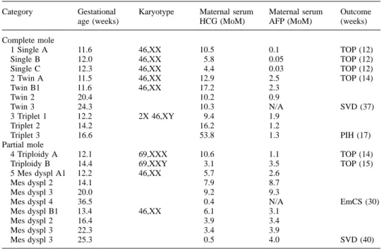
Table I. Clinical features in 10 pregnancies presenting with molar changes antenatally
Category Gestational Karyotype Maternal serum Maternal serum Outcome
age (weeks) HCG (MoM) AFP (MoM) (weeks)
Complete mole 1 Single A 11.6 46,XX 10.5 0.1 TOP (12) Single B 12.0 46,XX 5.8 0.05 TOP (12) Single C 12.3 46,XX 4.4 0.03 TOP (12) 2 Twin A 11.5 46,XX 12.9 2.5 TOP (14) Twin B1 11.6 46,XX 17.2 2.3 Twin 2 20.4 10.2 0.9 Twin 3 24.3 10.3 N/A SVD (37) 3 Triplet 1 12.2 2X 46,XY 9.4 1.9 Triplet 2 14.2 16.2 1.2 Triplet 3 16.6 53.8 1.3 PIH (17) Partial mole 4 Triploidy A 12.1 69,XXX 10.6 1.1 TOP (14)
Triploidy B 14.4 69,XXY 3.1 3.5 TOP (15)
5 Mes dyspl A1 12.2 46,XX 5.7 2.6
Mes dyspl 2 14.1 7.9 8.7
Mes dyspl 3 20.0 9.2 9.3
Mes dyspl 4 36.5 0.4 N/A EmCS (30)
Mes dyspl B1 13.4 46,XX 6.1 3.1
Mes dyspl 2 16.4 3.9 3.4
Mes dyspl 3 22.3 3.4 3.9
Mes dyspl 3 25.3 0.5 4.0 SVD (40)
HCG5 human chorionic gonadotrophin; AFP 5 alpha fetoprotein; Mes dyspl 5 mesenchymal dysplasia; MoM5 multiples of the median; TOP 5 termination of pregnancy; SVD 5 spontaneous vaginal delivery; PIH5 pregnancy-induced hypertension; EmCS 5 emergency caesarian section
maternal serum continued to be collected as part of our clinical protocol for the antenatal management of molar pregnancies includ-ing measurement of β human chorionic gonadotrophin (β-HCG) concentration and maternal thyroid function tests. Unused serum samples were kept frozen at –40°C until assayed.
The concentrations of intact HCG, freeα-HCG and free β-HCG subunits were measured using solid-phase two-site immunoradio-metric assay (IRMA) kits from BioMerieux (Marcy-l’Etoile, France). These assays, using monoclonal antibodies, were calibrated against the First International Reference Preparations 75/537 for HCG dimer, 75/569 for freeα-HCG subunits and 75/551 for freeβ-HCG subunits. Sensitivities were 1 mIU/ml for HCG and 0.03 mIU/ml for both free subunits. Intra- and interassay coefficient of variation were, respectively,,7% and ,10% for each assay. The alpha-fetoprotein (AFP) concentration was determined by an enzyme immunoassay with a fluorometric end-point (Hybritech Inc., San Diego, CA, USA) using the Stratus apparatus from Baxter (Dade Division, Miami, FL, USA). The intra-assay coefficient of variation was 5%.
Pregnancy-associated plasma protein A (PAPP-A) was measured in a double antibody sandwich enzyme immunoassay (ELISA) developed in our laboratory (Bersinger et al., 1995). Briefly, the polyclonal anti-PAPP-A antibody was purified in a negative affinity step using the,300 000 Da fraction of pooled pregnancy serum as a solid phase in order to remove contaminating antibodies. The purified immunoglobulin G was used as a coating antibody in the assay. Sera were diluted 1:100 to 1:1000 prior to assay. The intra-and inter-assay coefficients of variance were 4.3 intra-and 10.2%, respectively, for a PAPP-A concentration of 1.0 WHO mIU/ml. Pregnancy-specific β1-glycoprotein (SP1) was determined by microplate ELISA as previously described (Bersinger et al., 1994). Serum dilutions were 1:2600 to 1:10 000 in PBS buffer containing bovine serum albumin (BSA, 2% w/v) and Emulsit (0.05% v/v). Intra- and inter-assay coefficients of variance were 4.1 and 8.7%, respectively.
All serum marker concentrations were compared to the available
normal ranges for unaffected pregnancies (Bersinger et al., 1994; Nagy et al., 1994) and the results expressed as multiples of the median (MoM) and when appropriate, corrected for multiple pregnan-cies. Values.2.5 MoM were classified as high (Hsu et al., 1994; Benn et al., 1996).
Results
Six pregnancies were attributed to the classical (complete) mole group, including three singleton euploid complete moles and three cases of multiple pregnancy combining a complete mole and a normal gestational sac (Table I). The partial mole group consisted of four pregnancies including two cases of triploidy and two cases of euploid mesenchymal dysplasia. All pregnancies followed a spontaneous conception.
Singleton complete moles were evacuated surgically within 2 days of the ultrasound diagnosis. In cases where the molar sonographic changes were associated with a fetus, the parents were offered an invasive procedure to determine the fetal karyotype. The type of procedure, i.e. chorionic villous sampling (CVS), fetal blood sampling (FBS) or amniotic fluid sampling (AFS), was carried out according to the presence of co-existing fetal abnormalities, the gestational age at which the parents reached their decision and the location of placenta and molar mass inside the uterine cavity. Two fetuses of the partial mole group presented with abnormalities on ultrasound including increased nuchal thickness and holoprosencephaly. In both cases, karyotyping revealed a triploidy and the parents opted for a pregnancy termination. In one of the cases of twin molar pregnancy, the parents also opted for a pregnancy termination at 14 weeks because of the possible associated perinatal risks. Multicystic ovaries were observed in all cases of complete mole and in one case of partial triploid mole.
Figure 1. Maternal serum concentration ofβhuman chorionic gonadotrophin (HCG), HCG,α-HCG and alpha fetoprotein (AFP) in multiples of the median (MoM) at the time of the first ultrasound examination (11–13 weeks) in three cases of singleton complete mole (SCM), two cases of twin complete mole (TCM), one case of triplet complete mole (TrCM), two cases of triploidy (Trip) and two cases of mesenchymal dysplasia (MD).
One case of twin complete mole, the case of triplet complete mole and two cases of euploid partial mole were followed up. The twin pregnancy combining a complete mole resulted in a normal fetus at 37 weeks gestation. Chronic vaginal bleeding was observed from 13 weeks but the mother never presented with clinical or biological evidence of pregnancy-induced hypertension and her chest X rays were normal before delivery. The triplet pregnancy was complicated by severe pregnancy-induced hypertension at 17 weeks requiring pregnancy termina-tion. In both cases, the fetuses were anatomically normal at delivery and pathological examination of the placenta con-firmed the ultrasound diagnosis. In all cases of classical mole and partial triploid mole, the villi showed respectively generalized and focal hydropic changes and trophoblastic hyperplasia. The two pregnancies presenting with euploid partial moles had uncomplicated second trimesters. One case, presenting with polyhydramnios and premature labour at 30 weeks gestation and the fetus in breech position, was delivered by Caesarean section. The neonate was macrosomic (2.4 kg) with macroglossa and ear lobe creases and was diagnosed to be affected by Beckwith–Wiedemann syndrome. In these two cases, the placenta was large, weighing respectively 1535 g and 1430 g, and had a karyotype identical to that of the corresponding fetus. Histopathological examination showed aneurysmal dilatation of the chorionic vasculature and focal villous hydrops with no abnormal trophoblastic proliferation.
Maternal serum hormonal analysis indicated that free β -HCG and intact -HCG concentrations were very high in all cases. Free α-HCG concentration was only increased (.2.5 MoM) in the case of the triplet complete mole and was low
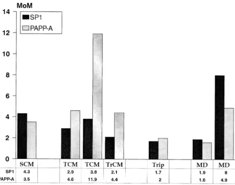
in one of the two cases of triploid partial mole (Figure 1). Maternal serum AFP concentration was extremely low (,0.5 MoM) in all cases of singleton complete moles and was high in one case of twin complete mole, in one case of triploid partial mole and the two cases of mesenchymal dysplasia. SP1 concentration was elevated in complete moles whereas PAPP-A concentration was elevated in all cases except in one case of partial triploid mole (Figure 2). At 17–20 weeks, in the euploid partial moles, maternal serum HCG and maternal serum β-HCG concentrations were within normal ranges and the AFP was more than 3.5 MoM whereas in the twin and triplet complete moles the maternal serum HCG and maternal serumβ-HCG were over 9 MoM and the AFP was within the normal range (Table I).
Discussion
Detailed ultrasound examination can identify placental molar transformation from the end of the first trimester (Jauniaux, 1998). The vast majority of classical and partial triploid moles miscarry before 10–12 weeks of gestation (Jeffers et al., 1993). In early pregnancy failure, the embryo often dies before the first ultrasound examination. Thus the differential diagnosis of molar changes in early pregnancy is essentially either classical mole, triploid partial mole or benign hydropic placental reten-tion. Although triploidy is less often associated with molar changes in the first trimester of pregnancy than in the second or third trimester, the use of standardized histological and/or cytogenetic criteria is both accurate and reproducible in the detection of complete and partial triploid mole (Jauniaux
Figure 2. Maternal serum concentration of pregnancy-specificβ1-glycoprotein (SP1) and pregnancy-associated plasma protein A (PAPP-A) in multiples of the median (MoM) at the time of the first ultrasound examination (11–13 weeks) in one case of singleton complete mole (SCM), two cases of twin complete mole (TCM), one case of triplet complete mole (TrCM), one case of triploidy (Trip) and two cases of mesenchymal dysplasia (MD).
The discovery, during the second trimester, of a pregnancy with ultrasound features suggesting placental molar trans-formation causes several diagnostic and management difficul-ties. Within this context, there are four main categories of pregnancy: classical hydatidiform mole; complete mole in a multiple pregnancy; partial triploid mole; and focal benign villous hydatidiform (pseudomolar) transformation (Jauniaux and Nicolaides, 1997). The first three can be complicated by persistent trophoblastic disease whereas the latter can be associated with Beckwith–Wiedemann syndrome as in one case in this series. An aberration in normal maternal suppression of the IGF-2 gene could account for the Beckwith–Wiedemann syndrome phenotype including the features of overgrowth (Morison and Reeve, 1998), which can be associated with congenital anomalies such as omphalocoele, mild microcephaly and ear lobe creases. In these cases, the placental anomaly appears to be a limited malformation of the extra-embryonic mesoderm involving the mesenchyme and the vessels of the stem villi of several cotyledons. It has been referred to as mesenchymal dysplasia and it can be found with a pheno-typically normal fetus (Jauniaux et al., 1997a) suggesting that in some cases the overgrowth is limited to the placental tissue. Confined placental diploidy or triploid mosaicism and tetraploidy may also appear as a partial mole and can theoretic-ally be complicated by persistent trophoblastic disease but these cases are exceptional (Jauniaux, 1998).
The ultrasound demonstration of classical hydatidiform mole is easy and accurate from 10 weeks of gestation. The presence of any form of placental molar change and a coexistent fetus continues to be referred to as partial mole (Nwosu et al., 1995). However, theoretically, the histopathological definition should only be applied when villous hydatidiform changes are associated with trophoblastic hyperplasia, which cannot be
demonstrated by ultrasound. If the placenta is partially molar and the fetus is growth retarded with structural anomalies, fetal karyotyping will usually demonstrate a triploidy (Jauniaux
et al., 1996b, 1997b). Thus the main difficulty will be the
differentiation between a complete mole coexisting with a normal pregnancy and non-triploid (euploid) partial moles. A classical mole may coexist with a normal fetus and placenta in cases of molar transformation of one ovum in a dizygotic twin or triplet pregnancy (Steller et al., 1994a). Because of a high maternal complication rate, including pre-eclampsia, hyperthyroidism, respiratory insufficiency and ovarian hyper-stimulation (bilateral multicystic ovaries), this condition must be distinguished from a true partial mole, as early in pregnancy as possible. At the beginning of the second trimester, the complete mole may be clearly separated from the normal placenta (Jauniaux and Nicolaides, 1997). However, the mole does not grow proportionally with the normal placenta and at 17–20 weeks it may partially cover the normal placenta, making it difficult to distinguish on ultrasound from a true partial mole.
The rate of differentiation of cytotrophoblast into syncytio-trophoblast appears to be the main factor that leads to the synthesis of HCG (Hay, 1988). HCG regulates the differenti-ation of cytotrophoblast via its receptors and thus its own synthesis (Rodway and Rao, 1995). Higher concentrations of HCG-receptor mRNA and protein are found in hyperplastic trophoblasts and HCG cannot self-regulate its synthesis under this condition (Rodway and Rao, 1995). We found that the concentration of intact HCG and free β-HCG was high (.2.5 MoM) in all categories of complete and partial moles. Although trophoblastic differentiation appears normal in mesenchymal dysplasia, the important placental overgrowth could explain the increased maternal serum HCG concentration
found in the two cases in this study. In complete moles, intact HCG and free β-HCG concentrations were higher than in partial moles and continued to increase as pregnancy advanced whereas freeα-HCG concentration was only increased in the case of triplet pregnancy combining a complete mole with a normal twin pregnancy. By contrast, the AFP concentrations were higher in most cases of partial mole. Fetal abnormalities in partial triploid moles and abnormal vascular permeability in mesenchymal dysplasia may explain the transfer of larger quantities of this protein from fetal into maternal circulation. Serum SP1 was high in complete moles whereas PAPP-A concentrations were increased in complete moles and in one case of mesenchymal dysplasia. Since the concentrations of these two proteins vary strongly throughout gestation, the measurement of these proteins is of limited value in the management of molar pregnancies. Other biological markers of trophoblastic cellular activity such as epidermal growth factor, progesterone or interleukines have been investigated
in vitro or in the post-molar surveillance (Steller et al., 1994a,
b; Vasilev et al., 1994) but PAPP-A or SP1 are likely to be of limited value during pregnancy. Investigation of the molecular heterogeneity of urinary or serum HCG (Hoermann et al., 1993) may help to further differentiate as well as follow up women presenting with molar pregnancies and a normal fetus. In cases of complete mole coexisting with a normal fetus and placenta, the mother must be counselled about possible perinatal complications and a therapeutic termination should be considered at all stages in cases of severe maternal com-plications. In ongoing pregnancies, an invasive procedure to establish the fetal karyotype should be performed. Placental biopsy may be technically difficult and misleading because of confined mosaicism while needle aspiration could be technically complicated by the presence of molar villi. Amnio-centesis can be proposed for partial moles but may be difficult in complete moles with a coexisting fetus due to the presence of intrauterine bleeding and the anterior location of a large molar mass. When the fetus is sonographically normal, fetal blood sampling is a possible alternative at 18–20 weeks of gestation. If the fetal karyotype was euploid, the fetus apparently anatomically normal and the mother’s clinical course stable, she should be offered bi-monthly follow-up visits. These visits must include sonographic evaluation of fetal anatomy and growth, blood pressure measurements, serum HCG concentration and urine analysis. If the maternal serum HCG concentration increases and the maternal serum AFP decreases, thus confirming a multiple pregnancy combining a classical mole and a normal conceptus, a monthly full blood count and thyroid functional tests should be obtained and a chest X ray should be performed every 3 months.
References
Bagshawe, K.D., Lawler, S.D., Paradinas, F.J. et al. (1990) Gestational trophoblastic tumours following initial diagnosis of partial hydatidiform mole. Lancet, 335, 1074–1076.
Benn, P.A., Horne, D., Briganti, S. et al. (1996) Elevated second-trimester maternal serum hCG alone or in combination with elevated alpha-fetoprotein.. Obstet. Gynecol., 87, 217–222.
Berkowitz, R.S. and Goldstein, D.P. (1996) Chorionic tumours. N. Eng. J. Med., 335, 1740–1748.
Bersinger, N.A., Brizot, M.L., Johnson, A. et al. (1994) First trimester maternal serum pregnancy-associated plasma protein A and pregnancy-specific β1-glycoprotein in fetal trisomies. Brit. J. Obstet. Gynecol., 101, 970–974. Bersinger, N.A., Zakher, A., Hucer, U. et al. (1995) A sensitive immunoassay
for pregnancy-associated plasma protein A(PAPP-A): a possible first trimester method of screening for Down syndrome and other trisomies. Arch. Gynecol. Obstet., 256, 185–192.
Chen, F.P. (1997) Molar pregnancy and living normal fetus coexisting until term: prenatal biochemical and sonographic diagnosis. Hum. Reprod., 12, 853–856.
Chen, C.P., Chern, S.R., Wang, T.Y. et al. (1997) Pregnancy with concomitant chorangioma and placental vascular malformation with mesenchymal hyperplasia. Hum. Reprod., 12, 2553–2556.
Hay, D.L. (1988) Placental histology and the production of human choriogonadotrophin and its subunits in pregnancy. Br. J. Obstet. Gynaecol.,
95, 1268–1275.
Hoermann, R., Spoettl, G., Grossmann, M. et al. (1993) Molecular heterogeneity of human chorionic gonadotropin in serum and urine from patients with trophoblastic tumours. Clin. Invest., 71, 953–960.
Hsu, C.D., Chan, D.W., Iriye, B. et al. (1994) Elevated serum human chorionic gonadotrophin as evidence of secretory response in severe preeclampsia. Am. J. Obstet. Gynaecol., 170, 1135–1138.
Jacobs, P.A., Szulman, A.E., Funkhouser, J. et al. (1982) Human triploidy: relationship between parental origin of the additional haploid complement and development of partial hydatidiform mole. Ann. Hum. Genet., 46, 223–231.
Jauniaux, E. (1998) Ultrasound diagnosis and follow-up of gestational trophoblastic disease. Ultrasound Obstet. Gynecol., 11, 367–377. Jauniaux, E. and Nicolaides, K.H. (1997) Early ultrasound diagnosis and
follow-up of molar pregnancies. Ultrasound Obstet. Gynecol., 9, 17–21. Jauniaux, E., Kadri, R. and Hustin, J. (1996a) Partial mole and triploidy:
screening in patients with first trimester spontaneous abortion. Obstet. Gynecol., 88, 616–619.
Jauniaux, E., Brown, R., Rodeck, C. and Nicolaides, K.H. (1996b) Prenatal diagnosis of triploidy during the second trimester of pregnancy. Obstet. Gynecol., 88, 983–989.
Jauniaux, E., Brown, R., Snijders, R.J.M. et al. (1997a) Early prenatal diagnosis of triploidy. Am. J. Obstet. Gynecol., 176, 550–554.
Jauniaux, E., Nicolaides, K.H. and Hustin, J. (1997b) Perinatal features associated with placental mesenchymal dysplasia. Placenta, 18, 701–706. Jeffers, M.D., O’Dwyer, P., Curran, B. et al. (1993) Partial hydatidiform mole:
a common but underdiagnosed condition. Int. J. Gynecol. Pathol., 12, 315–323.
Kajii, T. and Ohama, K. (1977) Androgenic origin of hydatidiform mole. Nature, 268, 633–634.
Morison, I.M. and Reeve, A.E. (1998) Insulin-like growth factor 2 and overgrowth: molecular biology and clinical implications. Molec. Med. Today, 3, 110–115.
Nagy, A-M., Glinoer, D., Delogne-Desnoeck, F.B. et al. (1994) Total amounts of circulatingαandβsubunits can be assessed throughout pregnancy using immunoradiometric assay calibrated with the unaltered and thermally dissociated heterodimer. J. Endocrinol., 140, 513–520.
Nwosu, E.C., Ferriman, E., McCormack, M.J. et al. (1995) Partial hydatidiform mole and hypertension associated with a live fetus. Variable presentation in two cases. Hum. Reprod., 10, 2459–2462.
Rodway, M.R. and Rao, C.V. (1995) A novel perspective on the role of human chorionic gonadotropin during pregnancy and in gestational trophoblastic disease. Early Pregn. Biol. Med., 1, 176–187.
Steller, M.A., Genest, D.R., Bernstein, M.R. et al. (1994a) Natural history of twin pregnancy with complete hydatidiform mole and coexisting fetus. Obstet. Gynecol., 83, 35–42.
Steller, M.A., Mok, S.C., Yeh, J. et al. (1994b) Effects of cytokines on epidermal growth factor expression by malignant trophoblast cells in vitro. J. Reprod. Med., 39, 209–216.
Szulman, A.E. and Surti, U. (1978a) The syndromes of hydatidiform mole. I. Cytogenetic and morphologic correlations. Am. J. Obstet. Gynecol., 131, 665–671.
Szulman, A.E. and Surti, U. (1978b) The syndromes of hydatidiform mole. II. Morphologic evolution of the complete and partial mole. Am. J. Obstet. Gynecol., 132, 22–27.
Vasilev, S.A., Schlaerth, J.B., Ahn, C. and Sauer, M.V. (1994) Serum progesterone monitoring in post-molar surveillance. Gynecol. Oncol., 54, 288–291.