EnzMet
™: An Enzymatic Metallography Reagent for Accurately Delineating
Neuronal Boundaries for Segmenting Gap Junction-Coupled Neurons in their
Three-dimensional Space
Irma I. Torres-Vazquez1*, Jose L. Serrano-Velez1*, François Orange2*, Maxime J.F. Guinel2, Ioannis Koutis3, Johanna Wolf4, Christoph Laimer4, Vishwas Joshi5, Richard D. Powell5 and
Eduardo Rosa-Molinar1
1
Biological Imaging Group, University of Puerto Rico-Rio Piedras, San Juan, Puerto Rico,
00931;2Department of Physics,University of Puerto Rico-Rio Piedras, San Juan, Puerto Rico,
00931;3Department of Computer Science, University of Puerto Rico-Rio Piedras, San Juan,
Puerto Rico, 00931, 4Bitplane AG, Zürich, Switzerland and 5Nanoprobes Incorporated, Yaphank,
NY 11980
Gap junctions have become a subject of intense investigation in neural circuits in which neural synchrony and oscillations may play an important part [1, 2]. Gap junction studies can be enhanced by correlative microscopy using laser scanning confocal microscopy (LSCM) and focused ion beam/scanning electron microscopy (FIB/SEM). However, the small number of correlative neural tract tracers and enhanced contrast reagents that can be imaged first by LSCM and then by FIB/SEM is hindering advances in understanding gap-junction coupled neural circuits. Here we report the outcome of our efforts to assess and optimize reliable correlative retrograde neural tract tracers and enhanced contrast reagents to facilitate high resolution 3D reconstruction of neurons imaged first by LSCM and then serially milled and block-face imaged
using FIB/SEM. Neurons were retrogradely labeled by combining Alexa Fluor® 594 biocytin
(Invitrogen Corporation., Carlsbad, CA) with immunohistochemistry using the
avidin-biotin-peroxidase complex method (Vectastain® Elite Kit, Vector Laboratories, Burlingame, CA)
visualized with EnzMet™ [3, 4, 5] (Nanoprobes Inc. Yaphank, NY) that utilizes probes labeled with peroxidase enzymes to reduce silver ions to elemental silver, resulting in the deposition of
silver metal particles at sites labeled by peroxidases. This labeling procedure was followed by en
bloc staining using heavy metal ions. The samples were oriented and flat embedded in a
modified epoxy resin. After polymerization, the blocks containing the embedded tissue were glued onto a sample stub using conductive silver paint. Using a 10-nA ion beam current, a first
coarse cross-section was milled using a Nova 600 NanoLab DualBeamTM system that integrates
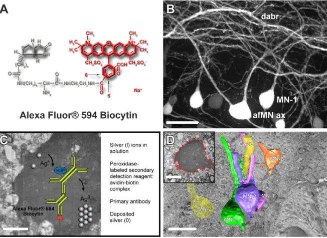
ion and electron beams for FIB and SEM functionality in one machine. The exposed surface of the cross-section was fine-polished by lowering the ion beam current to 200 pA. Layers from the fine polished cross-section were serially milled by scanning the ion beam parallel to the surface of the cutting plane, using the same ion beam current. After the removal of each slice, the milling process was paused and the freshly exposed surface was imaged with a low energy acceleration potential (~2keV) using the in-column Through-The-Lens (TLD) detector. For detecting the backscattered electrons, the TLD was set to repel all low energy electrons by setting the retardation voltage higher than 50eV. Combining Alexa Fluor® 594 biocytin with EnzMet dramatically increased the contrast within the neuronal soma and processes and enabled the selective identification of Alexa Fluor® 594 biocytin-labeled gap junction-coupled neurons within the spinal cord (see Figure 1A and 1B). The non-diffusing and sharply defined dense, black signal with low background yielded by EnzMet (see Figure 1C) provided superior spatial resolution for differentiation of gap junction-coupled neurons and permitted them to be
segmented easily and precisely using a new algorithm (see Figure 1D). Improvements in serial block-face imaging technologies, whose use in the study of the nervous system remains
uncommon, together with improved reagents for enhanced constrast and labeling specificty and new algorithms based on recent breakthroughs in spectral graph theory will enable large-scale high-throughput collection and analysis of gap junction-coupled neural circuits [6].
660
doi:10.1017/S1431927612005156 © Microscopy Society of America 2012Microsc. Microanal. 18 (Suppl 2), 2012
https://doi.org/10.1017/S1431927612005156
References
[*] Denotes that these individuals preformed equally as first authors to complete this work. [1] Leitch, B., Electron Microsc. Rev., 5 (1992) 11-339.
[2] Jermakowicz, W.J. and and Casagrande, V.A., Brain Res Rev. 55 (2007) 264–284. [3] Hainfeld, J.F., et al., Microsc. Microanal. 8 (Suppl. 2) (2002) 916 CD.
[4] Tubbs, R., et al., Appl. Immunohistochem. Mol. Morphol., 13 (2005) 371. [5] Powell, R., et al., Microsc. Microanal. 12 (Suppl. 2) (2006) 424CD.
[6] Partially supported by1018463 (IY), MH-086994 (SBIR grant to RDP and ERM), NSF-1039620 (ERM), and NSF-0964114 (ERM).
Figure 1A. Alexa Fluor® 594 biocytin was first visualized using laser scanning confocal microscopy. Figure 1B. One can see that Alexa Fluor® 594 biocytin readily crosses gap junctions, thus revealing extensive networks of coupled neurons (scale bar = 30ȝm). Figure 1C. Transported Alexa Fluor® 594 biocytin was visualized selectively with avidin-biotin-peroxidase/DAB and EnzMetTM (scale bar = 5ȝm). Figure 1D. There is excellent registration between all images within an FIB/SEM image stack. This allows simple exporting of the image files in order to easily contour, segment, and reconstruct 1 (Mn-1), 2 (Mn-2), and type-3 (Mn-type-3) spinal motor neurons and the axon of a commissarial primary ascending interneuron (CoPAax) with new algorithms based on recent breakthroughs in spectral graph theory (see insert; scale bar = 10 ȝm) and Imaris® (the 3D volume rendering of the neurons seen in Figure 1D was based on the first 200 serial optical cross sections within the 6,074 serial optical cross section volume). The CoPAax is located dorsally to the Mns in the spinal cord. The cell bodies of MN-1s, MN-2s, and MN-3s average about 16-20 ȝm; their axons are equally fine and lie in the ventral half of the cord (scale bar = 25 ȝm).
Microsc. Microanal. 18 (Suppl 2), 2012 661
https://doi.org/10.1017/S1431927612005156