HAL Id: hal-03057235
https://hal.archives-ouvertes.fr/hal-03057235
Submitted on 11 Dec 2020HAL is a multi-disciplinary open access archive for the deposit and dissemination of sci-entific research documents, whether they are pub-lished or not. The documents may come from teaching and research institutions in France or abroad, or from public or private research centers.
L’archive ouverte pluridisciplinaire HAL, est destinée au dépôt et à la diffusion de documents scientifiques de niveau recherche, publiés ou non, émanant des établissements d’enseignement et de recherche français ou étrangers, des laboratoires publics ou privés.
The ossicular chain of Cainotheriidae (Mammalia,
Artiodactyla)
Alexandre Assemat, Mickaël Mourlam, Romain Weppe, Jacob Maugoust,
Pierre-Olivier Antoine, Maeva Judith Orliac
To cite this version:
Alexandre Assemat, Mickaël Mourlam, Romain Weppe, Jacob Maugoust, Pierre-Olivier Antoine, et al.. The ossicular chain of Cainotheriidae (Mammalia, Artiodactyla). Journal of Anatomy, Wiley, 2020, 237 (2), pp.250-262. �10.1111/joa.13190�. �hal-03057235�
1
Short running page heading: Ossicular chain of Cainotheriidae
1 2
Title: The ossicular chain of Cainotheriidae (Mammalia, Artiodactyla)
3 4
Authors names: Assemat A.1, Mourlam M.J.1, Weppe R.1, Maugoust J.1, Antoine P.-O.1,
5
Orliac M.J.1
6 7
Corresponding author: Mourlam M.J. (mickael.mourlam@umontpellier.fr)
8 9
Authors affiliations:
10
1 – Institut des Sciences de l’Evolution, UMR 5554 (CNRS, IRD, EPHE), Université de
11
Montpellier, Place Eugène Bataillon, 34095 Montpellier cedex 5
12 13
Abstract:
14 15
This work describes an unparalleled sample of isolated fossil auditory ossicles of cainotheriid
16
artiodactyls from the Paleogene karstic infillings of Dams (Tarn-et-Garonne, Quercy, France).
17
This collection comprises a total of 18 mallei, 28 incudes and three stapedes. It allows the
18
documentation of both intra- and interspecific variability of ossicular morphology within
19
Cainotheriidae. We show that despite considerable intraspecific variability, the malleus, the
20
incus, and the stapes appear to be taxonomically informative at the Cainotheriidae scale. This
21
work further provides the first description of a reconstructed ossicular chain of a terrestrial
22
Paleogene artiodactyl species, found in a basicranium of the late Oligocene cainotheriine
23
Caenomeryx cf. procommunis (Pech Desse locality).
24 25
Key-Words: Morphometry, Malleus, Incus, Stapes, Paleogene, Quercy
26 27 28
2
Introduction
29 30
The middle ear ossicles - the malleus, the incus, and the stapes – form a bony chain
31
contained within the air-filled middle ear cavity. Their presence is a hallmark of Mammalia
32
(e.g., Simpson, 1959; Luo et al. 2001). The three ossicles are the smallest bones of the
33
mammalian skeleton and they play a fundamental role in hearing process. Indeed, the
34
ossicular chain transmits the airborne sound waves from the tympanic membrane (at the
35
interface between the outer and middle ear) to the fluid-filled cochlea of the inner ear, while
36
performing an anatomical impedance match between this two media (e.g., Wever &
37
Lawrence, 1954; Dallos, 1973; Schubert, 1978; Killion & Dallos, 1979; Peake & Rosowski,
38
1991; Hemilä et al. 1995; Nummela & Thewissen, 2008; Mason, 2016). Morphology of the
39
ossicular chain and its specificity within different mammalian groups has been intensely
40
studied for systematic purposes (e.g., Doran, 1878; Fleischer, 1973; Schmelzle et al. 2005;
41
Wible & Spaulding, 2012; Mason, 2013; Stoessel et al. 2016; Maier & Ruf, 2016a, 2016b;
42
Kerber & Sánchez-Villagra, 2018; Loza et al. 2018), or functional aspects (e.g., Fleischer,
43
1978; Nummela, 1995; Nummela & Sánchez-Villagra, 2006; Puria & Steele, 2010). Most of
44
these works deal with extant taxa, and, because of their fragility and small size, ossicles are
45
rarely preserved - or at least retrieved - in the fossil records. Yet, when retrieved, they bring
46
useful observations for the systematics or ecology of taxa, or both [e.g., systematic position of
47
Pakicetidae (Thewissen & Hussain, 1993) and hearing mechanisms in early cetaceans
48
(Nummela et al. 2004; Nummela et al. 2007) among Artiodactyla]. Still, fossil ossicles are
49
rarely preserved all three together, and almost never found in anatomical connection.
50
Here, we describe a broad sample of isolated ossicles of Cainotheriidae from Paleogene
51
karstic infillings from Quercy (Tarn-et-Garonne, France). Cainotheriids are an extinct family
52
of small artiodactyls (even-toed ungulates) documented in the fossil record between the late
53
Eocene and the middle Miocene in Western Europe (Blondel, 2005). Because of their unique
54
dental morphology, the phylogenetic position of Cainotheriidae within artiodactyls is still
55
debated. They have been related to different European endemic families (Romer 1966; Webb
56
& Taylor, 1980; Gentry & Hooker, 1988) without reaching a consensus, or to modern groups
57
of artiodactyls such as ruminants (Geisler & Uhen, 2005; O'Leary & Gatesy, 2007; Lihoreau
58
et al. 2015) or tylopods (Geisler & Uhen, 2003; Geisler et al. 2007; Thewissen et al. 2007).
59
The recent phylogenetic study of Weppe et al. (2019) retrieved Cainotheriidae closely related
60
to the European endemic families Mixtotheriidae, Anoplotheriidae and Robiacinidae.
3
Cainotheriidae are particularly abundant in karstic localities from Quercy, southwestern
62
France. This family, which includes at least five genera within two sub-families ranges from
63
rabbit-sized species to size of a small ruminant (Erfurt & Métais, 2007; Theodor, 2010).
64
Contrary to many European endemic ungulates which went extinct at the end of the Eocene
65
(Sudre & Legendre, 1992; Blondel, 2001), cainotheriids made it through the
66
Eocene/Oligocene transition and they are one of the very few artiodactyl groups to diversify
67
during Oligocene times (Blondel, 2005). The ossicles we describe here originate from two
68
loci from the karstic network of Dams, discovered in 2016. This karstic network was emptied
69
during the extensive phosphate exploitation that took place in Quercy during the late 19th
70
century. The network, however, still houses a great quantity of clay infillings including two
71
channels that yielded a great quantity of cainotheriid remains, namely DAM1 and DAM3.
72
These two infillings within Dams network bracket the Eocene-Oligocene transition (Weppe et
73
al. 2019) and they document a period that corresponds to a major faunal turnover in Western
74
Europe linked to climatic, geographic and oceanic circulation changes (Legendre, 1987;
75
Berggren & Prothero, 1992). Based on the unprecedented sample from Dams, including a
76
total of 18 mallei, 28 incudes and three stapedes, and on an in-situ ossicular chain from Pech
77
Desse (Quercy, France, late Oligocene), we discuss the intra- and interspecific variability of
78
ossicle morphology within Cainotheriidae and describe for the first time a reconstructed
79
ossicular chain for a Paleogene terrestrial artiodactyl species.
80 81
Material and methods
8283
Material
84 85
Most of the specimens included in this analysis come from the Dams karstic network located
86
near Caylus (Tarn-et-Garonne) in Quercy (SW France). All specimens studied are curated at
87
the University of Montpellier (UM). The Dams material was collected after screenwashing of
88
40kg from DAM1 and 30kg from DAM3 in 2016.Raw fossil material was first concentrated
89
by wet screening of the red clays collected in Dams locality, (0.7-mm mesh size), and then
90
picked up with smooth tweezers, under a stereomicroscope. The material consists of 16
91
mallei, 16 incudes, and two stapedes from DAM1 (late Eocene, Mammalian Paleogene
92
reference level 19 [MP19]; Weppe, 2018) and 12 incudes (among which two are in
93
connection with the malleus head), and one stapes from DAM3 (early Oligocene, MP22;
4
Weppe, 2018). The taxonomic identification of the isolated ossicles from Dams relies on a
95
strong corpus of evidences: 1) the relative abundance of mammalian fossil remains; 90% of
96
the remains (cranial, dental and postcranial) from DAM1 belong to the small cainotheriid
97
artiodactyl Paroxacron valdense. Therefore, all the mallei and 16 incudes upon 18, that can be
98
referred to the same morph, likely correspond, based on the relative abundance criterion, to
99
Paroxacron valdense; 2) Artiodactyla hallmark; the incus presents a processus longum
100
slightly smaller than the processus brevis which is a characteristic of Artiodactyla (Doran,
101
1878; Wilkie, 1936; Thewissen & Hussain, 1993; Thewissen, 1994; Milinkovitch &
102
Thewissen, 1997); 3) incudo-mallear joint association; the association between the malleus
103
and the incus, besides general size compatibility and match between the articular surfaces, is
104
based on fused incudo-mallear complexes found in DAM3.
105 106
Based on these criterions, the ossicles from DAM1 are all assigned to Paroxacron valdense,
107
the only cainotheriid species retrieved in this channel (Weppe, 2018). In contrast, five
108
different cainotheriid species co-occur in DAM3, making specific attribution of the isolated
109
ossicles impossible (in the current state of our knowledge). A list of the included material is
110
provided in supplementary information Table S1. Other incudes of similar size have been
111
found in both levels (DAM1 and DAM3), but they are not included in this study due to their
112
markedly different morphology that would point to rodents or chiropterans instead of
113
artiodactyls. In addition to the ossicles from Dams localities, we reconstruct in this work the
114
in-situ location of the cainotheriid ossicular chain based on a basicranium (UM PDS 3353)
115
from the late Oligocene locality of Pech Desse (MP28, Quercy; Hugueney, 1997) that
116
preserves the ossicles trapped in the middle ear cavity. Two cainotheriine species are retrieved
117
in Pech Desse, namely Plesiomeryx cf. cadurcensis and Caenomeryx cf. procommunis (Remy
118
et al. 1987). Based on the overall larger dimensions of the specimen, it is here referred to as
119
Caenomeryx cf. procommunis.
120 121
Micro CT scanning and virtual reconstruction
122 123
The ossicles were scanned, using the high-resolution micro CT-scanner EasyTom of the
124
technical facility of the Montpellier Rio Imaging platform, at a high voltage (150 kV) using a
125
copper filter and small sample holders (2 and 4 cm diameters), allowing to be close to the
X-126
ray source and therefore, to reach a voxel size of 11.89 µm for isolated specimens, and 23.81
127
µm for the partial cranium UM PDS 3353. They were reconstructed virtually in 3D using the
5
threshold tool of Avizo 9.5 (VSG-FEI) software. The specimens partly encrusted with matrix
129
were cleaned using the manual segmentation tool of the same software. The virtual
130
reconstruction of the in-situ location of the ossicular chain of cainotheriid from Pech Desse
131
was realized using the freeware MorphoDig (Lebrun, 2018). Anatomical terminology used in
132
this study mainly follows that of Wible and Spaulding (2012); orientations are based on the
133
reconstruction of the in-situ ossicle chain of the cranium UM PDS 3353.
134 135
Geometric morphometric analysis
136 137
Nine mallei and nine incudes from DAM1 and five incudes from DAM3 are included in the
138
geometric morphometric analyses, other specimens being discarded due to their fragmentary
139
condition. In addition, the incus and malleus from the Pech Desse specimen were also added
140
to the analyses. Only three stapedes were unearthed in Dams localities, which does not allow
141
a proper discussion of the morphometrical variability of this ossicle. To quantify the malleus
142
and incus shape variations, we digitized a set of 3D landmarks using MorphoDig software
143
(MorphoDig 0.8) (Fig. 1). Nine landmarks were placed on the malleus including five on the
144
articular area. The first one was placed at the highest point of the curve along the medial
145
margin of the articular area. The second one occupies the same position on the distal margin
146
of the articular area. The third landmark is located at the center of the ridge of the articular
147
area. The fourth and the fifth landmarks were placed on the deepest points along the dorsal
148
and ventral borders of the articular facet, respectively. The sixth landmark was placed at the
149
extremity of the muscular process and the seventh one is located on the notch between the
150
muscular process and the manubrium. The eighth is positioned at the deepest point of the
151
angulation formed by the neck of the malleus with the manubrium. The last one was placed at
152
the extremity of the anterior process. On the incus, we positioned nine landmarks, including
153
five on the articular area. The first one was placed on the deepest part on the lateral border of
154
the articular area and the second one at an equivalent location on the medial border.
155
Landmarks three and four were placed on the most salient points of the articular surface i.e.,
156
on the dorsal and ventral edge, respectively. The fifth landmark is located at the central
157
position of the bulge formed by the intersection of the two articular facets. Landmarks six and
158
seven are positioned on the incudal body. The sixth one is at the middle of the swelling along
159
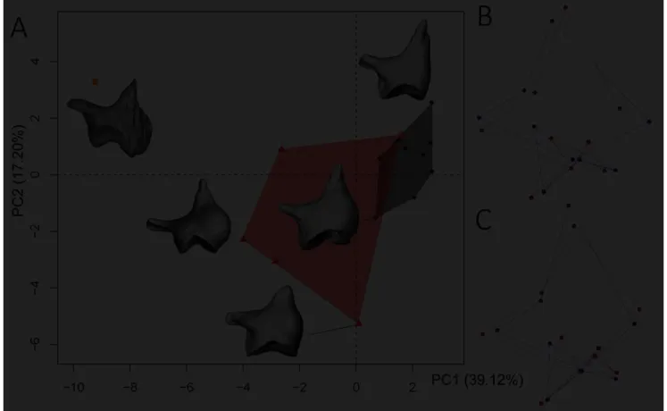
the upper border of the incudal body and the seventh was placed at the middle of the curve
160
joining the short and the long process posteroventrally. The eighth landmark was placed
161
internally to the long process where it becomes thinner, while the ninth one was positioned at
6
the extremity of the short process. The treatment of the raw dataset was performed using R
163
software version 3.4.4 (R Core Team, 2018). All 10 replicates were scaled to unit centroid
164
size (i.e. “the square root of the sum of squared distances from each landmark to the centroid
165
of the configuration” Claude, 2008:139), translated, rotated, and superimposed through the
166
Generalized partial Procrustes Analysis (pGPA) following Claude (2008, 2013; see also
167
Bookstein, 1990; Rohlf, 1990; Dryden & Mardia, 1998). Then, to apprehend the shape
168
variability of the malleus and incus, we performed a Principal Component Analysis (PCA;
169
Pearson type) on the Procrustes coordinates (resulting from the pGPA). Error measurement
170
follows Yezerinac et al. (1992; see also Claude et al. 2003; Claude, 2008: 65-66, 2013). Data
171
and script are available in supplementary material dataset 1.
172 173
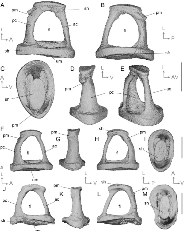
Data availability statement
174
All specimens studied are curated at the University of Montpellier (UM) and can be freely
175
consulted upon request. The 3D models of the middle ear reconstruction of Caenomeryx cf.
176
procommunis is available in open access on MorphoMuseuM (https://morphomuseum.com/ ;
177
Assemat, in press). The scripts used to perform the morphometric analyses are provided in
178 supplementary information. 179 180
Description
181 182 Malleus (Figs. 2-3) 183 184The description of the cainotheriid malleus (Fig. 2) primarily relies on material from
185
the DAM1 locality as DAM3 only yielded malleus articular surfaces fossilized in anatomical
186
connection with the incus. The malleus is the most lateral part of the ossicular chain. In life, it
187
contacts the tympanic membrane by a long, flat-shaped manubrium and a short and angular
188
lateral process. The malleo-incudal complex is unfused for all documented specimens from
189
DAM1. The globose head of the malleus bears the articular surface for the incus. The latter is
190
divided into two facets separate by a deep and wide groove; the superior articular facet for the
191
incus lies in the dorsal aspect, while the inferior articular facet, of about the same size, lies at
192
ca. 35° angle to it (Fig. 2A). The general shape of the articular surface is saddle-like and is
193
asymmetrical related to the fact that the groove is wider and shallower on the dorsomedial
7
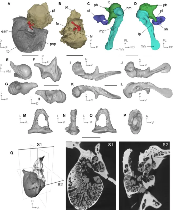
part. On the anterior surface, the head comes to a small, rounded point that we identify as a
195
blunt capitular spine (Wible & Spaulding, 2012). A thin and sharp bony crest joins the basis
196
of this spine to the anterior process, the outer lamella (Henson, 1961). Lateral to the capitular
197
spine, the surface of the head is carved by a small pit. The neck of the malleus is relatively
198
straight and it forms with the head an obtuse angle which confers to the malleus a general
199
sigmoidal shape. It is relatively broad and lines the osseous lamina on the posteromedial edge.
200
The osseous lamina consists of a particularly thin portion of bone bearing a depression on the
201
lateroventral aspect of the malleus (Fig. 2D); there is no clear demarcation with the basis of
202
the manubrium. The specimens of our sample seem to display a short blunt anterior process,
203
also known as processus gracilis, or prearticular, and mentioned in living artiodactyls (Wible
204
& Spaulding, 2012; Maier & Ruf, 2016b). It might have displayed a much thinner terminal
205
part but it would have been broken away during the fossilization process and left no trace. As
206
illustrated for carnivorans by Wible & Spaulding (2012), the base of the manubrium
207
corresponds to the confluence of the neck, lateral process, and ventral margin of the osseous
208
lamina. The manubrium is long with a flat and thin tympanic surface. The latter is much wider
209
than the lateral edge which bears a ridge becoming narrower at the manubrium’s extremity.
210
Unfortunately, the manubrium is partly broken on all isolated specimens. It displays a
well-211
developed lateral process at the posterior margin of its base. The medial margin of the bone
212
bears a strong and conical muscular process forming a 40° angle with the manubrium. The
213
course of the chorda tympani nerve is marked on the ventral aspect of the muscular process.
214 215
Comparison. The small and flat head of the malleus of the cainotheriids from Dams is closer
216
in proportion and shape to Ruminantia as illustrated in Capreolus and Giraffa by Fleischer
217
(1973) than to Sus, Hippopotamus, and Camelus. Like ruminants, they also display a long
218
neck and a wide osseous lamina. In suids, hippos, and camelids, the neck is shorter and the
219
osseous lamina remains smaller and closely appressed to the head. Compared to hippos and
220
suids, the cainotheriid malleus also displays a shorter but stockier muscular process which is
221
closer to the manubrium. The muscular process global shape in cainotheriids is relatively
222
similar to that of some living ruminants (e.g. Bos taurus, Ovis aries, and Cervus elaphus).
223 224
Inter- and intraspecific variation. The morphology of the 16 mallei from DAM1 shows a
225
noticeable variability mainly affecting the length of the malleus neck, the angulation of the
226
lateral process, the shape, depth and orientation of the articular surface, and also the location,
227
length and shape of the muscular process (Fig. 2G-H; Fig. S1A-H). In addition, in comparison
8
with the specimens from DAM1, the malleus from Pech Desse displays a different
229
morphology of its articular surface. In order to quantify this inter- and intraspecific shape
230
variation of the malleus, we performed two pGPAs, one with all available mallei (interspecific
231
analysis; see Material and methods) and one without the Pech Desse specimen (intraspecific
232
analysis). Due to small breakages on the latter specimen, interspecific shape variation has
233
been quantified by taking into account only the first seven landmarks. For the interspecific
234
analysis, measurement error is 6.92 % for centroid size and 16.88 % for shape. Inter-specimen
235
size and shape variations are stronger than the intra-specimen ones (specimen factor is
236
significant in both ANOVA (F = 135.4; p < 0.001) and Procrustes ANOVA (F = 50.23; p =
237
0.001); see also Claude, 2013). The Fig. 3A presents the projection of the specimens on the
238
first factorial plane. PC1 (52.96 % of the variance) clearly separates the Pech Desse specimen
239
from the other mallei and highlights deep morphological difference in term of shape and
240
orientation of the articular facets of the two species. The mallei from DAM1 present
241
intraspecific variation mainly on PC2 (19.72 % of the variance) that corresponds with small
242
size variation of the articular surface and slight variations of orientation of the body of the
243
malleus in regards to its head. More precisely, projection of individuals on the first factorial
244
plane (48.82 % of the variance) of the intraspecific analysis (Fig. 3D; where measurement
245
error for centroid size is 5.08 % with specimen factor significant (F = 187.7; p < 0.001) and
246
measurement error for shape is 23.41% with specimen factor significant (F = 33.72; p =
247
0.001)), underlines these variations. Indeed specimens from DAM1 are distributed more or
248
less randomly along the first two PCs that both stand for size variation of the articular surface.
249
Furthermore, PC1 also presents variation of orientation of the anterior process and PC2
250
highlights variation of orientation of the muscular process and of the main body in regards to
251
the malleus head. This disparity of orientation of the malleus body can also be observed
252
directly by superimposing all the mallei while keeping the same position for the articular
253
surface (Figs. 2G-H, 3E-F).
254 255
Incus (Figs. 4-5)
256 257
The incus is the middle ossicle joining the malleus and the stapes. It is conical and
258
stocky in shape (Fig. 4 A-B). The wide articular area of the incudomallear joint is composed
259
of two asymmetrical facets. It displays the same asymmetry as previously described for the
260
malleus. A salient edge, of different size depending on the specimen, separates the articular
261
surface from the incudal body (Fig. 4D). The dorsal aspect of the incus is variably convex,
9
from nearly flat to strongly domed. The cainotheriid incus displays two processes of similar
263
length, set apart by a wide angle; the processus brevis and the processus longum.
264
The processus brevis prolongs posteriorly the incudal body. The processus longum, which
265
connects the stapes by the lenticular apophysis, is located on the ventral edge of the incus,
266
posterior to the articular surface. The distal extremity of the processus longum of all incudes
267
of our sample is broken so that no lenticular apophysis is documented. A groove starts at the
268
base of the processus longum and runs all along of it. The separation between the two
269
processes is strengthened by the length of the body of the incus. The processus brevis of the
270
cainotheriid incus displays a thinning at its extremity while the processus longum is thicker.
271
The extremity of the processus longum ends in a small spike that corresponds to the
272
attachment of the broken lenticular apophysis.
273 274
Comparison. The cainotheriid incus is morphologically similar to that of other artiodactyls
275
with two processes of about the same length, whereas in most other mammals, the processus
276
longum is longer than the processus brevis (Doran, 1878). Cainotheriid incudes from Dams
277
differ from those of Camelus in having further apart processes, separated by a wider angle.
278
The articular area of cainotheriid incudes appears to be shallower than in camels, but deeper
279
than in bovines (Doran, 1878, Pl. 61). The body of studied incudes is close in shape to that of
280
Sus scrofa. It is more massive in the hippos and the llama which also display a more
281
cylindrical shape. Ruminants display a wide range of incudal body morphologies (Doran,
282
1878; Wilkie, 1925; Wilkie, 1936). The scarcity of illustrations available in the literature
283
prevents a broader comparison among artiodactyls.
284 285
Inter- and intraspecific variation. The incudes from DAM1 and DAM3 show a noticeable
286
variability of the depth of the articular facet and of the angulation between the processus
287
brevis and the processus longum. They also display a variation of their dorsal part bulge (Fig.
288
4F, Fig. 5A-B, Fig. S1I-M). In order to quantify inter- and intraspecific shape variations, we
289
performed a pGPA on a dataset of nine three-dimensional landmarks (Fig. 1B; see Material
290
and methods section, Geometric morphometric analysis) for a sample of 15 incudes from
291
DAM1, DAM3 and Pech Desse. For this analysis, measurement error is 3.14 % for centroid
292
size, 18.82 % for shape and inter-specimen size (F = 309.4; p < 0.001) and shape (F = 44.13;
293
p = 0.001) variations are significantly stronger than the intra-specimen ones. The two first
294
PCs explain 56.32 % of the variance (Fig. 5A). The main shape variation contributions of PC1
295
(39.12 % of the variance) correspond with an elongation of the processus brevis, correlated
10
with a smoother and thinner dorsal bulge toward the positive values (Fig. 5 B). PC1 also
297
displays shape and orientation variations of the articular facets following the same trends
298
observed for the interspecific variation of the malleus (see above). PC2 (17.20 % of the
299
variance) mainly highlights the variation of angulation between the two processes (Fig. 5 C).
300
The projection of the specimens on the first factorial plane results in two slightly overlapping
301
groups corresponding to DAM1 and DAM3 localities. DAM3’s morphospace, with a surface
302
that covers 41 % of the whole sample’s morphospace, is nearly five times more expanded
303
than that of DAM1 (that covers 8.6 %). Only one specimen from DAM3 lies within DAM1’s
304
morphospace. Incudes from DAM1 present intraspecific variability along PC1 and PC2,
305
highlighting mostly variation of the angulation between the two processes and also, small size
306
variation of the articular surface. PC1 also clearly isolates the Pech Desse specimen from the
307
incudes retrieved in Dams. Indeed, in terms of Mahalanobis distance on the first five PCs, the
308
specimens from DAM3 are always retrieved at least twice as far from the Pech Desse
309
specimen as from the centroid of DAM1 (see Tab. S2).
310
311
Stapes (Fig. 6)
312 313
The stapes is the most proximal element of the ossicular chain. Located
314
medioposteriorly relative to the other ossicles, it is composed of a head, two crura (anterior
315
and posterior), and a footplate. Regarding its connectivity, in life, the head articulates with the
316
lenticular apophysis of the incus while the footplate sits on the fenestra vestibuli, retained by
317
the annular ligament. The isolated nature of the stapes and the high resolution of µCT-scan
318
acquisition performed here permit us to describe the structure of this ossicle in cainotheriids
319
in more detail than was possible for Orliac and Billet (2016).
320
The stapedes from DAM1 present a global trapezoidal shape with two long crura
321
separated by a wide foramen intercrurale (Fig. 6 A-B). The latter has the same size on the
322
medial and lateral surface and it extends from the head to the large footplate. The two DAM1
323
stapedes are very similar in terms of size and shape of the foramen intercrurale. Their
324
footplate are roughly oval in shape and concavo-convex, with a prominent umbo. In the
325
specimen from DAM3, the footplate is bean-shaped (Fig. 6 C vs M). In the DAM1 stapes, the
326
rim is thick along its whole length, unlike that of DAM3, the rim of which is thicker
327
posteriorly and much thinner anteriorly. On the stapes head, the articular facet for the
328
lenticular apophysis of the processus longum is narrow and oval, slightly convex
11
anteroposteriorly and slightly concave mediolaterally (Fig. 6 A, D). The processus muscularis
330
stapedis, located on the top of the posterior crus, appears to be larger in the DAM1 stapedes
331
than in that from DAM3. The general shape of the bone is asymmetrical in specimens from
332
both DAM1 and DAM3, with the anterior crus longer and slenderer than the posterior one.
333
The posterior crus is slightly straighter in DAM1 stapedes.
334 335
Comparisons. The morphology of the cainotheriid stapedes from Dams differs from those of
336
the cow and llama which present a more rectangular general shape in lateral view, due to
337
more symmetrical crura and smaller footplates (Doran, 1878; Fleisher, 1973; Costeur et al.
338
2016). The cainotheriid stapedes from Dams, like in hippopotamids, display a wide foramen
339
intercrurale extending to footplate; hippopotamids however differ in having a smaller head
340
and a more elliptic (i.e. not asymmetrical) footplate (Fleisher, 1973, fig. 41; Orliac & Billet,
341
2016, fig. 2C-D). Asymmetrical crura as observed in cainotheriids are also observed in
342
camelids (Camelus bactrianus; Bai et al. 2009), suoids (Tayassu tajacu and Microstonyx
343
erymanthius; Orliac & Billet, 2016) and ruminants (e.g. Giraffa camelopardalis; Doran,
344
1878).
345 346
Reconstruction of the ossicular chain of the cainotheriin Caenomeryx filholi
347 348
The marked intra-specific variability of ossicle shape makes it difficult to reconstruct
349
an articulated ossicular chain based on composite material. We therefore performed a
350
reconstruction of the ossicular chain based on in-situ ossicles preserved in the middle ear
351
cavity of the basicranium UM PDS 3353 from Pech Desse (MP 28, Quercy). The malleus and
352
the stapes were preserved within the left bullar space, while the right side preserved the
353
malleus and the incus. Due to postmortem soft-tissue decay, the ossicles were no longer in
354
connection. A complete middle ear was therefore virtually reconstructed using the left bulla,
355
petrosal and incus and mirror-3D models of the right malleus and stapes. Based on the relative
356
position of the tympanic ring of the bulla and of the fenestra vestibuli of the petrosal, we
357
propose a reconstruction of the three-dimensional orientation of the ossicles within the middle
358
ear cavity (Fig. 7A-D). The malleus is positioned so that it closes anterodorsally the tympanic
359
ring with its anterior process, and that the manubrium contacts the tympanic membrane with
360
its flat part oriented ventrally. Unfortunately, the anterior process of the malleus is broken
361
away on both sides, and the connectivity with the bulla could not be fully assessed. The incus
362
contacts the malleus via the articular area, while it connects the petrosal by soft tissues fixed
12
on the processus brevis. The contact with the stapes is realized by the lenticular apophysis, a
364
very fragile structure that is not preserved here. Nevertheless, according to the location of the
365
fenestra vestibuli (determining the position of the stapes within the middle ear cavity), this
366
process seems to have been orthogonal to the processus longum of the incus. The 3D model of
367
the middle ear reconstruction is available on MorphoMuseuM (Assemat, in press).
368
Compared to the mallei from DAM1, the articular surface of the malleus from Pech
369
Desse presents a wider angle between the superior and inferior articular facets (nearly 90° for
370
Pech Desse). In addition, the shape of their articular surfaces are clearly distinct; while the
371
articular surface of the mallei of DAM1 (in posterior view) clearly disrupt from the neck of
372
the malleus with a subrectangular shape that extends ventrodorsally, the head of the malleus
373
of Pech Desse is much slender ventrodorsally and somehow extends continuously the neck of
374
the malleus medially (Fig. 2A-B vs Fig. 7I; see also Fig. 3A-C). The orientation of the
375
muscular process is also more medial. The malleus is generally more gracile in the specimen
376
from Pech Desse. It preserves the delicate structure of the manubrium’s extremity which is
377
spatulated (Fig. 7C-D), for the DAM1 material, it is difficult to assess if the manubrium is not
378
spatulated or if this feature was broken away on specimens. The shape of the incus from Pech
379
Dess reflects the differences observed at the level of the malleus articular surface. The stapes
380
from Pech Desse (Fig. 7M-P) also exhibits a quite different morphology from that of DAM1,
381
with a general profile slenderer anteroposteriorly, a smaller head (might be due to in situ
382
partial preservation), slight differences in crura width and orientation, and footplate outlines
383
that are oval instead of being bean-shaped. However, the morphology of the stapedial
384
footplate is very similar, being elongated, concavo-convex in dorsal view, and bearing a wide
385
stapedial footplate rim (Fig. 7M-P). The observation of the contact between the petrosal and
386
the bulla (Fig. 7Q) using µCT-scan data have not permitted us to confirm the presence of the
387
processus internus praearticularis (pipa; Maier & Ruf, 2016b), not visible in intracranial view
388
between the tegmen tympani and the basisphenoid bone because of tight contact between the
389 two structures. 390 391
Discussion
392 393Inter- and intraspecific variability of the shape of the ossicles has been described for a
394
few group of mammals such as the African mole-rats (Bathyergidae, Lange et al. 2007), the
395
hominoid primates (Stoessel et al. 2016) and the golden moles (Chrysochloridae, Mason et al.
13
2018). The sample from DAM1 composed of 16 mallei and 16 incudes, all referred to
397
Paroxacron valdense, allows the consideration of intraspecific variability of these ossicles in
398
this small extinct artiodactyl. The malleus shows a wide shape range; variations mainly affect
399
the size of the articular facet and the rotation of malleus head relative to the body (Fig. 2
G-400
H). This considerable variability of the shape might result in non-negligible variations in the
401
orientation of the ossicle chain in the auditory area or, in turn, in a similar variation range at
402
the incus level. Indeed, just as for the malleus, the incudes from DAM1, also all referred to
403
Paroxacron valdense, show a remarkable variability of shape (Fig. 4E-F; Fig. 5), mostly
404
concerning the length of the processus brevis, the angle between the two processes, the dorsal
405
bulging of the body, and the width of the articular facet.
406
Despite relatively significant shape variation, the morphospace of the incudes
407
specimens from DAM1 - representing one single species - is smaller than that of those from
408
DAM3, and rather well separated from it, except for one DAM3 specimen that lies within
409
DAM1 morphospace. This individual from DAM3 could be a representative of the genus
410
Paroxacron and could either document the species Paroxacron bergeri or Paroxacron sp.
411
retrieved in DAM3 (Weppe, 2018). Indeed, the genus Paroxacron crosses the
412
Eocene/Oligocene transition and is found both in DAM1 (with the single oxacronine
413
cainotheriid species Paroxacron valdense) and DAM3 fossiliferous levels. The larger
414
morphospace covered by the specimens from DAM3 compared to DAM1 could be explained
415
by the co-occurrence in DAM3 of three different cainotheriid genera (Oxacroninae:
416
Paroxacron; Cainotheriinae: Plesiomeryx and Caenomeryx), comprising five different
417
species. The morphology of the younger ossicles from Pech Desse, assigned to Caenomeryx
418
cf. procommunis, is also markedly different from those described from DAM1 and DAM3,
419
confirming the potential systematic and phylogenetic interest of ossicular morphology, as
420
shown by the wide array of morphologies observed within and between higher rank mammal
421
groups (e.g., Doran, 1878; Fleischer, 1973; Nummela, 1995; Schmelzle et al. 2005; Mason,
422
2013; Solntseva, 2013). The morphology of the stapes has, for example, been proposed as a
423
hallmark for major divisions among Placentalia (Novacek & Wyss, 1986). The variation of
424
shape observed among cainotheriid incudes between DAM1 and DAM3 supports the potential
425
interest of ossicles at the generic level. Yet, establishing a morphotype reference based on
426
ossicles for each cainotheriid genus seems most unlikely given the usual scarcity of these
427
smallest bones among the fossil material collected in localities. At a wider scale, ossicles are
428
likely to provide interesting signal at the Artiodactyla level and documentation of the
429
morphology of the cainotheriid ossicle chain could be of interest to address the phylogenetic
14
relationships of this extinct family. Actually, cainotheriids have been proposed to be closely
431
related to tylopods (e.g., Gentry & Hooker, 1988; Thewissen et al. 2007), or closer to
432
ruminants (e.g., Geisler & Uhen, 2005; O’Leary & Gatesy, 2007). Recent results based on
433
dental evidence placed them together with Anoplotheriidae and Mixtotheriidae, close to
434
Ruminantia (Weppe, 2018). One stapes has been described for Anoplotheriidae (Diplobune
435
minor; Orliac et al. 2017, fig. 3); it is morphologically very close to that described here for
436
Paroxacron valdense, with asymmetrical crura, a large slender head, a wide foramen
437
intercrurale, and a somewhat convex footplate. However, the general morphological signal
438
carried by the ossicular chain is difficult to interpret at the Artiodactyla level yet, because
439
modern groups also display a wide range of morphologies and specializations. The
440
morphology of the malleus of Dams cainotheriids is close in proportion and shape to that of
441
Ruminantia with a long neck, a wide osseous lamina, and a similar global shape of the
442
muscular process. Compared to camelids and hippos, the manubrium of Caenomeryx is much
443
more spatulated and shows a morphology close to rodents (e.g., Rattus Microtus, Fleischer
444
1973:fig.29, 31), carnivorans (e.g. Mustela, Fleischer 1973:fig.53) or primates (e.g., Galago,
445
Macaca, Fleischer 1973:fig.18-19); the spatulated aspect strongly recalls that of golden moles
446
(Willi et al., 2006). The meaning of spatulated manubrium in terms of sound transmission
447
remains unclear as it is found in a wide array of mammalian species.
448
The cainotheriid incus brings little information and is morphologically similar to that
449
of other artiodactyls with two processes of about the same length (Doran, 1878). Finally, the
450
stapedial morphology seems to be closer to that of Anoplotheriidae than to any modern
451
representatives of Artiodactyla illustrated in the literature. However, knowledge of early
452
members of Ruminantia and Camelidae is necessary before a proper discussion is engaged on
453
morphological proximity of Cainotheriidae with one modern group or the other.
454
The present cainotheriid sample provides a very first glimpse into Paleogene artiodactyl
455
ossicle evolution and variability. Compared to the ossicles from DAM1 (dated at ca. 35 Ma),
456
and DAM3 (ca. 32 Ma), the in-situ ossicular chain from Pech Desse (ca. 25 Ma) exhibits a
457
quite different morphology, with more gracile elements. These morphological changes could
458
echo a shift in cainotheriid ecological habits between the late Eocene and the late Oligocene.
459
This shift may relate with deep environmental changes, such as the opening of the vegetation
460
cover that occurred after the Grande Coupure in Western Europe (Collinson, 1992;
461
Cavagnetto & Anadón, 1996). However, documentation of the ossicular morphology of other
462
Oligocene–Miocene cainotheriid genera (i.e., Plesiomeryx and Cainotherium) is necessary to
15
properly address this question in the light of the phylogenetical signal, as revealed by
cranio-464 dental morphology. 465 466
Conclusions
467 468The unprecedented fossil ossicles sample from the karstic network of Dams, including
469
a total of 18 mallei, 28 incudes and three stapedes, allows the documentation of intra- and
470
interspecific variability of auditory ossicle morphology within Cainotheriidae. This
471
descriptive work constitutes the first description of a reconstructed ossicular chain of a
472
terrestrial Paleogene artiodactyl species. Despite considerable intraspecific variability, the
473
malleus, the incus, and the stapes appear to be taxonomically informative at the
474
Cainotheriidae scale. This highlights the interest of picking these tiny bones (~1-2 mm long)
475
when sorting out the sediments. Internal investigation of fossils by µCT-scan imaging will
476
certainly also allow for completing our knowledge of Paleogene artiodactyl ossicles, thereby
477
widening our observations and conclusions, notably from a phylogenetic perspective.
478 479
Acknowledgements
480481
We are especially grateful to Thierry Pélissié and Gilles Escarguel who organized the
482
field campaign in Dams (since 2016) and who are in charge of field excavation and
483
prospection in the Phosphorites of Quercy; we are also thankful to all the team of the Cloup
484
d’Aural and to the Quercy research team (C. Blondel, PALEVOPRIM, Poitiers; M. Godinot,
485
MNHN, Paris; S. Couette, EPHE, Dijon; Margot Bernardi, EPHE, Dijon; M. Vianey-Liaud,
486
ISEM, Montpellier; Christian Bousquet, Cloup d’Aural) for their work in the field. Many
487
thanks to M. Longuet for her help with sorting out the ossicles of DAM1. Finally, we are very
488
grateful to M.J. Mason, S. Nummela, and L. Costeur for their fruitful comments and
489
suggestion that helped us improving substantially this manuscript. This work was financially
490
supported by the ANR program DEADENDER (ANR-18-CE02-0003-01) - PI M.J. Orliac.
491
This is ISEM publication n° 2020-024.
492 493
Author contributions
494
A. Assemat performed the morphological study, the statistical analyses, and wrote the paper.
16
M. J. Mourlam supervised the statistical analyses and corrected different versions of the
496
manuscript.
497
R. Weppe and J. Maugoust collected the material in the field, sorted out the specimens, and
498
launched morphological analysis.
499
P.-O. Antoine collected the material in the field, supervised the field training and sediment
500
screenwashing, and corrected the manuscript.
501
M. J. Orliac designed research, sorted out the material, prepared the figures, and wrote the
502 manuscript. 503 504
Bibliography
505 506Assemat A, Orliac M.J. (in press) 3D models related to the publication: The ossicular chain of
507
Cainotheriidae (Artiodactyla, Mammalia). MorphoMuseuM
508
Bai ZT, Wang HJ, Yuan GQ, Ye WL, He JB, Wang JL (2009). A functional anatomy of the
509
external and middle ear of the bactrian camel (Camelus bactrianus). J Camel Prac Res
510
16(1), 115–120.
511
Berggren WA, Prothero DR (1992) Eocene-Oligocene climatic and biotic evolution: an
512
overview. In: Eocene-Oligocene climatic and biotic evolution, pp. 1-28. Oxford: Princeton
513
University Press.
514
Blondel C (2001) The Eocene-Oligocene ungulates from Western Europe and their
515
environment. Palaeogeogr Palaeoclimatol Palaeoecol 168(1-2), 125–139.
516
Blondel C (2005) New data on the Cainotheriidae (Mammalia, Artiodactyla) from the early
517
Oligocene of south-western France. Zool J Linn Soc 144(2), 145–166.
518
Bookstein FL (1990) Introduction to methods for landmark data. In: Rohlf FJ, Bookstein FL
519
(Eds.) Proceedings of the Michigan Morphometric Workshop, pp. 215–226. University of
520
Michigan Museum of Zoology Special Publication 2
521
Cavagnetto C, Anadón P (1996) Preliminary palynological data on floristic and climatic
522
changes during the Middle Eocene-Early Oligocene of the eastern Ebro Basin, northeast
523
Spain. Rev Palaeobot Palynol 92(3-4), 281–305.
524
Claude J (2008) Morphometrics with R. Springer Science & Business Media.
525
Claude J (2013) Log-shape ratios, Procrustes superimposition, elliptic Fourier analysis: three
526
worked examples in R. Hystrix It. J. Mamm 24(1), 94–102.
17
Claude J, Paradis E, Tong H, Auffray J-C (2003). A geometric morphometric assessment of
528
the effects of environment and cladogenesis on the evolution of the turtle shell. Biol. J.
529
Linn. Soc. Lond. 79, 485–501.
530
Collinson ME (1992) Vegetational and floristic changes around the Eocene/Oligocene
531
boundary in Western and Central Europe. In: Eocene-Oligocene climatic and biotic
532
evolution, pp. 437-450. Oxford: Princeton University Press.
533
Costeur L, Mennecart B, Müller B, Schulz G (2016) Middle ear bones of a mid-gestation
534
ruminant foetus extracted from X-ray computed tomography. Proc SPIE - Inter Soc Opt
535
Engin· DOI: 10.1117/12.2238119.
536
Dallos P (1973) The Auditory Periphery: Biophysics and Physiology. New York: Academic
537
Press.
538
Doran AHG (1878) Morphology of the mammalian ossicula auditus. Trans Linn Soc Lond
539
Zool 1, 371–497.
540
Dryden IL, Mardia KV (1998) Statistical Shape Analysis. Wiley, Chichester.
541
Erfurt J, Métais G (2007) Endemic European Paleogene Artiodactyls: Cebochoeridae,
542
Choeropotamidae, Mixtotheriidae, Cainotheriidae, Anoplotheriidae, Xiphodontidae, and
543
Amphimerycidae. In: Prothero DR, Foss SE (Eds.) The Evolution of Artiodactyls, pp. 59–
544
84. Baltimore, Maryland: The Johns Hopkins University Press.
545
Fleischer G (1973) Studien am Skelett des Gehörorgans der Säugetiere, einschließlich des
546
Menschen. Säugetierkund Mitteil 21, 131–239.
547
Fleischer G (1978) Evolutionary principles of the mammalian middle ear. Adv Anat Embryol
548
Cel 55, 1–70.
549
Geisler JH, Theodor JM, Uhen MD, Foss SE (2007) Phylogenetic Relationships of Cetaceans
550
to Terrestrial Artiodactyls. In: Prothero DR, Foss SE (Eds.) The Evolution of Artiodactyls,
551
pp. 19–31. Baltimore, Maryland: The Johns Hopkins University Press.
552
Geisler JH, Uhen MD (2003) Morphological support for a close relationship between hippos
553
and whales. J Vertebr Paleontol 23, 991–996.
554
Geisler JH, Uhen MD (2005) Phylogenetic relationships of extinct cetartiodactyls: results of
555
simultaneous analyses of molecular, morphological, and stratigraphic data. J Mammal Evol
556
12, 145–160.
557
Gentry AW, Hooker JJ (1988) The phylogeny of the Artiodactyla. In: The phylogeny and
558
classification of the tetrapods: Mammals, pp. 235-272. Oxford: Clarendon Press.
18
Hemilä S, Nummela S, Reuter T (1995) What middle ear parameters tell about impedance
560
matching and high-frequency hearing. Hear Res 85, 31–44.
561
Henson OW Jr (1961) Some morphological and functional aspects of certain structures of the
562
middle ear in bats and insectivores. Univ. Kans. Sci. Bull. 42(3), 151–255.
563
Hugueney M (1997) Biochronologie mammalienne dans le Paléogène et le Miocène inférieur
564
du Centre de la France : synthèse réactualisée. In: Actes du Congrès BiochroM’97, pp.
565
417-430. Montpellier : Mémoires et Travaux de l’Institut de Montpellier de l’Ecole
566
Pratique des Hautes Etudes 21.
567
Kerber L, Sánchez-Villagra MR (2018) Morphology of the Middle Ear Ossicles in the Rodent
568
Perimys (Neoepiblemidae) and a Comprehensive Anatomical and Morphometric Study of
569
the Phylogenetic Transformations of these Structures in Caviomorphs. J Mammal Evol
1-570
16. doi.org/10.1007/s10914-017-9422-9.
571
Killion MC, Dallos P (1979). Impedance matching by the combined effects of the outer and
572
middle ear. J Acoust Soc Am 66, 599–602.
573
Lebrun R (2018) MorphoDig, an open-source 3D freeware ded- MorphoDig, an open-source
574
3D freeware ded- icated to biology. 5th International Paleontological Congress, Paris.
575
Legendre S (1987) Les communautés de mammifères d'Europe occidentale de I'Eocène
576
supérieur et Oligocène: structures et milieux. Münchr geowiss Abh 10(A), 310–312.
577
Lihoreau F, Boisserie J-R, Manthi FK, Ducrocq S (2015) Hippos stem from the longest
578
sequence of terrestrial cetartiodactyl evolution in Africa. Nat Commun 6. doi:
579
10.1038/ncomms7264.
580
Loza CM, Reutimann O, Sánchez-Villagra MR, Carlini AA, Aguirre-Fernández G (2018)
581
Evolutionary transformations of the malleus in pinnipeds, with emphasis on Southern
582
Hemisphere taxa. Contrib Zool 87(2), 75–85.
583
Luo ZX, Crompton AW, Sun AL (2001) A new mammaliaform from the early Jurassic and
584
evolution of mammalian characteristics. Science 292(5521), 1535–1540.
585
Maier W, Ruf I (2016a) Evolution of the mammalian middle ear: a historical review. J Anat
586
228, 270–283.
587
Maier W, Ruf I (2016b) The anterior process of the malleus in Cetartiodactyla. J Anat 228,
588
313–323.
589
Mason MJ (2013) Of mice, moles and guinea-pigs: functional morphology of the middle ear
590
in living mammals. Hear Res 301, 4–18.
591
Mason MJ (2016) Structure and function of the mammalian middle ear. II: Inferring function
592
from structure. J Anat 228, 300–312.
19
Milinkovitch MC, Thewissen JGM (1997) Evolutionary biology. Even-toed fingerprints on
594
whale ancestry. Nature 388, 622–624.
595
Novacek MJ, Wyss A (1986) Origin and transformation of the mammalian stapes. Rocky
596
Mount Geol 24, 35–53.
597
Nummela S (1995) Scaling of the mammalian middle ear. Hear Res 85(1), 18–30.
598
Nummela S, Sánchez‐ Villagra MR (2006) Scaling of the marsupial middle ear and its
599
functional significance. J Zool 270(2), 256–267.
600
Nummela S, Thewissen JGM, Bajpai S, Hussain ST, Kumar K (2004) Eocene evolution of
601
whale hearing. Nature 430(7001), 776.
602
Nummela S, Thewissen JGM, Bajpai S, Hussain ST, Kumar K (2007) Sound transmission in
603
archaic and modern whales: anatomical adaptations for underwater hearing. Anat Rec
604
290(6), 716–733.
605
Nummela S, Thewissen JGM (2008) The Physics of Sound in Air and Water. In: Thewissen
606
JGM, Nummela S (Eds.) Sensory Evolution on the Threshold: Adaptations in Secondarily
607
Aquatic Vertebrates, pp. 175-182. University of California Press.
608
O’Leary MA, Gatesy J (2007) Impact of increased character sampling on the phylogeny of
609
Cetartiodactyla (Mammalia): combined analysis including fossils. Cladistics 23, 1–46.
610
Orliac MJ, Araújo R, Lihoreau F (2017) The petrosal and bony labyrinth of Diplobune minor,
611
an enigmatic Artiodactyla from the Oligocene of Western Europe. J Morphol 278, 1168–
612
1184.
613
Orliac MJ, Billet G (2016) Fallen in a dead ear: intralabyrinthine preservation of stapes in
614
fossil artiodactyls. Paleovertebrata 40(1), 1–10.
615
Peake WT, Rosowski JJ (1991) Impedance matching, optimum velocity, and ideal middle
616
ears. Hear Res 53, 1–6.
617
Puria S, Steele C (2010). Tympanic-membrane and malleus–incus-complex co-adaptations for
618
high-frequency hearing in mammals. Hear res 263(1-2), 183-190.
619
R Core Team (2018) R: a language and environment for statistical computing. Version 3.4.4.
620
R foundation for statistical computing, Vienna, Austria. https://www.r-project.org/
621
Remy JA, Crochet JY, Sigé B, Sudre J, Bonis L, Vianey-Liaud M, Godinot M, Hartenberger,
622
JL, Lange-Badré B, Comte B (1987) Biochronologie des phosphorites du Quercy: mise à
623
jour des listes fauniques et nouveaux gisements de mammifères fossiles. Münchner
624
Geowiss Abh 10(A), 169–188.
20
Rohlf FJ (1990) Rotational fit (Procrustes) Methods. In: Rohlf FJ, Bookstein FL (Eds.)
626
Proceedings of the Michigan Morphometric Workshop, pp. 227–236. University of
627
Michigan Museum of Zoology Special Publication 2.
628
Romer AS (1966) Vertebrate paleontology. Third Edition. Chicago: University of Chicago
629
Press, 468 p.
630
Schmelzle T, Nummela S, Sánchez-Villagra MR (2005) Phylogenetic transformations of the
631
ear ossicles in marsupial mammals, with special reference to diprotodontians: a character
632
analysis. Ann Carnegie Mus 74(3), 189–200.
633
Schubert ED (1978) History of research on hearing. In: Carterette EC, Friedman MP (Eds.)
634
Hearing, Handbook of Perception Volume IV, pp. 41–80. New York, San Francisco,
635
London: Academic Press.
636
Simpson GG (1959) Mesozoic Mammals and the Polyphyletic Origin of Mammals. Evolution
637
13(3), 405–414.
638
Solntseva G (2013) Adaptive features of the middle ear of mammal in ontogeny. Act Zool
639
Bulgarica 65, 101–116.
640
Stoessel A, Gunz P, David R, Spoor F (2016) Comparative anatomy of the middle ear ossicles
641
of extant hominids-Introducing a geometric morphometric protocol. J hum evol 91, 1–25.
642
Sudre J, Legendre S (1992) Ungulates from Paleogene of Western Europe: relationships
643
between their evolution and environmental changes during that period. In:
644
Ongulés/Ungulates, pp 15-25. Toulouse: SFEPM-IRGM.
645
Theodor JM (2010) Micro-Computed Tomographic Scanning of the Ear Region of
646
Cainotherium: Character Analysis and Implications. J Vert Paleontol 30(1), 236–243.
647
Thewissen JGM (1994) Phylogenetic aspects of Cetacean origins: A morphological
648
perspective. J Mamm Evol 2, 157–184.
649
Thewissen JGM, Cooper LN, Clementz MT, Bajpai S, Tiwari BN (2007) Whales originated
650
from aquatic artiodactyls in the Eocene epoch of India. Nature 450, 1190–1195.
651
Thewissen JGM, Hussain ST (1993) Origin of underwater hearing in whales. Nature
652
361(6411), 444.
653
Visualization Sciences Group – an FEI Company (2018) Avizo: 3D Analysis Software for
654
Scientific and Industrial Data.
655
Webb SD, Taylor BE (1980) The phylogeny of hornless ruminants and a description of the
656
cranium of Archaeomeryx. Bull Am Museum Nat Hist 167, 117–158.
657
Weppe R (2018) Cainotheriidés (Mammalia, Artiodactyla) et Grande Coupure : nouveau
658
matériel des phosphorites du Quercy. MSc thesis, Université de Montpellier.
21
Weppe R, Blondel C, Vianey-Liaud M, Escarguel G, Pélissié T, Antoine P-O, Orliac MJ
660
(2019) Cainotheriidae (Mammalia, Artiodactyla) from Dams (Quercy, SW France):
661
phylogenetic relationships and evolution around the Eocene–Oligocene transition (MP19–
662
MP21). J Syst Palaeontol 1–32.
663
Wever EG, Lawrence M (1954) Physiological Acoustics. London: Princeton University Press.
664
Wible JR, Spaulding M (2012) A reexamination of the Carnivora malleus (Mammalia,
665
Placentalia). PLoS ONE 7, e50485.
666
Wilkie HC (1925) The Ossicula Auditûs of the Sheep (Ovis aries). J Comp Pathol Ther 38,
667
298–301.
668
Wilkie HC (1936) The Auditory Organ of the Ox (Bos taurus ). Proc Zool Soc London 106,
669
985–1009.
670
Willi UB, Bronner GN, Narins PM (2006). Ossicular differentiation of airborne and seismic
671
stimuli in the Cape golden mole (Chrysochloris asiatica). J. Comp. Physiol. A, 192(3),
672
267–277.
673
Yezerinac SM, Lougheed SC, Handford P (1992) Measurement error and morphometric
674
studies: statistical power and observer experience. Syst Biol 41(4), 471–482.
675 676
Figure captions
677678
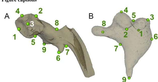
Figure 1. Position of landmarks used in the Generalized partial Procrustes analysis (pGPA)
679
and principal component analysis (PCA) on the malleus (A) and the incus (B) of late Eocene
680
and early Oligocene Cainotheriidae from Dams localities (DAM1 and DAM3, Phosphorites of
681
Quercy, SW France).
682 683
22 684
685
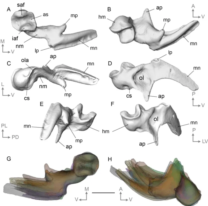
Figure 2. Left cainotheriid malleus from Dams DAM1 (DAM1 330) in (A) posterior, (B)
686
medial, (C) anterior, (D) lateral, (E) medioventral, and (F) dorsolateral views. G-H,
687
Illustration of morphological variability as observed in (G) posterior and (H) dorsomedial
688
views of a right malleus). Abbreviations: ap, anterior process; as, articular surface; cs,
689
capitular spine; hm, head of malleus; iaf, inferior articular facet; lp, lateral process; mn,
690
manubrium; mp, muscular process; nm, neck of malleus; ol, osseous lamina; ola, outer
691
lamella; saf, superior articular facet. Orientations: A, anterior; D, dorsal; L, lateral; M, medial;
692
P, posterior; V, ventral. Scale bar = 1 mm.
693 694
23 695
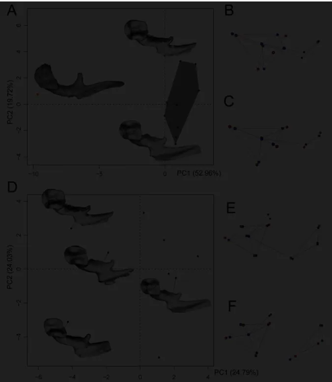
Figure 3. PCAs on Procrustes coordinates illustrating morphological variability of the
696
cainotheriid malleus from DAM1 (late Eocene, Phosphorites of Quercy, SW France). (A)
697
Inter- and intraspecific variation of the shape of the malleus based on 7 landmarks; projection
698
of specimens from DAM1 (in black) and Pech Desse (in orange) on the first factorial plane;
699
(B-C; E-F) Patterns of variation along PCs with maximal values in red and minimal in blue;
700
(B) shape variation on PC1 of (A); (C) shape variation on PC2 of (A); (D) Intraspecific
701
variation of the shape of the malleus based on 9 landmarks; projection of specimens from
702
DAM1 on the first two PCs; (E) shape variation on PC1 of (D); (F) shape variation on PC2 of
703
(D). See text for more details.
24 705
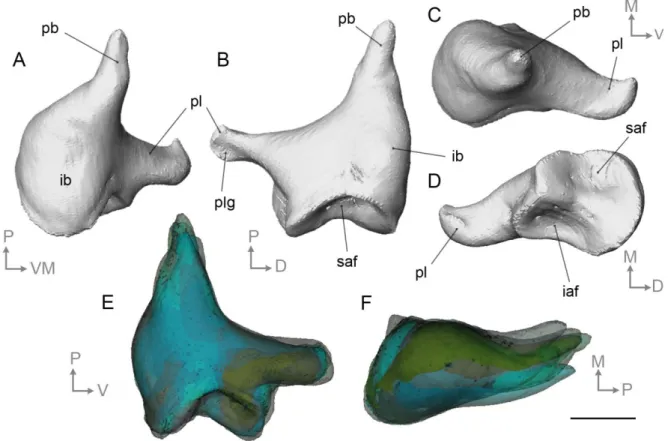
Figure 4. Left cainotheriid incus from Dams DAM1 (DAM 1 307) in (A) dorsomedial, (B)
706
lateral, (C) posterior, (D) anterior, views. E-F, Illustration of morphological variability range
707
as observed in (E) lateral and (F) dorsal views. Abbreviations: iaf, inferior articular facet; ib,
708
incus bulge; pb, processus brevis; pl, processus longum; plg, processus longum groove; saf,
709
superior articular facet. Orientations: A, anterior; D, dorsal; L, lateral; M, medial; P, posterior;
710
V, ventral. Scale bar = 0.5 mm.
711 712
25 713
Figure 5. PCA on Procrustes coordinates illustrating morphological variability of the
714
cainotheriid incus from the localities of DAM1 (grey surface; late Eocene), DAM3 (pink
715
surface; early Oligocene) and Pech Desse (orange square; late Oligocene). (A) Projection of
716
individuals on the first factorial plane; (B-C) Patterns of variation along PC1 (B) and PC2 (C)
717
with maximal values in red and minimal in blue.
26 719
Figure 6. Right stapes of Paleogene Cainotheriidae from the Phosphorites of Quercy, SW
720
France. (A-I) Paroxacron valdense from DAM1, late Eocene (A-E, DAM 1 316,) (F-I. DAM
721
1 317) – A, ventral view; B, dorsal view; C, lateral view; D, posterior view; E, antero dorsal
722
view. DAM 1 317 (F-I) – F, ventral view; G, posterior view; H, dorsal view; I, lateral view.
723
(J-M) Cainotheriidae indet. from DAM3, early Oligocene (DAM 3 13) J, ventral view; K,
724
posterior view; L, dorsal view; M, lateral view; Abbreviations: ac, anterior crus; fi, foramen
725
intercrurale; pc, posterior crus; pm, processus muscularis; sfr, stapedial footplate rim; sh,
726
stapes head; um, umbo. Orientations: A, anterior; D, dorsal; L, lateral; M, medial; P,
727
posterior; V, ventral. Scale bars = 0.5 mm.
27 729
Figure 7. 3D reconstruction of the middle ear of Caenomeryx cf. procommunis from Pech
730
Desse, late Oligocene, Phosphorites of Quercy, SW France (UM PDS 3353). A-B: Left
731
middle ear with in-situ ossicles; C-D: virtually reassembled composite left ossicle chain in
732
ventral (C) and dorsal (D) views; E-H: incus; I-L, malleus; M-P, stapes; Q, Petro-tympanic
733
complex of UM PDS 3353 showing orthogonal slices (S1 and S2) at putative location of the
734
processus internus prearticularis (pipa). Abbreviations: eam, external auditory meatus; fc,
735
fenestra cochleae; fv, fenestra vestibuli; hm, head of malleus; ib, incus bulge; lp, lateral
736
process of malleus; mn, manubrium; mp, muscular process of malleus; pb, processus brevis of
737
the incus; pl, processus longum of the incus; pop, paroccipital process; pt, petrosal bone; sf,
28
stapedial footplate; sh, stapes head; tb, tympanic bulla. Orientations: A, anterior; D, dorsal; L,
739
lateral; M, medial; P, posterior; V, ventral. Scale bars, A-B = 5 mm; C-P = 1 mm.
740 741 742