Visual exploration behaviour during clock reading
in Alzheimer's disease
U. P. Mosimann,
1,2* J. Felblinger,
1,²P. Ballinari,
2C. W. Hess
1and R. M. MuÈri
1,21Perception and Eye Movement Laboratory, Departments of Neurology and Clinical Research, University of Bern and 2Memory Clinics, Departments of Neurology and Psychiatry, University of Bern, Switzerland
*Present address: Institute for Ageing and Health, Wolfson Research Centre, Newcastle General Hospital, Newcastle, UK
²Present address: Department of Radiology, University Hospital of Nancy, Nancy, France
Correspondence to: PD Dr R. M. MuÈri, Perception and Eye Movement Laboratory, Departments of Neurology and Clinical Research, University of Bern, Inselspital, 3010 Bern, Switzerland
E-mail: rene.mueri@insel.ch
Summary
Eye movement behaviour during visual exploration of 24 patients with probable Alzheimer's disease and 24 age-matched controls was compared in a clock reading task. Controls were found to focus exploration on dis-tinct areas at the end of each clock hand. The sum of these two areas of highest ®xation density was de®ned as the informative region of interest (ROI). In Alzheimer's disease patients, visual exploration was less focused, with fewer ®xations inside the ROI, and the time until the ®rst ®xation was inside the ROI was
sig-ni®cantly delayed. Changes of ®xation distribution cor-related signi®cantly with the ability to read the clock correctly, but did not correlate with dementia severity. In Alzheimer's disease patients, ®xations were longer and saccade amplitudes were smaller. The altered visual exploration in Alzheimer's disease might be related to parietal dysfunction or to an imbalance between a degraded occipito-parietal and relatively preserved occi-pito-temporal visual network.
Keywords: Alzheimer's disease; clock reading; visual exploration
Abbreviations: CFP = central ®xation point; ChE-I = cholinesterase inhibitors; DSM-IV= Diagnostic and Statistical Manual of Mental Disorders (4th edition); MMSE = Mini-Mental State Examination; NINCDS-ADRDA = National Institute of Neurological and Communicative Disorders and Stroke and Alzheimer's Disease and Related Disorders Association; PPC = posterior parietal cortex; ROI = region of interest
Received May 26, 2003. Revised August 8, 2003. Accepted October 13, 2003. Advanced Access publication December 22, 2003
Introduction
Visual information is processed in multiple cortical areas. Temporal and parietal association areas include two highly interconnected visual pathways, which extend from the occipital to the frontal lobes (Haxby et al., 1991; Knierim and Van Essen, 1992; Wilson et al., 1993; Ungerleider and Haxby, 1994; Bullier et al., 1996). The parietal `where' pathway is important for spatial perception, internal image representation and sensomotor integration, and the more ventral temporal `what' pathway for object recognition (Wolpert et al., 1998; Mellet et al., 2002). Next to the regions of visual perception, the posterior-parietal cortex (PPC) closely links attentional and eye-movement networks (Leichnetz and Goldberg, 1988; Kowler et al., 1995; Corbetta et al., 1998; Perry and Zeki, 2000) and is activated during the
shifting of visuospatial attention (Corbetta et al., 2000) and the triggering of visually guided saccades (Pierrot-Deseilligny et al., 2002).
In Alzheimer's disease, progressive neuropathological changes (i.e. death of neurons, neuro®brillary tangles and amyloid plaques) affect certain laminae and cell types within the neocortex, and this may lead to cortico-cortical discon-nections (Braak and Braak, 1997; Newell et al., 1999; Grady et al., 2001). Pathology preferentially involves temporo-parietal association areas, whereas primary motor, somato-sensory and visual cortices are typically spared until the very late stages of the disease (Morrison et al., 1986; Lewis et al., 1987). This makes Alzheimer's disease patients prone to visual, attentional and eye movement disturbances. Visual Brain Vol. 127 No. 2 ã Guarantors of Brain 2003; all rights reserved
disturbances include impairments in spatial and/or object vision (Mendez et al., 1990; Cronin-Golomb et al., 1991; Hof and Bouras, 1991; Fujimori et al., 1997, 2000; Tetewsky and Duffy, 1999; Rizzo et al., 2000a). Common visuospatial attentional de®cits (Perry and Hodges, 1999; Rizzo et al., 2000b) manifest with impaired disengagement of attention (Parasuraman et al., 1992), impaired target selection (Parasuraman et al., 1995) or impaired shifting between global and focal attention (Filoteo et al., 1992; Slavin et al., 2002). Most studies that have assessed visually guided saccades in Alzheimer's disease patients have reported prolonged saccade latencies and inaccurate saccades (Pirozzolo and Hansch, 1981; Fletcher et al., 1986; Hotson and Steinke, 1988; Bylsma et al., 1995; Moser et al., 1995; Schewe et al., 1999; Abel et al., 2002).
Visual exploration, i.e. the sequence of ®xations and saccades, is crucial for perception and is a very effective and selective way to sample information (Noton and Stark, 1971; Rayner and Pollatsek, 1992; Land and Furneaux, 1997; Henderson and Hollingworth, 1999; Gilchrist and Harvey, 2000; Hodgson et al., 2000). Visual information is processed during ®xation, and to change ®xation, saccades direct the fovea towards a particular element of interest. Fixation behaviour is the end result of a complex interaction of features of the explored picture (`bottom up' processing) and the instruc-tion or quesinstruc-tion to be solved by the explorer (`top down' processing) (Yarbus, 1967; Rayner and Pollatsek, 1992; Henderson and Hollingworth, 1999). The analysis of ®xation distribution during exploration provides an indir-ect, non-verbal neurophysiological measure of this com-plex interaction. In Alzheimer's disease, visual exploration has been employed to measure spatial attention (Scinto
et al., 1994), and to characterize exploration during visual search and during reading of text or emotional facial expressions (Daffner et al., 1992, 1999; Moser et al., 1995; Lueck et al., 2000; Ogrocki et al., 2000; RoÈsler et al., 2000). Most of these studies reported longer ®xation duration and less systematic exploration during visual search.
The present study compares the visual exploration of Alzheimer's disease patients with that of controls during clock reading, a daily relevant, over-learned task, which is often impaired during the progressive course of the disease. We assumed that healthy controls explore clocks non-randomly, and hence wanted to ®nd out which areas of the clock face are normally targeted as most informative to read the time. Measuring saccade and ®xation parameters enabled a quantitative comparison of Alzheimer's disease patients and controls. To exclude impaired saccade motoricity as a possible cause of exploration changes in Alzheimer's disease patients, saccade triggering and accuracy were tested separ-ately in a gap and overlap task.
Methods
Subjects
A randomized sample of 24 consecutive outpatients with the diagnosis of probable Alzheimer's disease was recruited in the Memory Clinics at the University Hospital in Bern. Diagnosis was based on the criteria for dementia outlined in the Diagnostic and Statistical Manual of Mental Disorders, 4th edition (DSM-IV) (American Psychiatric Association, 1994) and by the criteria for probable Alzheimer's disease developed by the National Institute of Neurological and Communicative Disorders and Stroke and Alzheimer's Disease and Related Disorders Association (NINCDS-ADRDA) (McKhann et al., 1984). In accordance with these criteria patients were excluded if: (i) they suffered any medical conditions that could account for, or interfere with, their cognitive decline; (ii) had evidence of vascular lesions in computed tomography or MRI; (iii) had a Hachinski Ischaemic Score (Hachinski et al., 1975) > 4; or (iv) had evidence for an Axis I diagnosis (e.g. major depression or drug abuse) as de®ned by DSM-IV. To be eligible for the study, patients had to have at least one caregiver providing regular care and support. Patients taking cholinesterase inhibitors (ChE-I) were only included if they were not in the dose escalation phase and if the dose has remained unchanged for at least 6 weeks prior to inclusion. None of the subjects was taking hypnotics, sedative drugs or major tranquillizers. The control group consisted of elderly volunteers recruited from relatives and friends of the patients. By history, they had no known neurological and psychiatric disease, and no evidence of cognitive decline or impairment in activities of daily living. Controls had to score at least 28 out of 30 points in the Mini-Mental State Examination (MMSE) (Folstein et al., 1975), 4 points in the clock drawing (Shulman, 2000) and at least seven correct answers in the clock reading task (for description of these tasks, see below). The ethics committee of the University of Bern approved the study. All patients and their caregivers, and all control subjects gave written informed consent prior to inclusion into the study.
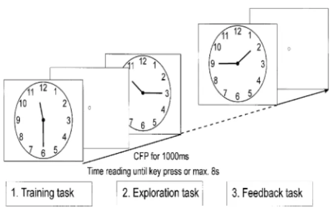
Fig. 1 The clock reading task included: (i) a training task to ensure that the subjects understood the instruction and were able to use the mouse button; (ii) the exploration task where eye movements were recorded; and (iii) a feedback task to assess the number of correctly read clocks. Each time was presented until the subject pressed the mouse button or until 8 s had elapsed. Between the times a grey screen with a CFP was presented for 1 s.
Testing procedure
Neuro-ophthalmological assessment
Clinical neuro-ophthalmological screening included a detailed history of current or past visual disturbances, the assessment of visual acuity, saccadic and pursuit eye movements, and visual ®eld examination by digital confronting test.
Additional neuropsychological testing
All subjects were assessed with the MMSE (Folstein et al., 1975) and the clock drawing test. Scoring was according to Shulman (2000), i.e. 5 points for a perfect clock and 0 for the inability to make any representation of a clock.
Clock reading experiment
The clock reading experiment consisted of three consecutive parts (Fig. 1). In the training task, eight times (i.e. 1:40, 2:30, 3:45, 5:15, 7:15, 8:50, 10:10, 11:40) were presented, and the instruction was to read and state the time, then to press the mouse button to see the next clock. All subjects did this training task before the main exploration experiment to ensure that they understood the instruction and were able to use the mouse button correctly.
Eye movements were recorded during the exploration task, while subjects were required to read eight different times (11:30, 1:45, 7:20, 10:15, 4:40, 8:55, 2:05, 3:40). Talking was not allowed, in order to avoid concomitant head movements. Therefore, the instruction was to read the time without saying it, and then to press the mouse button to see the next clock.
The feedback task helped to assess how many times shown during the exploration task were read correctly. The instruction was to read the time aloud and then to press the mouse button, to see the next clock. The times were the same as those for the exploration task (11:30, 1:45, 7:20, 10:15, 4:40, 8:55, 2:05, 3:40) and were used to de®ne the percentage of correctly read times. In all three tasks, each time was presented until the subject pressed the mouse button or until 8 s elapsed. The clock face was the same during the whole experiment and was presented in a visual angle of 18° (Fig. 1). Each time was followed by a 1 s grey screen presentation with a central ®xation point (CFP).
Analysis of visual exploration during clock reading
During the exploration task, ®xation duration, saccade length and the exploration time were assessed. Exploration time was the time
interval between the start of a clock presentation and the mouse button response, or the elapsing of 8 s. For each time, we used ®xation density plots to present areas with high ®xation density, since such areas have been considered to be informative (Loftus and Mackworth, 1978). Density plots of the control group were used to de®ne the region of interest (ROI) a posteriori. For each time, the ROI included the two areas of highest ®xation density at the end of the clock hands, containing at least 50% of all ®xations. The size corresponded to 16% of the total clock face size. The percentage of ®xations inside the ROI and the time elapsed before the ®rst ®xation inside the ROI were calculated.
Visually guided saccades: gap and overlap task
Fifty-six saccades were tested in the gap and overlap task (i.e. four blocks of 14 saccades). In the gap task, the CFP disappeared 200 ms before the target appeared (i.e. temporal gap) (Saslow, 1967). In the overlap task, however, the CFP remained visible during target presentation. The timing of CFP presentation (minimum 2000 ms, maximum 3000 ms), and the direction (left, right) and amplitude (minimum 3.7°, maximum 9.1°) of the lateral targets were kept unpredictable. The target was presented for 1000 ms. Subjects were instructed to look as precisely and as fast as possible at the targets. The latency of the ®rst saccade and the gain (i.e. saccade amplitude/ target amplitude) of the ®rst saccade and ®nal eye position were calculated.
Recording of eye movements
Eye movements were recorded with a commercially available, video-based infrared system (EyelinkÔ; SensoMotorik Instruments, Berlin, Germany). This system allows recording eye movements at a sampling rate of 250 Hz with a spatial resolution of <0.1°. To avoid head movements, subjects were asked to position their chin on a rest. They were seated 70 cm in front of the 19-inch (36 3 27 cm) colour screen. The refreshing rate of the screen was 120 Hz. The visual ®eld was 27° in the horizontal and 21° in the vertical plane. Repeated calibration procedures were used before each experimental block.
Statistics
All data were tested for normal distribution (Kolmogorov±Smirnov test). Distribution and dispersion measures for parametric data were calculated as mean and SD, and for non-parametric data as median and range. Distribution measures were calculated per subject, and two-group comparison was made either with parametric (t-tests for
Table 1 Description of study groups
Alzheimer's disease patients Controls P Demographic data n 24 24 ns Male:female ratio 11:13 15:9 ns Age (years)* 74.3 (6.3) 72.9 (6.9) ns Years of education* 13.1 (3.2) 13.5 (2.7) ns Neuropsychological data
MMSE (Folstein et al., 1975)* 20.1 (5.4) 29.1 (0.8) < 0.001 Clock drawing (Shulman, 2000)* 2 (1.9) 4.6 (0.5) < 0.001 Clock reading [% correct (range)] 56 (0±100) 97 (88±100) < 0.001 *Mean (SD); ns = non-signi®cant.
dependent or independent samples) or non-parametric tests (Mann± Whitney and Wilcoxon rank tests). A P-value of < 0.05 was considered statistically signi®cant, and all reported P-values were two-tailed. Bivariate Spearman rank correlations were used to correlate exploration data and neuropsychological data.
Results
Demographic and neuropsychological data of Alzheimer's disease patients and controls are summarized in Table 1. No
group differences were found for gender, age or years of education. As expected, the groups were signi®cantly differ-ent in the MMSE and clock drawing and reading tasks.
Clinical neuro-opthalmological examination did not reveal any major abnormalities in patients or controls, and mean visual acuity did not differ between the groups (Alzheimer's disease: 0.6 6 0.1); controls: 0.5 6 0.2; t-test: not signi®-cant).
Visual exploration during clock reading
Exploration data are summarized in Table 2.
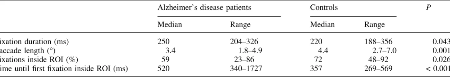
Median ®xation duration was longer (Mann±Whitney test: P = 0.043) and saccade length was shorter (Mann±Whitney test: P = 0.001) in the Alzheimer's disease group than in controls. In the control group, ®xation density plots revealed two areas of highest ®xation density at the end of the clock hands. Figure 2 shows a representative example of ®xation density plots for the control and Alzheimer's disease group.
In Alzheimer's disease patients, the time until the ®rst ®xation was inside the ROI was longer (Mann±Whitney test: P < 0.001) and the percentage of ®xations inside the ROI was lower (Mann±Whitney test: P = 0.026) compared with controls. Furthermore, the percentage of correctly read clocks correlated negatively with the time until the ®rst ®xation was inside the ROI (Spearman rank correlation: r = ±0.56, P = 0.005), and correlated positively with the percentage of ®xations inside the ROI (Spearman rank correlation: r = 0.45, P = 0.028). Such correlations were not found for the MMSE and other exploration data. Exploration time was longer in the Alzheimer's disease group (median 5.8 s; range: 2.4±7.9 s) compared with the control group (median 2.0 s; range: 0.8± 6.8 s) (Mann±Whitney test: P < 0.001).
Visually guided saccades: gap and overlap task
In Alzheimer's disease patients latency and gain of the ®rst saccade and ®nal eye position in the gap and overlap task was not signi®cantly different from controls. These results are summarized in Table 3. As expected, the gap latency was shorter than the overlap saccade latency in Alzheimer's disease patients (Wilcoxon rank test: P < 0.001) and controls (Wilcoxon rank test: P < 0.001).
Thirteen of the 24 Alzheimer's disease patients were treated with ChE-I (galantamine 1, donepezil 7, rivastigmine Table 2 Analysis of visual exploration during clock reading
Alzheimer's disease patients Controls P Median Range Median Range
Fixation duration (ms) 250 204±326 220 188±356 0.043 Saccade length (°) 3.4 1.8±4.9 4.4 2.7±7.0 0.001 Fixations inside ROI (%) 59 23±86 72 48±92 0.026 Time until ®rst ®xation inside ROI (ms) 520 340±1727 357 269±569 < 0.001
Fig. 2 An example of ®xation density plots of (A) the control group and (B) the Alzheimer's disease group. The highest ®xation density in the control group was found at the end of the clock hands.
5) for an average duration of 20.1 months (SD 13 months). No differences were found between patients with, and those without, ChE-I for all eye movement variables (i.e. saccade latency, accuracy, ®xation duration and saccade length) and neuropsychological test scores (i.e. MMSE, clock reading and drawing).
Discussion
The present study assessed visual exploration in healthy controls and Alzheimer's disease patients during clock reading and revealed the following. (i) Visual exploration of controls was non-random and areas of high ®xation density at the end of the clock hands de®ned ROI. (ii) In Alzheimer's disease patients the exploration was less focused on the ROI. in controls, the percentage of ®xation inside the ROI was lower and the time until the ®rst ®xation was inside the ROI was longer. These changes did not correlate with the global cognitive impairment, but correlated signi®cantly with the impaired ability to read the clock. (iii) During exploration, Alzheimer's disease patients showed shorter saccade ampli-tudes and longer ®xations than controls, but the two groups were not different in latency and accuracy of visually guided saccades.
To our knowledge, this is the ®rst study showing how healthy controls explore clock faces during time reading. Fixations are focused on areas at the end of each clock hand, and such areas are explored during the very early course of exploration. This observation suggests a speci®c exploration strategy for ef®cient clock reading in healthy subjects, and is in agreement with previous studies which suggested that visual exploration may be task speci®c (Buswell, 1935; Mackworth and Morandi, 1967; Yarbus, 1967; Antes, 1974; Loftus and Mackworth, 1978). The importance of a speci®c exploration strategy for successful task performance has been shown in other over-learned, `automatic' activities of daily living (Land and Furneaux, 1997; Land et al., 1999). Recently, Hodgson et al. (2000) revealed the importance of a selective strategy for successful performance in a neuropsychological task. They found that subjects who
made errors spend more time looking at irrelevant items. This also seems to be the case for clock reading: patients with impaired clock reading presented a reduced strategy for gazing at relevant items of the clock face compared with controls. Moreover, the two studies using the Tower of London task (Hodgson et al., 2000, 2002) suggested that exploration of healthy controls and patients with Parkinson's disease was more in¯uenced by problem-solving strategies than the salience of the presented objects.
In healthy subjects, parieto-frontal networks are activated when imagining a visual image (Spivey and Geng, 2001; Mellet et al., 2002), and in particular during imagination of clocks (Trojano et al., 2000). Neuropathological (Morrison et al., 1986; Lewis et al., 1987) and neuroimaging studies reported pronounced parietal dysfunction in Alzheimer's disease patients (Meltzer et al., 1996; Bartenstein et al., 1997; Jagust et al., 1997; Pietrini et al., 2000), which makes Alzheimer's disease patients prone to impaired internal representation and reduced `top down' control of the exploration strategy (Fujimori et al., 1997, 2000; Tetewsky and Duffy, 1999; Rizzo et al., 2000a). In agreement with previous studies (Daffner et al., 1992; Ogrocki et al., 2000), the changes in explorative strategy found in Alzheimer's disease patients did not correlate with global cognitive impairment, but were related to the ability or inability to read the clock: in our patients a signi®cant correlation was found between clock reading capacity and the time until the ®rst saccade was inside the ROI, and the percentage of ®xation inside the ROI.
The occipito-temporal network is important for central vision and the generation of small saccades, and the occipito-parietal network for spatial global vision and the generation of long saccades (Ungerleider and Haxby, 1994; Bullier et al., 1996). An imbalance between the two networks with a more pronounced occipito-parietal dysfunction, and a relatively spared occipito-temporal function, may lead to predomin-antly shorter saccade amplitudes and longer ®xations during exploration. This hypothesis is supported by a recent fMRI study, which found a reduced parietal activation and increased temporal activation during visuospatial processing Table 3 Saccade latency and accuracy of visually guided saccades
Alzheimer's disease patients Controls P Median Range Median Range
Gap task
Latency (ms) 165 136±318 159 92±237 0.23 (ns) Gain: ®rst saccade 0.86 0.60±1.01 0.9 0.73±0.99 0.15 (ns) Gain: ®nal eye position 0.97 0.87±1.11 0.99 0.92±1.09 0.85 (ns) Overlap task
Latency (ms) 293 189±518 258 153±367 0.14 (ns) Gain: ®rst saccade 0.90 0.56±1.05 0.94 0.72±1.01 0.33 (ns) Gain: ®nal eye position 0.99 0.90±1.07 0.99 0.94±1.04 0.67 (ns) ns = not signi®cant; gain = saccade amplitude/target amplitude.
in Alzheimer's disease patients (Prvulovic et al., 2002). Longer ®xations are in agreement with impaired parietal function due to impaired disengagement of ®xation, as reported in previous studies (Daffner et al., 1992; Moser et al., 1995; Lueck et al., 2000; RoÈsler et al., 2000). Smaller saccades may also be the consequence of a reduced visual area from which information can be acquired within one ®xation, i.e. reduced functional ®eld of view (Ball et al., 1988; Rizzo et al., 2000a, b), or impaired shifting between focal and global vision (Filoteo et al., 1992; Parasuraman et al., 2000; Slavin et al., 2002).
Eye movements of Alzheimer's disease patients taking ChE-I were not different from Alzheimer's disease patients not on such medication, and therefore exploration changes revealed in Alzheimer's disease patients are unlikely to be related to the presence or absence of medication. Impaired motor output of the saccadic eye movement system is another unlikely explanation for longer ®xation and smaller saccade amplitudes during exploration, since saccade latency and gain of visually guided saccades were normal in our Alzheimer's disease patients. This dissociation of normal saccade latency in a visually guided saccade task and prolonged ®xation during exploration can be due to the fact, that visually guided saccades are mainly driven `bottom up' by the visual stimulus, whereas the exploration of a clock face needs more `top down' control for target selection and ®xation disengagement. This notion is well in line with the ®nding that response selection and shifting between spatial locations are particularly vulnerable in Alzheimer's disease, whereas cue-driven shifting of attention is only minimally affected (Rizzo et al., 2000b).
In conclusion, the Alzheimer's disease group showed a distinct pattern of exploration changes during clock reading, which can be related to a parietal dysfunction in terms of an imbalance between the dorsal and ventral visual pathways, with degraded occipito-parietal and relatively preserved occipito-temporal visual pathway. The changes were not related to global cognitive impairments, but rather to impaired clock reading. Our results con®rm the importance of eye movements in daily relevant tasks, and when previous results are taken into consideration (Land and Furneaux, 1997; Land et al., 1999; Hodgson et al., 2002) there is increasing evidence that eye movement behaviour is expli-citly related to speci®c action in daily life, even for over-learned and `automatic' tasks. Furthermore, there is a close relationship between successful performance and eye move-ment behaviour. We may speculate that the combination of impaired spatial orientation (i.e. changes of ®xation distribu-tion), and loss of exploration strategy, shorter saccade amplitudes and longer ®xation duration, may put Alzheimer's disease patients at a disadvantage for many daily tasks associated with visual exploration demand. The present study also showed that the quantitative assessment of visual exploration behaviour is well tolerated by and feasible for Alzheimer's disease patients.
Acknowledgements
We wish to thank the patients, caregivers and control subjects who made this work possible. We thank S. Guyer and Dr K. Aebi for their assistance in recruiting patients and controls. The study was supported by a grant from the Swiss Foundation for Clinical Research in Neurodegenerative Disorders (`Fondazione per lo studio delle malattie neurode-generative delle persone adulte e dell' anziano', Lugano, Switzerland).
References
Abel LA, Unverzagt F, Yee RD. Effects of stimulus predictability and interstimulus gap on saccades in Alzheimer's disease. Dement Geriatr Cogn Disord 2002; 13: 235±43.
American Psychiatric Association. Diagnostic and statistical manual of mental disorders: DSM-IV. 4th ed. Washington (DC): American Psychiatric Association; 1994.
Antes JR. The time course of picture viewing. J Exp Psychol 1974; 103: 62± 70.
Ball KK, Beard BL, Roenker DL, Miller RL, Griggs DS. Age and visual search: expanding the useful ®eld of view. J Opt Soc Am A 1988; 5: 2210±9.
Bartenstein P, Minoshima S, Hirsch C, Buch K, Willoch F, Mosch D, et al. Quantitative assessment of cerebral blood ¯ow in patients with Alzheimer's disease by SPECT. J Nucl Med 1997; 38: 1095±101. Braak H, Braak E. Staging of Alzheimer-related cortical destruction. Int
Psychogeriatr 1997; 9 Suppl 1: 257±61.
Bullier J, Schall JD, Morel A. Functional streams in occipito-frontal connections in the monkey. Behav Brain Res 1996; 76: 89±97. Buswell GT. How people look at pictures. A study of the psychology of
perception in art. Chicago: University of Chicago Press; 1935. Bylsma FW, Rasmusson DX, Rebok GW, Keyl PM, Tune L, Brandt J.
Changes in visual ®xation and saccadic eye movements in Alzheimer's disease. Int J Psychophysiol 1995; 19: 33±40.
Corbetta M, Akbudak E, Conturo TE, Snyder AZ, Ollinger JM, Drury HA, et al. A common network of functional areas for attention and eye movements. Neuron 1998; 21: 761±73.
Corbetta M, Kincade JM, Ollinger JM, McAvoy MP, Shulman GL. Voluntary orienting is dissociated from target detection in human posterior parietal cortex. Nat Neurosci 2000; 3: 292±7.
Cronin-Golomb A, Corkin S, Rizzo JF, Cohen J, Growdon JH, Banks KS. Visual dysfunction in Alzheimer's disease: relation to normal aging. Ann Neurol 1991; 29: 41±52.
Daffner KR, Scinto LF, Weintraub S, Guinessey JE, Mesulam MM. Diminished curiosity in patients with probable Alzheimer's disease as measured by exploratory eye movements. Neurology 1992; 42: 320±8. Daffner KR, Mesulam MM, Cohen LG, Scinto LF. Mechanisms underlying
diminished novelty-seeking behavior in patients with probable Alzheimer's disease. Neuropsychiatry Neuropsychol Behav Neurol 1999; 12: 58±66.
Filoteo JV, Delis DC, Massman PJ, Demadura T, Butters N, Salmon DP. Directed and divided attention in Alzheimer's disease: impairment in shifting of attention to global and local stimuli. J Clin Exp Neuropsychol 1992; 14: 871±83.
Fletcher WA, Sharpe JA. Saccadic eye movement dysfunction in Alzheimer's disease. Ann Neurol 1986; 20: 464±71.
Folstein MF, Folstein SE, McHugh PR. `Mini-mental state'. A practical method for grading the cognitive state of patients for the clinician. J Psychiatr Res 1975; 12: 189±98.
Fujimori M, Imamura T, Yamashita H, Hirono N, Mori E. The disturbances of object vision and spatial vision in Alzheimer's disease. Dement Geriatr Cogn Disord 1997; 8: 228±31.
Fujimori M, Imamura T, Hirono N, Ishii K, Sasaki M, Mori E. Disturbances of spatial vision and object vision correlate differently with regional
cerebral glucose metabolism in Alzheimer's disease. Neuropsychologia 2000; 38: 1356±61.
Gilchrist ID, Harvey M. Re®xation frequency and memory mechanisms in visual search. Curr Biol 2000; 10: 1209±12.
Grady CL, Furey ML, Pietrini P, Horwitz B, Rapoport SI. Altered brain functional connectivity and impaired short-term memory in Alzheimer's disease. Brain 2001; 124: 739±56.
Hachinski VC, Iliff LD, Zilhka E, Du Boulay GH, McAllister VL, Marshall J, et al. Cerebral blood ¯ow in dementia. Arch Neurol 1975; 32: 632±7. Haxby JV, Grady CL, Horwitz B, Ungerleider LG, Mishkin M, Carson RE, et al. Dissociation of object and spatial visual processing pathways in human extrastriate cortex. Proc Natl Acad Sci USA 1991; 88: 1621±5. Henderson JM, Hollingworth A. High-level scene perception. [Review].
Annu Rev Psychol 1999; 50: 243±71.
Hodgson TL, Bajwa A, Owen AM, Kennard C. The strategic control of gaze direction in the Tower-of-London task. J Cogn Neurosci 2000; 12: 894± 907.
Hodgson TL, Tiesman B, Owen AM, Kennard C. Abnormal gaze strategies during problem solving in Parkinson's disease. Neuropsychologia 2002; 40: 411±22.
Hof PR, Bouras C. Object recognition de®cit in Alzheimer's disease: possible disconnection of the occipito-temporal component of the visual system. Neurosci Lett 1991; 122: 53±6.
Hotson JR, Steinke GW. Vertical and horizontal saccades in aging and dementia ± failure to inhibit anticipatory saccades. Neuro-ophthalmology 1988; 8: 267±73.
Jagust WJ, Eberling JL, Reed BR, Mathis CA, Budinger TF. Clinical studies of cerebral blood ¯ow in Alzheimer's disease. Ann NY Acad Sci 1997; 826: 254±62.
Knierim JJ, Van Essen DC. Visual cortex: cartography, connectivity, and concurrent processing. Curr Opin Neurobiol 1992; 2: 150±5.
Kowler E, Anderson E, Dosher B, Blaser E. The role of attention in the programming of saccades. Vision Res 1995; 35: 1897±916.
Land MF, Furneaux S. The knowledge base of the oculomotor system. [Review]. Philos Trans R Soc Lond B Biol Sci 1997; 352: 1231±9. Land MF, Mennie N, Rusted J. The roles of vision and eye movements in the
control of activities of daily living. Perception 1999; 28: 1311±28. Leichnetz GR, Goldberg ME. Higher centers concerned with eye movement
and visual attention: cerebral cortex and thalamus. Rev Oculomot Res 1988; 2: 365±429.
Lewis DA, Campbell MJ, Terry RD, Morrison JH. Laminar and regional distributions of neuro®brillary tangles and neuritic plaques in Alzheimer's disease: a quantitative study of visual and auditory cortices. J Neurosci 1987; 7: 1799±808.
Loftus GR, Mackworth NH. Cognitive determinants of ®xation location during picture viewing. J Exp Psychol Hum Percept Perform 1978; 4: 565±72.
Lueck KL, Mendez MF, Perryman KM. Eye movement abnormalities during reading in patients with Alzheimer's disease. Neuropsychiatry Neuropsychol Behav Neurol 2000; 13: 77±82.
Mackworth NH, Morandi AJ. The gaze selects informative details within pictures. Percept Psychophys 1967; 2: 547±52.
McKhann G, Drachman D, Folstein M, Katzman R, Price D, Stadlan EM. Clinical diagnosis of Alzheimer's disease: report of the NINCDS-ADRDA Work Group under the auspices of Department of Health and Human Services Task Force on Alzheimer's Disease. Neurology 1984; 34: 939±44.
Mellet E, Bricogne S, Crivello F, Mazoyer B, Denis M, Tzourio-Mazoyer N. Neural basis of mental scanning of a topographic representation built from a text. Cereb Cortex 2002; 12: 1322±30.
Meltzer CC, Zubieta JK, Brandt J, Tune LE, Mayberg HS, Frost JJ. Regional hypometabolism in Alzheimer's disease as measured by positron emission tomography after correction for effects of partial volume averaging. Neurology 1996; 47: 454±61.
Mendez MF, Mendez MA, Martin R, Smyth KA, Whitehouse PJ. Complex visual disturbances in Alzheimer's disease. Neurology 1990; 40: 439±43. Morrison JH, Rogers J, Scherr S, Lewis DA, Campbell MJ, Bloom FE, et al.
The laminar and regional distribution of neocortical somatostatin and neuritic plaques: implications for Alzheimer's disease as a global neocortical disconnection syndrome. In: Scheibel AB, Wechsler AF, editors. The biological substrates of Alzheimer's disease. Orlando: Academic Press; 1986. p. 115±31.
Moser A, KoÈmpf D, Olschinka J. Eye movement dysfunction in dementia of the Alzheimer type. Dementia 1995; 6: 264±8.
Newell KL, Hyman BT, Growdon JH, Hedley-Whyte ET. Application of the National Institute on Aging (NIA) ± Reagan Institute criteria for neuropathological diagnosis of Alzheimer disease. J Neuropathol Exp Neurolol 1999; 58: 1147±55.
Noton D, Stark L. Scanpaths in eye movements during pattern perception. Science 1971; 171: 308±11.
Ogrocki PK, Hills AC, Strauss ME. Visual exploration of facial emotion by healthy older adults and patients with Alzheimer's disease. Neuropsychiatry Neuropsychol Behav Neurol 2000; 13: 271±8. Parasuraman R, Greenwood PM, Haxby JV, Grady CL. Visuospatial
attention in dementia of the Alzheimer type. Brain 1992; 115: 711±33. Parasuraman R, Greenwood PM, Alexander GE. Selective impairment of
spatial attention during visual search in Alzheimer's disease. Neuroreport 1995; 6: 1861±4.
Parasuraman R, Greenwood PM, Alexander GE. Alzheimer disease constricts the dynamic range of spatial attention in visual search. Neuropsychologia 2000; 38: 1126±35.
Perry RJ, Hodges JR. Attention and executive de®cits in Alzheimer's disease. A critical review. [Review]. Brain 1999; 122: 383±404. Perry RJ, Zeki S. The neurology of saccades and covert shifts in spatial
attention: an event-related fMRI study. Brain 2000; 123: 2273±88. Pierrot-Deseilligny CH, Ploner CJ, Muri RM, Gaymard B, Rivaud-Pechoux
S. Effects of cortical lesions on saccadic eye movements in humans. Ann NY Acad Sci 2002; 956: 216±29.
Pietrini P, Alexander GE, Furey ML, Hampel H, Guazzelli M. The neurometabolic landscape of cognitive decline: in vivo studies with positron emission tomography in Alzheimer's disease. Int J Psychophysiol 2000; 37: 87±98.
Pirozzolo FJ, Hansch EC. Oculomotor reaction time in dementia re¯ects degree of cerebral dysfunction. Science 1981; 214: 349±51.
Prvulovic D, Hubl D, Sack AT, Melillo L, Maurer K, FroÈlich L, et al. Functional imaging of visuospatial processing in Alzheimer's disease. Neuroimage 2002; 17: 1403±14.
Rayner K, Pollatsek A. Eye movements and scene perception. Can J Psychol 1992; 46: 342±76.
Rizzo M, Anderson SW, Dawson J, Nawrot M. Vision and cognition in Alzheimer's disease. Neuropsychologia 2000a; 38: 1157±69.
Rizzo M, Anderson SW, Dawson J, Myers R, Ball K. Visual attention impairments in Alzheimer's disease. Neurology 2000b; 54: 1954±9. RoÈsler A, Mapstone ME, Hays AK, Mesulam MM, Rademaker A, Gitelman
DR, et al. Alterations of visual search strategy in Alzheimer's disease and aging. Neuropsychology 2000; 14: 398±408.
Saslow MG. Effects of components of displacement-step stimuli upon latency for saccadic eye movement. J Opt Soc Am 1967; 57: 1024±9, Schewe HJ, Uebelhack R, Vohs K. Abnormality in saccadic eye movement
in dementia. Eur Psychiatry 1999; 14: 52±3.
Scinto LF, Daffner KR, Castro L, Weintraub S, Vavrik M, Mesulam MM. Impairment of spatially directed attention in patients with probable Alzheimer's disease as measured by eye movements. Arch Neurol 1994; 51: 682±8.
Shulman KI. Clock-drawing: is it the ideal cognitive screening test? Int J Geriatr Psychiatry 2000; 15: 548±61.
Slavin MJ, Mattingley JB, Brandshaw JL, Storey E. Local-global processing in Alzheimer's disease: an examination of interference, inhibition and priming. Neuropsychologia 2002; 40: 1173±86.
Spivey MJ, Geng JJ. Oculomotor mechanisms activated by imagery and memory: eye movements to absent objects. Psychol Res 2001; 65: 235± 41.
Tetewsky SJ, Duffy CJ. Visual loss and getting lost in Alzheimer's disease. Neurology 1999; 52: 958±65.
Trojano L, Grossi D, Linden DE, Formisano E, Hacker H, Zanella FE, et al. Matching two imagined clocks: the functional anatomy of spatial analysis in the absence of visual stimulation. Cereb Cortex 2000; 10: 473±81. Ungerleider LG, Haxby JV. `What' and `where' in the human brain.
[Review]. Curr Opin Neurobiol 1994; 4: 157±65.
Wilson FA, Scalaidhe SP, Goldman-Rakic PS. Dissociation of object and spatial processing domains in primate prefrontal cortex. Science 1993; 260: 1955±8.
Wolpert DM, Goodbody SJ, Husain M. Maintaining internal representations: the role of the human superior parietal lobe. Nat Neurosci 1998; 1: 529± 33.
Yarbus AL. Eye movements during ®xation on stationary objects. In: Yarbus AL, editor. Eye movements and vision. New York: Plenum Press; 1967. p. 103±27.