REVIEW
Immune sensing of nucleic acids in inflammatory skin diseases
Olivier Demaria&Jeremy Di Domizio&Michel GillietReceived: 20 August 2014 / Accepted: 20 August 2014 / Published online: 16 September 2014 # Springer-Verlag Berlin Heidelberg 2014
Abstract Endosomal and cytosolic nucleic acid receptors are important immune sensors required for the detection of infect-ing or replicatinfect-ing viruses. The intracellular location of these receptors allows viral recognition and, at the same time, avoids unnecessary immune activation to self-nucleic acids that are continuously released by dying host cells. Recent evidence, however, indicates that endogenous factors such as anti-microbial peptides have the ability to break this protective mechanism. Here, we discuss these factors and illustrate how they drive inflammatory responses by promoting immune recognition of self-nucleic acids in skin wounds and inflam-matory skin diseases such as psoriasis and lupus.
Introduction
The skin is the largest organ of the human body. One of its main functions is the protection of internal organs from exter-nal insults. In addition to providing mechanical and chemical protection, the skin contains numerous resident immune cells that patrol the tissue under steady state conditions and recruit additional cells from the blood in the context of injury or infection. A first line of immune defense of the skin is pro-vided by cells of the innate immune system. These innate immune cells express germ-line-encoded pathogen recogni-tion receptors (PRRs) specialized in the detecrecogni-tion of patho-gens. Activation of PRRs triggers dendritic cell activation and maturation as well as cytokine production leading to inflam-matory responses in the skin that clear infections and restore
homeostasis. PRRs recognize pathogen-associated molecular patterns (PAMPs) which are of lipid, protein, or nucleic acid origin. Whereas lipid and protein PAMPs are highly specific for pathogens, the discrimination of pathogen-derived from host-derived (self) nucleic acids is less clear. Indeed, there is increasing evidence that, under certain circumstances, nucleic acid receptors can be activated by host-derived self-DNA and self-RNA. Such recognition appears to be involved in skin inflammation in response to wounding but may also be central in the initiation and maintenance of chronic inflammatory skin diseases such as psoriasis and lupus.
Nucleic acid receptors
During evolution, the immune system has developed an efficient machinery to detect infections. Nucleic acid re-ceptors that sense viral genomes (viral DNA and RNA, single- or double-stranded) represent a main component of this system. Nucleic acid receptors are commonly di-vided in two subgroups based on their endosomal or cytosolic distribution. In the endosomes, nucleic acid re-ceptors include Toll-like receptor (TLR) 3, TLR7, TLR8, and TLR9. Viral nucleic acids can access endosomal compartments through scavenger receptor-mediated endo-cytosis and phagoendo-cytosis or Fc receptor-mediated uptake of the infecting viruses. In addition, viral nucleic acids can be re-directed from the cytosol to endosomes by the process of autophagy, a mechanism that allows TLR rec-ognition of replicating viruses. In the cytosol, nucleic acid receptors include the retinoic acid-inducible gene I (RIG-I)-like receptors, the cytosolic DNA sensors and receptors involved in inflammasome formation. Cytosolic receptors typically detect viral nucleic acids that accumulate in the cytoplasm during viral replication.
This article is a contribution to the special issue on Mechanisms of Tissue Injury in Autoimmune Diseases - Guest Editor: Dan Eilat
O. Demaria
:
J. Di Domizio:
M. Gilliet (*)Department of Dermatology, Lausanne University Hospital CHUV, Lausanne, Switzerland
Endosomal Toll-like receptors
The expression pattern of endosomal TLRs is different among immune cells allowing a specialization of each cell type at recognizing various endocytosed microbes. In humans, TLR7 and TLR9 expression is restricted to plasmacytoid dendritic cells (pDCs) and B cells, TLR8 expression is found in mono-cytes and conventional dendritic cells (cDCs), and TLR3 is expressed by cDCs [1].
TLR9
TLR9 senses unmethylated CpG-containing DNA motifs which are abundant in microbial DNA [2]. TLR9 activation leads to the formation of a multiprotein signaling complex composed of the adaptor molecule myeloid differentiation primary response protein 88 (MyD88) adaptor molecule. Ac-tivation of the MyD88 complex leads to the acAc-tivation of IκB kinase (IKK) complex/nuclear factor-κB (NF-κB) and mitogen-activated kinases (MAPKs)/activator protein 1 (AP1) pathways to induce proinflammatory responses. TLR9 triggering also induces the nuclear translocation of interferon regulatory factor 7 (IRF7) that leads to the produc-tion of type I interferons (IFNs). To note, human pDCs, the main type I IFN producers of the human body, harbor a unique and original spatiotemporal regulation of the MyD88-IRF7 pathway that allows retention of TLR9 and TLR7 ligands in endosomes and subsequent robust type I IFN induction [3] [4] (Fig.1). Synthetic oligodeoxynucleotides (ODN) have been designed to specifically stimulate TLR9, including monomer-ic CpG ODN type B (CpG-B) and multimermonomer-ic CpG ODN type A (CpG-A).
TLR7 and TLR8
TLR7 and TLR8 sense single-stranded RNA [5,6]. Similarly to TLR9, they signal through MyD88 to activate the NF-κB and IRF7 pathways leading to the production of proinflam-matory cytokines and type I IFN, respectively. Synthetic li-gands for TLR7 and TLR8 have been designed, among which imiquimod is a selective agonist of TLR7.
TLR3
TLR3 recognizes viral double-stranded RNA [7], an interme-diate product of viral replication. TLR3 signals through the TIR-domain-containing adaptor protein inducing IFN-β (TRIF) that in turn activates the TANK-binding kinase 1 (TBK1)/interferon regulatory factor 3 (IRF3) pathway to pro-duce type I IFN and also activates IKK complex/NF-κB to induce the production of proinflammatory cytokines [8]. Polyinosine-polycytidylic acid (poly(I:C)), a synthetic analog
of double-stranded RNA (dsRNA), is used as a ligand for TLR3.
Cytosolic DNA sensors
Several cytosolic DNA sensors have been recently identified. These include IFI16, DAI, cyclic GMP-AMP synthase (cGAS), and RNA polymerase III, which all signal through stimulator of interferon gene (STING) and TBK1-IRF3. Others, such as Ku70, DHX9/DHX36, and AIM2, induce different signaling pathways. STING is a transmembrane protein of the endoplasmic reticulum, which upon activation triggers TBK1-dependent phosphorylation of IRF3 to initiate type I IFN production [9] [10] [11]. STING-deficient mice have been shown to present altered type I IFN production upon bacterial, viral, and mammalian DNA challenge [9]. In addition, STING can directly interact with bacterial cyclic dinucleotides c-di-AMP and c-di-GMP, suggesting that it can be considered a bona fide cytosolic DNA sensor [12].
IFI16
IFI16 belongs to the PYHIN protein family called HIN-200 in human and IFI200 in mice. IFI16 binds both ssDNA and double-stranded DNA (dsDNA) [13] and utilizes two distinct signaling pathways. On one hand, IFI16 can recruit ASC to form an inflammasome that triggers maturation and secretion of IL-1β and IL-18 [14]. On the other hand, IFI16 can pro-mote STING recruitment and subsequent activation of the TBK1-IRF3 signaling pathway to produce type I IFNs [13].
DAI
The DNA-dependent activator of IFN-regulatory factors (DAI) recognizes cytosolic dsDNA and induces type I IFN in a TBK1-IRF3-dependent manner [15]. It also activates NF-κB via the receptor-interacting proteins 1 and 3 (RIP1, RIP3) [16]. Most probably, DAI transmits the signal through STING although this has not been formally demonstrated [18].
cGAS
Cyclic GMP-AMP synthase (cGAS) is a nucleotidyltransferase that forms an oligomeric complex with cytosolic DNA [17–19]. Upon dsDNA ligation, cGAS can generate the second messenger cGAMP from GTP and ATP [20], which physically interacts and activates STING [21–23], leading to type I IFN production via TBK1/IRF3 activation [24]. Cells deficient in cGAS do not produce type I IFN after infection by HIV, murine leukemia virus, or simian immunodeficiency virus [25]. In addition, cGAS-deficient mice are more susceptible to herpes simplex virus-1 (HSV-1) infection in comparison with WT mice [26].
DDX41 is another sensor that recognizes cyclic nucleotides. DDX41 contains a DEAD (Asp-Glu-Ala-ASP) box domain, which has been identified as a receptor for the bacterial second messengers cyclic-di-GMP or cyclic-di-AMP [27].
Ku70
Ku70 belongs to the heterodimeric Ku complex made of Ku70 and Ku80. Ku complex is involved in nuclear processes such as in non-homologous end-joining pathway of DNA repair [28], V(D)J recombination, and telomerase maintenance but can also function as a receptor for DNA sensing by inducing
the production of type III IFN via the activation and nuclear translocation of IRF1 and IRF7 [29].
DHX9 and DHX36
DHX9 and DHX36 are helicases containing a DExD/H domain-like RIG-I-like receptors (RLRs) that can sense CpG-containing DNA sequences in pDCs. DHX9 binds to CpG-B and induces the production of proinflammatory cyto-kines through MyD88-dependent NF-κB activation. DHX36 recognizes CpG-A and is associated with type I IFN produc-tion via MyD88-dependent nuclear translocaproduc-tion of IRF7 [30].
LGP2 c-di-GMP c-di-AMP cGAMP TRIF MyD88 dsDNA DAI AIM2 MDA5 IPS-1 IKKα IRF7 IFNα IRF7 IKKβ TNFα, IL6 CpG DNA ssRNA dsRNA
TBK1 IRF3 IFNβ dsRNA ASC Pro -caspase 1 caspase 1 Pro-IL-1β Pro-IL-18 IL-1β IL-18 NEMO NFκ-B RIG-I STING βcatenin DHX9 DHX36 MyD88 DDX41 Ku70 plasmid DNA IFNλ1 IRF1 IFI16 cGAS RNA Pol III MyD88 NFκ-B IRF3 IRF1 βcatenin LRRFIP1 TLR9 TLR7 TLR8 TLR3
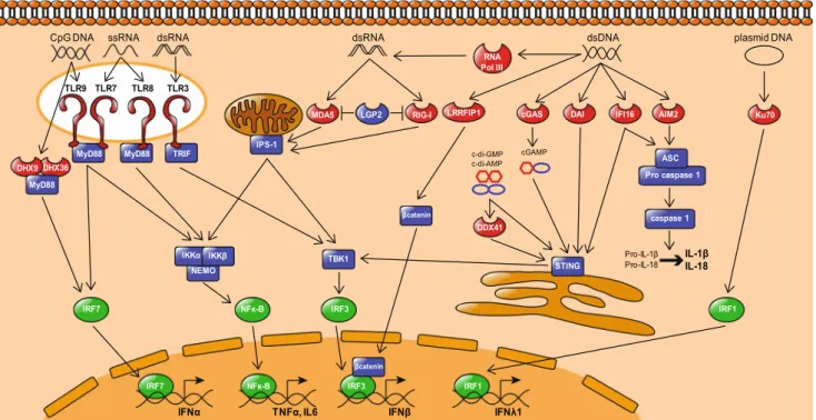
Fig. 1 Nucleic acids and their immune sensors. Multiple receptors allow the cell to sense the presence of microbial nucleic acids and to induce the production of proinflammatory cytokines. In endosomes, TLR9 senses the presence of CpG-containing DNA whereas TLR7 and TLR8 recog-nize ssRNA, leading to activation of the IKK complex via the MyD88 adaptor molecule and the subsequent activation of the NF-κB pathway, inducing the transcription of proinflammatory cytokines like TNF-α or IL6. TLR9 and TLR7 triggering also induces the translocation of IRF7 into the nucleus to initiate the expression of IFN-α. The endosomal TLR3 recognizes dsRNA and uses the adaptor molecule TRIF to induce the TBK1-dependent activation of IRF3 and the subsequent transcription of the type I IFN-β. In addition to endosomal recognition, cytosolic recep-tors can sense the presence of nucleic acids in the cytoplasm. IFI16 and AIM2 sense dsDNA and recruit the adaptor protein ASC to form an inflammasome complex that activates the caspase 1 enzyme, leading to the processing and release of IL1β and IL18. Similarly to the dsDNA sensor DAI, IFI16 also activates the ER-associated adaptor molecule STING that induces the nuclear translocation of IRF3 via TBK1 and the transcription of IFN-β. LRRFIP1 potentiates IRF3 activity by acti-vatingβ-catenin upon dsDNA sensing. Recently, dsDNA-activated cGAS was shown to catalyze cGAMP synthesis from GTP and ATP to induce STING-TBK1-IRF3-dependent IFN-β expression. Other cyclic nucleotides (i.d. c-di-GMP and c-di-AMP) produced by bacteria were
described to activate the STING signaling pathway following recognition by the DDX41 helicase. The RNA polymerase III converts dsDNA into dsRNA to permit its recognition by MDA-5 and RIG-I. Both sensors signal through the mitochondria-associated IPS-1 that leads to the acti-vation of the NF-κB and TBK1-IRF3 pathways and the expression of proinflammatory cytokines and IFN-β, respectively. The LGP2 RNA helicase is thought to compete with MDA-5 and RIG-I and inhibit their downstream signaling. Furthermore, new DNA helicases have been added to the cytosolic nucleic acids sensing machinery. Ku70 recognizes the presence of plasmid DNA and activates IRF1 to induce IFNλ1 expression, and DHX9 and DHX36 are able to sense CpG DNA in the cytosol to induce the transcription of IFN-α via the MyD88-IRF7 path-way. TLR Toll-like receptor, MyD88 myeloid differentiation primary response gene 88, IRF IFN-regulatory factor, TRIF TIR-domain-contain-ing adaptor-inducTIR-domain-contain-ing interferon-β, TBK1 TANK-bindTIR-domain-contain-ing kinase 1, IFI16 IFN-γ-inducible protein 16, AIM2 absent in melanoma 2, DAI DNA-dependent activator of IRF, LRRFIP1 leucine-rich repeat (in FLII) interacting protein 1, cGAS cyclic GMP-AMP synthase, cGAMP cyclic guanosine monophosphate-adenosine monophosphate, DDX DEAD-box protein, DHX DEAH-box protein, MDA-5 melanoma differentiation-as-sociated protein 5, RIG-I retinoic acid-inducible gene 1, LGP2 laboratory of genetics and physiology 2
DHX9 has also been shown to participate in type I IFN production after dsRNA activation in myeloid DC in an IPS-1-dependent manner [31].
AIM2
AIM2 preferentially binds dsDNA due to conserved motifs facilitating DNA binding [32] [33, 34]. Similarly to IFI16, a f t e r b i n d i n g c y t o s o l i c d sD N A , AI M 2 f o r m s a n inflammasome by recruiting ASC and caspase 1 to induce IL-1β and IL-18 maturation and secretion [33,34].
Cytosolic RNA sensors
RIG-like receptors RLRs are cytosolic pattern recognition receptors broadly expressed by immune and non-immune cells and specialized in sensing RNA viruses. They belong to the DExD/H-box helicase superfamily comprising RIG-I, melanoma differentiation-associated protein 5 (MDA-5), and laboratory of genetics and physiology 2 (LGP2). RIG-I recognizes viral dsRNA [35] with a preference for the 5′-triphosphate part of the viral RNA [36]. In-vivo RIG-I is essential for the detection of several viruses such as Sendai virus and vesicular stomatitis virus. MDA-5 preferentially senses long dsRNA molecules [37] and is needed to detect picornavirus [38] and measles virus. However, RIG-I and MDA-5 may also allow detection of small host-derived self-RNA generated by the cytosolic ribonuclease RNAse L [39]. The exact contribution of LGP2 to cytosolic RNA sensing remains unclear, but crystal structure and NMR studies predict that LGP2 is likely to interact with a variety of RNA species [40–42]. Upon activation, RLRs signal through their caspase acti-vation and recruitment domain (CARD) by binding to the IFN-β-promoter stimulator 1 (IPS1, aka MAVS) [8], an adaptor molecule that in turn activates IRFs and NF-κB transcription factors leading to the production of type I IFNs and proinflammatory cytokines. LGP2 lacks CARDs and thus is also considered as a negative regulator of RIG-I and MDA-5.
RNA polymerase III
The DNA-dependent RNA polymerase III has been described as a B-form dsDNA sensor. It can transcribe AT-rich DNA sequence in a promoter-independent manner into 5′pppRNA that in turn can activate RIG-I-dependent pathway and induce type I IFN production [43,44]. RNA polymerase III is re-quired for IFN-β induction by intracellular bacteria and DNA viruses such as Legionella pneumophila, adenovirus, HSV-1, or Epstein-Barr virus (EBV) [44].
LRRFIP1
Leucine-rich repeat flightless-interacting protein 1 (LRRFIP1) directly binds to exogenous dsRNA and B- or Z-form dsDNA to enhance IFN-β gene expression. LRRFIP1 uses a unique signaling pathway since it binds β-catenin to promote its activation. Subsequently,β-catenin activates IRF3 that facil-itates the recruitment of p300 acetyltransferase to the IFN-β1 promoter. p300 acetyltransferase increases hyperacetylation of histones H3 and H4 and thus enhances IFN-β expression [45]. LLRFIP1 has been shown to participate in the nucleic acid sensing of vesicular stomatitis virus and Listeria monocytogenes by macrophages [45].
Discrimination between microbial and self-nucleic acids Despite the efficient recognition of microbial nucleic acids by intracellular nucleic acid receptors, self-nucleic acids (self-DNA and self-RNA) released into the extracellular environ-ment by dying host cells typically remain immunologically inert. Three distinct mechanisms are in place to ensure re-sponses to microbial nucleic acids and, at the same time, avoid unnecessary sensing of self-nucleic acids:
1. Chemical differences between pathogen and self-nucleic acids. TLR9 recognizes preferentially unmethylated CpG motifs, which are abundant in pathogen-derived DNA. However, it has become clear that self-DNA also contains a certain degree of unmethylated CpG islets that could activate TLR9. Furthermore, the phosphodiesteric back-bone present in both pathogen-derived and host-derived self-DNA was shown to be a direct ligand for TLR9 [46]. RIG-I recognizes 5′-triphosphates present in viral RNA, which are usually masked or absent in self-RNA [36]. However, cytosolic DNA sensors recognize B-form DNA, which is common to both pathogen-derived and self-DNA independently of its sequence or methylation status. It becomes clear, therefore, that other mechanisms must be in place to avoid a continuous and unnecessary sensing of extracellular self-nucleic acids.
2. Rapid extracellular degradation of free nucleic acids by nucleases. Nucleic acids released into the extracellular environment by dying cells are rapidly eliminated via degradation by tissue nucleases. As a result, extracellular self-nucleic acids fail to gain access to immune cells that express nucleic acid sensors. Accordingly, deficiency of the DNA exonuclease Trex1 allows internalization and cytosolic accumulation of DNA, leading to uncontrolled STING activation [47–49]. Furthermore, mice lacking DNAse II develop an inflammatory syndrome due to uncontrolled STING activation [50,51].
3. Intracellular sequestration of nucleic acid receptors. The intracellular (endosomal or cytosolic) location of nucleic acid receptors allows sensing of microbial nucleic acids transported into intracellular compartments during the infectious process. By contrast, self-nucleic acids released in the extracellular environment by dying cells fail to spontaneously enter cells. An elegant study has demon-strated that enforced cell surface expression of TLR9 triggers uncontrolled sensing of self-nucleic acids with development of autoimmunity [52].
Breaking innate tolerance to self-nucleic acids
In the past few years, it has become clear that under certain circumstances, these protective mechanisms can be circumvented resulting in the conversion of otherwise non-stimulatory self-nucleic acids into potent activators of innate immunity. These circumstances are associated with the devel-opment of autoimmunity in psoriasis and lupus erythematosus and are linked to the expression of several endogenous factors including anti-microbial peptides, high-mobility group box 1 (HMGB1)/RAGE, autoantibodies, and amyloid precursors.
Cationic anti-microbial peptides
Cationic anti-microbial peptides (AMPs) belong to the first line of defense of the skin against bacterial, viral, and fungal invasion [53]. There are many different AMPs including the cathelicidins and the defensins, but they are all amphiphatic, containing a patch of positive charge clustered on one side of the molecule. This unique feature of AMPs confers them with antibiotic activity through binding of negatively charged phospholipids leading to the disruption of the microbial mem-brane. This feature also allows AMPs to form complexes with extracellular human nucleic acid fragments (DNA and RNA) and to form aggregated particles that are protected from ex-tracellular degradation. In addition, exposed cationic residues in these particles allow the attachment of the nucleic acid complexes to heparin sulfate proteoglycans on the cell mem-brane leading to endocytosis of the complexes. Finally, the nucleic acid complexes gain access to endosomal compart-ments where they trigger activation of TLR7 and TLR9 in pDCs and TLR8 in myeloid dendritic cells (mDCs) [54,55]. In addition, AMP-nucleic acid complexes can also be transported into the cytosol to activate STING/TBK1-dependent cytosolic DNA sensors in monocytes [56]. Recent evidence suggests that the unique structural features of nucleic acid-AMP complexes permit interdigitation of endosomal TLRs, contributing to the strong efficiency of innate activation to self-nucleic acids.
HMGB1 and RAGE
HMGB1 is an important DNA binding protein that is present in the nucleus of cells where it regulates DNA accessibility to transcription factors [57]. HMGB1 is released into the extra-cellular environment during cell necrosis [58], where it may bind to free DNA through two DNA binding HMG box domains. HMGB1-DNA complexes were shown to induce type I IFN production by pDCs and to activate autoreactive B cells via TLR9 [59]. The receptor for advanced glycation end products (RAGE) is a receptor for HMGB1, although its role as a scavenger for DNA-HMGB1 complexes has not been demonstrated [60]. Interestingly, there is evidence that RAGE can directly bind nucleic acids, raising the possibility that it functions as a transporter itself [61].
Amyloid fibrils
Amyloid fibrils are stable insoluble aggregates of misfolded protein products with extensive β-sheet structures [62], in-volved in the pathogenesis of human diseases like Alzheimer’s disease. Interestingly, these amyloid precursors are able to interact with nucleic acids [63] and to form com-plexes that protect DNA or RNA from degradation. Similarly to anti-microbial-nucleic acid complexes, complexes of amy-loid precursors with DNA can be taken up by immune cells such as pDCs to trigger endosomal TLR9, leading to the production of type I IFN [64]. Although the link between the production of amyloid precursors and nucleic acid-mediated innate immune responses needs to be fully elucidat-ed, an interesting study has demonstrated that beta amyloid in Alzheimer’s disease can trigger inflammasome activation [65], potentially via AIM2.
Autoantibodies to DNA or RNA complexes
In certain human autoimmune diseases, autoantibodies direct-ed against nucleic acids and associatdirect-ed proteins like histones or ribonucleoproteins (RNPs) are being produced. These au-toantibodies bind nucleic acids released by dying cells or released in the context of NETosis. On B cells, DNA or RNA containing immune complexes can bind to their specific BCR and be internalized to reach endosomal compartments where they can activate TLR7 or TLR9 signaling to induce autoreactive B cell proliferation [66, 67]. On antigen-presenting cells, DNA or RNA containing immune complexes can also bind to low affinity FcγRs that recognize the Fc fragment of antibodies and be engulfed within cells by endo-cytosis or phagoendo-cytosis to activate TLR7, TLR8, or TLR9. Such mechanisms have been demonstrated in mDCs [68] and pDCs [69–72]. In addition, the presence of AMPs such as LL37 in autoantibody complexes enhances immunogenicity of self-DNA [71, 72]. It is likely that aggregation and
protection of self-nucleic acids induced by AMPs (described earlier) promote nucleic acid stability and cell internalization via the process of receptor-mediated endocytosis.
Role of nucleic acid sensing in skin inflammation and autoimmunity
Skin wounds
Following skin injury, an inflammatory response is mounted to prevent infection and to initiate the re-epithelialization process. Wounded human skin was found to contain large numbers of activated pDCs, which specifically express TLR7 and TLR9 [1,73–75]. A mouse model of skin injury indeed demonstrated that pDCs infiltrate injured skin and are activated to produce type I IFNs via TLR7 and TLR9. pDC-derived type I IFNs were found to be crucial in driving the inflammatory response and the re-epithelialization process in wounded skin [73], although the link between type I IFNs and wound healing remains to be fully elucidated. Thus, sterile skin injury triggers TLR7 and TLR9 activation of pDCs, suggesting that this process is mediated via sensing of self-nucleic acids. But how are pDCs activated in the context of skin injury? One possibility is that AMPs produced by injured keratinocytes break innate tolerance to self-nucleic acids re-leased into the wounds by dying cells. In accordance with this, AMPs are rapidly and transiently expressed in wounded skin, and their expression appears to parallel the IFN-expression by pDCs [73]. Another possibility is that neutrophils, which infiltrate injured skin along with pDCs, activate pDCs via the release of neutrophil extracellular traps (NETs) containing AMP-nucleic acid complexes (Fig.2). This is in accordance with the finding that neutrophils in injured skin are activated to produce NETs and that NETs contain AMP-DNA com-plexes that activate pDCs.
Psoriasis
Psoriasis is a chronic inflammatory skin disease that affects 2– 3 % of the population worldwide and presents, in its most prevalent form, with scaly erythematous plaques. Psoriatic skin lesions are triggered by the aberrant activation of dermal dendritic cells that stimulate autoreactive Th17 cells. Activat-ed Th17 cells migrate from the dermis into the epidermis and produce large amounts of Th17 cytokines, which induce keratinocyte activation with production of AMPs as well as the typical epidermal hyperproliferation that is characteristic of psoriatic plaques. Psoriatic lesions are also characterized by the presence of large numbers of activated pDCs [76]. In contrast to the transient infiltration and activation of pDCs in skin wounds, psoriatic pDCs were found to be chronically
activated and to produce large amounts of type I IFNs that trigger unabated activation of dermal dendritic cells and autoreactive Th17 cells, leading to the development of the psoriatic plaque [76]. But what is the stimulus for pDC acti-vation in psoriatic skin? Cationic AMPs such as LL37 and β-defensin, which are overexpressed in psoriatic skin, were found to constantly activate pDCs as well as conventional DCs by breaking innate tolerance to host nucleic acids. As described above, this process involves the complex formation between AMPs and free self-DNA and self-RNA, leading to activation of TLR7 and TLR9 in pDCs and TLR8 in conven-tional DCs [54,55] (Fig.2). So, whereas the transient AMP expression and pDC activation in skin wounds lead to an adequate inflammatory response that promotes re-epithelialization of wounds, the constant AMP overexpression triggers sustained pDC activation and chronic inflammation that sustain keratinocyte hyperproliferation and development of the psoriatic plaque. But what are the mechanisms that stimulate chronic AMP overexpression in psoriatic skin? Th17 cytokines IL-17 and IL-22 are overexpressed in psoriatic skin due to an enhanced Th17 differentiation in psoriasis and can directly stimulate keratinocytes to produce AMPs [77,78]. A genetic basis for the enhanced Th17 differentiation was revealed in the finding of IL-23 and IL-23R gene polymor-phisms [79]. Furthermore, copy number polymorphisms in the β-defensin gene are directly associated with psoriasis, suggest-ing an additional link to AMP overexpression in psoriasis [80]. The role of endosomal TLRs in the triggering of psoriasis was further demonstrated by the observation that topical ap-plication of the TLR7 agonist imiquimod (Aldara 5 %) to the skin exacerbates human psoriasis [81] and the finding that repetitive application of Aldara to mouse skin induces a psoriasiform epidermal hyperproliferation [82,83]. This pro-cess is driven by cDCs since depletion of CD11c-positive cells abrogates psoriasis development [83] and specific expression of MyD88 in dendritic cells of MyD88-deficient mice restores imiquimod-induced psoriasis [84]. Furthermore, specific de-pletion of ABIN1, a negative regulator of MyD88 and TLR signaling in the dendritic cell compartment, exacerbates pso-riasis induced by imiquimod [85]. Similarly to the human disease, imiquimod model in mice shows strong involvement of the IL-23/IL-17 axis [84,86]. Also, mice deficient in IL-17a, IL-17F, IL-17c, and IL-22 are somehow protected against imiquimod-induced psoriasis [82, 87, 88], pointing out the role of these cytokines in this model. Whereas the source of Th17 cytokines in mice is gamma delta T cells [83,86] and innate lymphoid cells [87], in humans, αβ-T lymphocytes seem to be the main cell type producing Th17 cytokines. Lupus erythematosus
Lupus erythematosus (LE) is a complex autoimmune disease that primarily affects the skin but can become a multiorgan
disease affecting the joints, heart, lungs, kidneys, and central nervous and hematologic systems [89]. Systemic LE is char-acterized by the presence of circulating immune complexes (ICs) formed by self-nucleic acids complexed with autoanti-bodies. These ICs are typically deposited in various tissues leading to detrimental inflammation that initiates and sustains autoimmunity. In the skin, ICs are deposited along the basal membrane of the epidermis and trigger a cytotoxic response leading to vacuolization of the basal keratinocyte layer. A key question in the pathogenesis of LE is the origin of ICs. In the skin, self-DNA may be released by dying cells during skin infection or ultraviolet light irradiation. LE patients were indeed found to have an intrinsic defect in clearing dying cells, leading to the accumulation of remnants containing self-nucleic acids [90]. Another source of self-DNA is neu-trophil extracellular traps (NETs) released by neuneu-trophils [71,
91]. NETs are filaments of nuclear DNA covered with gran-ular proteins released by neutrophils during a cell death
process called NETosis [92]. This process is dependent of the production of reactive oxygen species (ROS) and is due to the nuclear translocation of elastase, which decondenses nuclear DNA [93,94]. The expanding nuclear DNA breaks into the cytosol, mixes with the granular content of neutrophil, and is ultimately extruded as NETs into the extracellular space [94]. Interestingly, LE patients display increased numbers of NETs due to both enhanced formation [71, 72] as well as decreased nuclease-mediated degradation [95] (Fig.2). These NETs were found to activate TLR9 in pDCs and trigger the production of type I IFNs [71].
Type I IFN production by pDCs is directly linked to disease exacerbation and maintenance of lupus. Indeed, a dominant type I IFN signature was identified in PBMC of SLE patients and was shown to correlate with disease activity and severity [96]. Whether activation of pDCs and their production of type I IFN can lead to the onset of the disease has been long debated. Recent work demonstrates that depletion of pDCs pDC
type I IFNs
TLR7 TLR9 injury AMPsPsoriasis
Wound healing
Lupus Erythematosus
AMPs
DNA RNA
DNA- and RNA-containing ICs
IFN signaling (STAT4, IRF5, MDA5) B cells and pDCs responsiveness
(TLR7, TLR9)
B cells activation (PTPN22, BLK) ICs uptake (FcγR2a, ITGAM)
anti-NETs auto-antibodies NETting neutrophils wound re-epithelialization mDC Th17 Th17 Autoimmunity Autoimmunity
IFN signaling (TYK2, MDA5) T cells activation (HLA-Cw6)
IL23R, IL23A) AMPs (β-defensin) AMP production GENETICS NET formation NET degradation B Cell GENETICS
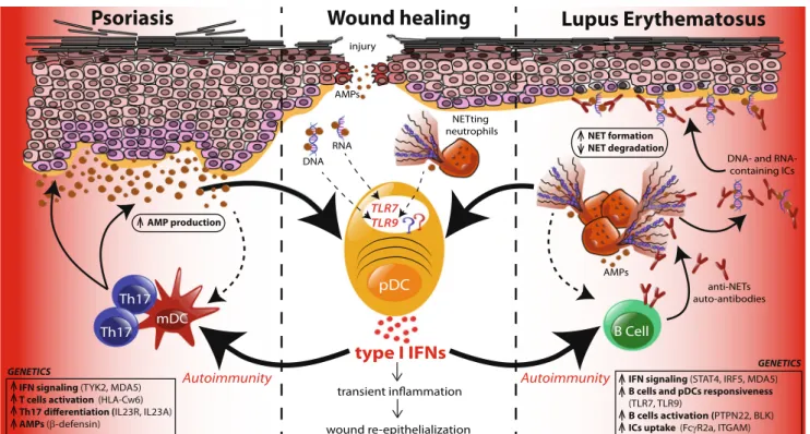
Fig. 2 TLR activation of pDCs in healthy and diseased skin. Upon skin injury, neutrophils first infiltrate the wound followed by pDCs. Anti-microbial peptides (AMPs) that are transiently produced by injured keratinocytes bind self-DNA and self-RNA released by dying cells and activate pDCs via TLR7 and TLR9. Neutrophil extracellular traps (NETs) extruded by neutrophils to prevent the spread of potential infection also trigger TLR9 in pDCs. Activated pDCs produce type I IFNs that initiate a transient inflammation leading to the recruitment of additional immune cells that will participate in the re-epithelialization of the wound. Some genetic variations are associated with increased susceptibility to develop autoimmune diseases in response to environmental triggers. Psoriasis patients have a genetic predisposition that enhances the response to IFN signaling, reduces the threshold of T cell activation, and favors the differentiation of Th17 cells. Infiltrating Th17 cells produce cytokines, namely, IL-17 and IL-22, which increase the proliferation of
keratinocytes and their production of AMPs leading to enhanced stimu-lation of pDCs that sustain inflammation. Moreover, dermal DCs process AMPs as autoantigens to generate AMP-specific autoreactive T cells that perpetuate autoimmunity. Lupus patients have genetic variants that am-plify IFN signaling and responsiveness of pDCs and B cells, leading to greater amounts of type I IFN that drive the generation of autoantibodies. Autoantibodies targeting NET-associated proteins and nucleic acids form immune complexes (ICs) that further deposit in the basal membrane of the dermo-epidermal junction of the skin and lead to keratinocyte necrosis and decreased epidermal thickness. Lupus patients also have a defect in the clearance of ICs which stimulates neutrophils to undergo NETosis. Increased NET formation and AMP production by neutrophils coupled to a defective NET degradation enhance pDC activation via TLR9 and favor the production of autoantibodies by B cells, sustaining the inflammatory response that participates in systemic autoimmunity
or treatment of mice with a bifunctional TLR7/TLR9 inhibitor prevents the development of skin lesions in lupus-prone mice [97]. Altogether, these data suggest that pDC activation seems to be an important step in the pathogenesis of lupus in these mice.
Several genetic studies that analyzed polymorphisms in TLR7, TLR8, and TLR9 genes in Caucasian and Asian pop-ulations revealed that some SNPs could be considered as SLE susceptibility factors [98,99]. It was shown that total PBMCs from SLE patients have higher expression of the TLR7 and TLR9 mRNAs compared to healthy donors that was positive-ly correlated with IFN-α expression [100]. Increased expres-sion of TLR7 in mice harboring duplication of the TLR7 gene is sufficient to drive the development of lupus, and deletion of this gene can improve the clinical disease [101]. By contrast, mouse models of deficiency for TLR9 and TLR8 demonstrat-ed an exacerbation of the disease because of increasdemonstrat-ed TLR7 response [102,103]. This was explained by a recent study showing that TLR8 controls TLR7 function on dendritic cells and TLR9 restrains TLR7 response on B cells [104]. Thus, alteration in the expression of TLR7, TLR8, or TLR9 can favor the loss of tolerance against self-nucleic acids. Impor-tantly, injection of immunoregulatory sequences (IRSs) that inhibit both TLR7 and TLR9 signaling in lupus-prone mice can prevent the production of autoantibodies [105]. This dual inhibitor, called DV1179, is currently tested in a human clin-ical trial.
Autoreactive B cells produce antibodies targeting nucleic acids or associated proteins, including AMPs present in NETs that can be taken up by pDCs via FcγRII leading to TLR7 and TLR9 activation [106, 107]. Anti-NETs antibodies such as anti-LL37 enhance NET formation by neutrophils through yet unclear mechanism [71]. This process provides an explanation for the sustained NETosis in SLE patients. Furthermore, anti-NETs antibodies such as anti-DNA and anti-LL37 antibodies enhance IC uptake by pDCs, resulting in enhanced type I IFN production. Type I IFN production facilitates autoantibody production by autoreactive B cells that recognize ICs via TLR9 [67,108]. Thus, autoantibodies targeting NETs enhance NETosis leading to the generation of autoreactive B cells, creating a self-sustaining loop that fuels the autoimmune response (Fig.2).
Beyond TLRs, activation of other nucleic acid-sensing receptors may participate in the development of lupus. Strik-ingly, loss of STING function could inhibit self-DNA-induced inflammation in DNase II-deficient mice [51], suggesting that cytosolic DNA sensor pathways may be involved. Interest-ingly, an increased expression of HIN-200, the human gene encoding IFI16, was found in leukocytes of SLE patients and was correlated with anti-nuclear antibody titers [109]. More-over, a strong sex bias was observed in the expression of IFI16 that correlates with a prevalence of women developing SLE [110]. However, the exact mechanism by which IFI16 is
activated in LE is unknown. Furthermore, SLE PBMCs as well as lupus-prone mice have enhanced expression of DAI [111].
Genetic studies revealed that variants in MDA-5 are asso-ciated with autoantibody production in SLE patients [112] and increased type I IFNs [113]. Furthermore, a variant of IPS-1, an essential protein regulating MDA-5/RIG-I signaling, was associated with a subset of SLE patients [114]. Importantly, mutant mice harboring a constitutive activated MDA-5 spon-taneously develop lupus-like autoimmune symptoms that can be ameliorated by intercrossing those mice with IFNAR-deficient mice [115]. This supports the idea that cytosolic DNA and RNA recognition are also involved in lupus pathogenesis.
Furthermore, naïve mice injected with DNA-containing amyloids develop symptoms resembling lupus in a pDC-dependent manner [64], indicating that amyloid precursors may also enhance self-DNA sensing in the course of lupus disease. Whether SLE patients have circulating amyloid pre-cursors remains to be investigated. Intriguingly, neutrophil NETs appear to contain amyloidogenic proteins.
Other diseases with skin involvement related to cytosolic DNA sensors
Aicardi-Goutières syndrome (AGS) is a genetically deter-mined inflammatory disease that develops in early childhood and manifests with inflammatory encephalopathy and SLE-like skin lesions on the fingers, toes, and ears. Like SLE, AGS is associated with a strong type I IFN signature in PBMCs [116]. Mutations in nuclease genes including the exonuclease TREX1 (AGS1), the ribonucleases RNASEH2 (AGS2, AGS3, AGS4), and SAMHD1 (AGS5) have been identified [117, 118]. These mutations lead to deficiency in nucleases and cellular accumulation of DNA or RNA leading to overactivation of cytosolic sensors and induction of type I IFN. As described earlier, mouse studies have shown that TREX1 deficiency drives STING activation and type I IFN-induced inflammation. Gain of function mutations in the MDA-5 gene has also been associated with the AGS, as well as other, yet undefined neurologic and immunological syn-dromes [119].
Recently, patients with mutations in the exon 5 of the TMEM173 gene, coding for the STING molecule, were identified. These patients presented cutaneous vasculitic lesions resembling AGS along with pulmonary inflam-mation. The syndrome was called STING-associated vas-culopathy with onset in infancy (SAVI) [120]. STING is principally expressed by immune cells but is also widely expressed by fibroblast and endothelial cells. The muta-tion observed in SAVI leads to constitutive STING acti-vation with spontaneous type I IFN production. As a major pathogenic event, STING activation of endothelial
cells drives endothelial cell death and vascular damage through the excessive production of type I IFN [120].
Conclusion and perspectives
Nucleic acid receptors capable of detecting the presence of DNA and RNA in endosomes and cytosol have been recently identified. In addition to their role in anti-viral immunity, these receptors can, under certain circumstances, detect self-DNA and self-RNA released by dying host cells. Several endoge-nous factors enable immune recognition of self-nucleic acids. These factors include cationic AMPs, HMGB1 and its recep-tor RAGE, autoantibodies, and amyloid precursors. Dysregu-lated expression of these factors drives continuous sensing of self-nucleic acids and chronic nucleic acid receptor activation leading to autoimmunity and development of diseases like psoriasis and LE. Several key questions still remain to be answered: What controls the expression of these factors and why is there a dysregulated expression in psoriasis and LE? What is the role of structural cells of the skin in the recognition of self-nucleic acids? Psoriatic keratinocytes were found to express high levels of AIM2 and to produce high levels of IL-1β in response to cytosolic DNA, raising the possibility for their contribution to the pathogenesis of psoriasis [121]. Fur-thermore, what is the factor that induces NETosis in both the injured healthy and diseased skin? As microbes can activate neutrophils to produce NETs, is there a role for the skin microbiome? Finally, is there a differential role of distinct AMPs or factors in targeting cell types or nucleic acid recep-tors? The answer to these questions will provide us with therapeutic targets to enhance wound healing or inhibit auto-immune inflammation of the skin.
References
1. Kadowaki N et al (2001) Subsets of human dendritic cell precursors express different toll-like receptors and respond to different micro-bial antigens. J Exp Med 194(6):863–869
2. Hemmi H et al (2000) A Toll-like receptor recognizes bacterial DNA. Nature 408(6813):740–745
3. Okuya K et al (2010) Spatiotemporal regulation of heat shock protein 90-chaperoned self-DNA and CpG-oligodeoxynucleotide for type I IFN induction via targeting to static early endosome. J Immunol 184(12):7092–7099
4. Guiducci C et al (2006) Properties regulating the nature of the plasmacytoid dendritic cell response to Toll-like receptor 9 activa-tion. J Exp Med 203(8):1999–2008
5. Hemmi H et al (2002) Small anti-viral compounds activate immune cells via the TLR7 MyD88-dependent signaling pathway. Nat Immunol 3(2):196–200
6. Heil F et al (2004) Species-specific recognition of single-stranded RNA via toll-like receptor 7 and 8. Science 303(5663):1526–1529 7. Alexopoulou L et al (2001) Recognition of double-stranded RNA and activation of NF-kappaB by Toll-like receptor 3. Nature 413(6857):732–738
8. Kawai T, Akira S (2010) The role of pattern-recognition receptors in innate immunity: update on Toll-like receptors. Nat Immunol 11(5): 373–384
9. Ishikawa H, Ma Z, Barber GN (2009) STING regulates intracellular DNA-mediated, type I interferon-dependent innate immunity. Nature 461(7265):788–792
10. Zhong B et al (2008) The adaptor protein MITA links virus-sensing receptors to IRF3 transcription factor activation. Immunity 29(4): 538–550
11. Tanaka Y, Chen ZJ, 214 (2012) STING specifies IRF3 phosphory-lation by TBK1 in the cytosolic DNA signaling pathway. Sci Signal 5:ra20
12. Burdette DL et al (2011) STING is a direct innate immune sensor of cyclic di-GMP. Nature 478(7370):515–518
13. Unterholzner L et al (2010) IFI16 is an innate immune sensor for intracellular DNA. Nat Immunol 11(11):997–1004
14. Kerur N et al (2011) IFI16 acts as a nuclear pathogen sensor to induce the inflammasome in response to Kaposi Sarcoma-associated herpesvirus infection. Cell Host Microbe 9(5): 363–375
15. Takaoka A et al (2007) DAI (DLM-1/ZBP1) is a cytosolic DNA sensor and an activator of innate immune response. Nature 448(7152):501–505
16. Rebsamen M et al (2009) DAI/ZBP1 recruits RIP1 and RIP3 through RIP homotypic interaction motifs to activate NF-kappaB. EMBO Rep 10(8):916–922
17. Zhang X et al (2014) The cytosolic DNA sensor cGAS forms an oligomeric complex with DNA and undergoes switch-like conformational changes in the activation loop. Cell Rep 6(3):421–430
18. Li X et al (2013) Cyclic GMP-AMP synthase is activated by double-stranded DNA-induced oligomerization. Immunity 39(6): 1019–1031
19. Civril F et al (2013) Structural mechanism of cytosolic DNA sensing by cGAS. Nature 498(7454):332–337
20. Gao P et al (2013) Cyclic [G(2′,5′)pA(3′,5′)p] is the metazoan second messenger produced by DNA-activated cyclic GMP-AMP synthase. Cell 153(5):1094–1107
21. Ablasser A et al (2013) cGAS produces a 2′-5′-linked cyclic dinucleotide second messenger that activates STING. Nature 498(7454):380–384 22. Zhang X et al (2013) Cyclic GMP-AMP containing mixed
phos-phodiester linkages is an endogenous high-affinity ligand for STING. Mol Cell 51(2):226–235
23. Diner EJ et al (2013) The innate immune DNA sensor cGAS produces a noncanonical cyclic dinucleotide that activates human STING. Cell Rep 3(5):1355–1361
24. Sun L et al (2013) Cyclic GMP-AMP synthase is a cytosolic DNA sensor that activates the type I interferon pathway. Science 339(6121):786–791
25. Gao D et al (2013) Cyclic GMP-AMP synthase is an innate immune sensor of HIV and other retroviruses. Science 341(6148):903–906 26. Li XD et al (2013) Pivotal roles of cGAS-cGAMP signaling in
antiviral defense and immune adjuvant effects. Science 341(6152): 1390–1394
27. Parvatiyar K et al (2012) The helicase DDX41 recognizes the bacterial secondary messengers cyclic di-GMP and cyclic di-AMP to activate a type I interferon immune response. Nat Immunol 13(12):1155–1161
28. Walker JR, Corpina RA, Goldberg J (2001) Structure of the Ku heterodimer bound to DNA and its implications for double-strand break repair. Nature 412(6847):607–614
29. Zhang X et al (2011) Cutting edge: Ku70 is a novel cytosolic DNA sensor that induces type III rather than type I IFN. J Immunol 186(8):4541–4545
30. Kim T et al (2010) Aspartate-glutamate-alanine-histidine box motif (DEAH)/RNA helicase A helicases sense microbial DNA in human plasmacytoid dendritic cells. Proc Natl Acad Sci U S A 107(34): 15181–15186
31. Zhang Z et al (2011) DHX9 pairs with IPS-1 to sense double-stranded RNA in myeloid dendritic cells. J Immunol 187(9): 4501–4508
32. Albrecht M, Choubey D, Lengauer T (2005) The HIN domain of IFI-200 proteins consists of two OB folds. Biochem Biophys Res Commun 327(3):679–687
33. Hornung V et al (2009) AIM2 recognizes cytosolic dsDNA and forms a caspase-1-activating inflammasome with ASC. Nature 458(7237):514–518
34. Burckstummer T et al (2009) An orthogonal proteomic-genomic screen identifies AIM2 as a cytoplasmic DNA sensor for the inflammasome. Nat Immunol 10(3):266–272
35. Yoneyama M et al (2004) The RNA helicase RIG-I has an essential function in double-stranded RNA-induced innate antiviral re-sponses. Nat Immunol 5(7):730–737
36. Hornung V et al (2006) 5′-Triphosphate RNA is the ligand for RIG-I. Science 314(5801):994–997
37. Kato H et al (2008) Length-dependent recognition of double-stranded ribonucleic acids by retinoic acid-inducible gene-I and melanoma differentiation-associated gene 5. J Exp Med 205(7): 1601–1610
38. Gitlin L et al (2006) Essential role of mda-5 in type I IFN responses to polyriboinosinic:polyribocytidylic acid and en-cephalomyocarditis picornavirus. Proc Natl Acad Sci U S A 103(22):8459–8464
39. Malathi K et al (2007) Small self-RNA generated by RNase L amplifies antiviral innate immunity. Nature 448(7155):816–819 40. Murali A et al (2008) Structure and function of LGP2, a DEX(D/H)
helicase that regulates the innate immunity response. J Biol Chem 283(23):15825–15833
41. Pippig DA et al (2009) The regulatory domain of the RIG-I family ATPase LGP2 senses double-stranded RNA. Nucleic Acids Res 37(6):2014–2025
42. Takahasi K et al (2009) Solution structures of cytosolic RNA sensor MDA5 and LGP2 C-terminal domains: identification of the RNA recognition loop in RIG-I-like receptors. J Biol Chem 284(26): 17465–17474
43. Ablasser A et al (2009) RIG-I-dependent sensing of poly(dA:dT) through the induction of an RNA polymerase III-transcribed RNA intermediate. Nat Immunol 10(10):1065–1072
44. Chiu YH, Macmillan JB, Chen ZJ (2009) RNA polymerase III detects cytosolic DNA and induces type I interferons through the RIG-I pathway. Cell 138(3):576–591
45. Yang P et al (2010) The cytosolic nucleic acid sensor LRRFIP1 mediates the production of type I interferon via a beta-catenin-dependent pathway. Nat Immunol 11(6):487–494
46. Haas T et al (2008) The DNA sugar backbone 2′ deoxyribose determines toll-like receptor 9 activation. Immunity 28(3):315–323 47. Stetson DB et al (2008) Trex1 prevents cell-intrinsic initiation of
autoimmunity. Cell 134(4):587–598
48. Yan N et al (2010) The cytosolic exonuclease TREX1 inhibits the innate immune response to human immunodeficiency virus type 1. Nat Immunol 11(11):1005–1013
49. Morita M et al (2004) Gene-targeted mice lacking the Trex1 (DNase III) 3′–5′ DNA exonuclease develop inflammatory myocarditis. Mol Cell Biol 24(15):6719–6727
50. Kawane K et al (2001) Requirement of DNase II for definitive erythropoiesis in the mouse fetal liver. Science 292(5521):1546– 1549
51. Ahn J et al (2012) STING manifests self DNA-dependent inflam-matory disease. Proc Natl Acad Sci U S A 109(47):19386–19391 52. Barton GM, Kagan JC, Medzhitov R (2006) Intracellular
localiza-tion of Toll-like receptor 9 prevents recognilocaliza-tion of self DNA but facilitates access to viral DNA. Nat Immunol 7(1):49–56 53. Zasloff M (2002) Antimicrobial peptides of multicellular
organ-isms. Nature 415(6870):389–395
54. Ganguly D et al (2009) Self-RNA-antimicrobial peptide complexes activate human dendritic cells through TLR7 and TLR8. J Exp Med 206(9):1983–1994
55. Lande R et al (2007) Plasmacytoid dendritic cells sense self-DNA coupled with antimicrobial peptide. Nature 449(7162):564–569 56. Chamilos G et al (2012) Cytosolic sensing of extracellular
self-DNA transported into monocytes by the antimicrobial peptide LL37. Blood 120(18):3699–3707
57. Stott K et al (2006) Structure of a complex of tandem HMG boxes and DNA. J Mol Biol 360(1):90–104
58. Scaffidi P, Misteli T, Bianchi ME (2002) Release of chromatin protein HMGB1 by necrotic cells triggers inflammation. Nature 418(6894):191–195
59. Tian J et al (2007) Toll-like receptor 9-dependent activation by DNA-containing immune complexes is mediated by HMGB1 and RAGE. Nat Immunol 8(5):487–496
60. Sirois CM et al (2013) RAGE is a nucleic acid receptor that promotes inflammatory responses to DNA. J Exp Med 210(11): 2447–2463
61. Sims GP et al (2010) HMGB1 and RAGE in inflammation and cancer. Annu Rev Immunol 28:367–388
62. Sideras K, Gertz MA (2009) Amyloidosis. Adv Clin Chem 47:1–44
63. Di Domizio J et al (2012) Binding with nucleic acids or glycosami-noglycans converts soluble protein oligomers to amyloid. J Biol Chem 287(1):736–747
64. Di Domizio J et al (2012) Nucleic acid-containing amyloid fibrils potently induce type I interferon and stimulate systemic autoimmu-nity. Proc Natl Acad Sci U S A 109(36):14550–14555
65. Halle A et al (2008) The NALP3 inflammasome is involved in the innate immune response to amyloid-beta. Nat Immunol 9(8):857–865 66. Leadbetter EA et al (2002) Chromatin-IgG complexes activate B
cells by dual engagement of IgM and Toll-like receptors. Nature 416(6881):603–607
67. Lau CM et al (2005) RNA-associated autoantigens activate B cells by combined B cell antigen receptor/Toll-like receptor 7 engage-ment. J Exp Med 202(9):1171–1177
68. Boule MW et al (2004) Tolllike receptor 9dependent and i n d e p e n d e n t d e n d r i t i c c e l l a c t i v a t i o n b y c h r o m a t i n -immunoglobulin G complexes. J Exp Med 199(12):1631–1640 69. Means TK et al (2005) Human lupus autoantibody-DNA complexes
activate DCs through cooperation of CD32 and TLR9. J Clin Invest 115(2):407–417
70. Lovgren T et al (2006) Induction of interferon-alpha by immune complexes or liposomes containing systemic lupus erythematosus autoantigen- and Sjogren’s syndrome autoantigen-associated RNA. Arthritis Rheum 54(6):1917–1927
71. Lande R et al (2011) Neutrophils activate plasmacytoid dendritic cells by releasing self-DNA-peptide complexes in systemic lupus erythematosus. Sci Transl Med 3(73):73ra19
72. Garcia-Romo GS et al (2011) Netting neutrophils are major in-ducers of type I IFN production in pediatric systemic lupus erythe-matosus. Sci Transl Med 3(73):73ra20
73. Gregorio J et al (2010) Plasmacytoid dendritic cells sense skin injury and promote wound healing through type I interferons. J Exp Med 207(13):2921–2930
74. Liu YJ (2005) IPC: professional type 1 interferon-producing cells and plasmacytoid dendritic cell precursors. Annu Rev Immunol 23: 275–306
75. Jarrossay D et al (2001) Specialization and complementarity in microbial molecule recognition by human myeloid and plasmacytoid dendritic cells. Eur J Immunol 31(11):3388–3393 76. Nestle FO et al (2005) Plasmacytoid predendritic cells initiate
psoriasis through interferon-alpha production. J Exp Med 202(1): 135–143
77. Zheng Y et al (2007) Interleukin-22, a T(H)17 cytokine, mediates IL-23-induced dermal inflammation and acanthosis. Nature 445(7128):648–651
78. Liang SC et al (2006) Interleukin (IL)-22 and IL-17 are coexpressed by Th17 cells and cooperatively enhance expression of antimicro-bial peptides. J Exp Med 203(10):2271–2279
79. Capon F et al (2007) Sequence variants in the genes for the interleukin-23 receptor (IL23R) and its ligand (IL12B) confer pro-tection against psoriasis. Hum Genet 122(2):201–206
80. Stuart PE et al (2012) Association of beta-defensin copy number and psoriasis in three cohorts of European origin. J Investig Dermatol 132(10):2407–2413
81. Gilliet M et al (2004) Psoriasis triggered by toll-like receptor 7 agonist imiquimod in the presence of dermal plasmacytoid dendritic cell precursors. Arch Dermatol 140(12):1490–1495
82. van der Fits L et al (2009) Imiquimod-induced psoriasis-like skin inflammation in mice is mediated via the IL-23/IL-17 axis. J Immunol 182(9):5836–5845
83. Tortola L et al (2012) Psoriasiform dermatitis is driven by IL-36-mediated DC-keratinocyte crosstalk. J Clin Invest 122(11):3965– 3976
84. Wohn C et al (2013) Langerin(neg) conventional dendritic cells produce IL-23 to drive psoriatic plaque formation in mice. Proc Natl Acad Sci U S A 110(26):10723–10728
85. Callahan JA et al (2013) Cutting edge: ABIN-1 protects against psoriasis by restricting MyD88 signals in dendritic cells. J Immunol 191(2):535–539
86. Cai Y et al (2011) Pivotal role of dermal IL-17-producing gammadelta T cells in skin inflammation. Immunity 35(4):596–610 87. Pantelyushin S et al (2012) Rorγt+ innate lymphocytes and gammadelta T cells initiate psoriasiform plaque formation in mice. J Clin Invest 122(6):2252–2256
88. Van Belle AB et al (2012) IL-22 is required for imiquimod-induced psoriasiform skin inflammation in mice. J Immunol 188(1):462– 469
89. Croker JA, Kimberly RP (2005) SLE: challenges and candidates in human disease. Trends Immunol 26(11):580–586
90. Munoz LE et al (2010) The role of defective clearance of apoptotic cells in systemic autoimmunity. Nat Rev Rheumatol 6(5):280–289 91. Kaplan MJ (2011) Neutrophils in the pathogenesis and
manifesta-tions of SLE. Nat Rev Rheumatol 7(12):691–699
92. Brinkmann Vet al (2004) Neutrophil extracellular traps kill bacteria. Science 303(5663):1532–1535
93. Fuchs TA et al (2007) Novel cell death program leads to neutrophil extracellular traps. J Cell Biol 176(2):231–241
94. Papayannopoulos V et al (2010) Neutrophil elastase and myeloperoxidase regulate the formation of neutrophil extracellular traps. J Cell Biol 191(3):677–691
95. Hakkim A et al (2010) Impairment of neutrophil extracellular trap degradation is associated with lupus nephritis. Proc Natl Acad Sci U S A 107(21):9813–9818
96. Baechler EC et al (2003) Interferon-inducible gene expression signature in peripheral blood cells of patients with severe lupus. Proc Natl Acad Sci U S A 100(5):2610–2615
97. Guiducci C et al (2010) Autoimmune skin inflammation is depen-dent on plasmacytoid dendritic cell activation by nucleic acids via TLR7 and TLR9. J Exp Med 207(13):2931–2942
98. Lee YH et al (2012) Associations between TLR polymorphisms and systemic lupus erythematosus: a systematic review and meta-anal-ysis. Clin Exp Rheumatol 30(2):262–265
99. dos Santos BP et al (2012) TLR7/8/9 polymorphisms and their associations in systemic lupus erythematosus patients from southern Brazil. Lupus 21(3):302–309
100. Komatsuda A et al (2008) Up-regulated expression of Toll-like receptors mRNAs in peripheral blood mononuclear cells from pa-tients with systemic lupus erythematosus. Clin Exp Immunol 152(3):482–487
101. Fairhurst AM et al (2008) Yaa autoimmune phenotypes are conferred by overexpression of TLR7. Eur J Immunol 38(7):1971–1978
102. Wu X, Peng SL (2006) Toll-like receptor 9 signaling protects against murine lupus. Arthritis Rheum 54(1):336–342
103. Demaria O et al (2010) TLR8 deficiency leads to autoimmunity in mice. J Clin Invest 120(10):3651–3662
104. Desnues B et al (2014) TLR8 on dendritic cells and TLR9 on B cells restrain TLR7-mediated spontaneous autoimmunity in C57BL/6 mice. Proc Natl Acad Sci U S A 111(4):1497–1502
105. Barrat FJ et al (2007) Treatment of lupus-prone mice with a dual inhibitor of TLR7 and TLR9 leads to reduction of autoantibody production and amelioration of disease symptoms. Eur J Immunol 37(12):3582–3586
106. Bave U et al (2003) Fc gamma RIIa is expressed on natural IFN-alpha-producing cells (plasmacytoid dendritic cells) and is required for the IFN-alpha production induced by apoptotic cells combined with lupus IgG. J Immunol 171(6):3296–3302
107. Lovgren T et al (2004) Induction of interferon-alpha production in plasmacytoid dendritic cells by immune complexes containing nucleic acid released by necrotic or late apoptotic cells and lupus IgG. Arthritis Rheum 50(6):1861–1872
108. Viglianti GA et al (2003) Activation of autoreactive B cells by CpG dsDNA. Immunity 19(6):837–847
109. Kimkong I, Avihingsanon Y, Hirankarn N (2009) Expression profile of HIN200 in leukocytes and renal biopsy of SLE patients by real-time RT-PCR. Lupus 18(12):1066–1072
110. Choubey D et al (2011) Emerging roles for the interferon-inducible p200-family proteins in sex bias in systemic lupus erythematosus. J Interferon Cytokine Res 31(12):893–906
111. Zhang W et al (2013) DNA-dependent activator of interferon-regulatory factors (DAI) promotes lupus nephritis by activating the calcium pathway. J Biol Chem 288(19):13534–13550 112. Molineros JE et al (2013) Admixture mapping in lupus identifies multiple
functional variants within IFIH1 associated with apoptosis, inflammation, and autoantibody production. PLoS Genet 9(2):e1003222
113. Robinson T et al (2011) Autoimmune disease risk variant of IFIH1 is associated with increased sensitivity to IFN-alpha and serologic autoimmunity in lupus patients. J Immunol 187(3):1298–1303 114. Pothlichet J et al (2011) A loss-of-function variant of the antiviral
molecule MAVS is associated with a subset of systemic lupus patients. EMBO Mol Med 3(3):142–152
115. Funabiki M et al (2014) Autoimmune disorders associated with gain of function of the intracellular sensor MDA5. Immunity 40(2):199–212 116. Lebon P et al (2002) Interferon and Aicardi-Goutieres syndrome. Eur J
Paediatr Neurol 6(Suppl A):A47–A53, discussion A55–8, A77–86 117. Rice G et al (2007) Heterozygous mutations in TREX1 cause
familial chilblain lupus and dominant Aicardi-Goutieres syndrome. Am J Hum Genet 80(4):811–815
118. Rice GI et al (2009) Mutations involved in Aicardi-Goutieres syn-drome implicate SAMHD1 as regulator of the innate immune re-sponse. Nat Genet 41(7):829–832
119. Rice GI et al (2014) Gain-of-function mutations in IFIH1 cause a spectrum of human disease phenotypes associated with upregulated type I interferon signaling. Nat Genet 46(5):503–509
120. Liu Y et al (2014) Activated STING in a vascular and pulmonary syndrome. N Engl J Med 371:507–518
121. Dombrowski Y, 82 et al (2011) Cytosolic DNA triggers inflammasome activation in keratinocytes in psoriatic lesions. Sci Transl Med 3:82ra38