Publisher’s version / Version de l'éditeur:
ACS Applied Materials & Interfaces, 9, 28, pp. 23731-23740, 2017-06-29
READ THESE TERMS AND CONDITIONS CAREFULLY BEFORE USING THIS WEBSITE. https://nrc-publications.canada.ca/eng/copyright
Vous avez des questions? Nous pouvons vous aider. Pour communiquer directement avec un auteur, consultez la
première page de la revue dans laquelle son article a été publié afin de trouver ses coordonnées. Si vous n’arrivez pas à les repérer, communiquez avec nous à PublicationsArchive-ArchivesPublications@nrc-cnrc.gc.ca.
Questions? Contact the NRC Publications Archive team at
PublicationsArchive-ArchivesPublications@nrc-cnrc.gc.ca. If you wish to email the authors directly, please see the first page of the publication for their contact information.
NRC Publications Archive
Archives des publications du CNRC
This publication could be one of several versions: author’s original, accepted manuscript or the publisher’s version. / La version de cette publication peut être l’une des suivantes : la version prépublication de l’auteur, la version acceptée du manuscrit ou la version de l’éditeur.
For the publisher’s version, please access the DOI link below./ Pour consulter la version de l’éditeur, utilisez le lien DOI ci-dessous.
https://doi.org/10.1021/acsami.7b05159
Access and use of this website and the material on it are subject to the Terms and Conditions set forth at
Transition metal ions enable the transition from electrospun prolamin
protein fibers to nitrogen-doped freestanding carbon films for flexible
supercapacitors
Wang, Yixiang; Yang, Jingqi; Du, Rongbing; Chen, Lingyun
https://publications-cnrc.canada.ca/fra/droits
L’accès à ce site Web et l’utilisation de son contenu sont assujettis aux conditions présentées dans le site LISEZ CES CONDITIONS ATTENTIVEMENT AVANT D’UTILISER CE SITE WEB.
NRC Publications Record / Notice d'Archives des publications de CNRC:
https://nrc-publications.canada.ca/eng/view/object/?id=850157cb-041e-4c64-8c04-111bd07f6f66 https://publications-cnrc.canada.ca/fra/voir/objet/?id=850157cb-041e-4c64-8c04-111bd07f6f66Transition Metal Ions Enable the Transition from Electrospun
Prolamin Protein Fibers to Nitrogen-Doped Freestanding Carbon
Films for Flexible Supercapacitors
Yixiang Wang,
†Jingqi Yang,
†Rongbing Du,
‡and Lingyun Chen
*
,† †Department of Agricultural, Food and Nutritional Science, University of Alberta, Edmonton, Alberta, Canada T6G 2P5
‡
National Institute for Nanotechnology, University of Alberta, Edmonton, Alberta, Canada T6G 2G2
*
S Supporting InformationABSTRACT: Flexible carbon ultrafine fibers are highly desirable in energy storage and conversion devices. Our previous finding showed that electrospun hordein/zein fibers stabilized by Ca2+were successfully transferred into nitrogen-doped carbon ultrafine fibers for supercapacitors. However, their relatively brittle nature needed to be improved. Inspired by this stabilizing effect of Ca2+, in this work, four transition metal divalent cations were used to assist the formation of flexible hordein/zein-derived carbon ultrafine fibers. Without alteration of the electrospinnability, adequate amounts of zinc acetate and cobalt acetate supported the fibrous structure during pyrolysis. This resulted in flexible freestanding carbon
films consisting of well-defined fibers with nitrogen-doped graphitic layers and hierarchical pores. These carbon films were easily cut into small square pieces and directly applied as working electrode in the three-electrode testing system without the need for polymer binders or conducting agents. Notably, the hz-Zn0.3-p electrode, synthesized with 0.3 mol/L Zn2+ and post-acid treatment, exhibited a specific capacitance of 393 F/g (at 1 A/g), a large rate capability (72.3% remained at 20 A/g), and a capacitance retention of ∼98% after 2000 charging−discharging cycles at 10 A/g. These superior electrochemical properties were attributed to the synergistic effects of the well-developed graphitic layers induced by Zn2+, the nitrogen-decorated carbon structure, and the interconnected channels generated by HCl treatment. This research advances potential applications for prolamin proteins as nitrogen-containing raw materials in developing carbon structures for high-performance supercapacitors. KEYWORDS: prolamin proteins, transition metal ions, nitrogen-doped carbon ultrafine fibers, freestanding, flexible supercapacitors
1. INTRODUCTION
Supercapacitors have gained much attention as alternative of conventional capacitors and secondary batteries.1 The energy
storage mechanisms in electrochemical capacitors are faradic and nonfaradic in nature, where faradic supercapacitors usually consist of metal oxides and metal hydroxides and nonfaradic ones are mainly based on carbon materials or derivatives.2Due to high specific surface area, conductivity, chemical stability, and power density, various carbon materials have been prepared by arc discharge, catalytic chemical vapor deposition, laser vaporization, and template methods, which suffer from complex processes and high costs.3 It is convenient to use electrospinning to fabricate fibers with much smaller diameters than the ones obtained from traditional processes,4,5 and the electrospun fibers are overlaid to form a three-dimensional (3D) porous network.6 After carbonization, the resultant carbon fiber mesh has a large surface area and high porosity, which offer some advantages in the preparation process, ion transmission, and cost. These advantages may be crucial to real-world viability in supercapacitor applications.7
Different polymers, for example, polyacrylonitrile (PAN), poly(vinyl alcohol), and polybenzimidazole, have been used to fabricate carbon fibers through electrospinning and subsequent heat treatment.8PAN especially has good spinnability and high carbon yield, and PAN fibers retain their shape after heating; therefore PAN is usually employed to provide carbon and nitrogen in carbon fiber production.9 However, N,N-dimethylformamide (DMF) has been widely employed as a solvent for PAN in electrospinning, and there is a health concern that DMF vapor can induce hepatic disorders, pancreatic disorders, and intolerance to alcohol.10,11 The
current research focuses on the biomedical and food applications of electrospun protein fibers.12Hordein and zein, which are byproducts of food processing,13can be dissolved in ethanol or acetic acid aqueous solutions to prepare electrospun fibers.14−16 The obtained protein fabrics have well-defined
porous structure and contain about 16% nitrogen, which can Received: April 12, 2017
Accepted: June 29, 2017
Published: June 29, 2017
Research Article
www.acsami.org
© 2017 American Chemical Society 23731 DOI:10.1021/acsami.7b05159
ACS Appl. Mater. Interfaces 2017, 9, 23731−23740
Downloaded via NATL RESEARCH COUNCIL CANADA on June 27, 2018 at 18:52:05 (UTC).
decorate the carbon structure to improve its wettability, electron conductivity, and capacitance.17,18Therefore, it is of interest to synthesize nitrogen-doped carbon fibers from prolamin proteins for supercapacitors. This seldom-reported strategy would provide opportunities for adding value to these byproducts of food processing.
In our previous work, we demonstrated the possibility of using prolamin proteins in supercapacitor applications.19 Electrospun hordein/zein fabrics lost their fibrous structure during pyrolysis, so calcium salt was added to the prolamin protein fibers to improve their thermal stability. This permitted the successful preparation of nitrogen-doped carbon fibers by heating under continuous argon environment. These fibers maintained their shape well and possessed good energy storage capacity. However, these fibers were relatively brittle, which prevented the formation of large carbon films, and the addition of poly(vinylidene difluoride) (PVDF) binder was necessary to make the working electrode. Flexible carbon films are desirable in numerous portable electronic facilities. It has been reported that transition metal ions were frequently involved in electrospun PAN fibers to improve their capacitive perform-ance.20,21Further to the success achieved by calcium induced thermal stability, we proposed that transition metal divalent cations could enhance both the capacitive performance and film flexibility of protein fibers. Therefore, in this study, the effect of four transition metal ions (Co2+, Ni2+, Cu2+, and Zn2+) on the transition from electrospun prolamin protein fibers to carbon films through pyrolysis was investigated. We explored the opportunity to make nitrogen-doped freestanding carbon films for flexible supercapacitors. The structure and electrochemical properties of the resultant carbon films were carefully characterized and discussed.
2. EXPERIMENTAL METHODS
2.1. Materials. Hordein was extracted from barley grains (Falcon),22and zein (F4000) was purchased from Freeman Industries LLC (New York, NY). A nitrogen analyzer (FP-428, Leco Corp., St. Joseph, MI) was used to test the protein content, which was 92% for both hordein and zein. Cobalt acetate tetrahydrate, nickel acetate tetrahydrate, copper acetate, zinc acetate dihydrate, potassium hydroxide, hydrochloric acid, and acetic acid were purchased from Sigma−Aldrich Canada Ltd. (Oakville, ON, Canada), and were used as received unless otherwise described. Ultrapure water obtained from a Milli-Q Advantage A10 system (EMD Millipore Corp.) was used throughout.
2.2. Electrospun Prolamin Protein/Transition Metal Fiber Preparation. Hordein (0.3 g), zein (0.3 g), and transition metal acetate were added to an acetic acid solution (3 mL, 90% v/v), and the mixture was stirred for 4 h at 25 °C. To reveal the interactions between transition metal ions and prolamin proteins, the complex viscosity of each suspension was tested at 25 °C by DHR-3 rheometer (TA Instruments). Prolamin protein/transition metal fibers were prepared with the customized digital electrospinning apparatus EC-DIG (IME Technologies, Eindhoven, The Netherlands) at 25 °C. The prepared suspensions were fed at the rate of 1.6 mL/h. The distance between the needle (0.8 mm diameter) and drum collector (10 cm diameter) was 15 cm, and a voltage of 25 kV was applied. The obtained samples were coded as hz, hz-Zn0.1, hz-Zn0.2, and hz-Zn0.3, corresponding to Zn concentrations in suspension of 0, 0.1, 0.2, and 0.3 mol/L, respectively, and additionally Co0.2, Ni0.2 and hz-Cu0.2 samples at 0.2 mol/L.
2.3. Nitrogen-Doped Carbon Fiber Synthesis.Nitrogen-doped carbon fibers were synthesized by heating in a tubular furnace (GSL-1100X-NT-UL, MTI Corp.) under continuous 5% H2/95% argon environment following our previous procedure: (1) the temperature was maintained at 25 °C for 10 h, (2) the temperature was raised from
25 to 200 °C at a speed of 5 °C/min, (3) the temperature was maintained at 200 °C for 2 h, (4) the temperature was increased from 200 to 300 °C at a speed of 1 °C/min, (5) the temperature was continuously increased from 300 to 800 °C at a speed of 2 °C/min, (6) the temperature was held at 800 °C for 2 h, and (7) the sample was cooled down naturally.19 Since the degradation of prolamin protein fiber network took place at around 200−300 °C, the temperature was held at 200 °C for 2 h and the heating rate within this temperature range was slowed down to prevent rapid collapse.17,19 For hz-Zn0.3 and hz-Co0.2, the prepared carbon films were further immersed in hydrochloric acid solution (10 M) for 72 h to remove zinc and cobalt nanoparticles7and then washed with ultrapure water to give hz-Zn0.3-p and hz-Co0.2-p.
2.4. Structural Characterization. Morphology observations of electrospun prolamin protein/transition metal fibers and nitrogen-doped carbon films with and without acid etching were carried out with a Zeiss Sigma field-emission scanning electron microscope (FESEM, Zeiss, Germany) at an acceleration voltage of 5 kV. ImageJ image-visualization software, developed by the National Institutes of Health, was used to measure the fiber diameter.23 A high-resolution transmission electron microscope (HRTEM; JEOL 2100, JEOL Inc.) was used to study the inner microstructure and composition of carbon fibers, and the samples were ground into powder before deposition onto 300-mesh Cu/Pd grids for observation.
Thermogravimetric analysis (TGA) of electrospun prolamin protein/transition metal fibers was conducted on a thermogravimetric analyzer (TA Instruments, Q500) under argon, following the same heating procedure described for carbon fiber synthesis. For hz-Zn0.3-p and hz-Co0.2-p samples, X-ray powder diffraction (XRD) patterns were recorded on a Bruker AXS D8 Discover diffractometer, using Cu Kα radiation (λ = 0.154 nm) at 40 kV and 44 mA (2θ was from 10° to 80°) with a scan rate of 1°/min. Raman spectra were obtained by a Nicolet Omega XR Raman microscope (Thermo Fisher Scientific). The excitation wavelength of the near-infrared laser was set at 532 nm, and data points were recorded at 0.5 cm−1 intervals. AutosorbiQ (Quantachrome Instruments) was employed to measure the N adsorption−desorption isotherm, specific surface area, and porous texture at 77 K. Specific surface area and pore-size distribution were calculated from Brunauer−Emmett−Teller (BET) theory and density functional theory (DFT), respectively, with ASiQWin software. X-ray photoelectron spectroscopy (XPS) was obtained by use of an Axis Ultra spectrometer (Kratos Analytical, Manchester, U.K.) with X-radiation Al Kα (hν = 1486.6 eV). The C 1s, O 1s, and N 1s core levels were fitted by use of CasaXPS software.
2.5. Mechanical Properties and Wettability.Tensile testing of the carbon fibers before and after acid corrosion was carried out on an Instron 5967 universal testing machine (Instron Corp.). Five carbon films from each sample were measured according to the ASTM D-638-V standard.24Wettability of the hz-Zn0.3-p and hz-Co0.2-p electrode surfaces was evaluated by water contact-angle measurements (Kruss DSA 10) via the sessile droplet technique (2 μL droplet volume) with the goniometer at 25 °C.
2.6. Electrochemical Measurements.Electrochemical measure-ments were carried out in 6 M KOH aqueous electrolyte at 25 °C. Synthesized freestanding hz-Zn0.3-p (0.29 mg, thickness of 0.07 mm) and hz-Co0.2-p (0.68 mg, thickness of 0.1 mm) samples with the same area (19.62 mm2) were used as working electrode and were kept in 6 M KOH for 2 h before the measurements. The counter electrode was platinum wire, and the reference electrode was saturated Ag/AgCl electrode. The prepared electrodes were measured at the potential window of −1 to 0 V, by a cyclic voltammetry (CV) method at scanning rates from 10 to 100 mV/s and galvanostatic charge− discharge chronopotentiometry (CP) at current densities from 1 to 20 A/g on an electrochemical workstation (PGstat128N, Metrohm Canada, Inc.). The specific capacitance (Cs) was obtained fromeq 1:
= Δ Δ
C I t/(m V)
s (1)
where I is discharge current, Δt stands for discharging time, m is the weight of carbon films as working electrode, and ΔV represents the potential window.
2.7. Statistical Analysis.The results were evaluated by analysis of variance (ANOVA), and the subsequent Duncan’s test was set at the 5% level (p < 0.05) to compare the means (SAS statistical software, SAS Institute, Inc., Cary, NC). The results were expressed as mean ± standard deviation (five repeats).
3. RESULTS AND DISCUSSION
3.1. Carbon Fibers with Transition Metal Ion Formation.According to our previous work,17 a
hordein-to-zein ratio of 50:50 was selected to prepare electrospun hordein/zein fibers as carbon and nitrogen source since it achieved the continuous production of ultrafine fibers at 25 kV. Transition metal/prolamin protein fiber morphology before and after pyrolysis was observed by SEM, and the fiber size -distribution was analyzed byeq 2:25
= − −
D Aexp[ (lnx ln ) / ]m2 s2 (2)
where s2 indicates variance and m stands for mode. The diameter polydispersity index (Pd) was calculated by dividing the diameter-weighted average values (dl) by the number-average diameters (dn). All these parameters are summarized in
Table 1. As shown in Figure 1a, hz fibers without transition metal ions were homogeneous and smooth (0.39 ± 0.09 μm, s2 value of 0.05, and Pd value of 1.06), suggesting good electrospinnability of hordein/zein in 90% acetic acid solution at 25 kV. With the addition of transition metal ions (Co2+, Ni2+,
Cu2+, or Zn2+), similar electrospun transition metal/prolamin protein ultrafine fibers were successfully prepared (Figure S1), but their diameters increased to different extents (0.45−0.66 μm). Especially, the samples containing Ni2+ and Cu2+ had relatively higher Pd values of 1.22 and 1.12, indicating an unstable electrospinning process compared to other samples. After heating under continuous 5% H2/95% argon environ-ment, no fibers could be observed in the hz sample without metal ions, due to excessive shrinkage (Figure 1e). It is worth noting that the incorporation of transition metal ions enabled the formation of well-defined carbon fibers. Due to pyrolysis, the diameters of carbon fibers shrank to 0.24−0.37 μm. A certain amount of metal ions was necessary to maintain fibrous structure during the carbonization process. It was observed that the film completely lost its fibrous structure when Zn2+content was 0.1 mol/L (Figure 1b). The hz-Zn0.2 sample with 0.2 mol/ L Zn2+kept the fiber shape, but it had a high P
dvalue of 1.14 and the bulk electrospun film lost its shape after heating due to excessive shrinkage. With the addition of 0.3 mol/L Zn2+, a flat film composed of carbon fibers with relatively uniform size distribution (Pdvalue of 1.09) was obtained. Then Co2+, Ni2+, and Cu2+ were applied to the protein fibers at the metal ion concentration of 0.2 mol/L. Only hz-Co0.2 formed stable fibers after carbonization, while the porous network of hz-Cu0.2 completely collapsed and the hz-Ni0.2 film suffered from Table 1. Parameters of Electrospun Transition Metal/Prolamin Protein Fibers before and after Pyrolysis
before pyrolysis after pyrolysis
sample m, μm s2 d n, μm dl, μm Pd m, μm s2 dn, μm dl, μm Pd hz 0.38 0.05 0.39 ± 0.09 0.41 1.06 nda nd nd nd nd hz-Zn0.1 0.45 0.08 0.45 ± 0.10 0.47 1.05 nd nd nd nd nd hz-Zn0.2 0.47 0.03 0.48 ± 0.11 0.51 1.05 0.22 0.07 0.24 ± 0.09 0.28 1.14 hz-Zn0.3 0.51 0.04 0.53 ± 0.12 0.55 1.05 0.34 0.06 0.37 ± 0.11 0.40 1.09 hz-Co0.2 0.64 0.09 0.66 ± 0.20 0.72 1.09 0.28 0.06 0.29 ± 0.08 0.32 1.09 hz-Ni0.2 0.42 0.21 0.47 ± 0.22 0.58 1.22 0.24 0.08 0.27 ± 0.09 0.29 1.10 hz-Cu0.2 0.46 0.12 0.47 ± 0.16 0.53 1.12 nd nd nd nd nd and, not determined.
Figure 1.SEM images of (a) electrospun hordein/zein fibers and (b−h) transition metal/prolamin protein fibers after pyrolysis: (b) hz-Zn0.1, (c) hz-Zn0.2, (d) hz-Zn0.3, (e) hz, (f) hz-Co0.2, (g) hz-Ni0.2, and (h) hz-Cu0.2. Scale bar = 2 μm.
ACS Applied Materials & Interfaces Research Article
DOI:10.1021/acsami.7b05159
ACS Appl. Mater. Interfaces 2017, 9, 23731−23740
excessive shrinkage. A similar phenomenon was observed when Ca2+was incorporated into electrospun prolamin protein fibers. Only the addition of an appropriate amount of Ca2+ allowed the formation of homogeneous smooth carbon fibers after carbonization: lower Ca2+ content led to collapsed networks, and excessive amounts of calcium acetate resulted in an irregular morphology of carbon fibers. It was believed that calcium acetate contributed to the thermal stability of prolamin protein fibers in two aspects: first, Ca2+ can promote protein intra- and intermolecular interactions by interacting with charged groups and hydrophobic residues on protein molecules, resulting in increased activation energy;26,27 and second, the existence of calcium salt/oxide can provide solid support during pyrolysis to restrict the melting of the protein network.28
To better understand the interactions induced by transition metal ions, the complex viscosity of different transition metal ion/prolamin protein solutions was studied, and the results are shown in Figure 2a. The hz solution exhibited shear thinning during the test, because the protein network was disrupted by the gradually increasing shear rate.15 All other solutions had much greater viscosities compared to the hz solution. Similar results were obtained when surface-modified cellulose nano-crystals were added to hordein/zein solution,16 and the increased viscosities suggests that transition metal ions also promoted stronger protein interactions. Considering the feeding rate of 1.6 mL/h, the solutions in the 0.8 mm needle
were under low shearing before being ejected and stretched in the electrostatic field. The hz-Zn0.3 and hz-Co0.2 solutions showed similar moderate viscosities in the low shear rate region, while Cu2+ and Ni2+ induced thicker solutions. It has been demonstrated that moderate viscosity was essential for producing electrospun prolamin protein nonwoven fabrics, and high or low viscosities resulted in a failure to obtain continuous fibers.13This explained why the electrospun Zn0.3 and hz-Co0.2 fibers had relatively uniform sizes, but the samples containing Ni2+ and Cu2+ suffered from an unstable electro-spinning process. In addition, the hz-Cu0.2 and hz-Ni0.2 films were not stable during carbonization. This suggests that a certain level of transition metal salt/oxide as solid support is required to maintain the fibrous structure during carbonization, as in the case of Ca2+, and the appropriate value may vary for different transition metal ions. A further increase of Ni2+ and Cu2+ contents may be beneficial to the thermal stability of prolamin protein fibers, but it would generate solutions too thick to be acceptable for electrospinning. Therefore, in this study, hz-Zn0.3 and hz-Co0.2 were selected to synthesize nitrogen-doped carbon films.
TGA measurement was subsequently employed to inves-tigate the changes during pyrolysis induced by Zn2+and Co2+. As shown inFigure 2b, the first weight loss of all three samples occurred between 50 and 180 °C, which corresponded to evaporation of moisture content and dehydration of zinc acetate dihydrate and cobalt acetate tetrahydrate.29 For hz
Figure 2.(a) Dependence of complex viscosity on angular frequency for various transition metal/prolamin protein blends in 90% (v/v) acetic acid at 25 °C. (b) TGA curves of electrospun transition metal/prolamin protein fibers hz, hz-Zn0.3, and hz-Co0.2.
Figure 3.Synthetic process of nitrogen-doped carbon fibers. (A) Digital photographs of electrospun transition metal/prolamin protein fibers hz-Zn0.3 and hz-Co0.2 before and after carbonization. (B) SEM images of hz-hz-Zn0.3 and hz-Co0.2. Scale bar = 100 nm. (C) SEM images of hz-hz-Zn0.3-p and hz-Co0.2-p. Scale bar = 100 nm. (D) TEM images of hz-Zn0.3-p and hz-Co0.2-p at different magnifications.
fibers, the major loss of weight was caused by thermal degradation and subsequent carbonization of proteins. There are often two stages in the thermal degradation of protein: degradation of protein matrix and subsequent degradation of the chemical bonding in protein molecules.30It was shown that the major weight loss amount and rate of Zn0.3 and hz-Co0.2 were reduced compared to those of hz. In particular, the
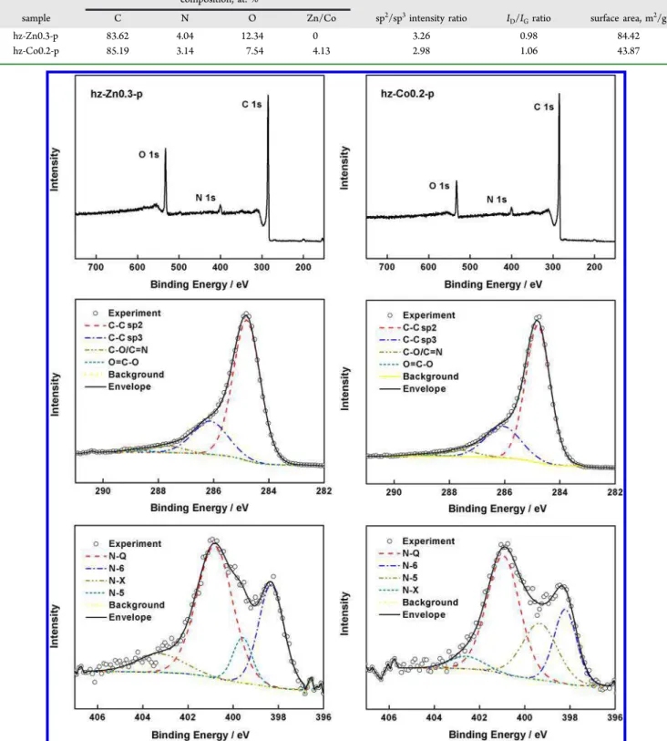
weight of hz, hz-Zn0.3, and hz-Co0.2 decreased about 45.91%, 26.30%, and 27.72% when the temperature rose from 200 to 300 °C, respectively. This confirms that the existence of Zn2+ and Co2+ largely impeded the early thermal decomposition of protein network by inducing stronger interactions and providing a solid support. This improved thermal stability Table 2. Atomic Concentrations of Elements, sp2/sp3Intensity Ratios, ID/IGRatios, and Surface Areas of Freestanding Carbon Films Synthesized from ZnAc and CoAc Precursors
composition, at. %
sample C N O Zn/Co sp2/sp3intensity ratio I
D/IGratio surface area, m2/g
hz-Zn0.3-p 83.62 4.04 12.34 0 3.26 0.98 84.42
hz-Co0.2-p 85.19 3.14 7.54 4.13 2.98 1.06 43.87
Figure 4.XPS spectra of (left) hz-Zn0.3-p and (right) hz-Co0.2-p: (top row) survey spectra, (middle row) deconvoluted C 1s peaks, and (bottom row) deconvoluted N 1s peaks.
ACS Applied Materials & Interfaces Research Article
DOI:10.1021/acsami.7b05159
ACS Appl. Mater. Interfaces 2017, 9, 23731−23740
explained well the retained film shape of hz-Zn0.3 and hz-Co0.2 with fibrous structures after pyrolysis, as observed by SEM.
3.2. Nitrogen-Doped Carbon Fiber Structure. In our preliminary experiment, the electrochemical properties of hz-Zn0.3 and hz-Co0.2 were not satisfactory, since the transition metal oxides suffer from poor electrical conductivity and limited electrochemical stability during cycles.2,20 Therefore, the
hz-Zn0.3 and hz-Co0.2 carbon fibers were further treated with hydrochloric acid solution for 72 h to remove the zinc and cobalt nanoparticles. Figure 3 shows the synthetic process of nitrogen-doped carbon fibers and their morphology before and after acid corrosion. The electrospun prolamin protein fabrics containing Zn2+ and Co2+ exhibited light yellow and pink colors, respectively, and formed the freestanding carbon films after carbonization. The obtained carbon films were highly flexible regardless of acid treatment. This feature was different from the carbon fibers prepared from PAN and cobalt, where the composite fibers were very brittle before acid corrosion.7 These carbon films were also much more flexible than the ones derived from electrospun prolamin protein fibers with Ca2+. This suggests that the composite structures formed by prolamin proteins with Zn2+ and Co2+ could more effectively release stress and avoid damage of carbon fibers during bending.31The results reveal that the prolamin proteins are promising hosts for fabricating metal oxide/carbon composites, and these flexible carbon films have potential to fulfill the rising global demand for wearable energy storage devices.32As shown inFigure 3B, some nanosized granules existed on the surfaces of hz-Zn0.3 and hz-Co0.2 carbon fibers. Those granules on fiber surfaces disappeared after acid corrosion, and hz-Zn0.3-p had a relatively smooth surface, whereas hz-Co0.2-p showed a sea island-like morphology. The diameters of carbon fibers also decreased from 0.37 ± 0.11 and 0.29 ± 0.08 μm to 0.26 ± 0.06 and 0.22 ± 0.05 μm, respectively. The altered surface morphology and reduced fiber diameters indicate that the outer layer of hz-Zn0.3 and hz-Co0.2 fibers was dissolved by HCl. The HCl solution was used not only for its ability to dissolve zinc and cobalt nanoparticles but also for its inertness toward carbon;7therefore, it could be deduced that the major components in the outer layer of hz-Zn0.3 and hz-Co0.2 fibers were transition metal oxides. This may be attributed to shrinkage of the carbon component during heating, which exposed some transition metal oxides and resulted in the poorly conductive fiber surface. It explained the unsatisfying electro-chemical properties of hz-Zn0.3 and hz-Co0.2 as revealed in our preliminary experiment. High-resolution TEM was subsequently employed to examine the microstructure and composition of hz-Zn0.3-p and hz-Co0.2-p. Clearly, there was a network of graphitic layers created in hz-Zn0.3-p (Figure 3D). These layers were parallel to each other along the axis of the
fiber, and the distance between them was 0.34 nm, which corresponds to the (002) plane of carbon. No zinc nano-particles could be observed inside hz-Zn0.3-p (Figure 3D inset andFigure S2). This demonstrated that the HCl solution not only etched the surface nanoparticles but also infiltrated into the internal region of the fibers. The complete removal of zinc nanoparticles could generate interconnected channels and inner pores in carbon fibers, which might facilitate the migration of ions through the fibers. At the same time, the graphitic layers could serve as the uninterrupted charge freeway network for quick electron transfer.33For hz-Co0.2-p, onionlike structures appeared and the distance between the carbon layers was 0.34 nm. During the carbonization process, the cobalt precursor was reduced to metallic cobalt, which could crystallize the adjacent amorphous carbon to form onionlike graphitic layers.34 The cobalt nanoparticles are located in the onionlike structure, and plenty of nanoparticles still existed in the fibers after acid corrosion for 72 h (longer than the suggested time of 48 h, as shown in Figure S2).7 It could be deduced that hz-Co0.2-p formed a denser structure to prevent the penetration of HCl, which could lead to the decrease in interconnected channels and the increased fiber density.
To investigate the chemical elements and bonding state, energy-dispersive spectrometric (EDS) and X-ray photo-electron spectroscopic (XPS) analyses were performed for hz-Zn0.3-p and hz-Co0.2-p. The percentages of C, N, and O in carbon fibers obtained by EDS are summarized inTable 2, and the XPS results are shown inFigure 4. Both samples had three obvious peaks, which were attributed to C, N, and O elements. Zn peak was absent from the survey spectrum as expected, but only 0.09% Co was detected. This was due to the very low penetration depth of X-ray photoelectrons, which could not reach the inner Co nanoparticles embedded in carbon fibers. Both samples displayed a similar narrow and asymmetric C 1s peak at 248.8 eV, corresponding to a graphite lattice structure.35 The deconvoluted C 1s spectrum displayed four
binding energies at 284.8, 286.1, 287.6, and 288.8 eV, which are assigned to sp2- and sp3-hybridized carbon and C−O and O C−O groups, respectively.36 The intensity ratio of sp2 to sp3 binding energies of the C 1s spectrum is known to be a good measure of the degree of graphitization.37The sp2to sp3ratios of hz-Zn0.3-p and hz-Co0.2-p were 3.26 and 2.98. This implies that a higher degree of graphitization occurred in the carbon fibers induced by Zn2+. Four types of N groups were observed in these two samples, namely, pyridinic N (N-6) at 398.2 eV, pyrrolic/pyridinic N (N-5) at 399.3 eV, quaternary N (N-Q) at 401.0 eV, and oxidized N (N-X) at 402.6 eV.38N-Q was the predominant N type for hz-Zn0.3-p and hz-Co0.2-p (52.23% and 48.62% of total N); it is located in the middle of the graphite plane and could enhance electronic conductivity by
assisting electron transfer through the carbon. The sum of N-5 and N-6 in hz-Zn0.3-p (38.36%) was less than that in hz-Co0.2-p (46.18%), and these two tyhz-Co0.2-pes of N grouhz-Co0.2-ps could contribute positively to the capacitance performance of carbon materials through faradic reaction-based pseudocapacitance. The hz-Zn0.3-p sample contained more N-X groups, and their hydrophilic nature could improve the wettability between electrode and electrolyte.
X-ray powder diffraction (XRD) patterns of hz-Zn0.3-p and hz-Co0.2-p are shown inFigure 5a. For hz-Zn0.3-p, two peaks centered at approximately 26° and 43.5° were distinctly detected, corresponding to the (002) and (100) planes of the graphite C 2H crystalline hexagonal structure (ICDD 00-041-1487).8,10From the θ value of (002) plane, the layer-to-layer distance (d spacing) between graphitic layers is calculated from
eq 3:
λ θ
=
d /(2 sin )
002 002 (3)
where λ is the X-ray wavelength used in the experiment. The distance between carbon layers was thus 0.34 nm, which was in accordance with TEM observation. No typical diffraction peaks of zinc nanoparticles were found. It confirmed that almost no residual zinc existed inside the hz-Zn0.3-p fibers. In addition to the peak at approximately 26°, the hz-Co0.2-p sample also showed three peaks at 44.3°, 51.6°, and 75.9°. These were identified as the (111), (200), and (220) planes of β-Co with a face-centered cubic (fcc) structure.39It meant that Co existed in the fibers as metallic nanocrystallites rather than oxides since it was embedded in carbon fibers after formation during the carbonization process. Raman spectroscopy was also employed to investigate the degree of graphitization. As shown inFigure 5b, two feature peaks emerged at 1340 and 1585 cm−1for both samples. The former corresponds to the A1gsymmetry related to local defects and disorder carbon with sp3 bonding (D band), and the latter to the E2gtangential stretching mode of an ordered graphitic structure with sp2hybridization (G band).40
A greater ratio of integrated intensity of D peak to G peak (ID/
IG) means a lesser degree of graphitization. The ID/IGvalues of hz-Zn0.3-p and hz-Co0.2-p were 0.98 and 1.06, respectively. This was consistent with the XPS results and confirms that a greater degree of graphitization occurred in the carbon fibers induced by Zn2+. The porous textures of nitrogen-doped freestanding carbon films were determined by nitrogen isothermal adsorption/desorption at 77 K (Figure 5c). The BET specific surface areas of hz-Zn0.3-p and hz-Co0.2-p were
84.42 and 43.87 m2/g, respectively. Nitrogen adsorption increased rapidly at low relative pressure (P/P0 < 0.01), suggesting the existence of nanopores (<2 nm). Both samples showed a typical type IV isotherm with a N2hysteresis loop, indicating the mesoporosity of carbon fibers.41 This was confirmed by the pore-size distribution, where these two samples contained mesopores with size ranges of 2.5−30 nm. The adsorption capacity exhibited sharp growth at high pressures (0.9 < P/P0< 1) without a plateau, indicating that macropores existed. According to the DFT method that has been suggested for calculating the pore-size distribution of carbons,42 the pore volumes of hz-Zn0.3-p and hz-Co0.2-p were 0.10 and 0.08 cm3/g, respectively. Therefore, it could be concluded that various pores existed in both samples to facilitate the diffusion of electrolyte. Although the acid corrosion removed the outside cobalt nanoparticles and generated the coarse surface of hz-Co0.2-p, its surface area and pore volume were still smaller than those of hz-Zn0.3-p. This was because some Co nanocrystals inside the fibers were embedded, which reduced the formation of interconnected channels and increased the fiber density.
3.3. Carbon Film Mechanical Properties and Wett-ability. The mechanical strength of carbon films before and after acid corrosion was investigated to study the handling properties. As shown in Figure 6a, all the samples exhibited multistage increases of stress, indicating that the carbon fibers were not tightly entangled and could slightly slide under stretching. This might be the reason for high flexibility of carbon films regardless of acid treatment. Films synthesized with Zn2+ were stronger than the cobalt ones (p < 0.05) because the diameters of hz-Zn0.3 and hz-Zn0.3-p fibers were relatively larger than those of hz-Co0.2 and hz-Co0.2-p fibers, respectively, which could allow better diffusion of stress. The presence of metal nanoparticles not only supported the carbon fibers but also affected the continuity of carbon structure. The complete removal of zinc nanoparticles resulted in hardly changed strength (p > 0.05) from 10.56 ± 0.82 to 10.88 ± 2.10 MPa, while the carbon fibers with embedded cobalt nano-particles had obviously decreased strength of 5.56 ± 0.92 MPa after acid corrosion.
Water contact angles were measured for Zn0.3-p and hz-Co0.2-p to evaluate their surface wettability. Since in this work an aqueous solution of KOH was used as electrolyte, carbon films with higher hydrophilicity are preferred. As shown in
Figure 6b, dry hz-Zn0.3-p and hz-Co0.2-p exhibited similar
Figure 6.(a) Typical stress−strain curves of Zn0.3, Co0.2, Zn0.3-p, and Co0.2-p. (b) Images of contact-angle measurements on hz-Zn0.3-p and hz-Co0.2-p.
ACS Applied Materials & Interfaces Research Article
DOI:10.1021/acsami.7b05159
ACS Appl. Mater. Interfaces 2017, 9, 23731−23740
contact angles of 130.4° ± 1.0° and 136.3° ± 0.7°, respectively, illustrating the hydrophobicity of the carbon films. This is expected because carbonaceous materials are intrinsically hydrophobic.43 Therefore, after acid treatment, hz-Zn0.3-p and hz-Co0.2-p were directly washed with ultrapure water and kept in 6 M KOH for 2 h before the electrochemical measurements, to facilitate the diffusion of aqueous electrolyte to the electrode surface.
3.4. Electrochemical Properties. The electrochemical properties of hz-Zn0.3-p and hz-Co0.2-p were tested in 6 M KOH at 25 °C to evaluate their performance as supercapacitor electrodes. These freestanding carbon films were easily cut into small square pieces and directly used as working electrode without addition of polymer binder or conducting agent.Figure 7a,b shows the CV curves at scan rates of 10, 25, 50, and 100
mV/s with a potential window ranging from −1.0 to 0 V. The hz-Zn0.3-p electrode displayed a quasi-rectangular pattern with slight distortion, indicating the double-layer capacitor nature of the charge−discharge process. It is worth noting that the specific current density of hz-Zn0.3-p was greater than that of hz-Co0.2-p when the scan rates were 50 and 100 mV/s (p < 0.05). This suggests that the hierarchical porous structure of hz-Zn0.3-p was more easily accessed by the electrolyte ions, and thus the hz-Zn0.3-p electrode material had the greater capacitance. This could be explained in three aspects: First, the addition of zinc acetate in prolamin protein fibers induced the higher degree of graphitization during pyrolysis as confirmed by XPS and Raman results. Second, the hz-Zn0.3-p samhz-Zn0.3-ple contained relatively large amounts of total N, N-Q, and N-X as revealed by XPS results. These nitrogen groups
Figure 7.(a, b) CV curves of (a) hz-Zn0.3-p and (b) hz-Co0.2-p at varied potential scan rates (10−100 mV/s). (c, d) Galvanostatic charge− discharge curves of (c) Zn0.3-p and (d) Co0.2-p at different current densities (1−20 A/g). (e) Specific capacitance of Zn0.3-p and hz-Co0.2-p as a function of current density. (f) Cycle stability and capacitance retention of hz-Zn0.3-p at 10 A/g.
assisted electron transfer through the carbon and improved the wettability between electrode and electrolyte. Third, hz-Zn0.3-p had relatively larger surface area and pore volume since the zinc nanoparticles in carbon fibers were removed by HCl solution, which generated the interconnected channels for electrolyte ions to migrate through the fibers and the inner pores. The galvanostatic charge−discharge curves of Zn0.3-p and hz-Co0.2-p at different current densities are shown inFigure 7c,d. These measurements are essential to predict the performance of carbon material in practical applications.20For both samples, the charge−discharge potential was nearly proportional to the charge−discharge time, revealing the fast I−V response. However, the charge−discharge time of hz-Zn0.3-p was longer than that of hz-Co0.2-p at the same current density. The specific capacitances were calculated fromeq 1on the basis of discharging time and are plotted inFigure 7e as a function of current density. The carbon film synthesized with the addition of zinc acetate delivered greater specific capacitance over the hz-Co0.2-p electrode material. When the current density rose from 1 to 2, 5, 8, 10, 15, and 20 A/g, the specific capacitance of hz-Zn0.3-p was gradually reduced from 393 to 368, 355, 324, 325, 292, and 284 F/g, respectively. It is well-known that a greater current density/scanning rate prevents the electrolyte ions from accessing the whole surface of electrode by reducing diffusion time, resulting in decreased capacitance. It was worth noting that the capacitance retention of hz-Zn0.3-p was 72.3% when the current density rose from 1 to 20 A/g, while the corresponding specific capacitance of hz-Co0.2-p was 291 F/g at 1 A/g, and the retention ratio was only 43.3% with the increase of current density to 20 A/g. These results were in accordance with the CV measurements. It should be mentioned that the all-carbon supercapacitors fabricated from electrospun PAN fibers combining with Co2+-assistant acid corrosion process exhibited a specific capacitance of 104.5 F/g (at 0.2 A/g) and a capacitance retention of 56.5% (at 10 A/g) in 0.5 M H2SO4;7 while the supercapacitor electrodes obtained from electrospun zinc acetate/PAN fibers showed a specific capacitance of 178.2 F/g (at 1 mA/cm2) and a capacitance retention of about 59% (at 20 mA/cm2) in 6 M KOH.20 In
consideration of the outstanding performance of hz-Zn0.3-p, its cycling stability was further tested at a current density of 10 A/g (Figure 7f). The specific capacitance was 97.8% retained after 2000 charging−discharging cycles. This result was similar to our previous work, where the carbon fibers stabilized by Ca2+ retained about 98% of capacitance after 5000 cycles of charging−discharging at 10 A/g. This excellent cycling stability suggests the potential long lifetime of prolamin protein-derived carbon materials in alkaline electrolyte, which makes them favorable for industrial and household applications.
4. CONCLUSION
Four transition metal ions (Co2+, Ni2+, Cu2+, and Zn2+) were incorporated in electrospun prolamin protein fibers. These ions not only promoted stronger interactions among protein molecules but also provided solid support to prevent the excessive shrinkage of protein fibers and induced the formation of graphitic layers during pyrolysis. By addition of Zn2+ and Co2+, flexible freestanding nitrogen-doped carbon films with a well-retained fibrous structure were successfully synthesized. Compared to hz-Co0.2-p, the hz-Zn0.3-p sample had a greater degree of graphitization; more total N, N-Q, and N-X; and larger surface area and pore volume. These features improved the wettability between electrode and electrolyte and facilitated
the migration of electrolyte ions, thus endowing hz-Zn0.3-p with better capacitance. The specific capacitance of hz-Zn0.3-p was 393 F/g at 1 A/g, and its capability retention was 72.3% at 20 A/g and 97.8% after 2000 charging−discharging cycles at 10 A/g. These findings reveal the promising applications of the binder-free, flexible carbon film derived from prolamin proteins in capacitive energy-storage devices. Moreover, its capacity can be improved by further activation steps in future investigations.
■
ASSOCIATED CONTENT*
S Supporting InformationThe Supporting Information is available free of charge on the
ACS Publications websiteat DOI:10.1021/acsami.7b05159. Two figures showing SEM images of electrospun fibers before pyrolysis and EDS spectra and mapping of carbon fibers after acid treatment (PDF)
■
AUTHOR INFORMATION Corresponding Author*Telephone +1-780-492-0038; fax +1-780-492-8914; e-mail
lingyun.chen@ualberta.ca.
ORCID
Yixiang Wang: 0000-0001-8386-7491
Lingyun Chen:0000-0002-8956-7358
Notes
The authors declare no competing financial interest.
■
ACKNOWLEDGMENTSThe Natural Sciences and Engineering Research Council of Canada (NSERC), Alberta Crop Industry Development Fund Ltd. (ACIDF), Alberta Innovates Bio Solutions (AI Bio), and Alberta Barley Commission are gratefully acknowledged for their financial support, as well as Canada Foundation for Innovation (CFI) for equipment support. L.C. is grateful to the Natural Sciences and Engineering Research Council of Canada (NSERC)-Canada Research Chairs Program for its financial support.
■
REFERENCES(1) Fan, L.; Yang, L.; Ni, X.; Han, J.; Guo, R.; Zhang, C. Nitrogen-Enriched Meso-Macroporous Carbon Fiber Network as a Binder-Free Flexible Electrode for Supercapacitors. Carbon 2016, 107, 629−637.
(2) Abouali, S.; Garakani, M. A.; Zhang, B.; Xu, Z.; Heidari, E. K.; Huang, J.; Huang, J.; Kim, J. Electrospun Carbon Nanofibers with in Situ Encapsulated Co3O4 Nanoparticles as Electrodes for High-Performance Supercapacitors. ACS Appl. Mater. Interfaces 2015, 7, 13503−13511.
(3) Li, J.; Liu, E.; Li, W.; Meng, X.; Tan, S. Nickel/Carbon Nanofibers Composite Electrodes as Supercapacitors Prepared by Electrospinning. J. Alloys Compd. 2009, 478, 371−374.
(4) Yang, H.; Hong, W.; Dong, L. A Controlled Biochemical Release Device with Embedded Nanofluidic Channels. Appl. Phys. Lett. 2012,
100, No. 153510.
(5) Lopez-Rubio, A.; Sanchez, E.; Wilkanowicz, S.; Sanz, Y.; Lagaron, J. M. Electrospinning as a Useful Technique for the Encapsulation of Living Bifidobacteria in Food Hydrocolloids. Food Hydrocolloids 2012,
28, 159−167.
(6) Yohe, S. T.; Colson, Y. L.; Grinstaff, M. W. Superhydrophobic Materials for Tunable Drug Release: Using Displacement of Air To Control Delivery Rates. J. Am. Chem. Soc. 2012, 134, 2016−2019.
(7) Liu, Y.; Zhou, J.; Chen, L.; Zhang, P.; Fu, W.; Zhao, H.; Ma, Y.; Pan, X.; Zhang, Z.; Han, W.; Xie, E. Highly Flexible Freestanding Porous Carbon Nanofibers for Electrodes Materials of
High-ACS Applied Materials & Interfaces Research Article
DOI:10.1021/acsami.7b05159
ACS Appl. Mater. Interfaces 2017, 9, 23731−23740
Performance All-Carbon Supercapacitors. ACS Appl. Mater. Interfaces 2015, 7, 23515−23520.
(8) Al-Enizi, A. M.; Elzatahry, A. A.; Abdullah, A. M.; AlMaadeed, M. A.; Wang, J.; Zhao, D.; Al-Deyab, S. Synthesis and Electrochemical Properties of Nickel Oxide/Carbon Nanofiber Composites. Carbon 2014, 71, 276−283.
(9) Huang, Y.; Lai, F.; Zhang, L.; Lu, H.; Miao, Y.; Liu, T. Elastic Carbon Aerogels Reconstructed from Electrospun Nanofibers and Graphene as Three-Dimensional Networked Matrix for Efficient Energy Storage/Conversion. Sci. Rep. 2016, 6, No. 31541.
(10) Nie, G.; Lu, X.; Chi, M.; Jiang, Y.; Wang, C. CoOxNanoparticles Embedded in Porous Graphite Carbon Nanofibers Derived from Electrospun Polyacrylonitrile@Polypyrrole Core-Shell Nanostructures for High-Performance Supercapacitors. RSC Adv. 2016, 6, 54693− 54701.
(11) Miyauchi, H.; Tsuda, Y.; Minozoe, A.; Tanaka, S.; Arito, H.; Tsukahara, T.; Nomiyama, T. Occupational Exposure to N,N-Dimethylformamide in the Summer and Winter. Ind. Health 2014,
52, 512−520.
(12) Scheibel, T. Protein Fibers as Performance Proteins: New Technologies and Applications. Curr. Opin. Biotechnol. 2005, 16, 427− 433.
(13) Wang, Y.; Chen, L. Electrospinning of Prolamin Proteins in Acetic Acid: The Effects of Protein Conformation and Aggregation in Solution. Macromol. Mater. Eng. 2012, 297, 902−913.
(14) Moomand, K.; Lim, L. T. Oxidative Stability of Encapsulated Fish Oil in Electrospun Zein Fibres. Food Res. Int. 2014, 62, 523−532. (15) Wang, Y.; Chen, L. Fabrication and Characterization of Novel Assembled Prolamin Protein Nanofabrics with Improved Stability, Mechanical Property and Release Profiles. J. Mater. Chem. 2012, 22, 21592−21601.
(16) Wang, Y.; Chen, L. Cellulose Nanowhiskers and Fiber Alignment Greatly Improve Mechanical Properties of Electrospun Prolamin Protein Fibers. ACS Appl. Mater. Interfaces 2014, 6, 1709− 1718.
(17) Wang, Y.; Yang, J.; Chen, L. Convenient Fabrication of Electrospun Prolamin Protein Delivery System with Three-Dimen-sional Shapeability and Resistance to Fouling. ACS Appl. Mater.
Interfaces 2015, 7, 13422−13430.
(18) Chen, L. F.; Zhang, X. D.; Liang, H. W.; Kong, M.; Guan, Q. F.; Chen, P.; Wu, Z. Y.; Yu, S. H. Synthesis of Nitrogen-Doped Porous Carbon Nanofibers as an Efficient Electrode Material for Super-capacitors. ACS Nano 2012, 6, 7092−7102.
(19) Yang, J. Investigation of Barley Proteins’ Interfacial Properties and Their Applications as Nanoscaled Materials. Doctoral dissertation, University of Alberta, Canada, 2016; pp 133−160, doi:10.7939/ R3736MC8K.
(20) Kim, C. H.; Kim, B. H. Zinc Oxide/Activated Carbon Nanofiber Composites for High-Performance Supercapacitor Electrodes. J. Power
Sources 2015, 274, 512−520.
(21) Iqbal, N.; Wang, X.; Ge, J.; Yu, J.; Kim, H. Y.; Al-Deyab, S. S.; El-Newehy, M.; Ding, B. Cobalt Oxide Nanoparticles Embedded in Flexible Carbon Nnaofibers: Attractive Material for Supercapacitor Electrodes and CO2Adsorption. RSC Adv. 2016, 6, 52171−52179.
(22) Wang, C.; Tian, Z.; Chen, L.; Temelli, F.; Liu, H.; Wang, Y. Functionality of Barley Proteins Extracted and Fractionated by Alkaline and Alcohol Methods. Cereal Chem. 2010, 87, 597−606.
(23) Silva, S. S.; Maniglio, D.; Motta, A.; Mano, J. F.; Reis, R. L.; Migliaresi, C. Genipin-Modified Silk-Fibroin Nanometric Nets.
Macromol. Biosci. 2008, 8, 766−774.
(24) Selling, G. W.; Woods, K. K.; Biswas, A. Electrospinning Formaldehyde-Crosslinked Zein Solutions. Polym. Int. 2011, 60, 537− 542.
(25) Elazzouzi-Hafraoui, S.; Nishiyama, Y.; Putaux, J.; Heux, L.; Dubreuil, F.; Rochas, C. The Shape and Size Distribution of Crystalline Nanoparticles Prepared by Acid Hydrolysis of Native Cellulose. Biomacromolecules 2008, 9, 57−65.
(26) Barreto, P. L. M.; Pires, A. T. N.; Soldi, V. Thermal Degradation of Edible Films Based on Milk Proteins and Gelatin in Inert Atmosphere. Polym. Degrad. Stab. 2003, 79, 147−152.
(27) Eckert, E.; Bamdad, F.; Chen, L. Metal Solubility Enhancing Peptides Derived from Barley Protein. Food Chem. 2014, 159, 498− 506.
(28) Lyckfeldt, O.; Brandt, J.; Lesca, S. Protein Forming − A Novel Shaping Technique for Ceramics. J. Eur. Ceram. Soc. 2000, 20, 2551− 2559.
(29) Li, X.; Mei, Q.; Dai, X.; Ding, G. Effect of Anaerobic Digestion on Sequential Pyrolysis Kinetics of Organic Solid Wastes Using Thermogravimetric Analysis and Distributed Activation Energy Model.
Bioresour. Technol. 2017, 227, 297−307.
(30) Torres-Giner, S.; Lagaron, J. M. Zein-Based Ultrathin Fibers Containing Ceramic Nanofillers Obtained by Electrospinning. I. Morphology and Thermal Properties. J. Appl. Polym. Sci. 2010, 118, 778−789.
(31) Cheng, Y.; Huang, L.; Xiao, X.; Yao, B.; Yuan, L.; Li, T.; Hu, Z.; Wang, B.; Wan, J.; Zhou, J. Flexible and Cross-Linked N-Doped Carbon Nanofiber Network for High Performance Freestanding Supercapacitor Electrode. Nano Energy 2015, 15, 66−74.
(32) Inagaki, M.; Yang, Y.; Kang, F. Carbon Nanofibers Prepared via Electrospinning. Adv. Mater. 2012, 24, 2547−2566.
(33) He, Y.; Chen, W.; Zhou, J.; Li, X.; Tang, P.; Zhang, Z.; Fu, J.; Xie, E. Constructed Uninterrupted Charge-Transfer Pathways in Three-Dimensional Micro/Nanointerconnected Carbon-Based Elec-trodes for High Energy-Density Ultralight Flexible Supercapacitors.
ACS Appl. Mater. Interfaces 2014, 6, 210−218.
(34) Barakat, N. A. M.; Kim, B.; Park, S. J.; Jo, Y.; Jung, M. H.; Kim, H. Y. Cobalt Nanofibers Encapsulated in a Graphite Shell by an Electrospinning Process. J. Mater. Chem. 2009, 19, 7371−7378.
(35) Takahagi, T.; Ishitani, A. XPS Study on the Surface Structure of Carbon Fibers Using Chemical Modification and Carbon-1s Line Shape Analysis. Carbon 1988, 26, 389−396.
(36) Diaz, J.; Paolicelli, G.; Ferrer, S.; Comin, F. Separation of the sp3 and sp2 Components in the C1s Photoemission Spectra of Amorphous Carbon Films. Phys. Rev. B: Condens. Matter Mater. Phys. 1996, 54, 8064−8069.
(37) Chu, P. K.; Li, L. Characterization of Amorphous and Nanocrystalline Carbon Films. Mater. Chem. Phys. 2006, 96, 253−277. (38) Ania, C. O.; Khomenko, V.; Raymundo-Piñero, E.; Parra, J. B.; Beguin, F. The Large Electrochemical Capacitance of Microporous Doped Carbon Obtained by Using a Zeolite Template. Adv. Funct.
Mater. 2007, 17, 1828−1836.
(39) Liu, Y.; Ling, J.; Li, W.; Zhang, X. Effective Synthesis of Carbon-Coated Co and Ni Nanocrystallites with Improved Magnetic Properties by AC Arc Discharge Under an N2 Atmosphere.
Nanotechnology 2004, 15, 43−47.
(40) Cesano, F.; Scarano, D.; Bertarione, S.; Bonino, F.; Damin, A.; Bordiga, S.; Prestipino, C.; Lamberti, C.; Zecchina, A. Synthesis of ZnO-Carbon Composites and Imprinted Carbon by the Pyrolysis of ZnCl2-Catalyzed Furfuryl Alcohol Polymers. J. Photochem. Photobiol., A 2008, 196, 143−153.
(41) He, J.; Zhou, M.; Wang, L.; Zhao, S.; Wang, Q.; Ding, B.; Cui, S. Electrospinning in Situ Synthesis of Graphene-Doped Porous Copper Indium Disulfide/Carbon Composite Nanofibers for Highly Efficient Counter Electrode in Dye-Sensitized Solar Cells. Electrochim. Acta 2016, 215, 626−636.
(42) Landers, J.; Gor, G. Y.; Neimark, A. V. Density Functional Theory Methods for Characterization of Porous Materials. Colloids
Surf., A 2013, 437, 3−32.
(43) Tai, M. H.; Gao, P.; Tan, B. Y. L.; Sun, D. D.; Leckie, J. O. Highly Efficient and Flexible Electrospun Carbon-Silica Nanofibrous Membrane for Ultrafast Gravity-Driven Oil-Water Separation. ACS