1
Allosteric activation of periplasmic HtrA proteases
byAnna Katherine de Regt B.S., Engineering Swarthmore College, 2009
Submitted to the Microbiology Graduate Program in partial fulfillment of the requirements for the degree of
DOCTOR OF PHILOSOPHY at the
MASSACHUSETTS INSTITUTE OF TECHNOLOGY February 2015
© 2015 Anna K. de Regt. All rights reserved.
The author hereby grants to MIT permission to reproduce and to distribute publicly paper and electronic copies of this thesis document in whole or in part in any medium now known or
hereafter created
Signature of Author _____________________________________________________________ Anna K. de Regt Microbiology Graduate Program September 30, 2014
Certified by ___________________________________________________________________ Robert T. Sauer Salvador E. Luria Professor of Biology Thesis Supervisor
Accepted by ___________________________________________________________________ Michael T. Laub Professor of Biology Director, Microbiology Graduate Program
3
Allosteric activation of periplasmic HtrA proteases
byAnna Katherine de Regt
Submitted to the Microbiology Graduate Program on September 26, 2014 in partial fulfillment of the requirements for the degree of Doctor of Philosophy
Abstract
Escherichia coli responds to outer-membrane stress using a signaling cascade initiated when
DegS, an HtrA-family periplasmic protease, cleaves RseA, a transmembrane anti-sigma factor, causing downstream release of the σE sigma factor and transcription of stress-response genes. Each subunit of the DegS trimer contains a trypsin-like protease domain and a PDZ domain. In the absence of stress, autoinhibitory interactions stabilize an inactive conformation of DegS. Allosteric activation occurs as a consequence of the linked binding of unassembled outer-membrane proteins to the PDZ domains and RseA to the DegS active sites.
In Chapter 2, I probe the pathway of communication between the PDZ and protease domains and identify a set of residues crucial for signal propagation across the enzyme. This set of residues is conserved across the HtrA protease family, including orthologs linked to human disease, and thus a common mechanism of allosteric activation appears to function in all family members. No satisfactory mechanism explains how OMP-peptide binding breaks autoinhibitory interactions. In Chapter 3, I present evidence that peptide binding initiates a steric clash between the side chains of a PDZ residue and an L3-loop residue that forces rearrangement of the loop and simultaneously breaks autoinhibitory interactions.
In Pseudomonas aeruginosa, a homologous signaling cascade controls the release of the transcription factor AlgU, which directs the production of alginate, an exopolysaccharide that protects the bacterium from antibiotics and the host immune system. The HtrA protease AlgW is activated by misfolded OMPs to cleave its transmembrane substrate MucA. In Chapter 4, I describe a collaboration that identifies the protein CupB5 as a novel regulator of this pathway. Peptides corresponding to a region of CupB5 needed for mucoidy activate AlgW in conjunction with OMPs to stimulate cleavage of MucA in vitro.
Finally, in Chapter 5, I summarize my findings on DegS and describe aspects of DegS activation that are yet to be resolved.
Thesis supervisor: Robert T. Sauer
Title: Salvador E. Luria Professor of Biology
5
Acknowledgements
Many people have supported me and helped me along in my graduate school adventure. First, I would like to thank my advisor, Bob Sauer, for his mentorship, his excitement about science and teaching, his open-door policy, and for striking the right balance between attention to detail and maintaining an appropriately messy bench and lab notebook. Thanks also to my co-advisor, Tania Baker, and my committee members, Leona Samson and Amy Keating, for always being there for me with advice on experiments, careers, or purple clothing - whatever I needed. Thank you to my labmates both past and present, and especially my baymates, Steve and Ben, for making this such a fun, productive and unique place to do science. And thank you to my postdocs, J, Randall, Steve and Santiago, for teaching me so much and then leaving me, forcing me to grow up as a scientist.
A special thanks goes to my undergradute advisor, Stephen Miller, who taught me that there is nothing cooler in the world than protein mechanism and set me on the biochemical path. My time in graduate school was profoundly enriched by my friends, particularly from sMITe, MIT's ultimate frisbee team, and MITOC, the outing club. They have kept me laughing, kept me challenged, kept me focused, and kept me out of lab. We have shared countless adventures that I will never forget.
And last but most importantly, unending thanks to my family, for their unconditional love and support. It has been incredible.
7 Table of Contents Title Page 1 Abstract 3 Acknowledgements 5 Table of Contents 7
List of Figures and Tables 10
Chapter 1: Introduction - The role of HtrA proteases in the outer-membrane stress response
13
Cellular proteolysis 14
HtrAs 14
Outer-membrane protein quality control 17
Gram negative outer envelope 17
Signal transmission across membranes 18
σE
pathway 19
The DegS protease 21
DegS allostery 20
DegS crystal structures 22
DegS activation via OMP-peptide binding 25
DegS binding to RseA substrate 26
The DegP protease 27
The AlgU RIP pathway in P. aeruginosa 29
Thesis overview 30
Referemces 31
Chapter 2: Conserved pathways of allosteric communication in HtrA-family proteases 41
Abstract 42
Introduction 43
Results 47
Residues that Play Major Roles in Allosteric Switching 49
Residues that Play Minor Roles or No Role in Allosteric Switching 50
8
Mutations that Block Allosteric Switching in DegP 55
Discussion 59
Methods 61
Protein Expression and Purification 61
Enzymatic and Biochemical Assays 62
Structural Refinement 62
Sequence Alignment 62
Accession Numbers 63
Acknowledgements 63
References 64
Chapter 3: Allosteric activation of the DegS stress-response protease by OMP-peptide binding requires a steric clash that destabilizes autoinhibitory interactions
69
Abstract 70
Introduction 71
Results 74
L3 interactions autoinhibit DegS 74
A steric-clash model of activation 79
Testing the clash model in vivo 82
Discussion 86
Methods 89
Protein Expression and Purification 89
Enzymatic and Biochemical Assays 89
Construction of L3 mutant library 90
β-galactosidase assay 90
Sequence alignment 91
Modeling 91
Acknowledgements 91
9
Chapter 4: Overexpression of CupB5 activates alginate overproduction in
Pseudomonas aeruginosa by a novel AlgW-dependent mechanism
95
Abstract 96
Introduction 97
Results 99
Overexpression of CupB5 induces mucoidy 99
Genetic requirements for CupB5-induced mucoidy 100
CupB5 contains an internal signal for alginate overproduction 101
CupB5 peptides stimulate OMP-activated AlgW cleavage of MucA and partially relieve MucB inhibition in vitro
103
Suppressing and enhancing alginate production 109
Discussion 112
Experimental procedures 115
Bacteria strains, plasmids, and growth conditions 115
Transposon mutagenesis 115
Genetic construction, transformation and conjugation 116
Protein preparation and Western blotting 116
Alginate assay 117
β-galactosidase assay 117
Proteins and peptides 117
Cleavage assays 119
MucA-resin assay 119
Acknowledgements 120
References 120
Supplementary figures and tables 124
Chapter 5: Conclusions, perspectives and future directions 131
Conclusions and perspectives 132
Future directions and unsuccessful experiments 134
10
Table of Figures and Tables
Fig. 1.1 The HtrA family of proteases. 15
Fig. 1.2 The E. coli periplasmic stress response 20
Fig. 1.3 Allosteric activation of DegS 23
Fig. 1.4 Residues important for stabilizing active vs. inactive conformations of DegS 23
Fig. 1.5 Two models for activation of DegS by OMP peptides 26
Fig. 1.6 Inactive DegP hexamers assemble into higher-order cages upon substrate binding
28
Fig. 2.1 The DegS trimer equilibrates between inactive and active structures 45
Fig. 2.2 Mutational effects on DegS cleavage of RseA 48
Fig. 2.3 Structural determinants of DegS activation 52
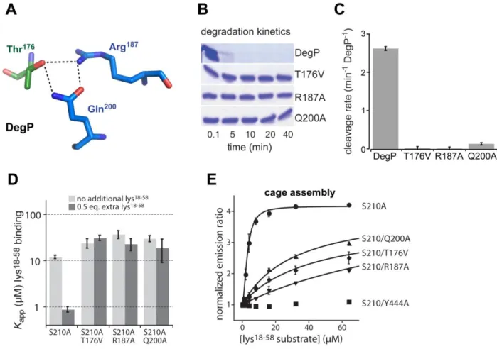
Fig. 2.4 Allosteric ligands and catalytic and regulatory elements in active DegS 54 Fig. 2.5 Effects of activation-cluster mutations on DegP activity and cage assembly 57 Fig. 2.6 Side chains important for DegS allostery are conserved in the HtrA family 60
Table 2.1 Biochemical characterization of DegS variants 67
Table 2.2 Re-refined structures statistics 68
Fig. 3.1 Allosteric activation of DegS 72
Fig. 3.2 The L3 loop makes autoinhibitory contacts 75
Fig. 3.3 Effects of L3 loop mutations on DegS activity and OMP-peptide binding 78
Fig. 3.4 The steric clash model of DegS activation 80
Fig. 3.5 Large side chains at the clashing L3 position support DegS activation 81 Fig. 3.6 Large side chains also support activation in vivo. 84
Fig. 3.7 Conservation of L3 loop residues. 87
11
Fig. 4.1 Overproduction of CupB5 causes mucoid conversion in PAO1 100 Fig. 4.2 Genetic requirements for CupB5-mediated alginate overproduction 101 Fig. 4.3 Delineation of the CupB5 signal that activates alginate production 103 Fig. 4.4 CupB5 peptides relieve MucB inhibition of MucA cleavage by AlgW 104 Fig. 4.5 CupB5 peptides do not compete with MucB for MucA binding but stimulate
WVF-activated AlgW cleavage of MucA.
107
Fig. 4.6 Inducing and suppressing alginate production in P. aeruginosa strains. 110 Fig. 4.S1 Alginate production and promoter activity of PalgU and PalgD in PAO1,
PAO1-VE2 (mucE overexpressed) and PAO1-VE22
124
Fig. 4.S2 CupB5 peptides do not relieve RseB inhibition of RseA cleavage by DegS 125 Fig. 4.S3 The CupB5 GYYYTVV motif is found in many orthologs in sequenced
strains of P. aeruginosa
126
Table 4.S1 Truncations of CupB5 to identify the signal that activates alginate production 127
Table 4.S2 Strains and plasmids used in this study 128
Fig. 5.1 Activation of DegS by OMP peptide 133
13
Chapter 1
Introduction:
The role of HtrA proteases in the outer-membrane
stress response
14
Cellular proteolysis
The proteome of a cell is in constant flux, as proteins are continuously synthesized, modified, and degraded. Some regulatory proteins must have their numbers tightly controlled so that pathway flux and cellular homeostasis are properly maintained. Other regulatory proteins are degraded in response to stress signals, allowing changes in transcription or translation. Protein degradation allows amino acids to be recycled from unnecessary or damaged proteins and used to synthesize new proteins, helping cells adapt to quickly changing environmental and/or developmental challenges. Finally, misfolded or unfolded proteins often need to be degraded before they aggregate and cause cell dysfunction or disease. Cellular quality control systems must ensure that misfolded or damaged proteins are degraded efficiently without rogue degradation of incorrect substrates. In times of stress, quality control proteases must be precisely activated to ensure a timely stress response, but also must be tightly regulated such that the response ceases upon the removal of stress.
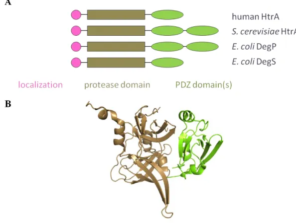
HtrAs
HtrA proteases represent a large family, with members in all domains of life. Each HtrA protease consists of an N-terminal localization domain that targets the enzyme to a cellular compartment, to a membrane, or to be secreted; a trypsin-like serine protease domain; and one or more C-terminal PDZ domains (Fig. 1.1). In some cases, additional functional domains are also present (Clausen et al., 2002). PDZ domains are an abundant class of small globular domains found in a wide variety of multidomain proteins, in which they usually play a regulatory role (Wang et al., 2010). The human genome appears to contain over 150 PDZ-containing proteins, with more than 250 non-redundant PDZ domains (Wang et al., 2010). PDZ domains generally bind C-terminal
15
peptide sequences, although some can bind internal sequences as well (Stiffler et al., 2007; Tonikian et al., 2008). The basic unit of HtrA function is a trimer, some of which further assemble into higher-order oligomers as a regulatory mechanism (Li et al., 2002; Walsh et al., 2004; Krojer et al., 2008; Kim et al., 2011; Truebestein et al., 2011).
Fig. 1.1. The HtrA family of proteases. (A) Each HtrA subunit contains an N-terminal localization domain, a trypsin-like protease domain, and one or two C-terminal PDZ domains. (B) The structure of one subunit of human HtrA2 (PDB ID: 1LCY).
HtrA stands for high-temperature requirement A, which reflects the knockout phenotype of the first family member identified, an Escherichia coli protein that is often called DegP (Lipinska et
al., 1989). Subsequently, HtrA proteases have been identified in many single- and multi-cellular
16
protein quality control (Ades et al., 1999; Huesgen et al., 2009; Mahomedmohaideen et al., 2008; Chien et al., 2009; Patel et al., 2014; Bakker et al., 2014). Humans have four HtrA proteins, HtrA1-4. HtrA1 is a secreted enzyme, which cleaves fibronectin and activates matrix metalloproteases and has been implicated roles in a wide range of diseases, including gingivitis, osteoarthritis, Alzheimer's disease, and preeclampsia (Grau et al., 2005; Grau et al., 2006; Hara
et al., 2009; Lorenzi et al., 2009; Lorenzi et al., 2014). HtrA2, a mitochondrial protein, is the
best-studied human HtrA. It was originally thought that its most important role is pro-apoptotic, as HtrA2 is released into the cytoplasm during stress, where it is processed into its active form and cleaves anti-apoptotic proteins (Suzuki et al., 2001; Cilenti et al., 2004). More recently, however, the focus has shifted to a role in cellular quality control, as HtrA2 can cleave the β-amyloid precursor protein (Gray et al., 2000; Huttenen et al., 2007; Park et al., 2006; Li et al., 2010). HtrA2 knockout mice have a neurodegenerative parkinsonian phenotype, and some Parkinson's Disease patients have mutations in the htrA2 gene (Jones et al., 2003; Martins et al., 2004; Strauss et al., 2005). HtrA3 is associated with many pregnancy- and cancer-related processes, such as placental development, endometrial cancer, ovarian cancer, and lung cancer, but almost nothing is known about its substrates or mechanism (Nie et al., 2003; Bowden et al., 2006; Nie et al., 2006; Narkiewicz et al., 2008; Narkiewicz et al., 2009; Beleford et al., 2010). HtrA4, the least-studied human family member, has high homology to HtrA1 and has also been suggested to be involved in preeclampsia (Wang et al., 2012).
Little detailed structural or mechanistic information is available for any of the human HtrA enzymes. Inactive and active crystal structures have been solved for HtrA1, and limited mutagenesis studies have been performed, revealing that neither the substrates tested, the PDZ
17
domain, nor the N-terminal domain seem to regulate enzyme activity (Truebestein et al., 2011; Eigenbrot et al., 2012). A crystal structure of HtrA2 in an inactive conformation has been solved, revealing a domain arrangement in which the PDZ domain packs against active site (Li et al., 2002). This arrangement suggests a regulatory mechanism in which activation induces a large conformational change in which the PDZ domain shifts away from the protease domain, but mutations that disrupt the protease-PDZ interface resulted in a decrease in proteolytic activity (Li
et al., 2002). A crystal structure and an NMR structure of the PDZ domain of HtrA3 have been
solved (Appleton et al., 2007). There are no structures of HtrA4.
At present, much of our understanding of HtrA proteolysis comes from studies of the structure and mechanism of two E. coli family members, the periplasmic proteases DegP and DegS. Together with a third family member, DegQ, these bacterial proteases are involved in protein quality control in the periplasm and in responding to stresses that affect the outer cell envelope.
Outer-membrane protein quality control
Gram negative outer envelope
Gram-negative bacteria have an envelope consisting of an outer membrane (OM), the periplasmic compartment, and an inner membrane (IM). A thin peptidoglycan layer runs through the periplasm, anchoring the OM and giving the cell its shape and rigidity. The OM serves as a protective barrier and problems with OM assembly or integrity serve as an important marker for environmental stress. Each face of the OM is chemically distinct, phospholipids comprising the inner leaflet and glycolipids comprising the outer leaflet. The glycolipids are overwhelmingly lipopolysaccharide (LPS), which consists of a glucosamine disaccharide with acyl chains
18
extending into the bilayer, a polysaccharide core, and the O-antigen, a long polysaccharide chain protruding to the outside of the cell (Raetz and Whitfield, 2002). LPS biogenesis begins on the cytoplasmic face of the inner membrane, and the full molecule is transported by a series of seven proteins across the IM, though the periplasm, and finally into the OM (Silhavy et al., 2010).
Outer-membrane proteins (OMPs), mostly transmembrane β-barrel porins, are the principal non-lipid component of the OM. These OMPs are tasked with controlling the permeability of the OM. Some are simple porins that enable passive diffusion of small molecules, whereas others allow passage only of specific substrates. The porins are mostly trimeric and are arranged such that their C-terminal tails are packed at the oligomeric interfaces and occluded from solvent (Cowan et al., 1992; Cowan et al., 1995). OMPs are synthesized in the cytoplasm, translocated across the IM, associate with periplasmic chaperones as they pass through this compartment, and are finally inserted into the OM by a complex protein machine (Silhavy et al., 2010; Hagan et
al., 2011). The OM is essential, and Gram-negative bacteria have evolved methods to monitor its
health and to respond to OM stress. As discussed below, these systems require the sensing of OM-specific signals in the periplasm and communication of this information across the inner membrane to the cytoplasm.
Signal transmission across membranes
Cells employ diverse systems for propagating external signals across a membrane to the cytoplasm. In eukaryotes, G-coupled protein receptors, transmembrane proteins located at the plasma membrane, bind extracellular small molecules, causing a conformational change that allows a partnered G-protein on the cytoplasmic face to hydrolyze GTP and initiate a signaling
19
cascade (Wettschureck and Offermanns, 2005). Many bacteria utilize two-component regulatory systems, in which a cytoplasmic domain of a transmembrane histidine kinase is autophosphorylated in response to an external signal and then transfers that phosphate to an associated response regulator, resulting in a conformational change in the response regulator that allows it to up- or down-regulate relevant genes (Mascher et al., 2006). Regulated membrane proteolysis (RIP), in which a transmembrane protein is sequentially cleaved by two proteases, resulting in the release of a cytoplasmic signal, is also widely used to propagate diverse signals across membranes (Ades 2008). For example, in the human sterol-sensing system, site-1-protease (S1P) and site-2-site-1-protease (S2P) in the Golgi membrane sequentially cleave a transmembrane protein in response to low-sterol conditions, releasing transcription factors that promote biosynthesis of sterols and fatty acids (Chen and Zhang, 2010). In E. coli and some other Gram-negative bacteria, a RIP cascade is used to sense and respond to OM stress.
σE
pathway
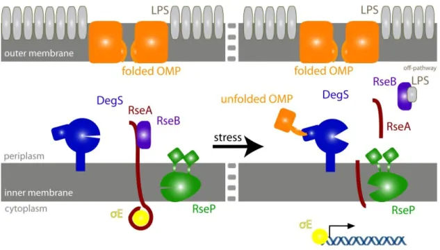
When E. coli are exposed to environmental conditions that threaten the OM, a RIP system propagates a periplasmic stress signal across the IM to the cytoplasm (Fig. 1.2). In the absence of stress, OMPs and LPS molecules undergo normal biogenesis, passing through the periplasm on their way to the OM; the alternative sigma factor σE is sequestered against the IM by the cytoplasmic domain of RseA, a transmembrane anti-sigma factor (De Las Peñas et al., 1997; Missiakas et al., 1997); RseB, a periplasmic protein with homology to members of the lipid-binding family, binds the periplasmic domain of RseA and protects it from proteolysis (Cezairliyan and Sauer, 2007); and DegS protease, which is anchored to periplasmic face of the IM, waits in a catalytically inactive state.
20
Fig. 1.2: The E. coli periplasmic stress response. In the absence of stress, σE is sequestered by RseA. Under stress conditions, unassembled OMPs bind and activate DegS, and periplasmic LPS binds RseB, releasing it from RseA. These dual signals induce cleavage of RseA and initiate a proteolytic cascade that frees σE and results in transcription of stress-response genes.
When OM biogenesis pathways are disrupted by stress, unassembled OMPs and intermediates in LPS biogenesis accumulate in the periplasm (Mecsas et al., 1993; Tam et al., 2005; Klein et al., 2009). The exposed C-termini of the unassembled periplasmic OMPs bind to the PDZ domains of DegS, activating its proteolytic activity (Walsh et al., 2003). Periplasmic LPS molecules compete for binding RseB with RseA, freeing the site-1 cleavage sequence (Lima et al., 2013). Activated DegS then cleaves the unprotected periplasmic domain of RseA at site 1, which initiates a proteolytic cascade. Site-1 cleavage of RseA by DegS recruits the integral-membrane RseP protease, which cleaves RseA at site 2 within the IM. This site-2 cleavage releases the inhibited complex of σE bound to the cytoplasmic domain of RseA from the inner membrane (Alba et al., 2002; Kanehara et al., 2002). σE is then released to up-regulate stress-response genes when cytoplasmic AAA+ proteases, such as ClpXP, unfold and degrade the cytoplasmic domain
21
of RseA (Chaba et al., 2007). σE regulates transcription of diverse genes relevant to cell-envelope health, including σE
itself, RseA, RseB, the periplasmic DegP protease, periplasmic chaperones (skp, dsbC, fkpA), and biogenesis genes for LPS, phospholipids, and IM and OM proteins (Rouviere et al., 1995; Raina et al., 1995; Dartigalongue et al., 2001; Raivio et al., 2001; Rezuchova et al., 2003).
The DegS protease
DegS allostery
As the site-1 protease, DegS is the gatekeeper for the OM-stress response. It must remain largely inactive in the absence of stress but cleave RseA when periplasmic OMPs accumulate. Like other HtrA proteases, DegS is a trimer. Proteolytic activation can be modeled by classic Monod-Wyman-Changeux (MWC) allostery, in which periplasmic OMPs and RseA bind more tightly to active than inactive DegS trimers and thus increase the equilibrium population of active enzymes (Monod et al., 1965; Sohn and Sauer, 2009). Both binding events are positively cooperative, allowing the switch to be ultra-sensitive with respect to OMP and RseA concentration (Sohn et
al., 2007).
Most E. coli OMPs have a YxF motif at the C terminus, where Y is tyrosine, x is any amino acid, and F is phenylalanine (Struyve et al., 1991). Peptides with C-terminal YxF sequences were initially found to bind to the PDZ domain of DegS and to active cleavage of RseA (Walsh et al., 2003). The identity of the “x” position affects the strength of binding to DegS, but saturating concentrations of peptide differing only at this penultimate position activate DegS cleavage to very similar extents (Sohn et al., 2007). Interestingly, OMP peptides with the same C-terminal
22
YxF tripeptide but with different lengths or N-terminal sequences have different affinities for active and inactive trimers of DegS and therefore activate the enzyme to different maximal extents (Sohn and Sauer, 2009). A YYF tripeptide is the most potent activator of DegS yet to be identified, and saturating concentrations increase DegS activity ~1000-fold over the basal cleavage rate (Sohn and Sauer, 2009). The detailed mechanism by which OMP-peptide binding to the PDZ domain activates DegS has been controversial, as discussed below, but studies of hybrid enzymes show that OMP-peptide binding to one PDZ domain activates both cis and trans protease domains (Mauldin and Sauer, 2012). Moreover, the peptide-free PDZ domain appears to inhibit DegS protease activity, as deletion of the PDZ domains results in a variant (DegS∆PDZ) with increased protease activity in vitro and in vivo (Walsh et al., 2003; Sohn et al., 2007).
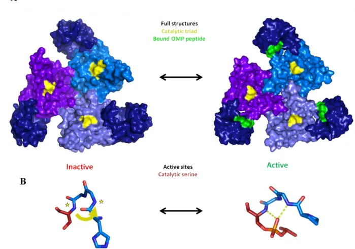
DegS crystal structures
Crystal structures of DegS trimers in an OMP-free inactive conformation and an OMP-bound active state established the overall architecture of the enzyme and revealed subtle but important differences between the two states (Wilken et al., 2004; Zeth, 2004). In both structures, the protease domains of DegS pack together in the center of the trimer with the PDZ domains on the outside (Fig. 1.3A). However, OMP-bound active DegS has a properly formed active-site oxyanion hole and thus is capable of stabilizing a tetrahedral intermediate and catalyzing proteolysis, whereas rotation about the 198-199 peptide bond in the inactive OMP-free structure destroys the oxyanion hole (Fig. 1.3B).
23
Fig. 1.3. Allosteric activation of DegS. (A) In inactive (1SOT) and active (3GDS) DegS, the protease domains (light colors) pack together to stabilize the trimer and the PDZ domains (dark blue) are on the periphery. The catalytic triad is shown in yellow. OMP peptides (green) are bound to the PDZ domains of active DegS. (B) Structures of residues 198-201 (catalytic Ser201 is red) in inactive and active DegS. A functional oxyanion hole is absent in inactive DegS. In active DegS, rotation of the peptide backbone between residue 198 and 199 creates a functional oxyanion hole. In the active structure, which also harbors the H198P mutation, the active site serine is modified with isopropyl-fluorophosphate and mimics a tetrahedral degradation intermediate.
Importantly, crystal structures predicted interactions that could be broken or introduced to bias the allosteric equilibrium in a particular direction and thereby increase or decrease DegS activity, many of which have been subsequently tested by mutational studies. For example, His198 lies near the catalytic site and its backbone atoms must be properly oriented to form a functional oxyanion hole and support catalysis. The His198Pro mutation allows the mutant side chain, to
24
of DegS (Fig. 1.4). DegSH198P has higher basal and peptide-stimulated activity than wild-type DegS in vitro and results in higher σE activity in vivo (Sohn et al., 2009; Chaba et al., 2011).
Mutations can also bias the equilibrium in the inactive direction. In active DegS, the hydroxyl group of the Tyr162 side chain makes a hydrogen bond with the side chain of Asn197. When Tyr162 is mutated to alanine, this interaction is abolished and the resulting Y162A enzyme has extremely low degradation activity both in the absence and presence of OMP peptide (Sohn et
al., 2007). Tellingly, neither adding in the activating H198P mutation nor deleting the PDZ
domain can activate the Y162A variant, and both Y162A DegSΔPDZ and H198P/Y162A DegSΔPDZ crystallize in the inactive conformation (Sohn et al., 2010).
Fig. 1.4. Residues important for stabilizing active versus inactive conformations of DegS. The catalytic triad is shown in red and sites of relevant mutations in green. The H198P mutation stabilizes the functional conformation of the oxyanion hole by introducing Pro198, which packs against the neighboring Tyr162. This conformation is also stabilized in wild type DegS by the hydrogen bond between the side chains of Tyr162 and Asn197, an interaction that is absent the Y162A mutant.
A large collection of crystal structures of full-length DegS or the ΔPDZ variant with different mutations and bound to various OMP peptides provide support for a two-state allosteric model.
25
All structures contain a set of "triangular base" residues in the protease domain that assume the same conformation in active and inactive structures (Sohn et al., 2010). Another set of protease-domain residues assume one conformation in active structures and another conformation in inactive structures.
DegS activation via OMP-peptide binding
Two models have been suggested for the mechanism of enzyme activation by OMP-peptide binding (Fig. 1.5). Both models involve the L3 loop (residues 176–189), which extends from the protease domain towards the PDZ domain and assumes different conformations in active versus inactive DegS. In the peptide-activation model, the penultimate residue of PDZ-bound OMP peptide makes specific activating contacts with the L3 loop that stabilize the active conformation of the protease domain (Wilken et al., 2004; Hasselblatt et al., 2007). Depending on the identity of the penultimate residue and its interactions, the protease domain assumes one of many different conformations that can vary from somewhat active to very active. However, in crystal structures of peptide-bound DegS, no consistent contacts between bound OMP peptide and the L3 loop of the protease domain are observed (Sohn et al., 2009). Furthermore, changing the penultimate residue of the OMP peptide affects its affinity for DegS but has little effect on the maximal level of DegS activation at peptide saturation (Sohn et al., 2009). As previously mentioned, DegS crystal structures also reveal two basic conformations. Although there are small differences in surface loops and side-chain conformations among each class of structures, there is no evidence for the proposed multitude of somewhat-active states with uniquely-tuned active sites of differing activity.
26
Fig. 1.5. Two models for activation of DegS by OMP peptides. In the relief-of-inhibition model, OMP binding to the PDZ domain breaks inhibitory interactions made by the L3 loop on the protease domain. The peptide-activation model posits that OMP peptide bound to the PDZ domain makes specific activating contacts with the L3 loop.
The relief-of-inhibition model for OMP-peptide activation is an extension of the two-state model and posits that inhibitory contacts between the unliganded PDZ domain and the L3 loop stabilize inactive DegS, with peptide binding breaking or destabilizing inhibitory interactions (Walsh et al., 2003; Sohn et al., 2007; Sohn and Sauer, 2009). The simplest evidence for this model is that deleting the entire PDZ domain or making mutations that remove inter-domain contacts that are observed in inactive but not active DegS results in enzyme activation (Sohn et al., 2007; Sohn et
al., 2009; Sohn et al., 2010).
DegS binding to RseA substrate
The RseA substrate also allosterically activates DegS, but much less is known about how RseA interacts with DegS than about how OMP peptides bind. The periplasmic domain of RseA (residues 120-216) is an intrinsically disordered polypeptide that is cleaved by DegS between Val148 and Ser149. However, an RseA peptide (residues 143-152) that contains the scissile peptide bond is cleaved by DegS far more slowly than the full periplasmic domain, indicating that RseA
27
residues distant from the cleavage site, called exosites, are important for tight binding and/or efficient cleavage by DegS. A potential RseA exosite (residues 160-189) that also binds the inhibitor RseB has been identified, but one or more additional sites probably exist (Cezairliyan and Sauer, 2007). Several glutamine-rich regions of RseA appear to inhibit cleavage of full-length RseA by RseP, but mutating these regions did not significantly affect DegS cleavage (Kanehara et al., 2003). An RseA peptide spanning residues 135-185, which includes the RseB interaction site, binds DegS very similarly to full-length periplasmic RseA and is efficiently cleaved by DegS (A.K. de Regt, unpublished). Further studies will be required to map the exosites of RseA and determine how they bind DegS and contribute to allosteric activation.
The DegP protease
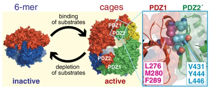
Studies of DegS have led to improved understanding of E. coli DegP, another periplasmic HtrA protease with a more complicated activation mechanism. DegP differs from DegS in having no membrane anchor and two PDZ domains per subunit instead of one (Waller and Sauer, 1996; Ponting, 1997). Like all HtrA proteases, DegP trimers appear to be the basic unit of function but can assemble into higher-order oligomers. For example, DegP forms inactive hexamers in which trimers pack face-to-face, but addition of protein or peptide substrates results in formation of catalytically active 12-, 18-, or 24-mer cages stabilized by interactions between the PDZ1 and PDZ2 domains (Fig. 1.6; Kim et al., 2012; Krojer et al., 2002; Jiang et al., 2008; Krojer et al., 2008). An allosteric model also describes DegP activity well, with substrate binding cooperatively and stabilizing the active conformation of trimers, which can then assemble into cages (Kim et al., 2011). The best DegP substrates contain a hydrophobic C-terminal residue that
28
binds to the PDZ1 domain of DegP and a small hydrophobic residue at the P1 position that binds to the active-site cleft (Krojer et al., 2008; Kim et al., 2011).
Fig. 1.6. Inactive DegP hexamers assemble into higher-order cages upon substrate binding. Cages are mediated by interactions between PDZ1 and PDZ2 and disassemble when substrate is depleted. (from Kim et al., 2012).
In wild type DegP, substrate binding is linked not only to proteolytic activation and formation of a functional oxyanion hole but also to cage formation, although these processes can be uncoupled by mutations, and substrate binding but not cage formation is required for protease activation (Kim et al., 2011; Kim and Sauer, 2012). DegP cages stay assembled until the concentration of intact substrates has been depleted, at which point they disassemble and revert back to inactive trimers or hexamers (Kim et al., 2011). This regulation of DegP activity is essential for cell viability, as DegP variants carrying double mutations that stabilize the active form and abolish cage formation are lethal to the cell (Kim and Sauer, 2014). Thus HtrA proteases have evolved a variety of mechanisms to regulate their activities.
29 The AlgU RIP pathway in P. aeruginosa
In Pseudomonas aerguinosa, a RIP system homologous to the E. coli σE pathway controls the release of AlgU, a stress-response transcription factor homologous to σE (Erickson and Gross, 1989; DeVries and Ohman, 1994; Rouvier et al., 1995; Ades, 2008). AlgW, which shares 44% sequence identity with DegS, is the site-1 protease, and MucA, a transmembrane anti-sigma factor with 34% sequence identity to RseA, is its substrate (Schurr et al., 1996; Cezairliyan and Sauer, 2009). The RseB homolog in the P. aeruginosa system is called MucB, and its inhibition of MucA cleavage by AlgW can be also be relieved by binding LPS (Cezairliyan and Sauer, 2007; Lima et al., 2013). A peptide corresponding to the C-terminus of MucE, a P. aeruginosa OMP, activates AlgW cleavage activity by binding to its PDZ domains (Cezairliyan and Sauer, 2009).
In addition to up-regulating stress-response genes, AlgU also directs transcription of the AlgD transcription factor, which in turn promotes transcription of genes for the biogenesis of alginate, an exopolysaccharide that renders the bacterium resistant to many antibiotics as well as to the human immune system (Govan and Deretic, 1996; Martin et al., 1993; Wood et al., 2006). For patients with cystic fibrosis, the production of alginate is a particular problem because their mucus-filled lungs have difficulty clearing chronic infections of P. aeruginosa (Folkesson et al., 2012). Over 80% of the alginate-producing clinical strains of P. aeruginosa have mutations in their mucA genes that cause constitutive activation of the AlgU pathway (Boucher et al., 1997).
30
Thesis overview
I have presented an introduction to the outer-membrane stress response in Gram-negative bacteria and to allostery in the E. coli DegS protease. In the following chapters, I further examine the activation mechanism of DegS and its homolog, P. aeruginosa AlgW. In Chapter 2, I examine the allosteric pathway for communication between the site of OMP peptide binding and the catalytic active site. Using mutagenesis and sequence alignments, I establish a network of conserved interactions that promote inter-subunit communication and stabilize the active conformation of DegS. Chapter 3 describes experiments seeking to understand the molecular mechanism by which OMP peptide activates DegS. I find that peptide binding causes a steric clash between a PDZ domain residue and a residue on the L3 loop, which forces the L3 loop to rearrange, breaking autoinhibitory interactions and activating the protease. In Chapter 4, I investigate a new protein activator of AlgW, periplasmic CupB5. I find that peptides corresponding to the activation signal on CupB5 bind to AlgW and stabilize the active form in conjunction with OMP peptide bound to the PDZ domain, a novel activation mechanism for this protein family. Finally, I conclude in chapter 5 with suggestions for further experiments to elucidate the activation mechanism of DegS.
31
References
Ades SE, Connolly LE, Alba BM, Gross CA. (1999). The Escherichia coli sigma(E)-dependent extracytoplasmic stress response is controlled by the regulated proteolysis of an anti-sigma factor. Genes Dev 13, 2449-61.
Ades SE. Regulation by destruction: design of the σE envelope stress response.(2008). Curr Opin Microbiol 11, 535-40.
Alba BM, Leeds JA, Onufryk C, Lu CZ, Gross CA. (2002). DegS and YaeL participate sequentially in the cleavage of RseA to activate the σE
-dependent extracytoplasmic stress response. Genes Dev 16, 2156-2168.
Appleton BA, Wiesmann C. (2007). Structural and functional analysis of the PDZ domains of human HtrA1 and HtrA3. Protein Sci 16, 2454-2471.
Bakker D, Buckley AM, de Jong A, van Winden VJ, Verhoeks JP, Kuipers OP, Douce GR, Kuijper EJ, Smits WK, Corver J. (2014). The HtrA-like protease CD3284 modulates virulence of Clostridium difficile. Infect Immun 02336-14.
Beleford D, Liu Z, Rattan R, Quagliuolo L, Boccellino M, Baldi A, Maguire J, Staub J, Molina J, Shridhar V. (2010). Methylation induced gene silencing of HtrA3 in smoking-related lung cancer. Clin Cancer Res 16, 398–409.
Boucher JC, Yu H, Mudd MH, Deretic V. (1997) Mucoid Pseudomonas aeruginosa in cystic fibrosis: Characterization of mucmutations in clinical isolates and analysis of clearance in a model of respiratory infection. Infect Immun 65, 3838–3846.
Bowden MA, Di Nezza-Cossens LA, Jobling T, Salamonsen LA, Nie G. (2006). Serine proteases HTRA1 and HTRA3 are down-regulated with increasing grades of human endometrial cancer. Gynecol Oncol 103, 253–60.
Cezairliyan BO, Sauer RT. (2007). Inhibition of regulated proteolysis by RseB. Proc Natl Acad Sci U S A 104, 3771-6.
Cezairliyan BO, Sauer RT. (2009). Control of Pseudomonas aeruginosa AlgW protease cleavage of MucA by peptide signals and MucB. Mol Microbiol 72, 368-79.
Chaba R, Alba BM, Guo MS, Sohn J, Ahuja N, Sauer RT, Gross CA. (2011). Signal integration by DegS and RseB governs the σE
-mediated envelope stress response in Escherichia coli. Proc Natl Acad Sci USA 108, 2106-11.
32
Chaba R, Grigorova IL, Flynn JM, Baker TA, Gross CA. (2007). Design principles of the
proteolytic cascade governing the sigmaE-mediated envelope stress response in Escherichia coli: keys to graded, buffered, and rapid signal transduction. Genes Dev 21, 124-36.
Chen G, Zhang X. (2010). New insights into S2P signaling cascades: regulation, variation, and conservation. Protein Sci 19, 2015-30.
Chien J, Ota T, Aletti G, Shridhar R, Boccellino M, Quagliuolo L, Baldi A, Shridhar V. (2009). Serine protease HtrA1 associates with microtubules and inhibits cell migration. Mol Cell Biol
29, 4177–4187.
Cilenti L, Soundarapandian MM, Kyriazis GA, Stratico V, Singh S, Gupta S, Bonventre JV, Alnemri ES, Zervos AS. (2004).Regulation of HAX-1 anti-apoptotic protein by Omi/HtrA2 protease during cell death. J Biol Chem 279, 50295–50301.
Clausen T, Southan C, Ehrmann M. (2002). The HtrA family of proteases: implications for protein composition and cell fate. Mol Cell 10, 443-55.
Cowan SW, Garavito RM, Jansonius JN, Jenkins JA, Karls-son R, Konig N, Pai EF, Pauptit RA, Rizkallah PJ, Rosenbusch JP, Rummel G, Schirmer T. (1995). The structure of OmpF porin in a tetragonal crystal form. Structure 3, 1041–1050.
Cowan SW, Schirmer T, Rummel G, Steiert M, Ghosh R, Pauptit RA, Jansonius JN, Rosenbusch JP. (1992). Crystal structures explain functional properties of two E. coli porins. Nature 358, 727-733.
Dartigalongue C, Missiakas D, Raina S. (2001). Characterization of the Escherichia coli σE
regulon. J Biol Chem. 276, 20866–20875.
De Las Peñas A, Connolly L, Gross CA. (1997). σE is an essential sigma factor in Escherichia coli. J Bacteriol 179, 6862–6864.
DeVries CA, and Ohman DE. (1994). Mucoid-to-nonmucoid conversion in alginate-producing
Pseudomonas aeruginosa often results from spontaneous mutations in algT, encoding a putative
alternative sigma factor, and shows evidence for autoregulation. J Bacteriol 176, 6677–6687.
Eigenbrot C, Ultsch M. (2012). Structural and functional analysis of HtrA1 and its subdomains. Structure 20, 1040-1050.
33
Erickson JW, and Gross CA. (1989). Identification of the σE
subunit of Escherichia coli RNA polymerase: a second alternate s factor involved in high-temperature gene expression. Genes Dev 3, 1462–1471.
Folkesson A, Jelsbak L, Yang L, Johansen HK, Ciofu O, Hoiby N, Molin S. (2012). Adaptation of Pseudomonas aeruginosa to the cystic fibrosis airway: an evolutionary perspective. Nat Rev Microbiol 10, 841-851.
Govan JRW, and Deretic V. (1996). Microbial pathogenesis in cystic fibrosis: mucoid
Pseudomonas aeruginosa and Burkholderia cepacia. Microbiol Rev 60, 539–574.
Grau S, Baldi A, Bussani R, Tian X, Stefanescu R, Przybylski M, Richards P, Jones
SA, Shridhar V, Clausen T, Ehrmann M.. (2005). Implications of the serine protease HtrA1 in amyloid precursor protein processing. Proc Natl Acad Sci USA 1026021–6026.
Grau S, Richards PJ, Kerr B, Hughes C, Caterson B, Williams AS, Junker U, Jones SA, Clausen T, Ehrmann M. (2006). The role of human HtrA1 in arthritic disease. J Biol Chem 2816124– 6129.
Gray CW, Ward RV, Karran E, Turconi S, Rowles A, Viglienghi D, Southan C, Barton A, Fantom KG, West A, Savopoulos J, Hassan NJ, Clinkenbeard H,Hanning C, Amegadzie B, Davis JB, Dingwall C, Livi GP, Creasy CL. (2000).Characterization of human HtrA2, a novel serine protease involved in the mammalian cellular stress response. Eur J Biochem 267, 5699– 5710 .
Hagan CL, Silhavy TJ, Kahne D. (2011). β-Barrel membrane protein assembly by the Bam complex. Annu Rev Biochem 80, 189-210.
Hara K, Shiga A, Fukutake T, Nozaki H, Miyashita A, Yokoseki A, Kawata H, Koyama A, Arima K, Takahashi T, Ikeda M, Shiota H, Tamura M, Shimoe Y,Hirayama M, Arisato T, Yanagawa S, Tanaka A, Nakano I, Ikeda S, Yoshida Y, Yamamoto T, Ikeuchi T, Kuwano R, Nishizawa M, Tsuji S, Onodera O. (2009). Association of HTRA1 mutations and familial ischemic cerebral small-vessel disease. N Engl J Med 360, 1729–1739.
Hasselblatt H, Kurzbauer R, Wilken C, Krojer T, Sawa J, Kurt J, Kirk R, Hasenbein S, Ehrmann M, and Clausen T. (2007). Regulation of the sigmaE stress response by DegS: how the PDZ domain keeps the protease inactive in the resting state and allows integration of different OMP-derived stress signals upon folding stress. Genes Dev. 21, 2659-2670.
34
Huesgen PF, Schuhmann H, Adamska I. (2009). Deg/HtrA proteases as components of a
network for photosystem II quality control in chloroplasts and cyanobacteria. Res Microbiol 160, 726–732.
Huttunen HJ, Guénette SY, Peach C, Greco C, Xia W, Kim DY, Barren C, Tanzi RE, Kovacs DM. (2007). HtrA2 regulates β-amyloid precursor protein (APP) metabolism through
endoplasmic reticulum-associated degradation. J Biol Chem 282, 28285–28295
Jiang J, Zhang X, Chen Y, Wu Y, Zhou ZH, Chang Z, Sui SF. (2008). Activation of DegP chaperone-protease via formation of large cage-like oligomers upon binding to substrate proteins. Proc Natl Acad Sci USA 105, 11939–11944.
Jones JM, Datta P, Srinivasula SM, Ji W, Gupta S, Zhang Z, Davies E, Hajnóczky G, Saunders TL, Van Keuren ML, Fernandes-Alnemri T, Meisler MH, Alnemri ES. (2003). Loss of Omi mitochondrial protease activity causes the neuromuscular disorder of mnd2 mutant
mice. Nature 425, 721–727.
Kanehara K, Ito K, Akiyama Y. (2002). YaeL (EcfE) activates the σE
pathway of stress response through a site-2 cleavage of anti-σE, RseA. Genes Dev 16, 2147-2155.
Kanehara K, Ito K, Akiyama Y. (2003). YaeL proteolysis of RseA is controlled by the PDZ domain of YaeL and a Gln-rich region of RseA. EMBO J 22, 6389-98.
Kim S, Grant RA, Sauer RT. (2011) .Covalent linkage of distinct substrate degrons controls assembly and disassembly of DegP proteolytic cages.Cell 145, 67–78.
Kim S, Sauer RT. (2012). Cage assembly of DegP protease is not required for
substrate-dependent regulation of proteolytic activity or high-temperature cell survival. Proc Natl Acad Sci USA 109, 7263-8.
Kim S, Sauer RT. (2014). Distinct regulatory mechanisms balance DegP proteolysis to maintain cellular fitness during heat stress. Genes Dev 28, 902-11.
Klein G, Lindner B, Brabetz W, Brade H, Raina S. (2009). Escherichia coli K-12 Suppressor-free Mutants Lacking Early Glycosyltransferases and Late Acyltransferases: minimal
lipopolysaccharide structure and induction of envelope stress response. J Biol Chem 284, 15369-89.
35
Krojer T, Garrido-Franco M, Huber R, Ehrmann M, Clausen T. Crystal structure of DegP (HtrA) reveals a new protease-chaperone machine. (2002). Nature 416, 455-9.
Krojer T, Sawa J, Schäfer E, Saibil HR, Ehrmann M, Clausen T. (2008). Structural basis for the regulated protease and chaperone function of DegP. Nature 453, 885–890.
Li W, Srinivasula SM, Chai J, Li P, Wu JW, Zhang Z, Alnemri ES, Shi Y. (2002). Structural insights into the pro-apoptotic function of mitochonrial serine protease HtrA2/Omi. Nat Struc Biol 9, 436-441.
Lima S, Guo MS, Chaba R, Gross CA, Sauer RT. (2013). Dual molecular signals mediate the bacterial response to outer-membrane stress. Science 340, 837-41.
Lipinska B, Fayet O, Baird L, Georgopoulos C. (1989). Identification, characterization, and mapping of the Escherichia coli htrA gene, whose product is essential for bacterial growth only at elevated temperatures. J Bacteriol 171, 1574–1584.
Lorenzi T, Marzioni D, Giannubilo S, Quaranta A, Crescimanno C, De Luca A, Baldi A, Todros T, Tranquilli AL, Castellucci M. (2009). Expression patterns of two serine protease HtrA1 forms in human placentas complicated by preeclampsia with and without intrauterine growth
restriction. Placenta 3035–40.
Lorenzi T, Niţulescu EA, Zizzi A, Lorenzi M, Paolinelli F, Aspriello SD, Baniţă M, Crăiţoiu S, Goteri G, Barbatelli G, Lombardi T, Di Felice R, Marzioni D, Rubini C, Castellucci M. (2014). The novel role of HtrA1 in gingivitis, chronic and aggressive periodontitis. PLoS One. 9, 96978.
Martin DW, Holloway BW, and Deretic V. (1993). Characterization of a locus determining the mucoid status of Pseudomonas aeruginosa: AlgU shows sequence similarities with a Bacillus sigma factor. J Bacteriol 175, 1153-1164.
Martins LM, Morrison A, Klupsch K, Fedele V, Noisoi N, Tesmann P, Abuin A, Grau E, Greppert M, Livi GP, Creasy CL, Martin A, Hargreaves I, Heales SJ, Okada H, Brandner S, Schulz JB, Mak T, Downward J. (2004). Neuroprotective role of the Reaper-related serine protease HtrA2/Omi revealed by targeted deletion in mice. Mol Cell Biol 24, 9848–9862.
Mascher T, Helmann JD, Unden G. (2006). Stimulus perception in bacterial signal-transducing histidine kinases. Microbiol Mol Biol Rev 70, 910-38.
36
Mauldin RV, and Sauer RT. (2013). Allosteric regulation of DegS protease subunits though a shared energy landscape. Nat. Chem. Biol. 9, 90-96.
Mecsas J, Rouviere PE, Erickson JW, Donohue TJ, Gross CA. (1993). The activity of sigma E, an Escherichia coli heat-inducible sigma-factor, is modulated by expression of outermembrane proteins. Genes Dev 7, 2618-28.
Missiakas, D., M. Lemaire, M. Mayer, C. Georgopoulos, and S. Raina. (1997). Modulation of the
Escherichia coli σE (RpoE) heat shock transcription factor activity by the RseA, RseB, and RseC proteins. Mol Microbiol 24, 355–372.
Monod J, Wyman J, Changeux JP. (1965). On the nature of allosteric transitions: A plausible model. J Mol Biol 12, 88-118.
Narkiewicz J, Klasa-Mazurkiewicz D, Zurawa-Janicka D, Skorko-Glonek J, Emerich J, Lipinska B. (2008). Changes in mRNA and protein levels of human HtrA1, HtrA2 and HtrA3 in ovarian cancer. Clin Biochem 41, 561–9.
Narkiewicz J, Lapinska-Szumczyk S, Zurawa-Janicka D, Skorko-Glonek J, Emerich J, Lipinska B. (2009). Expression of human HtrA1, HtrA2, HtrA3 and TGF-beta1 genes in primary
endometrial cancer. Oncol Rep 21, 1529–37.
Nie G, Li Y, Hale K, Okada H, Manuelpillai U, Wallace EM, Salamonsen LA. (2006). Serine peptidase HTRA3 is closely associated with human placental development and is elevated in pregnancy serum. Biol Reprod 74, 366–74.
Nie GY, Li Y, Minoura H, Batten L, Ooi GT, Findlay JK, Salamonsen LA. (2003). A novel serine protease of the mammalian HtrA family is up-regulated in mouse uterus coinciding with placentation. Mol Hum Reprod 9, 279–90.
Park HJ, Kim SS, Seong YM, Kim KH, Goo HG, Yoon EJ, Min do S, Kang S, Rhim H.. (2006). β-amyloid precursor protein is a direct cleavage target of HtrA2 serine protease. Implications for the physiological function of HtrA2 in the mitochondria. J Biol Chem 281, 34277–34287
Patel P, De Boer L, Timms P, Huston WM. (2014). Evidence of a conserved role for
Chlamydia HtrA in the replication phase of the chlamydial developmental cycle. Microbes Infect S1286-4579.
37
Ponting CP. (1997). Evidence for PDZ domains in bacteria, yeast, and plants. Protein Science 6, 464-468.
Raetz CR, Whitfield C. (2002). Lipopolysaccharide endotox-ins. Annu Rev Biochem 71, 635-700.
Raina S, Missiakas D, Georgopoulos C. (1995). The rpoE gene encoding the σE (σ24) heat shock sigma factor of Escherichia coli. EMBO J. 14, 1043–1055.
Raivio TL, Silhavy TJ. (2001). Periplasmic stress and ECF sigma factors. Annu Rev Microbiol 55, 591–624.
Rezuchova B, Miticka H, Homerova D, Roberts M, Kormanec J. (2003). New members of the Escherichia coli σE regulon identified by a two-plasmid system. FEMS Microbiol Lett. 225, 1-7.
Rouviere, PE, De Las Peñas A, Mecsas J, Lu CZ, Rudd KE, Gross CA. (1995). rpoE, the gene encoding the second heat-shock sigma factor, σE, in Escherichia coli. EMBO J. 14, 1032–1042.
Schurr MJ, Yu H, Martinez-Salazar JM, Boucher JC, Deretic V. (1996). Control of AlgU, a member of the sigma E-like family of stress sigma factors, by the negative regulators MucA and MucB and Pseudomonas aeruginosa conversion to mucoidy in cystic fibrosis. J Bacteriol. 16, 4997-5004.
Silhavy TJ, Kahne D, Walker S. The bacterial cell envelope. (2010). Cold Springs Harb Perspect Biol 5, 414.
Sohn J, Grant RA, Sauer RT. (2007). Allosteric activation of DegS, a stress sensor PDZ protease. Cell 131, 572-83.
Sohn J, Grant RA, Sauer RT. (2009). OMP peptides activate the DegS stress-sensor protease by a relief of inhibition mechanism. Structure 17, 1411-21.
Sohn J, Grant RA, Sauer RT. (2010). Allostery is an intrinsic property of the protease domain of DegS: implications for enzyme function and evolution. J Biol Chem 285, 34039-47.
Sohn J, Sauer RT. (2009). OMP peptides modulate the activity of DegS protease by differential binding to active and inactive conformations. Mol Cell 33, 64-74.
38
Stiffler MA, Chen JR, Grantcharova VP, Lei Y, Fuchs D, Allen JE, Zaslavskaia LA, MacBeath G. (2007). PDZ domain binding selectivity is optimized across the mouse proteome. Science
317, 364–369.
Strauss KM, Martins LM, Plun-Favreau H, Marx FP, Kautzmann S, Berg D, Gasser T, Wszolek Z, Müller T, Bornemann A, Wolburg H, Downward J, Riess O,Schulz JB, Krüger R.
(2005). Loss of function mutations in the gene encoding Omi/HtrA2 in Parkinson's disease. Hum Mol Genet 14, 2099–2111.
Struyve M, Moons M, and Tommassen J. (1991). Carboxy-termi-nal phenylalanine is essential for the correct assembly of a bacterial outer membrane protein. J Mol Biol 218, 141–148.
Suzuki Y, Imai Y, Nakayama H, Takahashi K, Takio K, Takahashi R. (2001). Serine protease, HtrA2, is released from the mitochondria and interacts with XIAP, inducing cell death. Mol Cell
8, 613–621
Tam C, Missiakas D. (2005). Changes in lipopolysaccharide structure induce the sigma(E)-dependent response of Escherichia coli. Mol Microbiol 55, 1403-12.
Tonikian R, Zhang Y, Sazinsky SL, Currell B, Yeh JH, Reva B, Held HA, Appleton BA,
Evangelista M, Wu Y, Xin X, Chan AC, Seshagiri S, Lasky LA, Sander C, Boone C, Bader GD, Sidhu SS. (2008). A specificity map for the PDZ domain family. PLoS Biol 6, e239.
Truebestein L, Tennstaedt A, Hauske P, Krojer T, Kaiser M, Clausen T, Ehrmann M. (2011). Substrate-induced remodeling of the active site regulates human HTRA1 activity. Nat Struct Mol Biol. 18, 386-388.
Waller PR, Sauer RT. (1996). Characterization of degQ and degS, Escherichia coli genes encoding homologs of the DegP protease. J Bacteriol 178, 1146-53.
Walsh NP, Alba BM, Bose B, Gross CA, Sauer RT. (2003). OMP peptide signals initiate the envelope-stress response by activating DegS protease via relief of inhibition mediated by its PDZ domain. Cell 113, 61-71.
Wang CK, Pan L, Chen J, Zhang M. (2010). Extensions of PDZ domains as important structural and functional elements. Protein Cell 1, 737-51.
39
Wang LJ, Cheong ML, Lee YS, Lee MT, Chen H. (2012). High-temperature requirement protein A4 (HtrA4) suppresses the fusogenic activity of syncytin-1 and promotes trophoblast invasion. Mol Cell Biol 32, 3707-17
Wilken C, Kitzing K, Kurzbauer R, Ehrmann M, Clausen T. (2004). Crystal structure of the DegS stress sensor: How a PDZ domain recognizes misfolded protein and activates a protease. Cell 117, 483-94.
Wood LF, Leech AJ, and Ohman DE. (2006). Cell wall-inhibitory antibiotics activate the
alginate biosynthesis operon in Pseudomonas aeruginosa: roles of σ22 (AlgT) and the AlgW and Prc proteases. Mol Microbiol 62, 412-426.
Wettschureck N, Offermanns S. (2005). Mammalian G proteins and their cell type specific functions. Physiol Rev 85, 1159-204.
Zeth K. (2004). Structural analysis of DegS, a stress sensor of the bacterial periplasm. FEBS Lett
41
Chapter 2
Conserved pathways of allosteric
communication in HtrA-family proteases
This work has been submitted to Structure as de Regt AK, Kim S, Sohn J, Grant RA, Baker TA, Sauer RT.
A.K.D. and J.S. purified and expressed recombinant proteins. A.K.D. performed all experiments involving DegS and wrote the initial manuscript. S.K. performed all experiments with DegP. R.A.G and R.T.S. re-refined the crystal structures.
42
Abstract
Escherichia coli responds to outer-membrane stress using a signaling cascade initiated when
DegS, an HtrA-family periplasmic protease, cleaves RseA, a transmembrane anti-sigma factor, causing downstream release of the σE
sigma factor and transcription of stress-response genes. Each subunit of the DegS trimer contains a trypsin-like protease domain and a PDZ domain. The trimeric protease domain is normally inactive because of intrinsic conformational preferences and autoinhibition mediated in part by the unliganded PDZ domains. Allosteric activation of RseA cleavage occurs as a consequence of the binding of unassembled outer-membrane proteins (OMPs) to the PDZ domains. Here, we identify a set of DegS residues that cluster together at subunit-subunit interfaces in the trimer, link the active sites and substrate-binding sites, and are crucial for stabilizing the active enzyme conformation in response to OMP signaling. These residues are conserved across the HtrA-protease family, including orthologs linked to human disease, and thus a common mechanism of allosteric activation in response to signaling appears to function in all family members. Indeed, mutation of residues at homologous positions in the DegP quality-control protease also eliminates allosteric activation.
43
Introduction
Allostery, a shift between distinct molecular structures, controls the biological activities of many enzymes, including proteases. In the periplasm of Escherichia coli, for example, heat and other environmental stresses allosterically activate the DegS protease, which is anchored to the inner membrane, and initiate an intramembrane proteolytic cascade that results in transcription of stress-response genes in the cytoplasm (Ades, 2008). DegS is a trimeric HtrA-family protease, in which each subunit contains a trypsin-like protease domain and a regulatory PDZ domain (Fig. 2.1A). The trimer is stabilized by packing between the protease domains. The principal substrate of DegS is the periplasmic domain of RseA, a transmembrane protein also containing a cytoplasmic domain that binds and inhibits the E transcription factor (Alba and Gross, 2004). Two stress-induced molecular signals are required for DegS cleavage of RseA in vivo. First, unassembled outer-membrane proteins (OMPs) accumulate in the periplasm during stress, and Tyr-Xxx-Phe (YxF) peptides at the C termini of these OMPs bind to the PDZ domains of DegS and stimulate proteolytic activity (Walsh et al., 2003). Second, lipopolysaccharides bind the RseB regulatory protein, releasing it from RseA, which allows cleavage of RseA at a single site by OMP-activated DegS (Lima et al., 2013). This initial cleavage event results in subsequent cleavage of RseA by other proteases, freeing σE to activate transcription of stress-response genes (Ades et al., 1999; Kanehara et al., 2002; Akiyama et al., 2004; Flynn et al., 2004; Chaba et al., 2007; Li et al., 2009).
In the absence of stress, DegS cleaves RseA at a low rate in vivo (Ades, 2008). Similarly, in the absence of OMP peptides in vitro, DegS cleaves the periplasmic domain of RseA much more slowly (>100-fold) than in their presence (Walsh et al. 2003; Sohn et al., 2007; Sohn and Sauer, 2009). Crystal structures of DegS or DegS∆PDZ trimers show two distinct conformations, one
44
inactive because of a malformed oxyanion hole and one with a functional His-Asp-Ser catalytic triad and oxyanion hole (Wilken et al., 2004; Zeth, 2004; Hasselblatt et al., 2007; Sohn et al., 2007; 2009; 2010). The PDZ domain has two major roles, keeping OMP-free DegS in an autoinhibited state (DegS∆PDZ has robust protease activity) and binding OMP peptide to allow activation. In structures of active DegS, the PDZ-bound OMP peptide is ~25 Å from the closest active site in the trimer. Allosteric activation over this distance is partially explained by remodeling of the protease-domain L3 loop, which is spatially adjacent to bound OMP peptide. This remodeling breaks autoinhibitory interactions between the L3 loop and the PDZ domain and allows formation of alternative contacts that appear to stabilize active DegS. Experiments using hybrid DegS enzymes with mixtures of full-length and ∆PDZ subunits show that all subunits in the trimer experience a strongly coupled energetic landscape, with peptide binding to a single PDZ domain stimulating proteolytic cleavage both in the attached protease domain and in neighboring domains (Mauldin and Sauer, 2013).
Proteolytic activation of DegS largely conforms to a two-state model of MWC allostery, in which the preferential binding of RseA substrates and OMP peptides to active DegS provides ~2.2 and ~4.4 kcal/mol of stabilization, respectively, relative to the inactive enzyme (Fig. 2.1B; Monod et al., 1965; Sohn and Sauer, 2009). Thus, only ~0.3% of substrate-saturated DegS assumes the active conformation, whereas ~83% of substrate-saturated and OMP-saturated DegS is active. DegS∆PDZ still behaves as an allosteric enzyme with respect to RseA cleavage (Sohn and Sauer, 2009; Sohn et al., 2007; 2010), indicating that the protease-domain trimer still adopts inactive and active conformations and thus contains the basic machinery required for allostery.
45
Fig. 2.1. The DegS trimer equilibrates between inactive and active structures. (A) Structure a DegS trimer in the active conformation with OMP peptides bound to the PDZ domains (pdb code 3GDS). Each DegS subunit (colored green, purple or blue) consists of a protease domain (lighter colors), which pack together to form the trimer, and a distal PDZ domain (darker colors). The catalytic triad is shown in light blue and YYF tripeptides bound to the PDZ domains are shown in red. (B) Free energies for conversion of inactive to active DegS in the free enzyme, the substrate-saturated enzyme, and the substrate and OMP-peptide saturated enzyme (Sohn and Sauer, 2009). These values would be decreased by ~4 kcal/mol for H198P DegS (Sohn et al., 2009; 2010). The column on the right lists the percentage of total DegS that assumes the active conformation for the unbound and ligand bound states.
46
Previous studies identified a handful of mutations that preferentially stabilize the inactive conformation compared to the active conformation of DegS or vice versa, and inspection of crystal structures suggested additional residues that may be involved in DegS allostery (Wilken
et al., 2004; Hasselblatt et al., 2007; Sohn et al., 2007; 2010; Sohn and Sauer, 2009). However,
there has been no comprehensive test of the importance of these residues in mediating the allosteric switch between DegS conformations. Here, we identify a critical set of amino acids needed for allosteric activation of DegS, providing a foundation for understanding regulation by substrate and OMP-peptide binding. These critical residues cluster at each subunit-subunit interface in the trimer and adjoin the active sites. Bound substrates would link these clusters and active sites around the trimer, providing a direct mechanism for allosteric activation. By contrast, bound OMP peptide appears to activate DegS indirectly by destabilizing autoinhibition. The cluster residues most important for DegS allostery are highly conserved within the HtrA-protease family, strongly suggesting that all of these enzymes function in similar fashions. Indeed, based on homology with DegS, we identify and characterize mutations that prevent allosteric activation of E. coli DegP, a trimeric HtrA quality-control protease that assembles into proteolytically active polyhedral cages in the presence of substrate proteins (Jiang et al. 2008; Krojer et al., 2008; Kim et al., 2011). These results have broad implications, as mutant forms of human HtrA homologs have been linked to cancer, vascular disease, arthritis, macular degeneration, and neurodegenerative disease (Grau et al., 2005; Coleman et al., 2008; Milner et al., 2008; Vande Walle et al., 2008; Chien et al., 2009; Hara et al., 2009; Bowden et al., 2010).
47
Results
Allosteric activation of DegS requires linkage between OMP-peptide binding to the PDZ domains, substrate binding to the protease domain, and the conformations of the active sites in each protease domain of the trimer (Sohn et al. 2007; Sohn and Sauer, 2009; Mauldin and Sauer, 2013). Based on interactions observed in DegS crystal structures, numerous side chains have previously been proposed to be important for allosteric switching or were selected for analysis in this study (Table 2.1). Most of these amino acids lie between the binding sites for OMP peptide in the PDZ domains and the proteolytic active sites. In the sections below, we discuss evidence for or against these residues playing important roles in allosteric switching.
To monitor proteolytic activity in vitro, we purified wild-type or mutant variants of DegS without the membrane anchor and determined second-order rate constants (kcat/KM) for cleavage
of sub-KM concentrations of a 35S-labeled protein fragment corresponding to the periplasmic
domain of RseA in the presence of saturating Tyr-Tyr-Phe (YYF) peptide (Sohn et al., 2007). The effects of different mutations were measured in otherwise wild-type backgrounds and also in combination with the H198P mutation, which stabilizes active DegS (Sohn et al., 2009; 2010) and increased the proteolytic activity of otherwise wild-type DegS ~6-fold under the assay conditions used (Fig. 2.2). The H198P mutation increased the relative proteolytic activities of some but not all mutations that diminish DegS activity (compare top and bottom panels in Fig. 2.2), allowing the relative importance of different mutations for proteolysis and allosteric switching to be determined.
48
Fig. 2.2. Mutational effects on DegS cleavage of RseA. Effects of DegS mutations in an otherwise wild-type background (upper panel) or in an H198P background (lower panel) on OMP-peptide stimulated proteolytic activity, expressed as the second-order rate constant (kcat/KM) for RseA cleavage. Values are averages of two or more
independent trials ± SEM. Cleavage reactions contained different sub-KM concentrations of 35
S-labelled RseA, DegS or variants (1 µM trimer), and YYF OMP peptide (230 µM). Initial cleavage rates normalized by total enzyme were plotted as a function of the RseA concentration, and the second-order rate constant was determined from the slope of a linear fit.