HAL Id: tel-02053165
https://tel.archives-ouvertes.fr/tel-02053165
Submitted on 1 Mar 2019
HAL is a multi-disciplinary open access archive for the deposit and dissemination of sci-entific research documents, whether they are pub-lished or not. The documents may come from teaching and research institutions in France or
L’archive ouverte pluridisciplinaire HAL, est destinée au dépôt et à la diffusion de documents scientifiques de niveau recherche, publiés ou non, émanant des établissements d’enseignement et de recherche français ou étrangers, des laboratoires
cross-talk with bitter taste and endocannabinoid
receptors
Lea Brissard
To cite this version:
Lea Brissard. Mechanisms of gustatory perception of dietary lipids : cross-talk with bitter taste and endocannabinoid receptors. Tissues and Organs [q-bio.TO]. Université Bourgogne Franche-Comté, 2018. English. �NNT : 2018UBFCK071�. �tel-02053165�
THESE DE DOCTORAT DE L’UNIVERSITE BOURGOGNE FRANCHE-COMTE PREPAREE A l’UFR Sciences de la Vie et de la Terre
Ecole doctorale n°554 Environnements - Santé En vue d’obtenir le grade de Docteur de l’Université de Bourgogne
Discipline : Sciences Vie
Spécialité : Physiologie de la Nutrition Par
Léa Brissard
Mechanisms of gustatory perception of dietary lipids: cross-talk with bitter taste
and endocannabinoid receptors
Thèse présentée et soutenue à Dijon, le 30 novembre 2018
Composition du Jury :
Pr. Degrace Pascal Pr UB-INSERM U1231, Dijon Président Dr. Cruciani-Guglielmacci Céline MCU Université Paris 7 UMR8251, Paris Rapporteur Dr. Goswami Nandu MCU Medical University of Graz Rapporteur
Titre : Mécanismes de perception gustative des lipides alimentaires : cross-talk avec les récepteurs du goût amer et des endocannabinoïdes.
Mots clés : Goût du gras, amer, endocannabinoïdes, cellules gustatives, HTC-8, CB1R
Introduction. L'obésité constitue l'un des principaux problèmes de santé publique en ce début du 21ème siècle. Sa prévalence augmente régulièrement, en particulier chez les enfants. Ce constat n'est pas anodin car l'obésité est généralement associée à diverses pathologies graves (diabète de type 2, hypertension et cancer,…). Ainsi, des investigations sur les mécanismes impliqués dans la perception gustative des lipides alimentaires pourraient éclairer leurs rôles dans l’incidence de l’obésité.
Buts et objectifs. Plusieurs études ont démontré le rôle des endocannabinoïdes et des aliments amers dans l’obésité. Ainsi, nous avons étudié l’interaction (cross-talk) des récepteurs cannabinoïdes et du goût amer avec le goût lipidique. Cette thèse comporte ainsi deux volets : les récepteurs cannabinoïdes (CB1R), le goût amer et leurs interactions avec les récepteurs lipidiques.
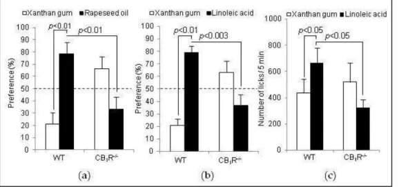
Résultats et discussion. Dans la première partie, nous avons étudié le rôle régulateur de CB1R. Ainsi, des tests comportementaux sur des souris CB1R-/- et des souris de type sauvage (WT) ont montré que l'invalidation du gène Cb1r était associée à une faible préférence pour les solutions contenant de l'huile de colza ou un acide gras à longue chaîne (AGLC) tel que l’acide linoléique (LA). L'administration de rimonabant, un agoniste-inverse de CB1R, chez la souris a également entraîné une faible préférence pour les acides gras alimentaires. Aucune différence dans l'expression des protéines CD36 et GPR120 n'a été observée dans les cellules des papilles gustatives des souris WT et CB1R-/-. La signalisation calcique via CD36 dans les cellules des papilles gustatives des souris CB1R-/- diminue de façon significative par rapport à celle observée dans les cellules gustatives des souris WT. Les cellules des papilles gustatives des souris CB1R-/- présentent également une diminution significative de l'ARNm de Pro-glucagon et de Glp-1r et un faible niveau basal de GLP-1. Nous rapportons que CB1R est impliqué dans la perception du goût du gras via la signalisation calcique et la sécrétion de GLP-1.
Résultats et discussion. Dans la seconde partie, nous avons d’abord caractérisé le phénotype de cellules fongiformes humaines (HTC-8). En effet, le projet de ma thèse comprend la caractérisation à l’échelle moléculaire des récepteurs amers et lipidiques et leur cross-talk dans ces cellules (collaboration BRAIN, Allemagne). Nous avons démontré que les cellules HTC-8 expriment PLCβ2 et l’α-gustducin à l’échelle des ARNm et des protéines. Elles expriment également TAS2R16 et TAS2R38 et ces mêmes cellules co-expriment CD36 et GPR120. Puis, nous avons étudié la signalisation via ces récepteurs en utilisant l’acide linoléique, un agoniste de CD36 et GPR120, la sinigrin, agoniste de TAS2R16 et TAS2R38, la salicin, agoniste du récepteur TAS2R16 et le phénylthiocarbamide, agoniste du récepteur TAS2R38. De plus, les études du signal calcique ont démontré que la signalisation en aval du goût gras partage une voie commune avec la signalisation en aval du goût amer, mettant en évidence un cross-talk entre ces deux modalités gustatives.
Bien que nous ayons montré le cross-talk entre les modalités gustatives amère et lipidique, il nous reste à étudier ces phénomènes à l’échelle de l’organisme. Ces résultats, d’ores et déjà, montrent que le goût amer et le récepteur cannabinoïde-1 sont liés à la sensibilité au goût du gras et doivent être pris en compte pour la gestion de l'obésité.
Abstract
Title : Mechanisms of gustatory perception of dietary lipids: cross-talk with bitter taste and endocannabinoid receptors.
Keywords : fat taste, bitter taste, endocannabinoids, taste cells, HTC-8, CB1R
Introduction. Obesity is one of the major public health problems at the beginning of the 21st century. Its prevalence is increasing steadily, especially among children. This observation is very important because obesity is generally associated with various serious pathologies (type 2 diabetes, hypertension and cancer, etc.). Thus, investigations into the mechanisms involved in the taste perception of dietary lipids could shed light on their roles in the incidence of obesity.
Aims and objectives. Several studies have demonstrated the role of endocannabinoids and bitter foods in obesity. Thus, we studied the cross-talk of cannabinoid receptors and bitter taste with lipid taste. This thesis consists of two components: cannabinoid (CB1R) and bitter taste receptors and their interactions with lipid receptors.
Results and discussion. In the first part, we studied the regulatory role of CB1R. The behavioural tests on CB1R-/- and wild-type (WT) showed that the invalidation of the Cb1r gene was associated with low preference for solutions containing rapeseed oil or a long chain fatty acid (LCFA) such as linoleic acid (LA). Administration of rimonabant, a CB1R inverse agonist, in mice also resulted in a low preference for dietary fatty acids. No differences in the expression of CD36 and GPR120 proteins were observed in the taste buds cells of WT and CB1R-/- mice. Calcium signalling via CD36 in the taste bud cells of CB1R-/- mice decreased significantly compared with those observed in the taste cells of WT mice. The taste bud cells of CB1R-/- mice also show a significant decrease in Pro-glucagon and Glp-1r mRNA and a low basal level of GLP-1. We report that CB1R is involved in the perception of fat taste via calcium signalling and secretion of GLP-1.
Results and discussion. In the second part, we first characterized the phenotype of human fungiform cells (HTC-8). Indeed, the project of my thesis includes molecular characterization of bitter and lipid receptors and their cross-talk in these cells (collaboration BRAIN, Germany). We have demonstrated that HTC-8 cells express PLCβ2 and α-gustducin at mRNA and protein levels. They also express TAS2R16 and TAS2R38 and the same cells co-express CD36 and GPR120. Then, we studied signalling via these receptors using linoleic acid, a CD36 and GPR120 agonist, sinigrin, TAS2R16 agonist and TAS2R38, salicin, TAS2R16 receptor agonist, and phenylthiocarbamide, TAS2R38 receptor agonist. In addition, calcium signal studies have shown that downstream fatty signalling shares a common path with downstream bitter taste signalling, highlighting a cross-talk between these two taste modalities.
Although we have shown the cross-talk between bitter and lipid taste modalities, we still have to study these phenomena at the level of the organism. These resultsshow that the bitter taste and the cannabinoid-1 receptor are related to the taste sensitivity of fat and must be taken into account for the management of obesity
Remerciements
Je tiens, tout d’abord, à remercier le Professeur Pascal Degrace qui me fait l’honneur de présider mon jury de thèse.
J’exprime, ensuite, toute ma reconnaissance aux Docteurs Céline Cruciani-Gugliani et Nandu Goswami pour avoir accepté d’être les rapporteurs de ma thèse, pour avoir consacré de leur temps à la lecture du manuscrit et pour leurs pertinents conseils.
Je souhaite remercier toutes les personnes qui m’ont aidée, de près ou de loin, pendant la réalisation de ma thèse.
Je remercie mon directeur de thèse, le Professeur Naim Khan, pour son accueil et pour m’avoir permis d’intégrer cette merveilleuse école de la vie qu’est la thèse.
Je remercie également le Docteur Aziz Hichami pour son aide.
Ce travail n’aurait pas été possible sans le soutien de l’Université de Bourgogne, du LipSTIC et de Brain Institute (Allemagne) qui m’ont permis de me consacrer sereinement à l’élaboration de ma thèse.
Je remercie également Julia Leemput, Sandrine Bellenger, Stéphanie Fioramonti, Line Folletet, Jean-Marc Blanche pour leur accueil chaleureux et leur disponibilité.
Aussi, ce travail n’aurait pas pu être mené à bien sans l’aide technique de Patricia Degrace et d’Adélie Dumont, ainsi que de l’aide du personnel de Cellimap, André Bouchot et Audrey Geissler.
De plus, je remercie toute l’équipe Padys pour son étroite collaboration sur le projet des endocannabinoïdes. Et bien sûr, tous les membres de mon équipe, l’équipe NUTox, particulièrement Marie-Claude Monnot, Catherine Berges, Jean-François Merlin et Guillaume Macquart.
Je remercie tous les membres enseignants et les étudiants du « couloir » pour leur bonne humeur et leur sympathie.
Pareillement, je remercie de tout cœur, Katja Riedel et Michael Krohn, pour leur disponibilité, leur soutien et conseil ainsi que pour leur intérêt pour mon projet.
Je n’oublie pas non plus Josette Théry, Corinne Aquilina et Thierry Rigaud, de l’Ecole Doctorale, qui m’ont suivie pendant ces 3 années et qui ont toujours fait preuve d’une grande réactivité et disponibilité. Ils ont
Enfin, je tiens à remercier toutes les personnes que j’ai rencontrées et côtoyées tout au long de ma thèse : Charline Daclin, Hélène Berger, Axelle Brulport, Anouchka Bories, Charmaine Bastian-Joseph, Zouheir Quarjouane ainsi que Fatima Zohra, Téma, Inchirah, Abdelhafid et Faten.
Une pensée particulière pour Julia et Sandrine qui, dans les moments difficiles, ont su m’épauler et me guider. Je les remercie sincèrement pour le temps qu’elles m’ont accordé et, plus particulièrement Julia, pour son écoute et ses judicieux conseils.
Au terme de ce parcours, je remercie enfin celles et ceux qui me sont chers et que j’ai quelque peu « ennuyés » ces derniers mois pour achever cette thèse. Leurs attentions et encouragements m’ont accompagnée tout au long de ces années. Je suis redevable à ma famille, particulièrement ma mère et ma sœur, pour leur soutien moral et matériel et leur confiance indéfectible dans mes choix.
Mechanisms of gustatory perception of dietary lipids: cross-talk with bitter taste
and endocannabinoid receptors
Remerciements ... 4
List of abbreviations ... 12
List of figures ... 15
List of tables ... 17
Introduction ... 19
First Part - State of the Art ... 22
Chapter 1. Physiology of the taste system ... 23
1. Gustative perception ... 23
1.1. General introduction ... 23
1.2. Taste physiology: from detection to perception ... 25
2. Orosensory detection of dietary lipids ... 26
2.1. Fat taste agonists ... 27
2.2. Fat taste receptors in taste bud cells ... 27
2.2.1. GPR40 and GPR120 ... 27
2.2.2. CD36 ... 29
2.2.3. Others candidates ... 31
2.3. Signalling transduction mediated by LCFA in taste bud cells ... 31
2.3.1. CD36/GPR120 cooperation hypotheses ... 33
2.3.1.1. Direct cooperation hypothesis ... 33
2.3.1.2. Indirect cooperation hypothesis ... 33
Chapter 2. Taste system and its regulatory pathways ... 39
1. Factors involved in impaired oro-sensory perception and obesity ... 39
1.1. Alteration of the salivary composition ... 39
1.2. Alteration in the oral microbiota ... 41
1.3. Inflammatory status ... 42
1.4. The “obese tongue” phenotype? ... 43
2. Detection of dietary lipids and bitterness by the olfactory system ... 45
2.1. Fat taste receptors ... 46
2.2. Bitter taste receptors ... 47
3. Existence of a taste system in the digestive tract ... 48
3.1. Digestive tract ... 48
3.2. The tongue-gut axis ... 51
4. Endocrine hormones ... 52
4.1. Oxytocin ... 52
4.1.1. Physiological role of oxytocin in feeding behaviour regulation ... 53
4.1.2. Oxytocin anorexigenic neural pathway ... 54
4.1.3. Oxytocin anorexigenic neuronal pathway and peripheral inputs ... 55
4.1.4. Oxytocin and reward-related brain regions ... 55
4.1.5. Fields of applications of oxytocin ... 57
4.2.7. Oxytocin in the peripheral taste system ... 59
4.2. Other endocrine hormones ... 60
4.2.1. NPY ... 60
4.2.2. Cholecystokinin ... 60
4.2.3. Ghrelin ... 61
4.2.4. GLP-1 ... 62
1.3. Peripheral control of food intake and endocannabinoids ... 67
2. Modulation of fat preference by bitter agonists ... 70
2.1. Detection threshold and fat sensitivity ... 70
2.2. Polymorphism ... 72
2.2.1. CD36 single nucleotide polymorphism ... 72
2.2.2 TAS2R38 single nucleotide polymorphism ... 72
2.3. Link between CD36/TAS2R38 ... 74
Working hypotheses ... 78
CB1R involvement in dietary fatty acids perception ... 78
Bidirectional communication between bitter and fat gustatory modalities in human gustatory cells... 79
Second part - Personal contributions ... 80
Modulation of fat preference by endocannabinoids... 81
1. Material & Methods ... 83
1.1. Ethical approval... 83
1.2. Behavioural experiments ... 83
1.2.1. Two-bottle preference tests ... 83
1.2.2. Licking tests ... 83
1.3. Papillae and taste buds isolation... 84
1.4. Real-time qPCR... 84
1.5. Western Blotting ... 85
1.6. Tissue culture of TBC and GLP-1 release ... 86
1.7. Measurement of Ca2+ signalling ... 86
1.8. Statistics ... 87
2. Results ... 89
2.1. The absence of CB1R gene induces a low preference for fatty solutions independently of postprandial factors ... 89
2.4. Cb1r gene invalidation induces a decrease in Pro-glucagon and Glp-1r mRNA and a low GLP-1
basal level ... 91
2.5. Both LA and cannabinoids induce CB1R-dependent calcium responses in TBC ... 92
2.6. CB1R blockade significantly decreases calcium responses triggered by LA, AEA and ACEA in WT TBC 94 2.7. AEA-induced Ca2+-signalling is PLC dependent ... 94
3. Discussion ... 97
Publication. Orosensory Detection of Dietary Fatty Acids IsAltered in CB1R-/- Mice ... 100
Modulation of fat preference by bitter agonists ... 113
1. Material & Methods ... 115
1.1. Cell culture ... 115
1.2. Immunocytochemistry ... 115
1.3. Real-time qPCR... 116
1.4. Western Blotting ... 117
1.5. Measurement of Ca2+ signalling ... 118
1.6. siRNA knockdown of Orai1 and Orai3 ... 119
1.7. Oxytocin mRNA essay and Oxytocin release ... 120
1.8. Statistics ... 120
2. Results ... 122
2.1. Expression of bitter and fat receptor genes and proteins in HTC-8 cells ... 122
2.2. Expression of bitter and fat receptor in HTC-8 cells ... 123
2.3 Expression of type I cell marker genes and proteins in HTC-8 cells ... 124
2.4 Expression of type II cell marker genes and proteins in HTC-8 cells... 124
2.5 Expression of type II cell markers... 125
2.6 Expression of type III cell marker genes and proteins in HTC-8 cells ... 126
2.7. Expression of hormones and their cognate receptor genes and proteins in HTC-8 cells ... 126
2.11 PTC, an agonist of TAS2R16, triggered Ca2+ signalling in HTC-8 cells ... 130
2.12 Salicin, an agonist of TAS2R16 and TAS2R38, triggered Ca2+ signalling in HTC-8 cells ... 131
2.13 CD36 and/or GPR120 inhibitors decrease fat and bitter Ca2+ signalling in HTC-8 cells ... 131
2.14 PLC inhibitor decreases fat and bitter Ca2+ signalling in HTC-8 cells ... 133
2.15 Fat and bitter agonists mobilize Ca2+ extra and intracellular pool in HTC-8 cells ... 133
2.16 Fat and bitter agonists trigger additive Ca2+ signalling in HTC-8 cells ... 135
2.17 Capacitative calcium influx blocker decreases fat and bitter Ca2+ signalling in HTC-8 cells ... 136
2.18 Receptor-mediated and voltage gated Ca2+ entry inhibitor decreases fat and bitter Ca2+ signalling in HTC-8 cells ... 137
2.19 CALHM1 inhibitor decreases fat and bitter Ca2+ signalling in HTC-8 cells ... 137
2.20 Ca2+/calmodulin-dependent protein kinase and Stim1 inhibitor decreases fat and bitter Ca2+ signalling in HTC-8 cells ... 138
2.21 Orai1 and 3 siRNA decrease fat and bitter Ca2+ signalling in HTC-8 cells ... 139
2.22 Oxytocin and its cognate receptor is expressed by human taste bud cell lines ... 140
2.23 Oxytocin triggered Ca2+ signalling in HTC-8 cells ... 141
2.24 GPR120 and CD36 are involved in the lipid-mediated release of oxytocin by HTC-8 cells ... 142
2.25 GPR120 and CD36 mediate the oxytocin mRNA expression in HTC-8 cells ... 142
3 Discussion ... 145
4 General discussion and perspectives ... 152
Résumé en français... 155
1. Perception gustative ... 156
1.1. Généralités ... 156
1.2. Perception orosensorielle des lipides alimentaires ... 157
1.2.1 Récepteur CD36 ... 157
1.2.2 Récepteur GPR120 ... 158
1.3. Mécanisme de signalisation ... 159
3. Comportement alimentaire et système endocannabinoïde ... 160
3.1. Système endocannabinoïde ... 160
3.2. Cannabinoïdes et prise alimentaire ... 161
4. Comportement alimentaire et SNP ... 161
Hypothèses de travail : ... 164
Implication de CB1R dans la perception des acides gras alimentaires ... 164
Résultats et Discussion ... 165
Hypothèse de travail : ... 168
Communication bidirectionnelle entre les modalités gustatives amères et lipidiques dans les cellules gustatives humaines... 168
Résultats et discussion ... 170
Discussion générale ... 175
List of abbreviations
2-AG 2-Arachidonoyl-Glycerol
5-HT 5-hydroxytryptamine - Serotonin
Δ9-THC 9-Tetrahydrocannabinol
A
AEA Anandamide - N-arachidonoyl ethanolamine
AFC Alternative force choice
AgRP Agouti-related peptide
AI Agranular insular cortex
ARC Arcuate nucleus
B
BAT Brown adipose tissue
bPRP basic proline-rich protein family
C
Ca2+ Calcium ion
[Ca2+]i Concentration of intracellular calcium
CALHM1 Calcium homeostasis modulator 1
CART Amphetamine-regulated transcript
Cav1 Caveolin-1
CA-VI Carbonic anhydrase VI
CB1R Cannabinoid receptor-1
CCK Cholecystokinin
CD36 Cluster of differenciation 36
CNS Central nervous system
CT Chorda-tympani
CVP Circumvallate papillae
D
DMSO Dimethyl sulfoxide
DRK Delayed rectifying potassium channels
E
ECS Endocannabinoid system
ER Endosplamic reticulum
F
FA Fatty acid
FFA Free fatty acid
Fop Frontal operculum
G
GH Growth hormone
GI/DI Granular/dysgranular insular cortex
GPR40 G protein-coupled receptor 40 H
HDL High density lipoprotein
HFD High fat diet
Hipp Hippocampus I IFN Interferon IL6 Interleukin 6 IP3 Inositol trisphosphate K KO/-/- Knock-out L LA Linoleic acid
LCFA Long-chain fatty acid
LDL Low density lipoprotein
LPS Lipopolysaccharide
M
MCFA Medium-chain fatty acid
MSH Melanocyte-stimulating hormone
N
NAc Nucleus accumbens
NE Norepinephrine
NPY Neuropeptide Y
NST Nucleus of the solitary tract
O
OBPIIa Odorant-binding protein IIa
OEA Oleylethanolamide
OFC Orbitofrontal cortex
Orai Calcium release-activated calcium channel protein
OTR Oxytocin receptor
P
PA Palmitic acid
PBN Parabrachial nucleus
PGC Primary gustatory cortex
P-GCG Pro-glucagon
PIP2 Phosphatidylinositol 4,5-bisphosphate
PLCβ2 Phospholipase C β2
POMc Proopiomelanocortin
PROP 6-n-Propylthiouracil
PTC Phenylthiocarbamide
S
siRNA Small interfering-RNA
SNAP25 Synaptic associated protein, 25kDa
SNP Single nucleotide polymorphism
SOC Stored operated calcium channels
SOCE Stored operated calcium entry
SON Supraoptic nucleus
Src-PTK Src protein-tyrosine kinase
SSO Sulfo-N-succinimidyl oleate
STIM1 Stromal interaction molecule
T
TAG Triacylglycerol
TAS1R Taste receptor type 1
TAS2R Taste receptor type
TBC Taste bud cell
TG Triglyceride
TLR Toll-like receptor
TNFα Tumor necrosis factor α
TRC Taste receptor cells
TRPM5 Transient receptor potential cation channel subfamily M member 5
V
VEG Von Ebner’s gland
VEGP Von Ebner’s gland protein
VIP Vasoactive intestinal peptide
VPMPC Ventral posterior medial nucleus parvocellular subdivision
VTA Ventral tegmental area
W
List of figures
Figure 1. Nerve pathways involved in taste perception ... 20
Figure 2. Sensory, genetic, personal and neuronal factors influencing the perception of fat taste ... 21
Figure 3. Taste qualities, taste receptors and natural stimuli ... 23
Figure 4. Peripheral gustatory system ... 24
Figure 5. Taste perception: emotional and metabolic brain involvement. ... 26
Figure 6. Predictive structure of human GPR120 ... 27
Figure 7. Structural features of CD36 ... 29
Figure 8. Comparison of the main characteristics of CD36 and GPR120 in the mouse ... 30
Figure 9. Localization of lingual von Ebner's gland ... 30
Figure 10. Signalisation induced by LCFA (working model) ... 32
Figure 11. Respective role of CD36 and GPR120 in taste bud cells: indirect cooperation hypothesis. .. 35
Figure 12. Schematic representation of the gustatory pathway activation involving metabolic, reward and learning, and memory processes ... 36
Figure 13. Mechanism occuring between the application of the bitter tastant and the generation of the behavioral response ... 38
Figure 14. Location of von Ebner’s glands in the circumvallate papillae ... 39
Figure 15. Three possible hypotheses by which oral bacteria could affect body weight and contribute to obesity ... 42
Figure 16. Involvement of the micro-environment surrounding the gustatory circumvallate papillae (CVP) in tasters (T) and non-tasters (NT) and their putative consequence on the fatty taste sensitivity (working model). ... 45
Figure 17. Orthonasal and retronasal smell mechanism in humans ... 46
Figure 18. Proposed mechanism for T2R and T1R signalling in sinonasal chemosensory cells ... 47
Figure 19. Role of the gut in lipid homeostasis... 48
Figure 20. Working hypothesis of the role de CD36/SR-B2 expressed along the tongue-gut axis on eating behaviour ... 50
Figure 21. Physiological consequences on the digestive function and eating behaviour of oral and post-oral detection of energy nutrients ... 52
Figure 22. Representative scheme of the oxytocin neuronal circuits controlling food intake ... 55
Figure 23. Proposed model for oxytocin-related neural pathways that regulate food intake. ... 56
Figure 24. The potential of exogenous oxytocin as an anti-obesity treatment. ... 58
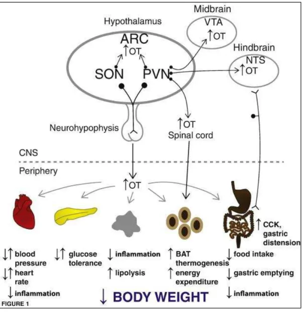
Figure 25. Schematic representation oxytocin involvement on energy homeostasis ... 59
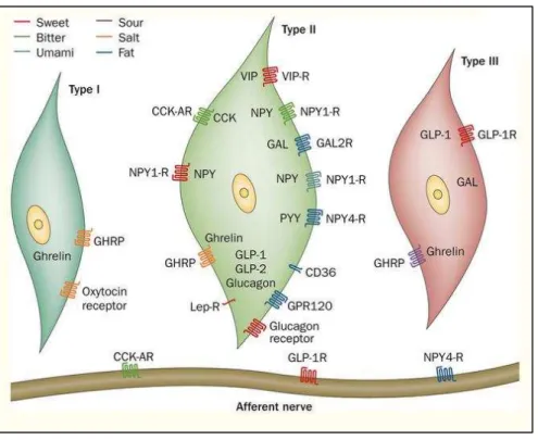
Figure 26. Expression of hormones and their receptors in the three subtypes of taste bud cells... 62
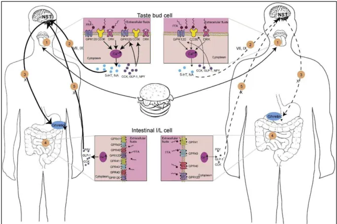
Figure 27. Proposed mechanisms of the systemic energy regulation via fat taste receptors ... 64
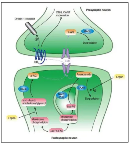
Figure 28. The endocannabinoid system in neurons: CB1 signalling affects the expression of orexigenic and anoretic mediators in the hypothalamus ... 66
Figure 29. Endocannabinoid system involvement ... 69
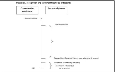
Figure 30. Relationship between chemical concentration, detection threshold and recognition threshold.. ... 71
Figure 36. Effect of rimonabant on preference for lipids, body weight and feeding behaviour ... 90
Figure 37. Impact of CB1R gene invalidation on CD36 and GPR120 protein expressions ... 91
Figure 38. Effect of CB1R gene invalidation on GLP-1 ... 92
Figure 39. Effects of linoleic acid (LA) and cannabinoids on Ca2+ signalling in mouse TBC. ... 93
Figure 40. Effects of rimonabant on linoleic acid in mTBC (LA) and cannabinoids-induced Ca2+ signalling in mTBC ... 94
Figure 41. Effects of DB-cAMP and U73122 on anandamide (AEA, 5 µM)-induced Ca2+ signalling in TBC ... 95
Figure 42. Schematic model of the proposed CB1R involvement in TBC ... 99
Figure 43. siRNA machinery... 119
Figure 44. Fat and bitter taste receptors expression ... 122
Figure 45. Immunostaining of fat taste receptors (CD36 and GPR120) and bitter taste receptors (TAS2R16 and TAS2R38). ... 123
Figure 46. Type I cell marker expression ... 124
Figure 47. Type II cell marker expression ... 125
Figure 48. Immunostaining of GNAT3 and PLCβ2. ... 125
Figure 49. Type III cell marker expression ... 126
Figure 50. Hormones and receptors expression. ... 127
Figure 51. Immunostaining of the bitter taste receptor TAS2R38 and the fatty taste receptor CD36. 128 Figure 52. Immunostaining of the bitter taste receptor TAS2R16 and the fatty taste receptor CD36. 128 Figure 53. LA induces increases in [Ca2+ ]i in HTC-8 cells. ... 129
Figure 54. Sinigrin induces increases in [Ca2+ ]i in HTC-8 cells ... 130
Figure 55. PTC induces increases in [Ca2+ ]i in HTC-8 cells ... 130
Figure 56. Salicin induces increases in [Ca2+ ]i in HTC-8 cells ... 131
Figure 57. Ca2+ imaging studies were performed in calcium-containing (100% Ca2+) buffer ... 132
Figure 58. U73122 induces reduced [Ca2+ ]i responses in HTC-8 cells. ... 133
Figure 59. Effects of LA, PTC, Sinigrin and Salicin on Ca2+ signalling in HTC-8 cells... 134
Figure 60. LA and bitter agonists induce additive [Ca2+ ]i responses in HTC-8 cells ... 135
Figure 61. Econazole induces reduced [Ca2+ ]i responses in HTC-8 cells ... 136
Figure 62. SKF96365 induces reduced [Ca2+ ]i responses in HTC-8 cells. ... 137
Figure 63. 2-APB induces reduced [Ca2+ ]i responses in HTC-8 cells. ... 138
Figure 64. ML-9 induces reduced [Ca2+ ]i responses in HTC-8 cells ... 139
Figure 65. Orai1 and Orai3 impact differently [Ca2+ ]i responses in HTC-8 cells ... 140
Figure 66. Oxytocin (OXT) and its cognate receptor (OTR) expression ... 141
Figure 67. OXT induces increases in [Ca2+ ]i in HTC-8 cells. ... 141
Figure 68. AH7614, a GPR120 inhibitor and SSO, a CD36 inhibitor increase active OXT release by HTC-8 cells. ... 142
Figure 69. LA decreases OXT mRNA expression in HTC-8 cells ... 143
Figure 70. Proposed model of the involvement of the oxytocin in TBC. ... 148
Figure 71. Proposed model of the cross-talk between fat and bitter taste modalities in TBC. ... 150
Figure 72. Proposed general hypothesis of the signalling pathway evoked by LCFA and bitter agonists in TBC. ... 152
Figure 73. LA, sinigrin, PTC and salicin induce increases in [Ca2+ ]i in mouse fungiform cell line ... 153
goût ... 159 Figure 78. Hypothèse proposée de la voie de signalisation induite par les AGLC et par les agonistes amers dans les cellules des bourgeons du goût. ... 175
List of tables
Modulation of fat preference by endocannabinoids
Table 1 : List of primers ... 84 Table 2 : List of primary antibodies used in Western blotting ... 85
Modulation of fat preference by bitter agonists
Table 3 : Liste of primary antibodies used in immunocytochemistry ... 115 Table 4 : List of primers ... 116 Table 5 : List of primary antibodies used in Western blotting ... 118
Introduction
Due to increasing abundance of food, the Western diet is constituted of almost 45% of fat (i.e. 10% beyond the nutritional recommandations), greatly contributing to the prevalence of obesity. In France, the obesity rate is increased from 8% in 1997 (first survey) to 15% in 2012 according to the last ObEpi report (OMS, 2012), involving serious diseases such as type II diabetes, atherosclerosis and hypertension. The epidemic of obesity is consequently considered as a major public health problem.
The availability of highly palatable foods rich in energy, fat and sugar has been associated with the risk for obesity (Erlanson-Albertsson, 2005). The hedonic property of this type of food is a driving force, promoting its preferential consumption. Mela and Sacchetti (1991) observed that obese subjects had a higher spontaneous preference for dietary lipids than normal subjects (Drewnowski et al., 1985). This suggests the existence of selective detection of these caloric / lipidic foods operating from the oral cavity.
Taste is one of the components that enables orosensory perception of foods and their composition. This plays a crucial role in food choices. Humans have a highly selective taste system that allows them to distinguish between nutritive and potentially toxic foods (Jyotaki et al., 2010). The perception of taste provides with the information on the quality of food ingested by the organism. The signals generated in the oral cavity will transmit the taste information via the central nervous system to different physiological targets like intestine via tongue-brain-intestine loop. The oral cavity, therefore, plays a significant role in the eating behaviour.
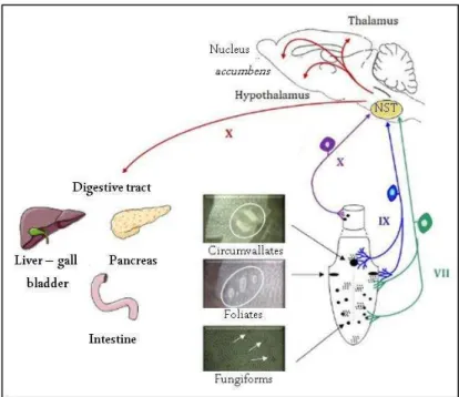
The taste buds localized in the lingual papillae relay the information. There are three main types of papillae: fungiform, foliated and circumvallate papillae (Figure 1). The binding of the tastant to its taste receptor located in the papillae induces, via [Ca2+], taste signals transmitted via the cranial nerve muscle IX and VII to the NTS, connected to different areas of the brain involved in food intake and the digestive tract.
Figure 1. Nerve pathways involved in taste perception. At the lingual level, the papillae are innervated by afferent nerve fibers belonging to two pairs of cranial nerves. Thus, the fungiform papillae and the anterior part of the foliated papillae establish synaptic contacts with the chorda-tympani nerve (CT, i.e., pairs of cranial nerves VII). The posteror part of the foliated papillae and the circumvallate papillae are innervated by fibers from the glossopharyngeal lingual branch (i.e., pair of cranial nerves IX). The few papillae present in the upper esophagus are connected to branches of the vagus nerve (nerve X). At the central level, the nucleus of the solitary tract (NST) is the first gustatory relay. The integration of tatse signals from the taste buds is mediated by brain areas (hypothalamus, nucleus accumbens, thalamus) whose role in the resultaion of the eating behavior is well established. The NST sends information back to the digestive tract through efferent vagal nerve fibers. VII tympanic chord, IX glossopharyngeal nerve, NST nucleus of solitary tract. Adapted from Martin et al. (2010).
Orosensory perception of dietary lipids has long been perceived as dependent only on their textural and olfactory properties. However, during the last recent years, the existence of a sixth taste modality, dedicated to the perception of dietary lipids, has been proposed. Indeed, the recent discovery of lipid receptors in rodents and humans suggests that lipids can be detected by the taste pathway (Mattes, 2011; Passilly-Degrace et al., 2009). Hence, CD36 (Cluster of differentiation 36) has been suggested to play the role of a lingual lipid receptor (Laugerette et al., 2005). In addition, it has been proposed, in the first place, that the GPR120 lingual receptor (G protein-coupled receptor 120) also has a role in lipid detection. However, it seems that this is not the case (Ancel et al., 2015). Indeed, GPR120 has been clearly demonstrated to play a “regulatory” role in the control of lipid feeding behaviour (Ozdener et al., 2014). Long-chain fatty acids (LCFA) have been shown to induce a cephalic response and influence the gastric motility of the gastrointestinal system as well as the hormonal cascade of
detection of fatty acids has two major roles: the cephalic phase activation and the regulation of fat and energy intake (Little and Feinle-Bisset, 2010). In this context, many studies aimed at analyzing lipid perception have been conducted to understand how individuals make high-fat dietary choices in terms of the quality and quantity they consume (Ebba et al., 2012). A large number of studies concluded that oral fat sensitivity is a key factor in food-selection (Keller, 2012; Keller et al., 2012; Melis et al., 2015a; Stewart et al., 2010, 2011a; Tepper and Nurse, 1997) (Figure 2).
Figure 2. Sensory, genetic, personal and neuronal factors influencing the perception of fat taste. BMI body mass index, CD36 cluster of differenciation 36, GPR120 G-protein coupler receptor 120, FFA free fatty acid, PROP 6-n-Propylthiouracil, SNP single nucleotide polymorphism, TAG triacylglycerol. Adapted from Heinze et al. (2015).
A defect in an individual's ability to detect dietary lipids would be linked to overconsumption of fatty foods (Keast et al., 2014).
First Part
State of the Art
Chapter 1
Physiology of the taste system
1. Gustative perception
1.1. General introduction
The primary organ responsible for the sense of taste is the tongue. It contains biological machinery for identifying non-volatile compounds in food and non-food products that are placed in the oral cavity (Keast and Costanzo, 2015).
Humans recognize and distinguish 5 basic taste qualities; sweet, umami, salty, sour and bitter. Basically, sweet detects energy-rich food; umami, the taste of L-glutamate reflecting a food’s
protein content; salty is responsible for the electrolyte balance; sour prevents ingestion of food that is damaged or not mature enough; finally, bitter warns about potential presence of harmfull compounds (Figure 3) (Chaudhari and Roper, 2010).
Figure 3. Taste qualities, taste receptors and natural stimuli. Human taste is characterized by 5 basic tastes qualities: sweet, umami, salty, sour and bitter. Recently, a sixth taste dedicated to the fat perception has been proposed. Adapted from Chaudhari and Roper (2010).
Taste perception relays the information triggered by ingested food from the oral cavity to the rest of the body. It occurs in the taste buds included in the papillae and mostly located, in the dorsal epithelium of the tongue. There are 3 main types of papillae: fungiform, foliated and circumvallate papillae (Figure 4). Each taste bud contains 30 to 100 taste cells. There are
glial-like cells; type II cells, corresponding to taste receptor cells, which express phospholipase Cβ2 (PLCβ2) and TRPM5 (Transient receptor potential cation channel subfamily M member 5) channels and type III cells which are pre-synaptic cells (Chaudhari and Roper, 2010). The taste bud cells (TBC) undergo continual turnover with a life span of about 10 days (Perea-Martinez et al., 2013). It has been recently shown that a lingual population of stem cells are involved in the production of mature TBC in the lingual papillae (Takeda et al., 2013). TBC derived from a pool of multipotent progenitor cells that are localized in the basal regions surrounding taste buds (Takeda et al., 2013; Yee et al., 2013). The binding of a sapid molecule to its receptor leads to changes in membrane potential and to an increase in the concentration of free calcium, [Ca2+]i. The depolarization of the taste cell
triggers the release of neurotransmitters such as serotonin (5-HT) (El-Yassimi et al., 2008). The taste signals from the oral cavity are then transmitted to cranial nerve VII (the cranial nerve tympanum) and IX (the glossopharyngeal lingual branch) and then to the nucleus of the solitary tract (NST) (Figure 4) (Gaillard et al., 2008). The NST is connected to different areas of the brain involved in food intake, reward, memory and the digestive tract (Besnard et al., 2010).
Figure 4. Peripheral gustatory system. A. Localization of circumvallate, fungiform and foliate papillae on the human tongue. B. Sagittal section of a circumvallate papilla (CVP) characterized by a dome-shaped structure highlighting the vicinity with the von Ebner glands. C. Schematic representation of a taste bud. CA-VI carbonic anhydrase, rNTS rostral nucleus of the solitary tract, VEGP protein of the
1.2. Taste physiology: from detection to perception
The oral detection of tastants by gustatory papillae, as mentioned above, triggers signals that are transmitted by the afferent gustatory fibers from the chorda-tympani (CT) and glossopharyngeal (GL) nerves (cranial nerve VII and IX, respectively) to the nucleus of tractus solitaries (NTS) (Figure 5-1). The NST receives information from the peripheral gustatory detection system, the digestive tract and the brain and is connected to different brain areas. The NST also sends gustatory signals towards the digestive tract via afferent vagal fibers (Figure 5-2). This cephalic phase reflex prepares, by anticipation, the digestive tract to food digestion (Power and Schulkin, 2008). The NTS projects taste signals to the primary gustatory cortex (PGC) constituted of the anterior insula and the frontal operculum (Fop). PCG is involved in identification and evaluation of intensity of tastants. Then, inputs from PGC reach the orbitofrontal cortex (OFC) which is involved in the evaluation of reward value associated with taste (Figure 5-3). The OFC is a sensory platform that receives inputs linked to food palatability such as smell or texture and send important informations with the reward pathway, constituted by the mesolimbic system, about hedonic experience (liking) and incentive salience (wanting) (Berridge, 1996). The mesolimbic system, an integrated pathway including the amygdala and hippocampus are involved in learned memory and the ventral tegmental as well as nucleus accumbens are mainly involved in the motivation of eating behaviour (Besnard, 2016). This system is responsible for taste perception and builds the hedonic value (Figure 5-4).
Post-ingestive regulatory signals provide real-time information about the feeding status to the hypothalamus that constitutes the “metabolic brain”. It is involved in the control of food intake and energy expenditure (Figure 5-5). It has been shown that taste-responsive neurons directly project to the lateral hypothalamus (Li et al., 2013) suggesting that orosensory signals are able to influence homeostatic functions of the hypothalamus and internal signals, in the same way, can modulate the “emotional brain” function (Figure 5-6). Finally, anatomical interconnections link the mesolimbic system and the hypothalamus (Figure 5-7). It emerges that the regulation of appetite is based on a subtle balance between the “emotional brain”, i.e. the hedonic hunger, and the “metabolic brain”.
Figure 5. Taste perception: emotional and metabolic brain involvement. 1. Oral detection of tastants by gustatory papillae. NTS sends gustatory signals toward the digestive tract allowing a digestive anticipation. 3. Cephalic integration of the taste signals. 4. The mesolimbic system is responsible for taste perception and builds the hedonic value. 5. Post-ingestive regulatory signals provide real-time information about the feeding status. 6. Orosensory signals influence homeostatic functions. 7. Anatomical interconnections link the mesolimbic system and the hypothalamus. NTS, nucleus of tractus solitarius; FOp, frontal operculum; OFC, orbito-frontal cortex. Adapted from Besnard (2016).
2. Orosensory detection of dietary lipids
"Fat" is the term used to refer to triglycerides and fats that are essential components of a traditional food intake in humans. Dietary fatty acid deficiency leads to impaired vision, stunting, skin lesions and reduced learning abilities (Connor et al., 1992). In contrast, overconsumption of fat induces negative health effects such as obesity (Stewart et al., 2011a; Swinburn et al., 2011), diabetes (Ravussin and Smith, 2002) and cancer (Giovannucci et al., 1993; Prentice and Sheppard, 1990).
Fat has been mentioned as a taste modality in 330 years before J-C by Aristotle (Aristotle, 350AD). Recent work has suggested that there is a sixth taste for lipid perception (Khan and Besnard, 2009; Laugerette et al., 2005). To define a flavor as a primary taste, it should meet 4
gustatory and physiological pathways. Indeed, the lipid perception system seems to respond to these conditions (Gilbertson and Khan, 2014).
2.1. Fat taste agonists
Dietary fats, i.e., triglyceride, do not appear to be effective stimuli. Indeed, compared to what is observed in intestinal fat sensing, the fat detection in the oral cavity appears to be dependent on the presence of fatty acids (FA). In humans, sensory detection of FA occurs within the millimolar range (0.02–6.4 mM) (Stewart et al., 2010). This detection threshold is consistent with the concentrations of FA naturally present in foods (0.76–3% w/v). It has been asserted that a lipolytic activity in saliva has been shown to be sufficient to produce micromolar amounts of fatty acids within the detectable range (Stewart et al., 2010). Furthermore, lingual lipase appeared to have an influence on fatty acid thresholds as the addition of orlistat (a lipase inhibitor) increased fatty acid thresholds (Pepino et al., 2012).
2.2. Fat taste receptors in taste bud cells
2.2.1. GPR40 and GPR120
G protein-coupled receptors (GPCR) belong to seven-transmembrane domain receptors. Sweet, bitter and umami taste perception is initiated by binding of tastants to specific GPCR. Two GPRCR, GPR120 and GPR40 (G protein-coupled receptor 40), were detected on mouse papillae. GPR120 was co-expressed with TRPM5, a marker of type II taste receptor cells (TRC) (Cartoni et al., 2010) whereas GPR40 was mainly found in type I cells which are not true TRC (Cartoni et al., 2010). Nevertheless, Galindo et al. (2012) failed to detected GPR40, at protein and mRNA levels, in human papillae and Hirasawa et al. (2008) also failed to detect GPR40 in rat gustatory papillae.
GPR120, also known as FFAR4 (free fatty acid receptor 4) has been identified as an orphan receptor in 2003 (Fredriksson et al., 2003) (Figure 6). GPR120 has been disorphaned in 2005. Indeed, GPR120 is activated by MCFA (Medium-chain fatty acid) and LCFA (Hirasawa et al., 2005). GPR120 is widely expressed in tissues such as brain, adipose tissue, heart and especially in lungs and gut (Hirasawa et al., 2005). GPR120 has been shown to mediate anti-inflammatory effects (Oh et al., 2010). Over the last few years, GPR120 has raised interest as it has been identified as a potential target for metabolic disorders such as type 2 diabetes mellitus or obesity (Mo et al., 2013; Ulven and Christiansen, 2015).
GPR120 was initially reported to be a plausible candidate for LCFA detection by taste buds because GPR120-/- mice were unable to properly detect an oily source during a behavioural test (Cartoni et al., 2010). Nevertheless, this finding has not been reproduced (Ancel et al., 2015; Sclafani et al., 2013). Moreover, healthy volunteers subjected to two-alternative force choice (AFC) tests were unable to detect an oral sensation consecutive to a pharmacological activation of GPR120 (Godinot et al., 2013). These inconsistencies strongly suggest that GPR120 is not a major player in oral fat detection. In contrast, an implication of GPR120 in the LCFA mediated release of the incretin hormone glucagon-like peptide-1 (GLP-1) by mouse TBC is likely, as previously demonstrated in enteroendocrine L-cells (Hirasawa et al., 2005). Indeed GPR120 and GLP-1 are found to be co-localized in a subset of mouse TBC (Martin et al., 2012). Moreover, LCFA elicit GLP-1 release by freshly isolated mouse circumvallate papillae (Martin et al., 2012). Ozdener et al. (2014) clearly demonstrated that GPR120 plays an alternative role in lingual detection of fatty acids. In fact, in the fasting situation, GPR120 is downregulated and in a fed-situation, it is upregulated in the raft fraction of mouse TBC.
2.2.2. CD36
Figure 7. Structural features of CD36. It is noteworthy the hair-pin loop structure of the extracellular
segment, suitable for the LCFA binding. Solid triangles show potential glycosylation sites. The red-coloured box shows a potential membrane-embedded domain. The green-red-coloured box on the outer membrane segment shows a proline rich region with the location of cysteine residues indicated within red circles. Adapted from Khan and Besnard (2009).
The Cluster of Differenciation 36 (CD36) formerly known as FAT for fatty acid translocase is a multifunctional glycoprotein. CD36 has been first identified 50 years ago (Silverstein and Febbraio, 2000) in mammary globules. The protein has been very well conserved over 300 million years (Ozbek et al., 2010). The first ligand has been described in 1993. Indeed, CD36 has been identified as a macrophage receptor for oxidized low density lipoprotein (Endemann et al., 1993).
CD36 is expressed in various tissues such as the heart, the kidney and the gut and is involved in various functions such as immunity, inflammation, angiogenesis, thrombosis or atherogenesis. CD36 has also been found to be a lipido-sensor in numerous organs including the brain, the lingual and the gut epithelium suggesting a functional continuity between these tissues (Martin et al., 2011a).
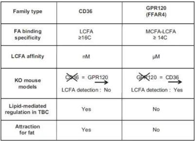
CD36 is an integral membrane ditopic glycoprotein with a large extracellular hydrophobic hair-spin structure between two short cytoplasmic tails (Figure 7) (Abumrad et al., 1993; Greenwalt et al., 1992). It belongs to scavenger receptor family and expresses the binding sites for a number of ligands such as fatty acids, thrombospondin-1, oxidized low-density lipoproteins (LDL), growth hormone (GH)-releasing peptides (Silverstein and Febbraio). CD36 exhibits high affinity (in the order of nanomolar) for fatty acids (Baillie et al., 1996) (Figure 8). CD36 appeared to be highly expressed on the apical region of mice circumvallate
extent. In human beings, CD36 has been detected in human foliate and circumvallate papillae (Simons et al., 2011).
Figure 8. Comparison of the main characteristics of CD36 and GPR120 in the mouse. FA, fatty acid; KO, knockout; LCFA, long-chain fatty acid; MCFA, medium-chain fatty acid; TBC, taste bud cells. Adapted from Besnard et al. (2016).
In mice, the CD36 deletion completely suppressed spontaneous preference for lipid solutions (Laugerette et al., 2005). This effect on feeding behaviour is lipid-specific since sweet preference and bitter aversion were not affected in these transgenic mice. It is remarkable that CD36 failed to influence post-oral fat conditioning in mice. Indeed, Sclafani and Ackroff (2012) and Sclafani et al. (2007) reported that CD36 deletion decreased fat preference and consumption but also enhanced the ability of mice to compensate for the calories provided by their optional fat intake in fat conditioned experiments. They also proposed that CD36 might be involved in fat “detection” when GPR120 whereas take part in the regulation of post-ingestive lipid behaviour.
The local release of lingual lipase by the von Ebner’s gland in the clefts of circumvallate papillae (Kawai and Fushiki, 2003) exposes the CD36 directly to a microclimate potentially rich in long-chain fatty acids (LCFA) (Figure 9). This organization allows the degradation of triglyceride into fatty acids (FA) that promote the fat detection in the oral cavity.
2.2.3. Others candidates
Other orphans GPCR have been suggested to bind fatty acids. GPR41and GPR43 are receptors for the short chain (C2:0–C6:0) saturated fatty acids (Galindo et al., 2012; Miyamoto et al., 2016; Ulven, 2012) whereas GPR84 appears to be predominately activated by medium chain (C8:0–C14:0) fatty acids (Venkataraman and Kuo, 2005; Wang et al., 2006). Besides, GPR41 and GPR84 have been shown to be abundantly expressed by posterior, probably circumvallate, papillae in rat (Gilbertson et al., 2010) whereas absent on fungiform papillae. As for GPR43, the receptor was present on both mouse fungiform and posterior papillae. There are currently no functional data on GPR41/43 in the taste system, preliminary data suggest that mouse TBC respond to medium chain fatty acids like myristic acid and lauric acid that are ligands of GPR84. However, the role of these TBC-expressed fatty acid-activated GPCR in fat taste perception remains to be elucidates in detail.
2.3. Signalling transduction mediated by LCFA in taste bud cells
During the eating period, LCFA bind to CD36, followed by the communication with GPR120. This binding triggers intracellular signal transduction involving successively the Src-PTK, phospholipase C (PLC) activation and inositol trisphosphate (IP3) production which induces
Ca2+ release by the endoplasmic reticulum (ER) (Figure 10-1). Ca2+ activates stromal interaction molecule (STIM1) reinforcing the [Ca2+]i accumulation by opening Orai1 and
Orai1/3 channels (Figure 10-2). The rise in [Ca2+]i induces a TBC depolarization via TRPM5
channels (Figure 10-3). LCFA also block the delayed rectifying potassium channels (DRK) channels via CD36 translocation or directly in order to maintain the cell activation (Figure 10-4). The Ca2+ rise produces cell depolarization leading to ATP efflux triggering serotonin (5-HT) release by type III cells via the calcium homeostasis modulator 1 (CALHM1) channels (Figure 10-5). Indeed, Subramaniam et al. (2016) showed that LA-evoked Ca2+ signalling and ERK1/2 phosphorylation were decreased in CALHM1-deficient TBC. Moreover, Erk1-/- and Calhm1-/- mice exhibit reduced preferences for oily solutions. These results suggested that
gustatory detection of dietary lipids (Subramaniam et al., 2016). ATP leads to the activation of primary gustatory neurons, type II and type III cells (Figure 10-6). Then, 5-HT mediates the modulation of the taste signal (Figure 10-7). It is noteworthy that FFA could also convey the taste signal to the NST via the non gustatory epithelium and the trigeminal endings (Figure 10-8).
Figure 10. Signalisation induced by LCFA (working model). 1. LCFA binds to CD36 and activates the cascade of PLCβ-IP3 thus releasing Ca2+ from the ER pool. 2. The depletion of the ER Ca2+ pool induces
the aggregation of STIM1 toward SOC channels like Orai1 and Orai1/3. 3. The Ca2+ increase also
activates the TRPM5 channels responsible for the cell depolarization. 4. The activation of the DRK channels is triggered by the Na+ influx. 5. Ca2+ rise produces cell depolarization leading to ATP efflux.
6. ATP leads to the activation of primary gustatory neurons, type II and type III cells and to 5-HT liberation from Type III cells. 7. 5-HT mediates the modulation of the taste signal. 8. FFA could also convey the taste signal to the NST via the non gustatory epithelium and the trigeminal endings Obligatory genes for the fatty taste transduction are shown framed. FFA, free fatty acids; LCFA, long-chain fatty acids; PUFA, polyunsatured fatty acids; 5-HT, serotonin; r-NTS, rostral nucleus of solitary tract; V,trigeminal nerve; VII, chorda tympani nerve; IX, glosso-pharyngeal nerve. Adapted from Besnard et al. (2016).
After cell depolarisation, neurotransmitters such as 5-HT are secreted toward the afferent gustatory nerve fibres (specifically VIIth and IXth cranial nerves) (Passilly-Degrace et al., 2014), which relay the signal to the nucleus of solitary tract (NST), and then to the brain stem and digestive tract (Besnard et al., 2016a; El-Yassimi et al., 2008; Gilbertson and Khan, 2014; Passilly-Degrace et al., 2014) (Figure 10).
2.3.1. CD36/GPR120 cooperation hypotheses
CD36 and GPR120 are co-expressed in mouse and human TBC (Martin et al., 2012; Ozdener et al., 2014) and both are able to bind and to be activated by LCFA (Baillie et al., 1996; Hirasawa et al., 2005). They are two putative gustarory lipid receptors however, a functional redundancy seems unlikely.
2.3.1.1. Direct cooperation hypothesis
This hypothesis suggests that the simultaneous presence of CD36 and GPR120 in TBC is absolutely required to properly detect the presence of LCFA in saliva. It was postulated that LCFA binding to CD36 should activate GPR120 to initiate the signal transduction cascade leading to transmitter release and, hence, fat detection (Gilbertson and Khan, 2014; Gilbertson et al., 2010). This prioritization is based on the fact that CD36 displays a greater binding affinity for LCFA than GPR120 and is known to act as a cofactor facilitating recognition of derived lipids by Toll-like receptors in the mouse (Hoebe et al., 2005). However, this scenario is challenged by behavioural studies performed using knockout mouse models Indeed, detection and preferential consumption of lipid sources are not affected by a targeted GPR120 gene deletion (Ancel et al., 2015; Sclafani et al., 2007), likely because CD36 gene expression in circumvallate papillae (CVP) is maintained in this mouse model (Ancel et al., 2015). Therefore, a role for GPR120 as a primary lipid receptor responsible for the oral fat detection appears to be largely speculative (Besnard et al., 2016).
2.3.1.2. Indirect cooperation hypothesis
An indirect cooperation between CD36 and GPR120 is suggested by several observations. Indeed, hormones and LCFA are known to regulate the membrane localization of CD36. In the digestive tract, the presence of LCFA in the intestinal lumen also induced a rapid disappearance of CD36 from the apical side of enterocytes, leading to a partial degradation by the ubiquitin-proteasome pathway (Tran et al., 2011). This ligand-mediated negative feedback likely constitutes a physiological desensitization mechanism that progressively abolishes the LCFA-mediated CD36 signalling in enterocytes during the postprandial period (Tran et al., 2011). A similar dynamic regulation of CD36 seems to exist in TBC. Indeed, lipid deposition onto the lingual epithelium is enough to induce a dramatic decrease in the CD36 protein levels in mouse CVP (Martin et al., 2011b). In line with this observation, CD36 in CVP displays a
period). It is tempting to speculate that this downregulation of CD36 might lead to a progressive decrease in the motivation to eat fatty foods during a meal as lingual CD36 is significantly involved in the avidity for lipids (Laugerette et al., 2005; Martin et al., 2011b; Ozdener et al., 2014; Sclafani et al., 2007). Conversely to CD36, GPR120 levels in the mouse CVP remains globally stable throughout the day and appear to be insensitive to the lipid content of the diet (Martin et al., 2011b).
It is also known that GPR120 is broadly expressed in the distal small intestine and colon, where it is coexpressed with GLP-1 in the enteroendocrine L cells (Hirasawa et al., 2005). During the prandial period, stimulation of GPR120 by LCFA promotes the secretion of intestinal GLP-1, increasing circulating insulin (Hirasawa et al., 2005). GLP-1 is known to modulate sweet and umami taste sensitivities in the mouse (Martin et al., 2009; Shin et al., 2008). Behavioural experiments, conducted using GLP-1R-/- mice, have shown a reduced capacity to detect low concentrations of rapeseed oil, demonstrating for the first time that GLP-1 also modulates the fat taste sensitivity (Martin et al., 2012). The molecular mechanism by which this control takes place is not yet fully established. However, the dynamic downregulation of CD36 in CVP, normally triggered by LCFA (Martin et al., 2011b), was lacking in GPL1-R-/- mice (Martin et al., 2012). This finding provides the first evidence that GRP120 might be able to modulate the CD36-mediated detection of oral lipid, via GLP-1 secretion.
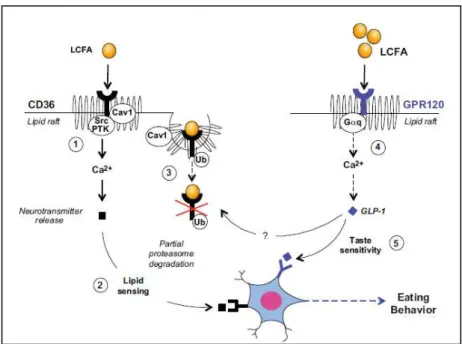
Thus, Besnard et al. (2016) proposed the following “indirect cooperation” scenario (Figure 11). At the beginning of a feeding period, LCFA binding to CD36 into lipid rafts (Pohl et al., 2005) results in the formation of a multimolecular complex of transmembrane proteins, including adaptors (Heit et al., 2013), members of Src protein-tyrosine kinase (Src-PTK) , and caveolin-1 (Cav1) (Fig 11-1). This multimolecular platform should be able both to trigger a downstream signal and to limit its duration by the partial degradation of the ligand-receptor complex. Indeed, because Src kinases activate PLC (Makranz et al., 2004), this event might result in a signalling cascade responsible for a rapid rise in Ca2+ levels, initiating neurotransmitter release and, in turn, transfering a lipid signal to the brain (Figure 11-2).
Figure 11. Respective role of CD36 and GPR120 in taste bud cells: indirect cooperation hypothesis. 1. LCFA binding to CD36 into lipid rafts results in the formation of a multimolecular complex of transmembrane proteins, including adaptors, members of Src protein-tyrosine kinase (Src-PTK) , and caveolin-1 (Cav1). 2. This multimolecular platform should be able both to trigger a downstream signal and to limit its duration by the partial degradation of the ligand-receptor complex. 3. Ubiquitination of CD36 induces a gradual decrease in the attraction for fat during the meal. 4. When the oral LCFA levels become sufficient to allow the binding and activation of GPR120 in lipid raft triggers the GLP-1 release by TBC. 5. GLP-1 acts on its cognate receptor located in gustatory nerve endings to modulate taste sensitivity. LCFA, long-chain fatty acid; Src PTK, Src protein tyrosine kinases; Cav1, caveolin 1; Ub, ubiquitination; G q, protein G; GLP-1, glucagon like peptide-1. Adapted from Besnard et al. (2016).
Ubiquitination of CD36, by inducing its progressive removal from the plasma membrane (Su and Abumrad, 2009), might allow its subsequent partial degradation by the proteasome pathway (Tran et al., 2011), inducing a gradual decrease in the attraction for fat during the meal (Martin et al., 2011b) (Figure 11-3). The molecular mechanism by which this negative feedback takes place in TBC remains to be elucidated. One potential route of internalization of CD36 is the caveolae-dependent pathway used by toll-like receptors (TLR) (Shuto et al., 2005). Studies showing that Caveolin-1 (Cav1) gene disruption is associated with altered subcellular localization and function of CD36 (Ring et al., 2006) are consistent with this hypothesis. When the oral LCFA levels become sufficient to allow the binding and activation of GPR120 in lipid raft (Ozdener et al., 2014) triggers the GLP-1 release by TBC (Martin et al., 2012; Ozdener et al., 2014) (Figure 11-4). GLP-1 acts on its cognate receptor located in gustatory nerve endings (Shin et al., 2008) to modulate taste sensitivity (i.e., fat, sweet, and umami tastes; Figure 11-5). The use of Glp-1r-/- mice has revealed an involvement of GLP-1 in the dynamic regulation of CD36 by LCFA (Martin et al., 2012). The mechanism by which
It seems that chemical information combines with textural signals to form the complete sensory perception of fat (Liu et al., 2016).
2.4. Gustatory and reward brain circuits
Peterschmitt et al. (2018) recently studied the implication of gustatory and reward brain circuits following the lingual application of LCFA (LA). They found that LA induced c-Fos expression in the NST, the parabrachial nucleus (PBN) and the ventroposterior medialis parvocellularis (VPMPC) of the thalamus, that are regions known to be activated by gustatory signals. LA also induced c-Fos expression in central amygdala and ventral tegmental area (VTA) involved in food reward. Finally, they observed that LA induced high expression of genes involved in memory consolidation in the arcuate nucleus (Arc) and hippocampus (Hipp). They consequently proposed the following model: LA deposition induces the activation of the gustatory pathway. The gustatory signal is conveyed to the NST that transmits the information to the PBN. Next, the gustatory part of the thalamus (VPMPC) is activated and the signal reaches the gustatory insular cortical areas (AI,GI/DI) with modulatory influences of the central amygaloid nucleus (CeA) and the posterior part of the lateral hypothalamus, the parasubthalamic nucleus/calbindin nucleus (PSTN/CbN) (Figure 12). Thus, they demonstrated that oral lipid taste perception triggers both gustatory and reward pathways.
Figure 12. Schematic representation of the gustatory pathway activation involving metabolic,
reward and learning, and memory processes. Acb, accumbens nucleus; AI, agranular insular cortex;
Arc, arcuate nucleus; BLA, basolateral amygdaloid nucleus; CeA, central amygdaloid nucleus; GI/DI, granular/dysgranular insular cortex; Hbn, habenula; Hipp, hippocampus; mPFC, medial prefrontal cortex; NST, nucleus of the solitary tract; PBN, parabrachial nucleus; PSTN/CbN, parasubthalamic
3. Orosensory detection of bitter compounds
3.1. Bitter taste agonists
The ability to taste bitterness is linked to a critical function for survival allowing animals to discriminate safe from potentially harmful foods. Thank to its innate negative hedonic value (Steiner et al., 2001), bitterness perception prevents feeding, and differences in bitter taste sensitivity influence human dietary behaviour. The bitter taste perception is controlled by the taste 2 receptor (TAS2R) gene family. There are 25 human TAS2R genes (Drayna, 2005) with many associated polymorphisms and a wide range of individual differences in bitter taste sensitivity (Chandrashekar et al., 2000). This explains in part why thousands of chemically dissimilar agonists can elicit a single taste quality. The most common role is played by TAS2R16 and TAS2R38.
3.1.1. TAS2R16
TAS2R16 is a bitter taste receptor gene located on chromosome 7, has traditionally been associated with the detection of toxic β–D-glucopyranosides, such as salicin from willow bark (Bufe et al., 2002; Greene et al., 2011; Soranzo et al., 2005). Several studies have also reported that bitter taste receptors are expressed in cell types that are not directly involved in oral sensory perception. Indeed, nucleotide polymorphisms in or near the coding exon of TAS2R16 have been identified as risk factors for alcohol and nicotine dependence (Hayes et al., 2011; Hinrichs et al., 2006; Mangold et al., 2008) in African Americans. These polymorphisms have also been correlated with differences in lifespan and senescence among individuals in an isolated European population from southern Italy (Campa et al., 2012). Thus, bitter taste receptors participate in a variety of biological processes, including metabolism and immune response.
3.1.2. TAS2R38
The TAS2R38 gene is one of the most widely studied. TAS2R38 encodes a seven transmembrane G protein-coupled receptor (Drayna, 2005). It binds to thiourea group contained in synthetic compounds such as phenylthiocarbamide (PTC) and 6- n- propylthiouracil (PROP) (Kim and Drayna, 2005).
Fox (1932) serendipitously discovered that phenylthiocarbamide (PTC) concentration that tasted extremely bitter for some people were virtually tasteless for others. Indeed, sensitivity to PTC, a compound containing an -N-C=S group, is used as a phenotypic marker for oral
3.2. Signal transduction mediated by bitter in taste bud cells
The generation of bitter taste starts when a bitter compound enters the oral cavity. The ligand binds to a TAS2R receptor expressed in the apical membrane of receptor cells in taste buds. It triggers a cascade of signalling events, involving α-gustducin, PLCβ2 and inositol trisphosphate (IP3) activation (Zhang et al., 2003b), leading to Ca2+ influx and ultimately to
the release of neurotransmitter. The neurotransmitters activate an afferent nerve fiber that transmits the signal via the cranial nerves VII and IX to the brain (Figure 13). The taste signals transit through the brain and provide input to circuits involed in various functions, such as physiological reflexes, discriminative perception, and affective processing. It illustrates the complexity of the mechanisms intervening between the application of the bitter stimulus and the generation of the behavioural response.
Figure 13. Mechanism occuring between the application of the bitter tastant and the generation of
the behavioral response. Ligand binds to a T2R G-protein coupled receptor expressed in the apical
membrane in taste buds triggereing a cascade of signaling events, leading to the release of neurotransmitters. This mechanism activates afferent nerve fibers that transmit the signal via the
Chapter 2
Taste system and its regulatory pathways
1. Factors involved in impaired oro-sensory perception and obesity
1.1. Alteration of the salivary composition
The flow of saliva is increased during a meal and it is utilized to form a moisturized bolus of foodstuff thus soaking the masticated food in salivary enzymes to increase its flavor perception, initiate the digestion, and promote swallowing (Zolotukhin, 2013). Saliva plays a significant role in the orosensory perception of dietary components. Indeed, during a meal, the major part of saliva is produced by the salivary glands and only a few is produced by minor salivary glands such as the von Ebner’s glands (VEG; Figure 14). VEG secrete directly into the clefts of papillae, known to house the great majority of taste buds (Figure 14). A preduodenal lipase (LIPPF) has been shown to be synthesized and secreted by VEG in rodents (Hamosh and Scow, 1973), its anatomical location highlights an involvement in promoting the enzymatic release of FFA from dietary triglycerides (TG) directly in the vicinity of taste buds, thus facilitating their detection by the TBC (Figure 14).
Figure 14. Location of von Ebner’s glands in the circumvallate papillae. Sagittal section of a circumvallate papillae (CVP) highlighting the vicinity of taste buds with the von Ebner’s glands in the clefts of the CVP. Adapted from Besnard et al. (2016).
LIPPF expression is lacking in human VEG (Spielman et al., 1993), nevertheless a similar mechanism involving a lipase activity leading to a partial TG breakdown of FFA is likely to occur in human saliva (Neyraud et al., 2012; Pepino et al., 2012; Stewart et al., 2010; Voigt et al., 2014). Indeed, a recent study revealed the presence of alternative lipase isoforms (LIPK, LIPM, LIPN) that are also known to induce TG cleavage (Voigt et al., 2014). The