HAL Id: hal-01633963
https://hal.sorbonne-universite.fr/hal-01633963
Submitted on 13 Nov 2017
HAL is a multi-disciplinary open access
archive for the deposit and dissemination of
sci-entific research documents, whether they are
pub-lished or not. The documents may come from
teaching and research institutions in France or
abroad, or from public or private research centers.
L’archive ouverte pluridisciplinaire HAL, est
destinée au dépôt et à la diffusion de documents
scientifiques de niveau recherche, publiés ou non,
émanant des établissements d’enseignement et de
recherche français ou étrangers, des laboratoires
publics ou privés.
Distributed under a Creative Commons Attribution| 4.0 International License
Limiting habenular hyperactivity ameliorates maternal
separation-driven depressive-like symptoms
Anna Tchenio, Salvatore Lecca, Kristina Valentinova, Manuel Mameli
To cite this version:
Anna Tchenio, Salvatore Lecca, Kristina Valentinova, Manuel Mameli. Limiting habenular
hyper-activity ameliorates maternal separation-driven depressive-like symptoms. Nature Communications,
Nature Publishing Group, 2017, 8, pp.1135. �10.1038/s41467-017-01192-1�. �hal-01633963�
Limiting habenular hyperactivity ameliorates
maternal separation-driven depressive-like
symptoms
Anna Tchenio
1,2,3,4
, Salvatore Lecca
1,2,3,4
, Kristina Valentinova
1,2,3,4
& Manuel Mameli
1,2,3,4
Early-life stress, including maternal separation (MS), increases the vulnerability to develop
mood disorders later in life, but the underlying mechanisms remain elusive. We report that
MS promotes depressive-like symptoms in mice at a mature stage of life. Along with this
behavioral phenotype, MS drives reduction of GABA
B-GIRK signaling and the subsequent
lateral habenula (LHb) hyperexcitability
—an anatomical substrate devoted to aversive
encoding. Attenuating LHb hyperactivity using chemogenetic tools and deep-brain
stimula-tion ameliorates MS depressive-like symptoms. This provides insights on mechanisms and
strategies to alleviate stress-dependent affective behaviors.
DOI: 10.1038/s41467-017-01192-1
OPEN
1Institut du Fer à Moulin, Paris 75005, France.2Inserm, UMR-S 839, Paris 75005, France.3Université Pierre et Marie Curie, Paris 75005, France. 4Department of Fundamental Neuroscience, The University of Lausanne, Lausanne 1005, Switzerland. Correspondence and requests for materials should be
addressed to M.M. (email:[email protected])
123456789
C
hildhood neglect (i.e., maternal separation, MS) is
aver-sive, has negative long-term repercussions on child
development and primes depression in adulthood
1,2. The
prolonged separation of newborn rodents from their mother
represents an animal model of severe early-life stress that
reca-pitulates aspects of child neglect. Animals undergoing MS present
deficits in coping with stressful events. Moreover, such paradigm
promotes anxiety, addictive and depressive-like behavioral
phe-notypes
3–5. This raises the hypothesis that MS-driven behavioral
adaptations may, at least partly, emerge from the dysfunction of
neural circuits devoted to aversion processing.
The lateral habenula (LHb) contributes to encode aversion and
negative reward prediction error as neurons in this structure are
excited by external aversive stimuli
6,7. The LHb provides such
aversive-related information to monoaminergic centers
suggest-ing a relevant role in motivated behaviors
8–10. Stressors of
dif-ferent nature increase the activity of LHb neurons, and lesioning
experiments indicate that the LHb regulates coping behaviors
when facing aversive stimuli
11–13. Furthermore, when such
stressful events (i.e., foot-shocks) become inescapable and
per-sistent, this produces aberrant LHb hyperactivity, which is
instrumental
for
the
emergence
of
depressive-like
symptoms
11,12,14,15.
However, whether chronic, painless stressful events, such as
MS, promote depressive-like phenotypes through LHb
adapta-tions remains elusive. If this holds true, we propose that reversal
strategies restoring LHb function can ameliorate MS-dependent
depressive-like symptoms.
Here, we demonstrate that, at a mature stage of life, mice
undergoing MS present depressive-like behaviors along with
increased LHb neuronal excitability. In addition, we show that
MS-mediated reduction in GABA
B-GIRKs signaling is causal for
such LHb hyperexcitability, and that limiting LHb neuronal
activity through chemogenetics or deep brain stimulation
ame-liorates MS-driven behavioral phenotypes. These
findings support
the notion that aberrant habenular neuronal hyperactivity
represents a neurobiological substrate underlying discrete
stress-driven (i.e., acute and chronic) depressive-like symptoms.
Results
MS drives depressive-like states and habenular
hyperexcit-ability. We examined the early-life stress-dependent behavioral
ramifications exposing mice to maternal separation (MS)
16(Methods, Fig.
1
a). Briefly, pups aged 7 days were, or not
(Control), removed from their litter, separated from their mother,
and kept in isolation for 6 h daily for a week. When tested three
weeks later, Control and MS mice had comparable weight,
locomotion and performance in the open
field (Supplementary
Fig.
1
a–c). In contrast, MS mice presented higher failure rates
when challenged with escapable foot-shocks (shuttle box),
beha-vioral despair (higher immobility in tail suspension test; TST), and
diminished sucrose preference (Fig.
1
b–d). Furthermore, ex-vivo
recordings in acute LHb-containing brain slices revealed that LHb
neurons from MS mice exhibit hyperexcitability, with no
altera-tions of the resting membrane potential (Fig.
1
e; Supplementary
Fig.
1
d). Altogether, these
findings suggest that MS promotes
depressive-like symptoms and LHb hyperactivity later in life.
Cellular mechanisms underlying MS-driven plasticity. Tonic
GABA
B-GIRK signaling controls LHb neuronal
firing and its
reduction promotes cell hyperexcitability
11. Patch-clamp
record-ings from LHb neurons revealed that bath application of the
GABA
B-R
agonist
baclofen
evoke
an
outward
current
Maternal separation (MS)
a
Shuttle box test TST Sucrose preference Ctrl 0 7 15 35 MS (6h/day) Behavior Birth 300 250 200 150 100 50 0 30 25 20 15 10 5 0 Failures 100 80 60 40 20 0 Sucrose preference, % Immobility, s
*
***
**
MS Shock Water Sucrose Day 30 20 10 0**
aCSF 50 pA 0 40 80 Action potentials Injected current, pA 0.2s 20 mVb
c
d
e
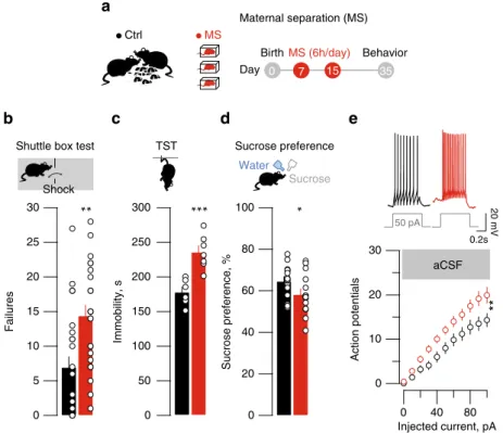
Fig. 1 MS-induced depressive-like symptoms and hyperexcitability. a MS protocol (all schematics are original drawing made by authors). b Bar graph and scatter plot of failures in the shuttle box for control (Ctrl) and MS mice (Ctrl vs. MS;nmice= 24, unpaired t-test, t45= 3.36 **p < 0.01). c Same as b for TST
immobility (Ctrl vs. MS;nmice= 6–7; unpaired t-test, t11= 4.63; ***p < 0.001). d Same as b but for sucrose preference (Ctrl vs. MS; nmice= 16; unpaired
t-test, t30= 32.12; *p < 0.05). e Top, sample traces from Ctrl and MS mice of current-evoked firing (50 pA step). Bottom, action potentials vs. injected
current (0–100 pA, steps of 10 pA) in all experimental groups (Ctrl vs. MS; aCSF, nmice = 6 per group; ncells = 23 per group; two-way ANOVA-RM, interaction, F(10;440)= 2.74; **p < 0.01). Scale bars=0.2 s and 20 mV
(I-Baclofen) readily reversed by the specific antagonist
CGP-54626 (Fig.
2
a). I-Baclofen was significantly reduced throughout
the LHb of MS mice (Fig.
2
b, Supplementary Fig.
2
a, b). The
reduction of I-Baclofen, with no evident territorial specificity,
suggests that MS reduces GABA
B-GIRKs independently of the
structures to which LHb neurons send their axons. To test this
prediction, we used
fluorescent tract tracing to retrogradely label
LHb neurons projecting either to the ventral tegmental area
(LHb
VTA) or the rostromedial tegmental nucleus (LHb
RMTg).
These are prominent habenular-midbrain projections that, if
SN 200 150 100 50 0 Ctrl MS Baclofen CGP 54626 CGP 54626
*
*
125 µm MHb MHb 0-25 pA 25-50 pA 50-75 pA 75-100 pA >100 pA >200 pA I-Baclofen recording MapI-Baclofen, pA
a
Ctrl MS*
I-Baclofen, pA Baclofen CGP 54626 50 pA 20 pA 400 300 200 100 0 4 min 0 I-GABA B-slo w /I-GABA A*
Baclofen CGP 54626 AAV2-hSyn-CoChR-GFP MS 0.5 s 10 pA Ctrl +Picrotoxin +CGP +Picrotoxin +CGP LHbRMTg LHbRMTg LHbVTA Ctrl MS Ctrl MS LHb MHb EPN 0.5 mm 200 µmb
c
d
Baclofen 5 min 5 min 20 pA PAG I-GABAA I-GABAB 500 µm RMTg MHb MHb LHbVTA PAG VTA 1.0 0.8 0.6 0.4 0.2f
e
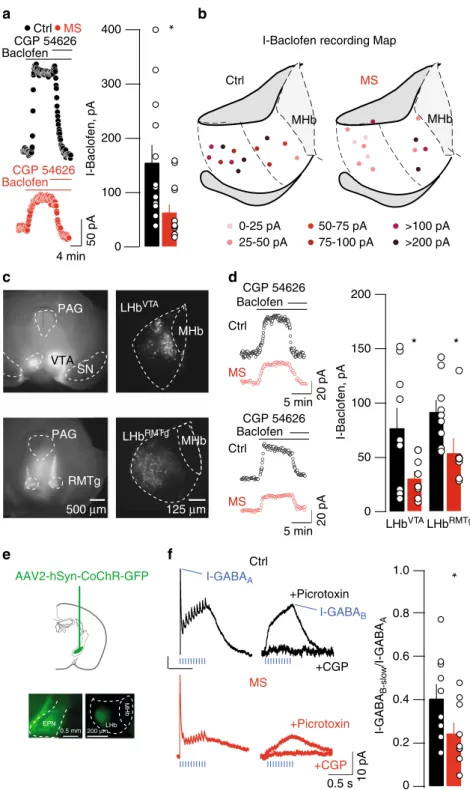
Fig. 2 MS drives GABAB-GIRK plasticity in LHb.a Sample traces, bar graph and scatter plots depicting CGP54626-sensitive I-Baclofen (I-Baclofen Ctrl vs.
MS;nmice= 5/6; ncells= 13; unpaired t-test, t24= 2.67 *p < 0.05). I-Baclofen was measured at steady state. b Territorial distribution of I-Baclofen showing
MS-dependent reduction of GABAB-GIRK signaling throughout the LHb.c Examples of the injection site of red retrobeads infused in the VTA (top) and in
the RMTg (bottom). Right, retrogradely labeled LHb neurons projecting to VTA or RMTg.d Sample traces, bar graph and scatter plots depicting CGP54626-sensitive I-Baclofen in LHbVTAand LHbRMTgneurons (LHBVTA: I-Baclofen Ctrl vs. MS;n
mice= 2; ncells= 9; unpaired t-test, t16= 2.37 *p < 0.05;
LHbRMTg: I-Baclofen Ctrl vs. MS;nmice= 2; ncells= 9 vs. 7; unpaired t-test, t14= 2.35 *p < 0.05). e Stereotactic infusion of AAV2-hSyn-CoChR-GFP within
the EPN, and opsin expression in terminals within the LHbf Opto-GABAA-IPSCs and opto-GABAB-IPSC sample traces and summary plot (Ctrl vs. MS;
nmice= 6/3; ncells= 9 per group; unpaired t-test, t16= 2.21; *p < 0.05). Values for opto-GABAA-IPSC and opto-GABAB-IPSC were taken at the maximal peak
disrupted, contribute to the establishment of depressive-like
symptoms
8, 12. MS significantly reduced I-Baclofen in both
LHb
VTAand LHb
RMTgneurons supporting a scenario in which
GABA
B-GIRK signaling reduction occurs throughout the LHb,
likely affecting multiple downstream targets (Fig.
2
c, d).
GABA
B-R activation within the LHb leads to cell
hyperpolar-ization through GIRK channels opening
11. Along with the
reduced I-Baclofen, MS also diminished GIRK currents. Indeed,
responses generated by the intracellular dialysis of the G-protein
activator GTPγS (I–GTPγS) and the bath application of the
specific GIRK1/2 activator ML-297 were smaller throughout the
LHb of MS mice (Supplementary Fig.
2
c, d)
17,18. These results
indicate that early-life stress, similarly to foot-shocks
11, 19,
reduces GABA
B-GIRK function within the LHb.
Activation of the GABA
B-GIRK signaling requires GABA
spillover from the presynaptic terminal
20. A major input source
of GABA onto LHb neurons arises from the entopeduncular
nucleus (EPN). Furthermore, the reduction of EPN-originating
inhibitory transmission within the LHb contributes to
depressive-like symptoms
15,21,22. We therefore set out to provide proof of
principle that MS reduces, not only agonist-evoked GABA
B-R
responses, but more precisely synaptically-relevant GABA
B-R
function. To probe synaptically-activated GABA
BRs, we
trans-duced EPN neurons with a rAAV2-hSyn-CoChR-eGFP allowing
for the expression of the excitatory opsin from Chloromonas
oogama (CoChR; Fig.
2
e)
23. CoChR-expressing EPN terminals
received trains of light-pulses (20 Hz), which elicited
fast-GABA
AR and slow-GABA
B-R outward currents (I-GABA
Aand
I-GABA
B-slowrespectively), the latter likely mediated by GABA
spillover diffusing to perisynaptic GABA
B-Rs (Fig.
2
f). We then
computed the I-GABA
B-slow/I-GABA
Aratios as a measure of the
postsynaptic strength of inhibitory transmission. MS diminished
the I-GABA
B-slow/I-GABA
Aratios without significant alterations
in miniature EPSCs or IPSCs, nor on presynaptic GABA
B-R
function (Fig.
2
f, Supplementary Fig.
3
a–d). Altogether, MS
diminishes postsynaptic GABA
B-GIRK signaling in LHb.
Thus, we predicted that MS-dependent LHb hyperexcitability
results from the above-described GABA
B-GIRK reduction.
Blocking GABA
ARs, AMPARs and NMDARs left MS
hyperexcit-ability unaffected. However, MS occluded GABA
BRs
antagonism-driven increased neuronal excitability observed in control slices
(Fig.
1
e, Fig.
3
a, b; (Ctrl, aCSF/CGP54626, F
(10;410)= 2.3 p < 0.05;
MS, aCSF/CGP54626, F
(10;410)= 0.37 p > 0.05; two-way ANOVA
RM, interaction). This suggests that MS leads to LHb
hyperexcit-ability via GABA
B-GIRK plasticity.
Limiting habenular activity ameliorates depressive-like
symp-toms. Is LHb hyperexcitability necessary for MS depressive-like
phenotypes? MS-driven adaptations occur throughout the LHb
(Fig.
2
b–d). Therefore, we sought to limit hyperactivity of a
large LHb neuronal population. Thus, we virally expressed the
inhibitory designer receptors exclusively-activated by designer
drugs (rAAV8-hSyn-HA-hM4Di-mCherry; Gi-DREADD) in the
LHb (Fig.
4
a, Supplementary Fig.
4
a). Ex-vivo, bath application of
the specific DREADDi ligand clozapine-N-oxide (CNO)
gener-ated a K
+-dependent outward current (I-CNO) and abolished
neuronal
firing (Supplementary Fig.
4
b, c). MS reduced I-CNO,
supporting a diminished GIRK function
24. We next examined
baseline neuronal activity using single unit recordings in
anes-thetized mice. The baseline
firing rate of MS mice was higher than
Control animals, an observation in line with the increased
excitability reported in acute slices. The systemic injection of
CNO diminished LHb neuronal
firing in
Gi-DREADD-expressing mice, and reduced the activity of MS-LHb neurons
to values not statistically different from Control animals
(Sup-plementary Fig.
4
d, e). This validates this strategy to limit
hyperexcitability in behaving mice. When exposed to escapable
foot-shocks (shuttle box), in the absence of CNO MS mice
(YFP/Gi-DREADD) expressed high failure rates (Fig.
4
b). After a
recovery period of 3 days, mice of all experimental groups
received a single systemic injection of CNO ~15 min prior the
test. CNO reduced the failure rate in the shuttle box only in
Gi-DREADD-expressing MS mice. However, 5 days later, after
complete clearance of CNO from the animal body, MS mice
displayed the depressive state (Fig.
4
b). Accordingly, in different
sets of mice treated with CNO (see Methods), TST immobility
and sucrose preference were comparable between
Gi-DREADD-expressing MS mice and Controls (Fig.
4
c, Supplementary
Fig.
4
f). In contrast, CNO treatment did not affect locomotor
activity measured in the open
field, arguing against compromised
motor function (Supplementary Fig.
4
f). Therefore, the
chemo-genetic reduction of LHb activity is sufficient to ameliorate
MS-driven depressive-like symptoms.
Individuals presenting symptoms of depression following
early-life stress poorly respond to antidepressants
25, heightening
the need of alternative treatments. Deep brain stimulation (DBS)
of the LHb ameliorates depressive symptoms, but its efficiency in
the context of MS is unknown
12,26. When applied ex-vivo within
the LHb, DBS-like stimulation (130 Hz)
12reduced glutamate
release, diminished neuronal
firing, hyperpolarized neurons and
decreased input resistance (Fig.
4
d, e). Accordingly, in
0.2s 20 mV 30 20 10 0
***
Picrotoxin +NBQX+APV Picrotoxin+NBQX APV+CGP54626 Injected current, pA 0 40 80 0 40 80 Action potentialsa
30 20 10 0 Action potentials 0.2s 20 mV 50 pA Ctrl MS 50 pA Ctrl MS Injected current, pAb
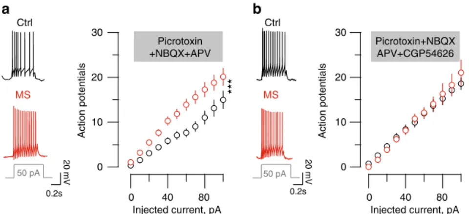
Fig. 3 MS-induced LHb neurons hyperexcitability requires reduced GABAB-GIRK signaling.a Left, sample traces for recordings in Ctrl and MS mice of a
current-evokedfiring (for a 50 pA step) in the presence of picrotoxin/NBQX. Graph representing the action potentials vs. injected current in all experimental groups. (picrotoxin/NBQX,nmice= 4/6; ncells= 20 per group; two-way ANOVA-RM, interaction F(10;380)= 3.52; ***p < 0.001) b Same than a
but in the presence of picrotoxin/NBQX/CGP54626,nmice= 5/6; ncells= 20 per group; two-way ANOVA-RM, interaction F(10;380)= 0.32). Scale bars=0.2
s and 20 mV
anesthetized mice, DBS lowered LHb activity (Fig.
2
f, g and
Supplementary Fig.
4
g). Therefore, DBS reduces neuronal activity
by engaging both presynaptic and postsynaptic mechanisms. We
then unilaterally implanted DBS electrodes in LHb (Fig.
4
f), and
examined its efficacy on MS-induced depressive-like behavior.
DBS reduced the number of failures in the shuttle box (Fig.
4
h).
This highlights DBS as a strategy, complementary to
pharmacol-ogy, to compensate for MS-driven LHb hyperexcitability and
ameliorate depressive states.
Discussion
Here we demonstrate that MS promotes LHb neuronal
hyper-activity by reducing GABA
B-GIRK function. This
hyperexcit-ability ultimately triggers depressive-like symptoms.
The GABA
B-GIRK signaling in the LHb tightly controls
neu-ronal activity
11and GABA
B-R internalization often occurs along
with GIRK trafficking
11,27,28. Consistently, we report that
MS-driven reduced GABA
B-Rs function occurs along with diminished
GIRK channels either expression or function. Internalization of
a
b
c
LHb LHb 3v pAAV-hSYN-HA-HM4D(Gi)-mCherry LHb LHb mHb ****** *** ** *** 300 250 200 150 Immobility, s 100 50 30 25 20 15 10 5 0 Failures LHb 0 7 15 24-28 Ctrl/MS Birth Day 47 50 55 57 Shuttle box CNO Tail suspension test CtrlMS CNO _+ _Shuttle box test Tail suspension test
Shock
AAV-YFP AAV-Gi-DREADD YFP Gi-DREADD Surgery
Single unit recordings in vivo
0 7 15 Ctrl/MS Birth
Day 35 39
Shuttle box Shuttle box Surgery 30 ** 12 8 4 0 Firing rate, Hz 25 DBS 130 Hz 5 min 10 s Pre-DBS Post-DBS MS ShamDBS Day 35 39
g
Normalized EPSC 5 4 3 2 1 Normalized EPSC,% 25 ms 100 pAd
e
LHb LHb 3v LHb DBS 0 *** 200 150 100 50 0 –50 30 25 20 15 10 5 0 b c a –55 mV –48.3 mV –48.2 mV 0.1 s 20 mVBaseline 5 min post DBS 10 min post DBS
30 25 20 15 10 5 0 Failures 100 µm 250 µm DBS electrode track * * Pulse number Time, min 5 4 3 2 1 a b c AAV: AAV-YFP AAV-Gi-DREADD 130Hz DBS 130 Hz 1 h _+_ _+_ _+_ CNO ++ ++ 500 400 300 200 100 0 Input resistance,MOhm Vm, mV 60 50 40 30 20 10 0 –60 –50 –40 –30 –20 –10 0 Action potential in 5s
Amplitude EPSC#1, % of baseline
** * * 160 140 120 100 80 60 40 20 0 *** 1.2 0.0 0.4 0.8
h
f
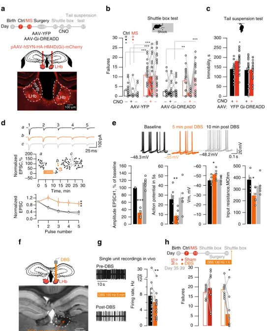
Fig. 4 Chemogenetic and DBS approaches reduce LHb activity and ameliorate MS-dependent depressive-like symptoms. a Schematic and image for Gi-DREADD LHb expression.b CNO effects on failures in the shuttle box (AAV-YFP, Ctrl vs. MS and AAV-Gi-DREADD, Ctrl vs. MS;nmice= 21/22/23/26;
two-way ANOVA RM, interaction, F(6,176)= 3.29, **p < 0.01). c Bar graph and scatter plot for TST immobility (AAV-YFP and AAV-Gi-DREADD, Ctrl vs.
MS: nmice= 23/25/25/ 25; two-way ANOVA, interaction, F(1,94)= 4, *p < 0.05). d DBS effect on 1st EPSC and PPR (5 pulses, 20 Hz. Normalized eEPSC
post-DBS,nmice= 2; ncells= 6; One-way ANOVA RM; F(1.23,6.13)= 99.65, ***p < 0.001; Normalized EPSCs, nmice= 2; ncells= 6; Two-way ANOVA RM; DBS
effect, F(2,50)= 25.1, ***p < 0.001). e Sample I-clamp recordings (5 superimposed-sweeps), and DBS effects on action potentials, resting membrane
potential and input resistance (Before vs. 5 min post-DBS vs. 10 min post-DBS,nmice= 2; ncells= 6: Action potential: F(1.29,6.49)= 13.5, **p < 0.01; Vm:
F(1.15,5.77)= 6.3, *p < 0.05; Ri: F(1.06,5.30)= 8.3,*p < 0.05; One-way ANOVA-RM) f DBS-electrode placement in LHb. g DBS-induced (130 Hz, 150 µA)
reduction of activity in vivo (Firing before vs. post-DBS,nmice= 4; ncells= 14; paired t-test, t6= 3.1;**p < 0.01) h DBS-driven reduction of failures in the
shuttle on MS mice (Sham vs. DBS;nmice= 8/9; Two-way ANOVA RM, interaction, F(1,15)= 9.476,**p < 0.01). Scale bars=, 100 μm a, 25 ms and 100 pA d,
GABA
B-Rs and GIRKs requires the activity of phosphatases
including PP2A, and a PP2A inhibitor presents antidepressant
properties
11,27, 28. However, whether PP2A contributes to
MS-driven GABA
B-GIRK plasticity in the LHb remains to be
established.
In line with a reduction of I-Baclofen, we observed a
dimin-ished I-GABA
B-slow/I-GABA
Aratios at EPN-to-LHb synapses.
These
findings provide new insights on (i) Physiological patterns
of presynaptic activity to elicit synaptic GABA
B-Rs in the LHb;
(ii) MS-mediated plasticity of synaptically-evoked GABA
B-Rs.
However MS-driven GABA
B-Rs reduction may also occur at
synaptic inputs other than the EPN.
Our data indicate that MS does not significantly affect mIPSCs,
however these events stem from unidentified inputs, leaving open
the possibility that MS also engages input-specific changes of fast
inhibitory transmission
15,21. In addition, the learned helplessness
depressive state potentiated fast excitatory transmission onto
LHb
VTAneurons
12. We cannot completely rule out that a similar
adaptation also occur in MS mice. The detection threshold for MS
plasticity of mEPSCs might be lowered by the different animal
model of depression employed or, alternatively, by circuit-specific
adaptations yet to be identified. Overall, plasticity of GABA
B-GIRKs
in the LHb emerges as a general cellular substrate underlying
stressor-driven depressive phenotypes as it is common to acute
traumatic events and chronic stressful conditions such as MS
11,19.
Reduced GABA
B-GIRK signaling mediates LHb neuronal
hyperactivity
11. Considering that MS-driven plasticity occurs in
VTA and RMTg-projecting LHb neurons, MS cellular
adapta-tions may remodel target midbrain circuits highlighting
LHb-to-midbrain relevance in mood disorders
3, 8, 12. The habenular
output connectivity is however complex indicating that
LHb-receiving structures other than the midbrain may contribute to
the MS-driven behavioral phenotypes. This is indeed likely, as the
inactivation of the LHb fails to rescue aberrant dopamine neurons
activity in the chronic mild stress model of depression
29.
Alto-gether, stress-driven depressive-like symptoms emerge from the
dysfunction of complex and parallel neuronal circuits (i.e.,
LHb-related and midbrain-LHb-related)
11,29–31.
MS-dependent hyperactivity can be limited by chemogenetic
and DBS approaches, which in turn are efficient to ameliorate
MS-mediated depressive-like phenotypes.
Our data provide an extensive description of Gi-DREADD
efficiency at the single-cell level ex-vivo and in-vivo within the
LHb. We report that Gi-DREADD activation in vivo in the LHb
reduces, but does not silence, neuronal activity. This highlights
Gi-DREADD efficacy in rescuing aberrant activity, without
abruptly altering the physiology of neural systems. These
prop-erties may differ according to neuronal populations, viral vector
properties or promoter employed.
In addition to a chemogenetic-based strategy we also
investi-gated the use of DBS approaches to reduce MS hyperactivity in
the LHb. The high-frequency DBS protocol reported in our work
was previously shown to also ameliorate depressive symptoms
emerging in congenital learned helplessness rats
12. Our data
suggest that DBS efficiency not only relies on reduced presynaptic
release
12, but also on postsynaptic mechanisms. The
DBS-mediated modulation of LHb activity is however transient,
sug-gesting that the MS depressive-like phenotypes can re-emerge
after the intervention
12. This indicates that DBS efficacy stems
from its pre/postsynaptic-mediated reduction in LHb neuronal
activity rather than on the MS-driven plasticity expression
mechanisms (i.e., GABA
B-GIRK plasticity).
Altogether, this study provides mechanistic insights underlying
MS-induced adaptations in the LHb. Moreover, limiting LHb
neuronal activity has potential therapeutic relevance in alleviating
affective symptoms of neuropsychiatric disorders.
Methods
Experimental subjects and MS paradigm. All procedures were used in accor-dance with the guidelines of the French Agriculture and Forestry Ministry for handling animals (Committee Charles Darwin #5, University Pierre et Marie Curie, Pairs). Part of the current study was carried out in the Department of Fundamental Neuroscience (Lausanne, Switzerland) under license and according to regulations of the Cantonal Veterinary Offices of Vaud and Zurich (Switzerland). Pregnant dams C57Bl/6J were received at the gestational stage E13–18 (Janvier Laboratories, France). Mothers were housed 2 per cages with access of food and water ad libitum. After birth, pups of either sex remained untouched until postnatal day (P) 7. At P7, litters were randomly divided in 2 groups. The maternal separation group consisted of pups removed from their litter and isolated in small compartments for 6 h per day (light phase 0800:1900 hours) repeated from P7 to P15 and followed by an early weaning at P17. During the separation, animals were maintained in heating plate and water was provided, maintaining constant temperature and humidity. The control group consisted of mice from independent litters, which were not manipulated until the regular weaning at P21 except during cage changing. During cage changing some old bedding and nest were transferred into the new cage in order to limit novelty stress. After weaning, mice where separated by sex and housed 6 per cage. Experiments were performed in mice aged 4–8 weeks. Surgery. Animals, aged at least 24 days were anesthetized with Ketamine (150 mg kg−1)/Xylazine (100 mg kg−1i.p.) before bilateral injection of rAAV8-Hsyn-Gi-DREADD-mCherry (University of Pennsylvania, US) in the LHb at the following coordinates (from bregma, in mm): A-P:–1.45; M-L:±0.45; D-V: –3.1. After three weeks, mice were subjected to CNO i.p injection (1 mg kg−1) for the DREADD activation. For optogenetic experiments rAAV2.1-hSyn-CoChr-eGFP (University of North Carolina, US) was infused in the entopeduncular nucleus (from bregma, in mm: A-P:–1.25; M-L:±1.8; D-V: –4.65). Recordings were performed 3 weeks after surgery. The injection sites were carefully examined and only animals with correct injections were kept for behavioral and electrophysiological analysis. DBS electrodes were unilaterally implanted using similar procedures and coordinates in the LHb. DBS electrodes were chronically implanted using a Superbond resin cements (Sun medical, Japan). For the experiment analyzing the output specificity of I-Baclofen, mice were bilaterally injected with a mixture of herpes simplex virus (McGovern Institute, USA) expressing enhanced GFP and red retrobeads (Lumafluor, US) into the RMTg or the VTA. The following coordinates were used (RMTg: from bregma, in mm: A-P:–2.9; M-L:±0.5; D-V: –4.3; VTA: from bregma, in mm: A-P:–2.4; M-L:±0.65; D-V: –4.9). Recordings from fluorescent LHb neurons were performed± 12 days following the surgery, and injection site were verified using the retrobeads labeling.
Behavioral paradigm. All experimental behaviors were performed during the light phase and experimenters were blind of their experimental group.
The shuttle box test was performed in a shuttle box (13 × 18 × 30 cm3) equipped
with an electrified grid floor and a door separating the two compartments. The test session consisted of 30 trials of escapable foot-shocks (10 s at 0.1–0.3 mA) separated by an interval of 30 s. The shock terminated any time that the animal shuttled in the other compartment. Failure is defined as the absence of shuttling to the other compartment within the 10 s shock delivery.
The tail suspension test was performed with mice being suspended by their tails with adhesive tape for a single session of 6 min. Immobility time of each animal was scored online by the experimenter. Mice were considered immobile only when they suspended passively and motionless.
The sucrose test preference was performed with mice being single-housed and habituated with two bottles of 1% sucrose for 2 days. At day 3 (test day) mice were exposed to two bottlesfilled with either 1% sucrose or water for 24 h. The sucrose preference was defined as the ratio of the consumption of sucrose solution vs. total intake (sucrose+water) during the test day and expressed as a percent.
Behavioral experiments in DREADD-injected animals were performed three weeks after viral infusion. For the shuttle Box, the tail suspension test, and the locomotor activity all the groups (YFP or DREADDi injected animals) were injected 15 min with CNO i.p. (1 mg kg−1). For sucrose preference experiments, all groups were injected with CNO i.p. (1 mg kg−1) every 3 h for the extent of the preference session (24 h) to maintain a constant DREADD-mediated inhibition. Electrophysiology. For in vitro recordings, animals were anesthetized with keta-mine and xylazine (i.p. 150 mg kg−1and 100 mg kg−1, respectively). Coronal brain slices (250µm) containing the LHb were prepared in bubble ice-cold 95% O2/5%
CO2-equilibrated solution containing: 110 mM choline chloride; 25 mM glucose;
25 mM NaHCO3; 7 mM MgCl2; 11.6 mM ascorbic acid; 3.1 mM sodium pyruvate;
2,5 mM KCl; 1.25 mM NaH2PO4; 0.5 mM CaCl2. Slices were then stored at room
temperature in 95% O2/5% CO2-equilibrated artificial cerebrospinal fluid (ACSF)
containing: 124 mM NaCl; 26.2 mM NaHCO3; 11 mM glucose; 2.5 mM KCl; 2.5 mM CaCl2; 1.3 mM MgCl2; 1 mM NaH2PO4. Recordings (flow rate of 2.5 ml min −1) were made under an Olympus-BX51 microscope (Olympus, France) at
30 °C. Currents were amplified, filtered at 5 kHz and digitized at 20 kHz. Access resistance and input resistance were monitored by a step of−4 mV (0.1 Hz). Experiments were discarded if the access resistance increased>20%.
The internal solution used to examine GABABand/or GIRK currents and
neuronal excitability contained: 140 mM potassium gluconate, 4 mM NaCl, 2 mM MgCl2, 1.1 mM EGTA, 5 mM HEPES, 2 mM Na2ATP, 5 mM sodium creatine
phosphate, and 0.6 mM Na3GTP (pH 7.3 with KOH). The liquid junction potential was ~12 mV. When we measured the synaptic inhibitory or excitatory release, the internal solution contained: 130 mM CsCl; 4 mM NaCl; 2 mM MgCl2; 1.1 mM
EGTA; 5 mM HEPES; 2 mM Na2ATP; 5 mM sodium creatine phosphate; 0.6 mM Na3GTP; and 0.1 mM spermine. The liquid junction potential was−3 mV. Whole-cell voltage-clamp recordings were achieved to measure GABAB-GIRK currents in
aCSF only. For agonist-induced currents, changes in holding currents in response to bath application of baclofen (100µM) were measured (at −50 mV every 5–10 s). The plotted values correspond to the difference between the baseline and the plateau (for the baclofen and ML297 experiments) or the difference between the plateau and the value of holding current after barium (for the I-GTP-γS) GABAB-GIRK currents were confirmed by antagonism with 10 μM of CGP54626. When stated, 100μM of GTP-γS was added to the internal solution in place of Na3GTP. Plateau currents were then reversed by 1 mM Barium application, a selective inhibitor of K+channels. Changes in holding currents in response to GIRK agonist were measured (at−50 mV every 5–10 s) by bath application of ML-297 (50 µM), a Selective GIRK1/2 channel activator then reversed by 1 mM Barium application. Synaptic GABABslow IPSCs were optically evoked by trains of 10 pulses delivered
at 20 Hz through a 470 nm LED. The fast GABA amplitude correspond to the amplitude of thefirst pic of the train, the slow GABA current instead were measured after picrotoxin bath application, and correspond to the
I-max. Miniature excitatory postsynaptic currents (mEPSCs) were recorded in voltage-clamp mode at−60 mV in the presence of bicuculline (10 μM), AP5 (50μM) and tetrodotoxin (TTX, 1 μM). Miniature inhibitory postsynaptic currents (mIPSCs) were recorded (−60 mV) in the presence of NBQX (20 μM) AP5 (50 μM) and TTX (1μM). EPSCs were evoked through an ACSF-filled monopolar glass electrode placed in the LHb. For the experiments in which high-frequency stimulation trains were used to determine presynaptic release probability (5 pulses at 20 Hz), QX314 (5 mM) was included in the internal solution to prevent the generation of sodium spikes.
Current-clamp experiments were performed using a series of current steps (from−80 to 100 pA or when the cell reached a depolarization block) injected to induce action potentials (10-pA injection current per step, duration of 500 ms). Cells were maintained at−55 mV throughout the experiment. When testing changes in tonicfiring, cells were depolarized to obtain stable firing activity in current-clamp mode.
When in vivo single unit recordings were performed, mice were anesthetized with isoflurane (induction: 2%; maintenance: 1–1.5%) using an anesthesia device for small animals (Univentor 410, Malta). We placed the mice in the stereotaxic apparatus (Kopf, Germany) and their body temperature was maintained at 36± 1 °C using a feedback-controlled heating pad (CMA 450 Temperature Controller, USA). The scalp was retracted and one burr hole was drilled above the LHb (A-P:–1.3/–1.6; M-L:±0.4/0.5) for the placement of a recording electrode. Single unit activity of neurons located in the LHb (Ventral 2.3–3.2 mm to cortical surface) was recorded extracellularly by glass micropipettesfilled with 2% pontamine sky blue dissolved in 0.5 M sodium acetate (impedance 3–6 MΩ). Signal was pre-amplified (DAM80, WPI, Germany), filtered (band-pass 500–5000 Hz) amplified (Neurolog System, Digitimer, UK), displayed on a digital storage oscilloscope (OX 530, Metrix, USA), and digitally recorded. Experiments were sampled on- and off-line by a computer connected to CED Power 1401 laboratory interface (Cambridge Electronic Design, Cambridge, UK) running the
Spike2 software (Cambridge Electronic Design). Single units were isolated and identified according to previously described electrophysiological characteristics (Meye et al.8) including a broad triphasic extracellular spike (>3 ms), and a tonic
regular, tonic irregular or bursting spontaneous activity.
Isolated LHb neurons were recorded for 5 min to establish the basal spontaneousfiring rate. CNO was administered i.p. (1 mg kg−1) and thefiring
activity of the neuron was monitoring every 5 m for total 40 m. When CNO was administered, only one cell was recorded per mouse.
DBS experiments were performed with a modified double barrel system allowing to stimulate in close proximity of the recording site: the stimulating electrode was attached to the recording one by using glass barrels (the 2 electrodes form an angle of±30°). The stimulating tip was glued above the recording tip (<300 µm). A stable spontaneous firing rate was recorded for 5 min before to start the DBS protocol (total duration: 2–5 m; Train pulses: 7; ITI: 40 ms; Frequency: 130 Hz; Intensity: 150µA). The firing activity recorded immediately after the protocol was compared with the respective baseline.
At the end of each experiment, the electrode placement was marked with an iontophoretic deposit of pontamine sky blue dye (−80 μA, continuous current for 35 min). Brains were then rapidly removed andfixed in 4% paraformaldehyde solution. The position of the electrodes was microscopically identified on serial sections (60μm).
Deep brain stimulation. MS mice for DBS experiments werefirst preselected on the basis of their failure rate in the Shuttle box test (A cutoff of 12 failures was used for the preselection). In total 50 mice were tested, and 17 of these animals met the criteria. Standard surgical procedures were used to implant bipolar concentric electrodes unilaterally into the LHb (coordinates−1.45 mm AP, ±0.45 mm ML and
−3.1 mm DV). After 5 days recovery from surgery, DBS or no (Sham) stimulation was applied for 1 h (seven stimulus trains of 130 Hz, separated by 40 ms intervals; 150µA intensity) prior testing each mouse in the shuttle box test.
Analysis and drugs. All drugs were obtained from Abcam (Cambridge, UK) and Hello Bio, and Tocris (Bristol, UK) and dissolved in water, except for TTX (citric acid 1%), ML297 and CNO (DMSO). Online/offline analysis were performed using IGOR-6 (Wavemetrics, US) and Prism (Graphpad, USA). Data analysis for in vivo electrophysiology was performed off-line using Spike2 (CED, UK) software. Sample size required for the experiment was empirically tested by running pilots experiments in the laboratory. While behavioral experiments were run in a single-to-triple trial, electrophysiological experiments were replicated at leastfive times. Experiments were replicated in the laboratory at least twice. Animals were ran-domly assigned to experimental groups. Data distribution was assumed to be normal, and single data points are always plotted. Compiled data are expressed as mean± s.e.m. All groups were tested with Grubbs exclusion test (limit set at 0.05) to determine outliers. Significance was set at p < 0.05 using Student’s t-test two-sided, Kolmogorov–Smirnov test, one-way, two-way or three-way Anova with multiple comparison when applicable.
Data availability. All relevant data are available from the authors upon request.
Received: 26 January 2017 Accepted: 24 August 2017
References
1. Norman, R. E. et al. The long-term health consequences of child physical abuse, emotional abuse, and neglect: a systematic review and meta-analysis. PLoS Med. 9, e1001349 (2012).
2. Tractenberg, S. G. et al. An overview of maternal separation effects on behavioural outcomes in mice: Evidence from a four-stage methodological systematic review. Neurosci. Biobehav. Rev. 68, 489–503 (2016). 3. Authement, M. E. et al. Histone deacetylase inhibition rescues maternal
deprivation-induced GABAergic metaplasticity through restoration of AKAP Signaling. Neuron 86, 1240–1252 (2015).
4. Marco, E. M., Adriani, W., Llorente, R., Laviola, G. & Viveros, M. P. Detrimental psychophysiological effects of early maternal deprivation in adolescent and adult rodents: altered responses to cannabinoid exposure. Neurosci. Biobehav. Rev. 33, 498–507 (2009).
5. Nishi, M., Horii-Hayashi, N. & Sasagawa, T. Effects of early life adverse experiences on the brain: implications from maternal separation models in rodents. Front Neurosci. 8, 166 (2014).
6. Matsumoto, M. & Hikosaka, O. Representation of negative motivational value in the primate lateral habenula. Nat. Neurosci. 12, 77–84 (2009).
7 Lecca, S. et al. Aversive stimuli drive hypothalamus-to-habenula excitation to promote escape behavior. eLife 6, e30697 (2016).
8. Meye, F. J. et al. Cocaine-evoked negative symptoms require AMPA receptor trafficking in the lateral habenula. Nat. Neurosci. 18, 376–378 (2015). 9. Proulx, C. D., Hikosaka, O. & Malinow, R. Reward processing by the lateral
habenula in normal and depressive behaviors. Nat. Neurosci. 17, 1146–1152 (2014).
10. Stamatakis, A. M. & Stuber, G. D. Activation of lateral habenula inputs to the ventral midbrain promotes behavioral avoidance. Nat. Neurosci. 15, 1105–1107 (2012).
11. Lecca, S. et al. Rescue of GABAB and GIRK function in the lateral habenula by protein phosphatase 2A inhibition ameliorates depression-like phenotypes in mice. Nat. Med. 22, 254–261 (2016).
12. Li, B. et al. Synaptic potentiation onto habenula neurons in the learned helplessness model of depression. Nature 470, 535–539 (2011).
13. Wilcox, K. S., Christoph, G. R., Double, B. A. & Leonzio, R. J. Kainate and electrolytic lesions of the lateral habenula: effect on avoidance responses. Physiol. Behav. 36, 413–417 (1986).
14. Li, K. et al.βCaMKII in lateral habenula mediates core symptoms of depression. Science 341, 1016–1020 (2013).
15. Shabel, S. J., Proulx, C. D., Piriz, J. & Malinow, R. Mood regulation. GABA/ glutamate co-release controls habenula output and is modified by antidepressant treatment. Science 345, 1494–1498 (2014).
16. George, E. D., Bordner, K. A., Elwafi, H. M. & Simen, A. A. Maternal separation with early weaning: a novel mouse model of early life neglect. BMC Neurosci. 11, 123 (2010).
17. Kaufmann, K. et al. ML297 (VU0456810), thefirst potent and selective activator of the GIRK potassium channel, displays antiepileptic properties in mice. ACS Chem. Neurosci. 4, 1278–1286 (2013).
18. Logothetis, D. E., Kurachi, Y., Galper, J., Neer, E. J. & Clapham, D. E. The beta gamma subunits of GTP-binding proteins activate the muscarinic K+channel in heart. Nature 325, 321–326 (1987).
19. Lecca, S., Trusel, M. & Mameli, M. Footshock-induced plasticity of GABAB signalling in the lateral habenula requires dopamine and glucocorticoid receptors. Synapse 71, e21948 (2017).
20. Lüscher, C., Jan, L. Y., Stoffel, M., Malenka, R. C. & Nicoll, R. A. G protein-coupled inwardly rectifying K+ channels (GIRKs) mediate postsynaptic but not presynaptic transmitter actions in hippocampal neurons. Neuron 19, 687–695 (1997).
21. Meye, F. J. et al. Shifted pallidal co-release of GABA and glutamate in habenula drives cocaine withdrawal and relapse. Nat. Neurosci. 19, 1019–1024 (2016). 22. Shabel, S. J., Proulx, C. D., Trias, A., Murphy, R. T. & Malinow, R. Input to the
lateral habenula from the basal ganglia is excitatory, aversive, and suppressed by serotonin. Neuron 74, 475–481 (2012).
23. Klapoetke, N. C. et al. Independent optical excitation of distinct neural populations. Nat. Methods 11, 338–346 (2014).
24. Pascoli, V., Terrier, J., Hiver, A. & Lüscher, C. Sufficiency of mesolimbic dopamine neuron stimulation for the progression to addiction. Neuron 88, 1054–1066 (2015).
25. Williams, L. M., Debattista, C., Duchemin, A. M., Schatzberg, A. F. & Nemeroff, C. B. Childhood trauma predicts antidepressant response in adults with major depression: data from the randomized international study to predict optimized treatment for depression. Transl. Psychiatry 6, e799 (2016).
26. Sartorius, A. et al. Remission of major depression under deep brain stimulation of the lateral habenula in a therapy-refractory patient. Biol. Psychiatry 67, e9–e11 (2010).
27. Hearing, M. et al. Repeated cocaine weakens GABA(B)-Girk signaling in layer 5/6 pyramidal neurons in the prelimbic cortex. Neuron 80, 159–170 (2013). 28. Padgett, C. L. et al. Methamphetamine-evoked depression of GABA(B) receptor
signaling in GABA neurons of the VTA. Neuron 73, 978–989 (2012). 29. Moreines, J. L., Owrutsky, Z. L. & Grace, A. A. Involvement of infralimbic
prefrontal cortex but not lateral habenula in dopamine attenuation after chronic mild stress. Neuropsychopharmacology 42, 904–913 (2017). 30. Tye, K. M. et al. Dopamine neurons modulate neural encoding and expression
of depression-related behaviour. Nature 493, 537–541 (2013).
31. Yang, L. M., Hu, B., Xia, Y. H., Zhang, B. L. & Zhao, H. Lateral habenula lesions improve the behavioral response in depressed rats via increasing the serotonin level in dorsal raphe nucleus. Behav. Brain. Res. 188, 84–90 (2008).
Acknowledgements
This work was supported by the European Research Council (Starting grant SalienSy 335333) to M.M. We are grateful to M. Creed and C. Lüscher for support on DBS approach. We thank C. Bellone, M. Creed, E. Schwartz, C. Lüscher. and the Mameli Laboratory for feedback on the manuscript. M.M. is a member of the Fens-Kavli Net-work of Excellence.
Author contributions
A.T. performed and analyzed in vitro recordings together with K.V. and M.M. S.L. performed in vivo recordings. A.T. performed behavioral experiments with M.M. and S.L. M.M. and A.T. designed the study and wrote the manuscript.
Additional information
Supplementary Informationaccompanies this paper at doi:10.1038/s41467-017-01192-1. Competing interests:The authors declare no competingfinancial interests.
Reprints and permissioninformation is available online athttp://npg.nature.com/ reprintsandpermissions/
Publisher's note:Springer Nature remains neutral with regard to jurisdictional claims in published maps and institutional affiliations.
Open Access This article is licensed under a Creative Commons Attribution 4.0 International License, which permits use, sharing, adaptation, distribution and reproduction in any medium or format, as long as you give appropriate credit to the original author(s) and the source, provide a link to the Creative Commons license, and indicate if changes were made. The images or other third party material in this article are included in the article’s Creative Commons license, unless indicated otherwise in a credit line to the material. If material is not included in the article’s Creative Commons license and your intended use is not permitted by statutory regulation or exceeds the permitted use, you will need to obtain permission directly from the copyright holder. To view a copy of this license, visithttp://creativecommons.org/ licenses/by/4.0/.
© The Author(s) 2017