HAL Id: hal-02568181
https://hal.archives-ouvertes.fr/hal-02568181
Submitted on 8 May 2020HAL is a multi-disciplinary open access archive for the deposit and dissemination of sci-entific research documents, whether they are pub-lished or not. The documents may come from teaching and research institutions in France or abroad, or from public or private research centers.
L’archive ouverte pluridisciplinaire HAL, est destinée au dépôt et à la diffusion de documents scientifiques de niveau recherche, publiés ou non, émanant des établissements d’enseignement et de recherche français ou étrangers, des laboratoires publics ou privés.
The Nanoscale Organization of the Plasma Membrane
and Its Importance in Signaling: A Proteolipid
Perspective
Yvon Jaillais, Thomas Ott
To cite this version:
Yvon Jaillais, Thomas Ott. The Nanoscale Organization of the Plasma Membrane and Its Importance in Signaling: A Proteolipid Perspective. Plant Physiology, American Society of Plant Biologists, 2020, 182 (4), pp.1682-1696. �10.1104/pp.19.01349�. �hal-02568181�
Title: The nanoscale organization of the plasma membrane and its importance in signaling – a proteolipid perspective
Running title: plasma membrane compartmentalization in signaling Yvon Jaillais1 and Thomas Ott2
1 Laboratoire Reproduction et Développement des Plantes, Université de Lyon, ENS
de Lyon, UCB Lyon 1, CNRS, INRA, F-69342 Lyon, France.
2 Cell Biology, Faculty of Biology, University of Freiburg, 79104 Freiburg, Germany
Address correspondence to yvon.jaillais@ens-lyon.fr and thomas.ott@biologie.uni-freiburg.de
ABSTRACT:
Plasma membranes provide a highly selective environment to a large number of transmembrane and membrane-associated proteins. While the lateral movement of proteins in this lipid bilayer is possible, it is rather limited in turgid and cell wall-shielded plant cells. However, membrane-resident signaling processes occur on sub-second scales that cannot be explained by simple diffusion models. Accordingly, several receptors and other membrane-associated proteins are organized and functional in membrane nanodomains. While their general presence has become a widely-accepted fact, fundamental functional aspects, the roles of individual lipid species and their interplay with proteins as well as aspects of nanodomain maintenance and persistence remain to be addressed sufficiently. Here, we review the current knowledge on nanodomain organization and function with a particular focus on signaling processes considering both proteins, lipids and their interactions. Furthermore, we propose new and hypothetical aspects of plant membrane biology that we consider important for future research.
ADVANCES:
• Virtually all proteins at the plasma membrane are segregated in
nanodomains, which are critical for signaling.
• The interplay between lipids and scaffolding proteins allows the
coexistence of a dynamic patchwork of plasma membrane nanodomains, which ensure both signal specificity and integration.
• The traditional view of plasma membrane compartmentalization in animal
systems needs to be revisited in plants because of the presence of the cell wall, which deeply impact protein and lipid diffusion.
• The plasma membrane may act as an analog-digital-analog converter
which allows high fidelity signaling with built-in tuning capacities through the use of lipids, scaffolding proteins and membrane skeletons.
Together with the cell wall, the plasma membrane forms the frontier of the cell. As such, it acts as a physical barrier and allows the generation and maintenance of chemical gradients between the outside and inside of the cell. At the same time, the plasma membrane is a critical checkpoint for the perception and integration of extracellular signals prior to signal transduction in the cytoplasm. The fluid mosaic model initially predicted that biological membranes are fluids, with the underlying assumption that their protein and lipid constituents can laterally diffuse in the plane of the membrane without major restrictions (Singer and Nicolson, 1972). According to this view, membrane-embedded receptors would distribute uniformly throughout the cell surface and irrespective of the plasma membrane proteome. The very opposite seems to be the case. Unequivocal evidence implies that the plasma membrane itself is highly compartmentalized into subdomains and that lateral segregation of proteins and lipids is a critical facet of cell surface signaling, modulating signal perception, specificity and integration.
This view of a compartmentalized plasma membrane first arose from biochemical fractionation, which could separate biological membranes in a binary manner between so called resistant (also referred to as insoluble) and detergent-sensitive membranes (Brown and Rose, 1992; Mongrand et al., 2004; Borner et al., 2005; Morel et al., 2006; Laloi et al., 2007; Lefebvre et al., 2007). However, fluorescent microscopy techniques with ever increasing resolution power have largely replaced biochemical fractionation as it rapidly became clear that plasma membrane subdomains are not binary but rather a part of a large patchwork of many subdomains that coexist on various spatial and temporal scales. Such view is supported by a wealth of data from colocalization analyses using confocal, total internal reflection fluorescence (TIRF) and super-resolution microscopy (Kleine-Vehn et al., 2011; Demir et al., 2013; Jarsch et al., 2014; Hosy et al., 2015; Bucherl et al., 2017; Martinière et al., 2019; Platre et al., 2019). Furthermore, the fact that different membrane constituents display varying diffusion patterns within the plane of the plasma membrane is also sustained by studies on the dynamics of protein/lipid lateral diffusion using fluorescence recovery after photobleaching (FRAP), single molecule imaging (e.g. single particle tracking PhotoActivated Localization Microscopy, sptPALM) and fluctuation correlation spectroscopy (FCS) (Li et al., 2011; Martiniere et al., 2012; Wang et al., 2013; Jarsch et al., 2014; Hosy et al., 2015; Wang et al., 2015; Li et al., 2016; Gronnier et al., 2017; Cui et al., 2018; Martinière et al., 2019; McKenna et al., 2019; Platre et al., 2019). Technically, receptor/scaffold complexes have also often been studied using distance-based imaging techniques such as fluorescence lifetime imaging- Förster resonance energy transfer (FLIM-FRET). However, a note of caution has recently been put forward, concerning the use of Bimolecular-Fluorescence
Complementation (BiFC) that can artificially stabilize membrane proteins in membrane contact sites of the endoplasmatic reticulum and the plasma membrane (Tao et al., 2019).
In this update, we review the current evidence for the coexistence of a patchwork of membrane nanodomains in the plant plasma membrane and their functional importance and assess the roles of lipids, the cell wall and the cytoskeleton in shaping this diverse plasma membrane landscape. Finally, we will discuss plausible scenarios for the functional importance of protein nanoclustering in signal transduction.
THE PLASMA MEMBRANE AS A PATCHWORK OF COEXISTING FUNCTIONAL MEMBRANE NANODOMAINS
Receptor-scaffolding at the nanoscale
Unequivocal evidence demonstrates that a significant number of membrane-resident proteins cluster in higher order structures that have been termed ‘membrane nanodomains’ or ‘membrane microdomains’, for which a nomenclature has been suggested recently (Ott, 2017). Briefly, nanodomains are submicron protein and/or lipid assemblies (roughly 20-300nm and below 1µm), while microdomains are significantly larger assemblies (>1µm, e.g. perimicrobial membranes, the Casparian strip domain, polar domains, plasmodesmata or membrane contact sites) (Ott, 2017). For the following of this Update, we will specifically focus on plasma membrane nanodomains, which have formerly often been termed ‘lipid rafts’. While the ‘lipid raft’ model was mainly based on biochemical evidence, recent cell biological approaches revealed that a number of proteins distribute heterogeneously on plant cell membranes mostly labelling puncta-like structures (Kleine-Vehn et al., 2011; Demir et al., 2013; Jarsch et al., 2014; Bucherl et al., 2017; Martinière et al., 2019; Platre et al., 2019). Over time, members of the flotillin (FLOT) and the plant-specific remorin (REM) protein families have been described as marker proteins for these membrane nanodomains (Raffaele et al., 2009; Jarsch and Ott, 2011; Li et al., 2012; Marin et al., 2012; Perraki et al., 2012; Hao et al., 2014; Jarsch et al., 2014; Wang et al., 2015; Bucherl et al., 2017; Gronnier et al., 2017; Liang et al., 2018) (Figure 1). This allowed comparative studies that revealed the co-existence of multiple nanodomains within the same cell (Jarsch et al., 2014). Flotilins comprise a small protein family with only two members in
Arabidopsis thaliana (Daněk et al., 2016), whereas at least 16 remorins have been
identified with an additional two legume-specific members (group 2) (Raffaele et al., 2007) (Figure 1). While the precise molecular function for most of these proteins remains unknown, mostly group 1 REMs were repeatedly shown to regulate viral spreading in leaves possibly by modulating plasmodesmatal conductance (Raffaele et al., 2009; Perraki et al., 2012; Ishikawa et al., 2017; Perraki et al., 2018). A recent pre-print reports on reduced numbers of plasmodesmata (PDs) in a rem1.2/rem1.3 double knock-out mutant, indicating a role of remorins in PD biogenesis (Wei et al., 2019). Additionally, remorins form higher order oligomers leading to filamentous structures in vitro (Bariola et al., 2004; Marin et al., 2012; Martinez et al., 2019) and in vivo (Wei et
al., 2019) and this may drive association of REMs with the PM (Legrand et al., 2019). REM oligomer formation itself is mainly mediated by the conserved C-terminal coiled-coil region, a hallmark for these proteins (Raffaele et al., 2009; Marin et al., 2012; Martinez et al., 2019) and further supported by the intrinsically disordered N-terminal region that harbors the majority of phosphorylation sites (Marin and Ott, 2012; Marin et al., 2012). The importance of REM phosphorylation on their association patterns with membrane nanodomains is, however, still elusive. However, functionality of REMs in plasmodesmata is hampered in phospho-mutants (Perraki et al., 2018). While REM phosphorylation was mostly studied in vitro, several REMs are able to associate or at least co-localize with a number of soluble or membrane-associated kinases such as the CALCIUM-DEPENDENT PROTEIN KINASES CPK3 (Perraki et al., 2018) and CPK21 (Demir et al., 2013), AVRPPHB SUSCEPTIBLE1 (PBS1) (Albers et al., 2019), SNF1 RELATED KINASE (SnRK1) (Son et al., 2014) and receptor-like kinases (RLKs) such as the A. thaliana brassinosteroid receptor BRASSINOSTEROID INSENSITIVE 1 (BRI1) and innate immune receptor FLAGELLIN SENSING 2 (FLS2) (Bucherl et al., 2017), the rice SOMATIC EMBRYOGENEIS RECEPTOR KINASE 1 (SERK1) and OsBRI1 (Gui et al., 2016) and the Medicago truncatula RLKs NOD FACTOR PERCEPTION (NFP), LYSIN MOTIF KINASE 3 (LYK3) and DOES NOT MAKE INFECTIONS 2 (DMI2) as well as their corresponding homologs in Lotus japonicus (Lefebvre et al., 2010; Toth et al., 2012; Liang et al., 2018). Increasing evidence suggests that remorins and flotillins play versatile roles in nanodomain stabilization (Huang et al., 2019a) and receptor recruitment into these structures (Haney et al., 2011; Bucherl et al., 2017; Liang et al., 2018).
Significant progress with respect to REM functionality and the functional relevance of receptor recruitment into nanodomains were made in rice, where ligand-induced phosphorylation of OsREM4.1 results in its dissociation from OsSERK1 and consequently allowing the assembly of the signaling-competent OsSERK1/OsBRI1 receptor complex (Gui et al., 2016). A different mode of maintaining an active receptor complex has been proposed in the legume Medicago truncatula, where receptor-mediated ligand perception results in transcriptional activation of the group 2 remorin SYMREM1, which in turn physically associates with the entry receptor LYK3. This interaction results in a stabilization and physical recruitment of LYK3 in a laterally stable and FLOT4-positive nanodomain. Genetic evidence further suggests that this process is required for receptor stabilization at the plasma membrane (Haney et al., 2011; Liang et al., 2018).
Nanodomain targeting
Even though the evidence is still slightly scattered, a number of studies suggest that receptor nanoclusters co-localize with one or the other scaffold belonging to the FLOT and/or REM protein families. This implies that these proteins may act as organizing centers as recently suggested for remorins (Gui et al., 2016; Liang et al., 2018; Huang et al., 2019a) and at least for the M. truncatula flotillin FLOT2/4 (Haney and Long, 2010; Haney et al., 2011; Liang et al., 2018).
Recruitment of REMs themselves to the plasma membrane and, in some cases, also to nanodomains is mediated by a C-terminal hydrophobic stretch, called the Remorin C-terminal anchor (REM-CA) (Figure 1). The REM-CA peptide specifically binds in a pH-dependent manner to sterols and PIPs and requires the simultaneous presence of both β-sitosterol and PIPs in the same nanodomain (Legrand et al., 2019). This demonstrates the tight link between specific lipid species and nanodomain recruitment of proteins. In addition, REM association with membranes is further supported by S-acylation of cysteine residues as well as protein-protein interactions (Raffaele et al., 2009; Perraki et al., 2012; Konrad et al., 2014; Gronnier et al., 2017; Legrand et al., 2019). It remains, however, still an open point of discussion whether S-acylation represents a major and general post-translational modification supporting nanodomain targeting of REMs and other proteins. Among the over 600 acylated proteins identified in A. thaliana is also the immune receptor FLS2 (Hemsley et al., 2013). While S-acylation on two serine residues (S830/S831) within the juxtamembrane domain of FLS2 have been mapped, these residues are not required for plasma membrane localization of the receptor per se (Hurst et al., 2019). However, a recent preprint suggests that they may contribute to the localization of FLS2 in nanodomains (Chen et al., 2019). This study also claims that S-acylation might be a hallmark for receptor targeting into nanodomains as a posttranslational modification-dependent recruitment to FLOT1-labelled nanodomains was also observed for the receptors CERK1 and P2K1 (DORN1) that mediate perception of chitin and extracellular ATP, respectively (Chen et al., 2019). However, these results were obtained using transient expression in protoplasts in which the formation of nanodomains is heavily impacted due to the absence of a cell wall (see below). Furthermore, nanodomains were visualized using deconvolution as an imaging post-processing method, rather than microscopy techniques that can actually resolve diffraction-limited structures. Therefore, these results should be taken with caution until they have been verified in situ. In addition, Hurst and co-workers found that FLS2 mutants, in which C830/831 were substituted by serines, were fully functional, arguing that S-acylation at these sites is dispensable for FLS2 signaling function. Receptor tagging and expression levels are critical for FLS2 function and therefore need to be considered in functional studies (Hurst et al., 2018). Similar concerns have been raised for the FLS2 co-receptor BAK1 (Ntoukakis et al., 2011).
IMPORTANCE OF LIPIDS IN PLASMA MEMBRANE LATERAL SEGREGATION Asymmetric localization of lipids in the two leaflets of the plasma membrane
Like proteins, lipids are also not uniformly localized in the plasma membrane. In animal cells, they display a strong asymmetry between the outer and inner membrane leaflets, which face the cell wall and the cytosol, respectively (Figure 2). The outer membrane leaflet is rich in glycosphingolipids and sterols as well as the phospholipid
phosphatidylcholine (PC). In plants, lipid asymmetry between the two membrane leaflets has not been extensively addressed experimentally, but was proposed to be largely similar to animal cells (Gronnier et al., 2018), with the main plant sphingolipids, glycosylinositol phosphorylceramides (GIPCs) (Cacas et al., 2016), being enriched in the outer membrane leaflet (Figure 2). By contrast, the inner leaflet is enriched in phospholipids, notably phosphatidylethanolamine (PE) and the minor anionic phospholipids, which include phosphatidic acid (PA), phosphatidylserine (PS), phosphatidylinositol (PI) and its phosphorylated derivative the phosphoinositides (PIPs) (Colin and Jaillais, 2019). These anionic lipids confer a strong electronegative property to the inner surface of the plasma membrane, which drives the identity of the plasma membrane and is crucial for the recruitment of many soluble or lipid-anchored proteins to this compartment (Simon et al., 2016; Noack and Jaillais, 2017; Platre et al., 2018) (Figure 2).
Lipid order in plasma membrane compartmentalization
Lipid segregation not only exists between the outer and inner plasma membrane leaflets but also laterally within each leaflet. The lipid raft hypothesis already postulated that lipids in biological membranes may be in a liquid-disordered or liquid-ordered phase (Kusumi et al., 2012). The liquid-ordered phase is enriched in sterols and glycosphingolipids, which largely corresponds to the lipid composition of the outer leaflets. Sphingolipids, such as GIPC, interact with phytosterols to increase lipid order (Cacas et al., 2016) (Figure 2). Evidence for different ordered phases at the plant plasma membrane recently emerged when using di-4-ANEPPDHQ (thereafter named ANEPP), a probe sensitive to the lipid order (Roche et al., 2008; Frescatada-Rosa et al., 2014; Zhao et al., 2015; Zhao et al., 2015; Gerbeau-Pissot et al., 2016; Gronnier et al., 2017; Grosjean et al., 2018; Huang et al., 2019a; Laurent et al., 2019; Pan et al., 2019). Its fluorescence emission spectrum is blue-shifted in liquid-ordered compared with liquid-disordered phases (Jin et al., 2005; Jin et al., 2006). Initially validated in
vitro in model membranes, this dye is also amenable to live imaging, as it is easy to
apply and can also detect both the liquid-ordered and liquid-disordered lipid phases in living cells (Gerbeau-Pissot et al., 2016). ANEPP staining in vivo shows heterogeneous labeling of the plasma membrane (Gronnier et al., 2017; Pan et al., 2019). It is sensitive to methyl-β-cyclodextrin (mßcd), a sterol depleting agent, which decreases the ANEPP-stained liquid-order phase at the expense of the liquid-disordered phase (Huang et al., 2019a). Similarly, a recent preprint suggests that the amount of staining of the liquid-ordered phases is reduced in the sterol biosynthesis mutant fackel-J79 (fk-J79) (Pan et al., 2019). Together, these data support the notion that sterols are required for the formation of liquid-ordered lipid phases in planta. However, these approaches also have limitations and conclusion obtained with either ANEPP or mßcd should be taken with caution. Indeed, ANEPP is a membrane-intercalating dye and may therefore impact membrane properties. In addition, mßcd has a massive impact on membrane structure and integrity, and protein-lipid/lipid-lipid interactions.
Roles of lipids in protein targeting in nanodomains
Recent data imply that liquid-ordered phases are relevant for protein localization, since the ANEPP-stained liquid-ordered phases colocalize with the group 1 REM StREM1.3 (Gronnier et al., 2017). Interestingly, the localization of StREM1.3 in nanodomains is sensitive to fenpropimorph, an inhibitor that affects the plasma membrane sterol composition but not the total amount of sterols (Gronnier et al., 2017). Below, we will use StREM1.3 as an example for the role of protein/lipid interactions in nanodomain localization, as this is to date the best studied case in plants (Figure 1). Although indirect, these data are consistent with the notion that sterols participate in the formation of liquid-ordered membrane domains, which are themselves required for localization of proteins, including REMs (Gronnier et al., 2017; Legrand et al., 2019). However, as mentioned above, sterols and sphingolipids mainly accumulate in the outer membrane leaflet, while remorins are inserted into the cytosolic leaflet (Cacas et al., 2016; Gronnier et al., 2017). This raises two questions: are there any lipids directly involved in remorin nanodomain localization in the cytosolic leaflet? And, if yes, how are the liquid-ordered domains formed in the outer leaflet influencing the nanoscale organization of the plasma membrane at the inner leaflet?
The first question is partially understood, at least for StREM1.3. Indeed, biophysical and modeling approaches showed that StREM1.3 is recruited to plasma membrane nanodomains via its C-terminal anchor and that it directly binds to phosphoinositides (Gronnier et al., 2017) (Figure 1). Accordingly, immuno-electron microscopy on purified membranes showed that the phosphoinositide PI(4,5)P2 localizes to
nanodomains of about 40nm in size (Furt et al., 2010). Furthermore, inducible genetic perturbation indicates that PI4P is required for StREM1.3 plasma membrane localization (Gronnier et al., 2017). Whether the liquid-ordered phase induced by sterols and GIPCs at the outer leaflet modulates the formation of nanodomains inside the cell remains unknown. However, studies in animal cells suggest transbilayer coupling between outer and inner leaflets lipids via very long chain fatty acids (Raghupathy et al., 2015). Indeed, GIPCs have very long chain fatty acids, which would perfectly fit such role in transbilayer coupling (Figure 2). From the cytosolic side, the most prominent phospholipid for transbilayer coupling is PS, which contains very long chain fatty acids of 20 to 24 carbons in plants (Mamode Cassim et al., 2019). Single molecule super-resolution imaging of a PS biosensor suggests that indeed, PS accumulates in nanodomains of about 50-70nm in the inner plasmalemma leaflet (Platre et al., 2019) (Figure 2). However, there are still many unanswered questions concerning these PS-containing domains as it is unclear whether i) they also contain phosphoinositides or sterols, ii) their formation depends on sterols and/or GIPCs, iii) there is a coupling between liquid ordered membrane domains on the outer leaflet and PS-containing domains in the inner leaflet, and iv) remorins themselves depend on PS for localization in nanodomains. In vitro analyses detected no interaction between PS and StREM1.3 (Legrand et al., 2019), but this does not exclude that PS could be indirectly involved in StREM1.3 localization in vitro and in vivo. Indeed, PS is required
to stabilize RHO-OF-PLANTS 6 (ROP6) in nanodomains in root epidermis (Platre et al., 2019). A recent preprint proposes that ROP6 nanoclustering in the leaf epidermis also depends on sterols (Pan et al., 2019). This suggests a combined action of PS and sterols for Rho GTPase clustering, although this needs to be investigated further in the same developmental context to be fully validated.
Proteins feedback on the lateral segregation of lipids
Lipid nano-patterning at the plasma membrane appears critical for the localization of proteins. However, the experimental evidence for the presence of lipids in different nanodomains is comparably scarce and the full extent of lipid segregation in nanodomains is not fully appreciated. Although lipids alone may cluster into distinct domains in vitro depending on the composition, it is also important to note that proteins highly influence such lateral sorting of lipids. For example, REMs themselves impact on the local membrane order (Huang et al., 2019a; Legrand et al., 2019). The underlying molecular basis is not fully understood but it is likely that REM oligomerization is important to remodel membrane properties and perhaps also induce local phosphoinositide clustering (Gronnier et al., 2018; Legrand et al., 2019; Martinez et al., 2019). Indeed, if each C-terminal anchor of StREM1.3 binds PI4P and considering that StREM1.3 is present as a trimer, this could induce local clustering of PI4P (Legrand et al., 2019). Similar mechanisms were proposed for the Sec14-Nodulin protein AtSfh1, which is critical for the focal accumulation of PI(4,5)P2 at the tip of
growing root hairs (Ghosh et al., 2015). Local PI(4,5)P2 accumulation requires both the
PI(4,5)P2 binding by a polycationic peptide at the C-terminus of AtSfh1 and the
homo-oligomerization activities of its nodulin domain. In the case of AtSfh1, this could act as part of a self-organizing system, since AtSfh1 stimulates PI(4,5)P2 synthesis, either
directly or by inducing PI4P production (Ghosh et al., 2015; Kf de Campos and Schaaf, 2017).
PLASMA MEMBRANE COMPARTMENTALIZATION: REVISITING THE ANCHORED PICKET FENCE CONCEPT
One of the most unifying models to explain the regulation of plasma membrane organization is the anchored picket fence model (Kusumi et al., 2012). In this model, the cortical cytoskeleton, made of actin microfilaments, defines membrane domains of about 40-300nm by acting as a fence which restricts lateral diffusion of proteins and lipids within these domains (Figure 3). As such, the cortical actin network is seen as a “membrane skeleton” which is key for plasma membrane organization (Figure 3). Because lipids in both inner and outer leaflets are fenced by this membrane skeleton, the model additionally postulates the existence of transmembrane proteins that acts as pickets. These pickets are then anchored either by the cytoskeleton in the cytosol or the extracellular matrix (Kusumi et al., 2012) (Figure 3). The diffusion of lipids, even on the outer leaflet, is thereby physically hindered by these anchored pickets and lipids
may also transiently interact with them. Although the picket fence model is useful when thinking about the organization of the plant plasma membrane, it also falls short of including some plant-specific features that may have a profound impact on the organization of the cell surface.
The role of the cytoskeleton in nanodomain organization
Plants not only have cortical actin but also cortical microtubules, and it is possible that microtubules could additionally act as a “membrane skeleton” (Figure 3). Indeed, a number of membrane nanodomain proteins align with or directly bind to intact actin or microtubule filaments (Homann et al., 2007; Jarsch et al., 2014; Gui et al., 2015; Szymanski et al., 2015; Liang et al., 2018). In addition, a recent preprint suggests that stimulus-dependent microtubule fragmentation by a member of the M. truncatula DEVELOPMENTALLY-REGULATED PLASMA MEMBRANE POLYPEPTIDE (DREPP) family might occur within SYMREM1/FLOT4 positive membrane nanodomains (Su et al., 2019).
In several cases, chemical disruption of these filaments resulted in a reduction or loss of nanodomain localization even though the used marker proteins themselves mostly resided at the plasma membrane (e.g. (Raffaele et al., 2008; Jarsch et al., 2014; Konrad et al., 2014; Szymanski et al., 2015; Bucherl et al., 2017; Lv et al., 2017). This indicates an organizing role of the cytoskeleton during nanodomain and/or protein complex assembly, which is supported by studies on the A. thaliana HYPERSENSITIVE INDUCED REACTION 1 (HIR1) (Lv et al., 2017). Nonetheless, it was recently shown that HIR1 as well as FLOT2-containing nanodomains remained intact upon chemical disruption of the cytoskeleton (Daněk et al., 2019). In addition, FRAP and single particle analyses showed a minor effect of the cytoskeleton on the lateral diffusion of transmembrane proteins such as the FLS2 receptor (McKenna et al., 2019). Given the size of these protein assemblies in the nanometer range, the usual width of the cytoskeleton network and the restricted data availability, a general mechanism on how the cytoskeleton supports or even mediates nanodomain assembly cannot be proposed to date. Overall, there is strong evidence that cortical cytoskeleton components participate in the dynamics and spatial repartitioning of plasma membrane nanodomains, but they may play a less prominent role in plasma membrane compartmentalization compared to animal cells.
The cell wall-plasma membrane continuum in shaping the plasma membrane landscape
In addition to the cytoskeleton in the cytosol, a key component to understand the nanoscale organization of the plasma membrane landscape in plants is the extracellular cell wall. Indeed, plant cells have a high turgor pressure that physically presses the plasma membrane to the rigid, yet porous, cell wall. This has a strong impact on the dynamic behavior of lipids and proteins at the plasma membrane-cell wall contacts and sets apart the plasma membrane from any other membranes of the
cell. Most importantly, the cell wall is a barrier to the free diffusion of plasma membrane molecules that are sticking out into the wall (Martiniere et al., 2012) (Figure 3). Using fluorescence recovery after photobleaching (FRAP) and particle tracking, it was demonstrated that synthetic reporters with an extracellular domain exhibit limited lateral diffusion at least on the minute timescale (Martiniere et al., 2012; McKenna et al., 2019). This is in great contrast to soluble cytosolic synthetic reporters that interact with the inner leaflet of the plasma membrane and which diffuse significantly faster (Martiniere et al., 2012). Disruption of the plasma membrane/cell wall continuum, by digestion of the cell wall, induction of plasmolysis or chemical perturbations of the cell wall synthesis, have a profound impact on the diffusion of cell surface proteins and the formation of nanodomains (Martiniere et al., 2012; Daněk et al.; McKenna et al., 2019). While the full extent of the importance of the plasma membrane/cell wall continuum for the organization of the cell surface is not understood and barely challenged experimentally, its existence points towards several predictions and hypotheses as well as differences with the canonical view of membrane organization derived from animal systems. Animal transmembrane proteins that stably and directly interact with extracellular matrix components are excellent candidates as anchored pickets (Freeman et al., 2016; Freeman et al., 2018), while proteins without interaction with the extracellular matrix are expected to diffuse within the boundary imposed by “membrane skeleton”. However, in plants, the anchored picket situation seems to be pushed to the extreme since virtually every protein with an extracellular domain could be seen as an anchored picket. Indeed, even the diffusion of proteins with few extracellular residues appears to be impacted by the presence of the cell wall (Martiniere et al., 2012). Pushing this reasoning further, GIPCs, which have a very large head group sticking out of the outer leaflet of the plasma membrane should also encounter limited diffusion because of the cell wall (Cacas et al., 2016; Gronnier et al., 2018) (Figure 2). Due to the high abundance of both membrane proteins with an extracellular domain and GIPCs they may have a very strong impact on the diffusion of plasma membrane constituents. It is even possible that components of the outer membrane leaflets are barely diffusing. This could explain observations made in plants on the diffusion of membrane proteins, which are basically “fixed” (i.e. non-diffusing), something that is rarely seen in animal systems (Martiniere et al., 2012). However, if receptors are fixed and unable to diffuse, one may wonder how protein complexes are formed upon ligand binding? It is possible that receptor complexes are pre-formed but inhibited and that ligand binding triggers the activation rather than oligomerization of receptor complexes. Such view is supported by in vivo interaction data using FRET-FLIM between BRI1 and BAK1 receptors (Bucherl et al., 2013). Indeed, such analyses suggest that BRI1/BAK1 heterodimers are present even in the absence of brassinosteroid and that the proportion of dimers is not induced by exogenous treatment with this ligand (Bucherl et al., 2013). These experiments have recently been confirmed using Selective Surface Observation Fluorescence Lifetime Imaging Microscopy (SSO-FLIM), where the confocal spot is placed perpendicular to the surface of the observed cells to reduce signal detection from structures below the PM
(Hutten et al., 2017). However, they contradict results obtained by co-immunoprecipitation and also models based on crystallographic data, which indicate that brassinosteroid acts as a molecular glue to bridge BRI1 and BAK1 extracellular domains (Belkhadir and Jaillais, 2015; Hohmann et al., 2017).
The idea that membrane proteins with an extracellular domain do not diffuse in plants because of the presence of the cell wall is perhaps too strong. Indeed, it is possible that we perceive an apparent lack of diffusion due to the resolution limits of the techniques used, notably FRAP and TIRF microscopy. The plant cell wall is to some extent porous, so it is possible that proteins are able to diffuse within the boundaries of this porous material. In such view, the cell wall could constitute a “membrane skeleton”, but it would act as an exoskeleton rather than a cytoplasmic cortical actin skeleton (Figure 3). From a regulatory point of view, this implies that modifications of the cell wall rather than (or in parallel) to the cytoskeleton could impact protein diffusion. In addition, the cell wall may form membrane domains that could be much smaller than the 300nm delimited by actin “membrane skeleton”. If this is the case, this could perhaps reconcile data obtained from FRET-FLIM and co-immunoprecipitation of receptor kinases (Wang et al., 2005; Wang et al., 2008; Bucherl et al., 2013; Hutten et al., 2017). Indeed, FRET is a proximity technique. Consequently, constitutive FRET could be observed even in the absence of physical interaction if receptors are enclosed in small membrane domains. If they exist, determining the size of such cell wall-delimited membrane domains and their composition are critical information that should be addressed in the future.
According to the scenario highlighted above, receptor proximity could be achieved by pre-formation of nanodomains during the insertion of transmembrane proteins into the endoplasmic reticulum membrane and thus prior to their exocytosis. Here, but also subsequently, scaffolding could be executed by proteins like the peptide-binding receptor FERONIA, which facilitates complex formation of EFR and FLS2 with their co-receptor BAK1 (Stegmann et al., 2017). Interestingly FER itself, or its protein partners, directly bind cell wall components (Li et al., 2016; Feng et al., 2018; Dunser et al., 2019), which could be an additional way to integrate the cell wall information (i.e. composition, remodeling, peptide signaling...) into nanodomain formation. However, whether FER also controls nanodomain patterning needs to be further tested. Interestingly, soluble scaffold proteins localizing to the inner membrane leaflet only access the receptor complex upon exocytotic vesicle fusion with the plasma membrane. This spatio-temporal association seems highly specific for scaffolds like REMs as the great majority of studies on REMs did not observe any labelling of intracellular membrane even though these proteins were often strongly over-expressed. In fact, REMs do not use the secretory pathway but are rather directly synthesized as soluble proteins in the cytosol and subsequently inserted in the inner plasma membrane leaflet via their REM-CA anchor (Gronnier et al., 2017) (Figure 1).
In addition, some proteins or lipids may act as real anchored pickets through direct interaction with wall components (Voxeur and Fry, 2014; Herger et al., 2019; Rui and Dinneny, 2019; Vaahtera et al., 2019). This is exemplified by extensin-like formins that bind the cell wall via a long extracellular domain or a comparably short proline-and serine-rich amino acid stretch in their N-terminus, which resembles an extensin-like motif (Martiniere et al., 2011; Herger et al., 2019). These domains are connected via a single-span transmembrane helix with an actin- or microtubule binding Formin Homology domain. As such, members of the formin family were shown to act as actin nucleation facilitators in tip growing systems such as root hairs (Yi et al., 2005). Therefore, it is impossible to fully uncouple the cell wall from the cytoskeleton. Furthermore, there is a continuum between cortical microtubules inside the cell, cellulose synthases in the plasma membrane and cellulose microfibrils in the cell wall. This three-way connection may impact membrane partitioning, but this has so far remained largely unexplored.
To conclude, the relative impact of the cytoskeleton and the cell wall on plasma membrane organization is poorly understood, but it is likely that they are different from animal systems.
THE PLASMA MEMBRANE AS A DIGITAL SCREEN FOR SIGNAL INTEGRATION: A 2D PERSPECTIVE
What is the function of plasma membrane compartmentalization in signal perception and integration? There is neither a single response to this question and nor satisfying exhaustive answers. During signaling, extracellular information is transmitted through the plasma membrane inside the cell. The signals perceived by single activated receptors have to be amplified. It becomes increasingly evident that such amplification steps often happen directly at the plasma membrane. Indeed, while the initial view of signaling “cascades” would place receptors at the plasma membrane and their downstream components in the cytosol, this is often not the case. For example, in the brassinosteroid pathway, nearly all downstream components of BRI1 signaling, including the transcription factors BES1/BZR1, have recently been shown to localize to the plasma membrane at some point during signal transduction (Amorim-Silva et al., 2019; Ren et al., 2019). This is achieved through the action of scaffolds that transiently recruit kinases/phosphatases and other signaling molecules (Amorim-Silva et al., 2019). It is likely that the mechanisms of lateral membrane organization discussed above contribute to the formation and scaffolding of active receptor complexes. In addition to signal amplification, clustering may also increase sensitivity. For example, clustered receptors are more likely to transduce information carried by ligands with weak binding properties. Furthermore, the localization of receptors in laterally segregated nanodomains may also increase signaling specificity by unmixing downstream signaling components. This is particularly relevant when different receptors share the same amplifying downstream components. For example, spatial
separation of BRI1 and FLS2 in distinct nanoclusters may ensure robust signaling from these receptors, even though they share several kinases such as BAK1 or BSKs (Bucherl et al., 2017). Another possible role of receptor clustering is their connection with intracellular trafficking, notably endocytosis, which is often associated with signal downregulation.
Modeling suggests that the plasma membrane may also act as a digital 2D screen that allows integrating analog information and convert it into digital pixels before relaying this information at high fidelity inside the cell (Tian et al., 2007; Tian et al., 2010). This has been mainly studied at experimental and theoretical levels for Ras signaling in animal cells (Harding and Hancock, 2008). Ras is a small GTPase from the Rho/Ras superfamily and is involved in canonical MAP Kinase signaling downstream of growth factor receptors (i.e. receptor tyrosine kinase) (Harding and Hancock, 2008). The idea behind digital signaling is that they rely on bistable switches that can only be in two states ‘OFF’ and ‘ON’ (Figure 4). This is opposed to analog circuits which transmit continuous information (input) into a proportional continuous output. Most information in biology is of analog nature, including the varying amounts of growth factors experienced by the cell (Figure 4). The challenge is to transmit such analog signal with high fidelity across the plasma membrane into a proportional output signal. Thus, Ras signaling works as an analog-digital-analog converter, which is one way to convey the signal across the membrane with high fidelity (Tian et al., 2007) (Figure 4). Indeed, membrane-localized Ras may be in two states: diffusing or in nanoclusters, which corresponds to its inactive state (‘OFF’) and active state (’ON’), respectively (Prior et al., 2001; Tian et al., 2007). As such, each Ras nanocluster can be seen as a digital pixel at the membrane, which relays the information to downstream MAPK. Ultimately, this digital information is processed in an analog (i.e. continuous) output signal, which is the amount of MAPK phosphorylation (Tian et al., 2007) (Figure 4). While it has been validated mathematically for Ras signaling, it is likely that such analog-digital-analog relays are widespread in signal transduction processes across the plasma membrane. One of the prerequisites for such system is that it indeed behaves as a binary system with none-clustered molecules being fully unable to transduce the signal. This is the case for the plant Rho GTPase ROP6, which belongs to the same GTPase superfamily as Ras and like Ras is not able to signal when it is not stably localized in nanoclusters, even when it is in a constitutive active GTP-loaded conformation (Platre et al., 2019). One of the interesting features of analog-digital-analog converters is that it is possible to adjust their “digital gain” (Harding and Hancock, 2008). In other words, it is possible to tune the sensitivity of the system by boosting or restricting the formation of the nanoclusters (Figure 4). This is well exemplified for ROP6, whose nanoclustering is regulated by PS (Platre et al., 2019). In the absence of PS, ROP6 is not stabilized in nanoclusters and does not signal. However, in the presence of low amounts of PS, ROP6 can be stabilized in nanoclusters but this requires an increased input signal (i.e. auxin) to do so (Platre et al., 2019). By contrast, in plants with elevated PS levels, ROP6 nanoclustering is boosted and requires less input signal. Therefore, variations in PS level acts as a
molecular rheostat that adjusts the “digital gain” of the system and as such may modulate the strength of the output signal while keeping the input signal constant (Platre et al., 2019) (Figure 4). Because PS levels at the plasma membrane vary during root epidermis differentiation, such variations are one way to modify cellular output according to the developmental context of the cell (Colin and Jaillais, 2019; Platre et al., 2019).
The comparison between the plasma membrane and a two-dimensional digital screen is useful as it allows conceptualizing how different membrane parameters may participate together in signaling. However, it should also be emphasized that this is a simplification, as the plasma membrane should not be seen isolated from its environment but rather be considered as part of a three-dimensional continuum including the extracellular cell wall (as discussed above) and the cytoplasm (see below).
LIQUID-LIQUID PHASE SEPARATION IN PLASMA MEMBRANE NANO-ORGANIZATION: HOW MUCH 3D IS IN A NANODOMAIN?
At present, we define nanodomains purely as membrane compartments, which ‘only’ extend into the cytosol by proteins interacting with the inner membrane leaflet or proteins within the complex. However, given the time scales of reactions, the lack of free diffusion in the cytosol due to tight packing, high net charges of lipids and proteins and the necessity to compartmentalize cellular processes in order to avoid unwanted cross talk between pathways favors a concept that spatially extends nanodomains significantly into the cytosol. Recently, ‘liquid-liquid phase separations’ (LLPS) have been proposed as a novel organization concept (Hyman et al., 2014; Banani et al., 2017; McSwiggen et al., 2019). These LLPS are dynamic cellular compartments that lack a membrane envelope and are also referred to as “membrane-less organelles”. Similar to reactions inside the nucleolus, proteins and nucleic acids can be sequestered into LLPS that has so far mostly been found in the nucleus or the cytosol and termed e.g. ‘stress granules’ or ‘liquid droplets’. In contrast to lipid droplets, LLPS rely on a reversible and protein-induced liquid unmixing providing almost viscoelastic properties to these compartments (Hyman et al., 2014). As such, four major characteristics of LLPS have been proposed: i) they are spheres with ii) a fluid content that iii) fuse upon contact and iv) can be actively reshaped by shear flow (Cuevas-Velazquez and Dinneny, 2018). They are most often organized by highly oligomeric proteins with long stretches of intrinsic disorder (ID). LLPS only occur under specific conditions and dissociation of proteins maintaining this phase separations results in total disintegration of the structure. While theoretically most ID proteins could contribute to LLPS formation some proteins have higher tendencies for self-coalescing at critical pH or salt concentrations or to bind nucleic acids or other proteins (Bergeron-Sandoval et al., 2016). Different to protein aggregates, LLPS-associated proteins can maintain native folding and activity upon liquid mixing and dissociation. These features
are well-exemplified by the nuclear A. thaliana RNA-binding protein FCA that interacts with RNA processing components (Fang et al., 2019).
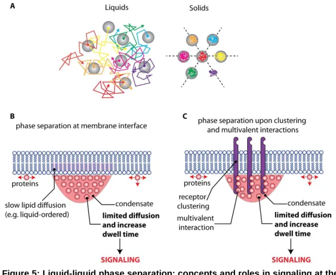
To date there is no convincing experimental evidence demonstrating the existence and functional importance of LLPS at or near the plant plasma membrane. However, the plasma membrane, as any membrane surface, restricts the molecular diffusion to a two-dimensional plane, which reduces the concentration threshold required for phase separation (Case et al., 2019; Snead and Gladfelter, 2019) (Figure 5). In addition, recent advances in mammalian systems suggest that LLPS may play a crucial role for cell surface signaling (Case et al., 2019; Martin and Mittag, 2019). Indeed, several examples were found in which multivalent interactions between transmembrane receptors and their downstream partners trigger phase transitions (Case et al., 2019; Huang et al., 2019b) (Figure 5). In this scenario, the clustered receptor-effector complexes separate from the rest of the cytosol in a membrane-less liquid compartment, which significantly enhances the residence time of the adaptors at the plasma membrane (Figure 5). The induction of the enzymatic activity of effector molecules is slow, and their recruitment to unclustered receptors is too transient to allow signal transduction (Huang et al., 2019b). By contrast, prolonged residence time in the biomolecular condensate at the receptor nanocluster phase favors signal activation (Figure 5). This model was coined “kinetic proofreading” as it directly relates signaling activity to the dwell time of membrane association of downstream receptor components (Figure 5). This dwell time is regulated by LLPS, which can be rapidly modulated by post-translational modification such as tyrosine phosphorylation (Huang et al., 2019b). Given the facts that i) the N-terminal regions of REM proteins are intrinsically disordered (Marin et al., 2012; Marin and Ott, 2014), ii) their C-terminal regions are able to form higher order oligomers (Bariola et al., 2004; Marin et al., 2012; Legrand et al., 2019; Martinez et al., 2019), iii) they actively bind polyanions, including anionic lipids (Gronnier et al., 2017; Legrand et al., 2019), and iv) they are phosphorylated (Perraki et al., 2018), make them promising candidates to induce LLPS at the plasma membrane interface and would support and extend a recently proposed model for a liquid-like receptor clustering compartments (Cuevas-Velazquez and Dinneny, 2018). Furthermore, multivalent association with the extracellular matrix in animal cell also potentiates LLPS at the extracellular surface of animal cells (Case et al., 2019). Accordingly, the extent of phase separations within the plant cell wall could also profoundly impact signaling and reciprocally plasma membrane organization could impact phase separation within the plant cell wall. For example, cellulose may be present in two states: amorphous or crystalline. In addition, cellulose microfibrils are embedded in a matrix composed of water, polysaccharides, proteins, and ions. This matrix is a biphasic mixture between a porous solid phase and a liquid one (Ali and Traas, 2016). One may envision that a change in the equilibrium between these different states may impact lateral diffusion of plasma membrane proteins/lipids. Conversely, the presence of anchored pickets could locally impact the equilibrium between the cell wall porous solid phase and the liquid one.
CONCLUDING REMARKS AND PERSPECTIVES
Altogether, accumulating evidence over the last years clearly demonstrates that the plant plasma membrane, like the plasma membrane in other eukaryotic cells, is highly compartmentalized. However, most of our knowledge originates either from biochemical fractionation of detergent resistant membranes or from diffraction-limited microscopy techniques. It is increasingly clear that these techniques lack the resolution to properly address the challenges behind the study of plasma membrane compartmentalization as the size of nanodomains are typically below the resolution limit of light microscopy. Nonetheless, in the past few years, several studies started to use super-resolution microscopy to address this problem in planta and we expect that they will rapidly expand in the future. So far, such techniques have mainly been used to localize one protein at a time and an important future development of super-resolution microscopy in plants will be to set-up colocalization analyses pipelines. As described above, the cell wall plays a crucial role in plasma membrane organization and the diffusion of cell surface proteins and lipids. This highlights the importance to study the cell in its native state inside its organism rather than in isolated cellular systems such as protoplasts. It also exemplifies the differences between plants and animals with respect to plasma membrane compartmentalization. A clear challenge for the coming years will be to better understand the potential coupling between the plasma membrane and the cell wall organization. This will notably require experimental approaches to characterize their respective physical state and to monitor the evolution of these states in both space and time. With that respect, new developments in the field of atomic force microscopy, Raman microscopy, Brillouin microscopy and mass spectrometry imaging may open new opportunities to challenge our current understanding of the cell wall/plasma membrane continuum. Since both systems are highly complex, we envisioned that such studies will likely require mathematical and/or physical modeling together with reconstitution experiments using minimal membrane/cell wall components. The differences between plants and animals do not only concern extracellular matrices but also include differences in their use of cytoskeleton components and the presence of different lipid species, notably sphingolipids, phytosterols and very-long chain phosphatidylserines. Therefore, although models based on animal or yeast studies are useful, they should not be taken at face value when thinking about the plant plasma membrane. It is likely that the plasma membrane greatly contributes to the phenotypic plasticity of plants. We therefore need to understand it on a functional level and address local and temporal specifications of the bilayer in response to the ever-changing environment. In comparison to many other model organisms, the ability to combine genetic, biochemical and cell biological approaches on tissue, organ and even organismic level provides an outstanding potential to mechanistically understand signal transduction across multiple scales.
• What are the mechanisms that allow the two membrane leaflets to talk to
each other and thereby to integrate extracellular and intracellular cues?
• To what extent does the cell wall act as a membrane exoskeleton which
fences plasma membrane nanodomains?
• What is the impact of cell wall remodeling, cytoskeleton dynamics and
membrane trafficking on membrane organization?
• How does phase separation within the bilayer and at its surface participate
in plasma membrane compartmentalization and signaling?
• To what extent does membrane organization at the nanoscale vary across
different cell types and tissues? And what are the underlying consequences on plant development, physiology and environmental interactions?
ACKNOWLEDGMENTS
We thank Julien Gronnier for his membrane lipid template and Alexandre Martinière, Matthieu Platre for critical comments on the manuscript. T.O. is funded by the German Research Foundation (DFG) under Germany’s Excellence Strategy (CIBSS – EXC-2189 – Project ID 39093984). Y.J. is funded by the French National Research Agency ANR caLIPSO 0025-02) and ANR STAYING-TIGHT (ANR-18-CE13-0016-02).
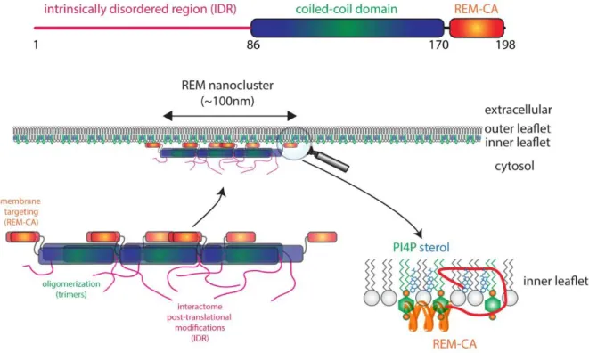
Figure 1: REM proteins and their targeting to plasma membrane nanodomains.
Remorins (REMs) have an N-terminal intrinsically disordered region (IDR), a central coiled-coil domain and a C-terminal membrane anchor (REM-CA). The IDR is regulated by post-translational modification such as phosphorylation and possibly modulates interaction with many different proteins. The coiled-coil domain is an oligomerization domain and mainly contributes to REM trimer formation and interactions with other proteins. REM-CA is required for both plasma membrane and nanodomain targeting via interaction with inner leaflet lipids such as sterols and PI4P. PI4P, phosphatidylinositol-4-phosphate.
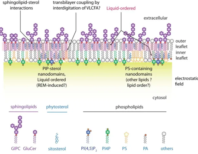
Figure 2: Schematic representation of the lipid distribution within a plant plasma membrane. Note the asymmetric repartition of lipids across the bilayer as well as
lateral segregation of lipids in both inner and outer membrane leaflets (Gronnier et al., 2018). PIP, phosphoinositides; PS, phosphatidylserine; GIPC, glycosylinositol phosphorylceramides; GluCer, Glucosylceramide; REM, Remorin; PI4P, phosphatidylinositol-4-phosphate; PI(4,5)P2, phosphatidylinositol-(4,5)-bisphosphate;
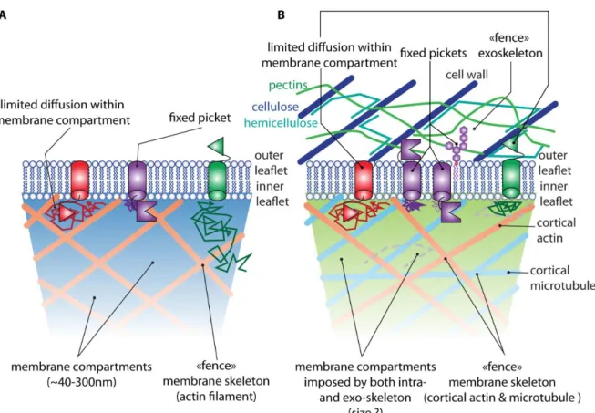
Figure 3: The picket fence model in animal and possible revision of the model in plants. A) The picket fence model for plasma membrane compartmentalization in
animal cells, with a prominent influence of cortical actin as a membrane skeleton (Kusumi et al., 2012). B) The picket fence model in plants is likely to involve both intra- (actin and microtubules) and extracellular (cell wall) skeletons (i.e. fences), which may explain the limited diffusion of plasma membrane proteins. Lines with arrows represent examples of diffusion trajectories of the respective plasma membrane proteins.
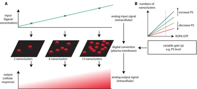
Figure 4: The plasma membrane may act as an analog-digital-analog converter for high fidelity signal transduction. A) During signaling both the input (i.e. ligand)
and output (i.e. cellular response such as phosphorylation) signals are analog by nature. However, small GTPases, such as ROPs, act in a binary fashion akin to a digital signal by being “OFF” when they are freely diffusing, and being “ON” when they are immobilized in nanodomains (Harding and Hancock, 2008). B) As such, molecules (such as phosphatidylserine in roots (Platre et al., 2019)) that favor or dampen the OFF (diffusing) or ON (nanodomains) state of the GTPase may act as a digital gain, which may modify the magnitude of the output signal even though the input signal is constant (Zhou and Hancock, 2015).
Figure 5: Liquid-liquid phase separation: concepts and roles in signaling at the plasma membrane/cytosol interface. A) Schematic representation of ideal liquids
and solids. In liquids (left), molecules diffuse to distances greater than their size. In solids (right), molecules are confined by their neighbors. Furthermore, for crystalline solids, positional order exists over long distances and it is possible to draw straight lines along which particles are equally spaced (dashed lines) (Hyman et al., 2014). In liquid-liquid phase separation (LLPS), two liquids are unmixed similar to a water/oil solution. This limits the diffusion of molecules in and out of each liquid phase and allows specific reactions to occur in each of these “membrane-less” compartments. B) Liquid-liquid phase separation in the cytosol may be coupled with lipid phase separation in membranes (Snead and Gladfelter, 2019). C) Phase separation may also occur upon clustering of receptors and adaptor molecules at the plasma membrane. The resulting biomolecular condensate will locally limit the diffusion of downstream signaling components, increasing their dwell time at the plasma membrane and thereby triggering signaling. This process was coined “kinetic proofreading” because it allows to filter out noise (i.e. uncontrolled recruitment of effectors at the plasma membrane) from real activation (i.e. induction of liquid-liquid phase separation upon receptor nanoclustering) (Huang et al., 2019b).
REFERENECES
Albers P, Ustun S, Witzel K, Kraner M, Bornke F (2019) A Remorin from Nicotiana
benthamiana Interacts with the Pseudomonas Type-III Effector Protein HopZ1a and is Phosphorylated by the Immune-Related Kinase PBS1. Mol Plant Microbe Interact 32: 1229-1242
Ali O, Traas J (2016) Force-Driven Polymerization and Turgor-Induced Wall Expansion.
Trends Plant Sci 21: 398-409
Amorim-Silva V, Garcia-Moreno A, Castillo AG, Lakhssassi N, Esteban Del Valle A, Perez-Sancho J, Li Y, Pose D, Perez-Rodriguez J, Lin J, Valpuesta V, Borsani O, Zipfel C, Macho AP, Botella MA (2019) TTL Proteins Scaffold Brassinosteroid
Signaling Components at the Plasma Membrane to Optimize Signal Transduction in Arabidopsis. Plant Cell 31: 1807-1828
Banani SF, Lee HO, Hyman AA, Rosen MK (2017) Biomolecular condensates: organizers
of cellular biochemistry. Nat Rev Mol Cell Biol 18: 285-298
Bariola PA, Retelska D, Stasiak A, Kammerer RA, Fleming A, Hijri M, Frank S, Farmer EE (2004) Remorins form a novel family of coiled coil-forming oligomeric and
filamentous proteins associated with apical, vascular and embryonic tissues in plants. Plant Mol Biol 55: 579-594
Belkhadir Y, Jaillais Y (2015) The molecular circuitry of brassinosteroid signaling. New
Phytol 206: 522-540
Bergeron-Sandoval LP, Safaee N, Michnick SW (2016) Mechanisms and Consequences of
Macromolecular Phase Separation. Cell 165: 1067-1079
Borner GH, Sherrier DJ, Weimar T, Michaelson LV, Hawkins ND, Macaskill A, Napier JA, Beale MH, Lilley KS, Dupree P (2005) Analysis of detergent-resistant membranes
in Arabidopsis. Evidence for plasma membrane lipid rafts. Plant Physiol 137: 104-116
Brown DA, Rose JK (1992) Sorting of GPI-anchored proteins to glycolipid-enriched
membrane subdomains during transport to the apical cell surface. Cell 68: 533-544
Bucherl CA, Jarsch IK, Schudoma C, Segonzac C, Mbengue M, Robatzek S, MacLean D, Ott T, Zipfel C (2017) Plant immune and growth receptors share common signalling
components but localise to distinct plasma membrane nanodomains. Elife 6
Bucherl CA, van Esse GW, Kruis A, Luchtenberg J, Westphal AH, Aker J, van Hoek A, Albrecht C, Borst JW, de Vries SC (2013) Visualization of BRI1 and BAK1(SERK3)
membrane receptor heterooligomers during brassinosteroid signaling. Plant Physiol
162: 1911-1925
Cacas JL, Bure C, Grosjean K, Gerbeau-Pissot P, Lherminier J, Rombouts Y, Maes E, Bossard C, Gronnier J, Furt F, Fouillen L, Germain V, Bayer E, Cluzet S, Robert F, Schmitter JM, Deleu M, Lins L, Simon-Plas F, Mongrand S (2016) Revisiting
Plant Plasma Membrane Lipids in Tobacco: A Focus on Sphingolipids. Plant Physiol
170: 367-384
Case LB, Ditlev JA, Rosen MK (2019) Regulation of Transmembrane Signaling by Phase
Separation. Annu Rev Biophys 48: 465-494
Case LB, Zhang X, Ditlev JA, Rosen MK (2019) Stoichiometry controls activity of
phase-separated clusters of actin signaling proteins. Science 363: 1093-1097
Chen D, Ahsan N, Thelen JJ, Stacey G (2019) <em>S</em>-Acylation of plant immune
receptors mediates immune signaling in plasma membrane nanodomains. bioRxiv: 720482
Colin LA, Jaillais Y (2019) Phospholipids across scales: lipid patterns and plant development.
Curr Opin Plant Biol 53: 1-9
Cuevas-Velazquez CL, Dinneny JR (2018) Organization out of disorder: liquid-liquid phase
separation in plants. Curr Opin Plant Biol 45: 68-74
Cui Y, Yu M, Yao X, Xing J, Lin J, Li X (2018) Single-Particle Tracking for the
Quantification of Membrane Protein Dynamics in Living Plant Cells. Mol Plant 11: 1315-1327
Daněk M, Angelini J, Malínská K, Andrejch J, Amlerová Z, Kocourková D, Brouzdová J, Valentová O, Martinec J, Petrášek J (2019) Cell wall contributes to the stability of
plasma membrane nanodomain organization of Arabidopsis thaliana FLOTILLIN2 and HYPERSENSITIVE INDUCED REACTION1 proteins. The Plant Journal 0
Daněk M, Valentová O, Martinec J (2016) Flotillins, Erlins, and HIRs: From Animal Base
Camp to Plant New Horizons. Critical Reviews in Plant Sciences 35: 191-214
Demir F, Horntrich C, Blachutzik JO, Scherzer S, Reinders Y, Kierszniowska S, Schulze WX, Harms GS, Hedrich R, Geiger D, Kreuzer I (2013) Arabidopsis
nanodomain-delimited ABA signaling pathway regulates the anion channel SLAH3. Proc Natl Acad Sci U S A 110: 8296-8301
Dunser K, Gupta S, Herger A, Feraru MI, Ringli C, Kleine-Vehn J (2019) Extracellular
matrix sensing by FERONIA and Leucine-Rich Repeat Extensins controls vacuolar expansion during cellular elongation in Arabidopsis thaliana. EMBO J 38
Fang X, Wang L, Ishikawa R, Li Y, Fiedler M, Liu F, Calder G, Rowan B, Weigel D, Li P, Dean C (2019) Arabidopsis FLL2 promotes liquid-liquid phase separation of
polyadenylation complexes. Nature 569: 265-269
Feng W, Kita D, Peaucelle A, Cartwright HN, Doan V, Duan Q, Liu MC, Maman J, Steinhorst L, Schmitz-Thom I, Yvon R, Kudla J, Wu HM, Cheung AY, Dinneny JR (2018) The FERONIA Receptor Kinase Maintains Cell-Wall Integrity during Salt
Stress through Ca(2+) Signaling. Curr Biol 28: 666-675 e665
Freeman SA, Goyette J, Furuya W, Woods EC, Bertozzi CR, Bergmeier W, Hinz B, van der Merwe PA, Das R, Grinstein S (2016) Integrins Form an Expanding Diffusional
Barrier that Coordinates Phagocytosis. Cell 164: 128-140
Freeman SA, Vega A, Riedl M, Collins RF, Ostrowski PP, Woods EC, Bertozzi CR, Tammi MI, Lidke DS, Johnson P, Mayor S, Jaqaman K, Grinstein S (2018)
Transmembrane Pickets Connect Cyto- and Pericellular Skeletons Forming Barriers to Receptor Engagement. Cell 172: 305-317 e310
Frescatada-Rosa M, Stanislas T, Backues SK, Reichardt I, Men S, Boutte Y, Jurgens G, Moritz T, Bednarek SY, Grebe M (2014) High lipid order of Arabidopsis cell-plate
membranes mediated by sterol and DYNAMIN-RELATED PROTEIN1A function. Plant J 80: 745-757
Furt F, Konig S, Bessoule JJ, Sargueil F, Zallot R, Stanislas T, Noirot E, Lherminier J, Simon-Plas F, Heilmann I, Mongrand S (2010) Polyphosphoinositides are enriched
in plant membrane rafts and form microdomains in the plasma membrane. Plant Physiol
152: 2173-2187
Gerbeau-Pissot P, Der C, Grebe M, Stanislas T (2016) Ratiometric Fluorescence Live
Imaging Analysis of Membrane Lipid Order in Arabidopsis Mitotic Cells Using a Lipid Order-Sensitive Probe. Methods Mol Biol 1370: 227-239
Ghosh R, de Campos MK, Huang J, Huh SK, Orlowski A, Yang Y, Tripathi A, Nile A, Lee HC, Dynowski M, Schafer H, Rog T, Lete MG, Ahyayauch H, Alonso A, Vattulainen I, Igumenova TI, Schaaf G, Bankaitis VA (2015) Sec14-nodulin
proteins and the patterning of phosphoinositide landmarks for developmental control of membrane morphogenesis. Mol Biol Cell 26: 1764-1781
Gronnier J, Crowet JM, Habenstein B, Nasir MN, Bayle V, Hosy E, Platre MP, Gouguet P, Raffaele S, Martinez D, Grelard A, Loquet A, Simon-Plas F, Gerbeau-Pissot P, Der C, Bayer EM, Jaillais Y, Deleu M, Germain V, Lins L, Mongrand S (2017)
Structural basis for plant plasma membrane protein dynamics and organization into functional nanodomains. Elife 6
Gronnier J, Gerbeau-Pissot P, Germain V, Mongrand S, Simon-Plas F (2018) Divide and
Rule: Plant Plasma Membrane Organization. Trends Plant Sci 23: 899-917
Grosjean K, Der C, Robert F, Thomas D, Mongrand S, Simon-Plas F, Gerbeau-Pissot P
(2018) Interactions between lipids and proteins are critical for organization of plasma membrane-ordered domains in tobacco BY-2 cells. J Exp Bot 69: 3545-3557
Gui J, Zheng S, Liu C, Shen J, Li J, Li L (2016) OsREM4.1 Interacts with OsSERK1 to
Coordinate the Interlinking between Abscisic Acid and Brassinosteroid Signaling in Rice. Dev Cell 38: 201-213
Gui J, Zheng S, Shen J, Li L (2015) Grain setting defect1 (GSD1) function in rice depends
on S-acylation and interacts with actin 1 (OsACT1) at its C-terminal. Front Plant Sci 6: 804
Haney CH, Long SR (2010) Plant flotillins are required for infection by nitrogen-fixing
bacteria. Proc Natl Acad Sci U S A 107: 478-483
Haney CH, Riely BK, Tricoli DM, Cook DR, Ehrhardt DW, Long SR (2011) Symbiotic
rhizobia bacteria trigger a change in localization and dynamics of the Medicago truncatula receptor kinase LYK3. Plant Cell 23: 2774-2787
Hao H, Fan L, Chen T, Li R, Li X, He Q, Botella MA, Lin J (2014) Clathrin and Membrane
Microdomains Cooperatively Regulate RbohD Dynamics and Activity in Arabidopsis. Plant Cell 26: 1729-1745
Harding A, Hancock JF (2008) Ras nanoclusters: combining digital and analog signaling. Cell
Cycle 7: 127-134
Hemsley PA, Weimar T, Lilley KS, Dupree P, Grierson CS (2013) A proteomic approach
identifies many novel palmitoylated proteins in Arabidopsis. New Phytol 197: 805-814
Herger A, Dunser K, Kleine-Vehn J, Ringli C (2019) Leucine-Rich Repeat Extensin Proteins
and Their Role in Cell Wall Sensing. Curr Biol 29: R851-R858
Hohmann U, Lau K, Hothorn M (2017) The Structural Basis of Ligand Perception and Signal
Activation by Receptor Kinases. Annu Rev Plant Biol 68: 109-137
Homann U, Meckel T, Hewing J, Hutt MT, Hurst AC (2007) Distinct fluorescent pattern of
KAT1::GFP in the plasma membrane of Vicia faba guard cells. Eur J Cell Biol 86: 489-500
Hosy E, Martiniere A, Choquet D, Maurel C, Luu DT (2015) Super-resolved and dynamic
imaging of membrane proteins in plant cells reveal contrasting kinetic profiles and multiple confinement mechanisms. Mol Plant 8: 339-342
Huang D, Sun Y, Ma Z, Ke M, Cui Y, Chen Z, Chen C, Ji C, Tran TM, Yang L, Lam SM, Han Y, Shu G, Friml J, Miao Y, Jiang L, Chen X (2019a) Salicylic acid-mediated
plasmodesmal closure via Remorin-dependent lipid organization. Proc Natl Acad Sci U S A
Huang WYC, Alvarez S, Kondo Y, Lee YK, Chung JK, Lam HYM, Biswas KH, Kuriyan J, Groves JT (2019b) A molecular assembly phase transition and kinetic proofreading
modulate Ras activation by SOS. Science 363: 1098-1103
Hurst CH, Turnbull D, Myles SM, Leslie K, Keinath NF, Hemsley PA (2018) Variable
Effects of C-Terminal Fusions on FLS2 Function: Not All Epitope Tags Are Created Equal. Plant Physiol 177: 522-531