HAL Id: inserm-01352196
https://www.hal.inserm.fr/inserm-01352196
Submitted on 5 Aug 2016
HAL is a multi-disciplinary open access archive for the deposit and dissemination of sci-entific research documents, whether they are pub-lished or not. The documents may come from teaching and research institutions in France or abroad, or from public or private research centers.
L’archive ouverte pluridisciplinaire HAL, est destinée au dépôt et à la diffusion de documents scientifiques de niveau recherche, publiés ou non, émanant des établissements d’enseignement et de recherche français ou étrangers, des laboratoires publics ou privés.
Copyright
Animal models to study AMPK
Benoit Viollet, Marc Foretz
To cite this version:
Benoit Viollet, Marc Foretz. Animal models to study AMPK. Mario D. Cordero ; Benoit Viollet. AMP-activated Protein Kinase, 107, Springer International Publishing, pp.441-469, 2016, Experientia Supplementum, 978-3-319-43587-9. �10.1007/978-3-319-43589-3�. �inserm-01352196�
HAL Id: inserm-01352196
http://www.hal.inserm.fr/inserm-01352196
Submitted on 5 Aug 2016
HAL is a multi-disciplinary open access archive for the deposit and dissemination of sci-entific research documents, whether they are pub-lished or not. The documents may come from teaching and research institutions in France or abroad, or from public or private research centers.
L’archive ouverte pluridisciplinaire HAL, est destinée au dépôt et à la diffusion de documents scientifiques de niveau recherche, publiés ou non, émanant des établissements d’enseignement et de recherche français ou étrangers, des laboratoires publics ou privés.
Copyright}
Animal models to study AMPK
Benoit Viollet, Marc Foretz
To cite this version:
Benoit Viollet, Marc Foretz. Animal models to study AMPK. Mario D. Cordero ; Benoit Viollet. AMP-activated Protein Kinase, 107, Springer International Publish-ing, 2016, Experientia Supplementum, 978-3-319-43587-9. <10.1007/978-3-319-43589-3>. <http://www.springer.com/gp/book/9783319435879#aboutBook>. <inserm-01352196>
Part VI: Methods of study in AMPK Animal models to study AMPK Benoit Viollet1,2,3 and Marc Foretz1,2,3 1 INSERM U1016, Institut Cochin, Paris, France 2 CNRS UMR 8104, Paris, France 3 Université Paris Descartes, Sorbonne Paris Cité, Paris, France
Correspondence: Benoit Viollet, Institut Cochin, Inserm U1016, CNRS UMR8104, Université Paris Descartes, 24 rue du faubourg Saint Jacques 75014 Paris, France. Phone + 33 1 44 41 24 01, Fax + 33 1 44 41 24 21, e‐mail: benoit.viollet@inserm.fr Running title: AMPK animal models Keywords: AMPK‐activated protein kinase; animal models; transgenic animals; energy metabolism; pharmacological drugs; therapeutics. Abstract AMPK is an evolutionary conserved energy sensor involved in the regulation of energy metabolism. Based on biochemical studies, AMPK has brought much of interest in the recent years due to its potential impact on metabolic disorders. Suitable animal models are therefore essential to promote our understanding of the molecular and functionnal roles of AMPK but also to bring novel information for the development of novel therapeutic strategies. The organism systems include pig (Sus scrofa), mouse (Mus
musculus), fly (Drosophila melanogaster), worm (Caenorhabditis elegans) and fish (Danio rerio) models. These animal models have provided reliable experimental evidence
demonstrating the crucial role of AMPK in the regulation of metabolism but also of cell polarity, autophagy and oxidative stress. In this chapter, we update the new development in the generation and application of animal models for the study of AMPK biology. We also discuss recent breakthroughs from studies in mice, fly and worms showing how AMPK has a primary role in initiating or promoting pathological or beneficial impact on health.
Introduction
AMP‐activated protein kinase (AMPK) is widely accepted as a sensor of cellular energy balance (Hardie 2014). At the cellular level, AMPK promotes ATP producing catabolic pathways, while simultaneously inhibiting ATP consuming anabolic pathways. At the organismal level, AMPK integrates stress responses such as exercise as well as nutrient and hormonal signals to control whole body energy expenditure and substrate utilization. As such a potent regulator of cellular and whole body metabolism, AMPK has become the focus of great deal of attention and appeared as an obvious target for treatment of metabolic disorders such as obesity and type 2 diabetes (Winder and Hardie 1999). In several rodents models of diabetes and obesity, pharmacological activation of AMPK results in the remodeling of a wide range of metabolic pathways and have led to substantial improvement of disease outcome (Buhl et al. 2002, Halseth et al. 2002, Song et al. 2002, Cool et al. 2006, Fullerton et al. 2013). In parallel with rapid scientific discovery in cell‐free and cellular systems, the development of animal models has been instrumental to the expanding field of AMPK (Viollet et al. 2009). Over the last decade, knockout (KO) mouse models for the different AMPK subunit isoforms as well as transgenic mouse models overexpressing loss of function or gain of function AMPK mutants have been developed to better understand the impact of AMPK on metabolic health and disease and have largely contributed to expand our knowledge on the relevance of AMPK in human disease (Figure 1). Conditional targeting approaches are now at the forefront of mechanistic studies to investigate the relevance and specificity of AMPK in the homeostasis of multiple organs. These mouse models are also critically important for pre‐clinical translational studies and early stage clinical investigation to progress. In addition, the use of model organisms such as Saccharomyces cerevisiae (yeast) [See chapter AMPK in yeast: the SNF1 (sucrose non‐fermenting 1) protein kinase complex], Drosophila melanogaster (fly) [See chapter The role of AMPK in Drosophila
melanogaster] and Caenorhabditis elegans (roundworm) [See chapter 5’‐AMP‐Activated
Protein Kinase Signalling in Caenorhabditis elegans] has played a key role in delineating the physiological role of AMPK pathway and has been instrumental to provide novel information on the biology of AMPK system (Figure 1). These various models helped to expand the paradigm of AMPK as a metabolic sensor and showed that AMPK has broad
effects on cellular function, regulating cell growth, autophagy, oxidative stress and cell polarity.
1‐ Naturally occurring mutations
Many genetically engineered animal models have been generated to investigate the physiopathological role of AMPK, but understanding of AMPK function has been also greatly advanced by studies of naturally occurring mutations in AMPK genes (Figure 1). These mutations are apparently fairly rare but can result in pronounced pathological changes (Milan et al. 2000, Blair et al. 2001, Gollob et al. 2001, Arad et al. 2002). Naturally occurring mutations have been initially characterized in the pig PRKAG3 gene and the human PRKAG2 gene (encoding the 3 and 2‐subunit of AMPK, respectively) and are associated with abnormally high glycogen accumulation that results in pathological changes to skeletal and cardiac muscle (Milan et al. 2000, Blair et al. 2001, Gollob et al. 2001, Arad et al. 2002). Later, a mutation in human PRKAG3 gene has been identified in association with an increase in skeletal muscle glycogen content and a decrease in intramuscular triglyceride (Costford et al. 2007).
• 1‐1‐ Human and pig PRKGA3 gene mutations and related mouse models
In 2000, a naturally occurring mutation in the 3‐subunit of AMPK, primarily expressed in white skeletal muscle (glycolytic, fast‐twitch type II), was identified in purebred Hampshire pigs by a positional cloning approach (Milan et al. 2000). A non‐conservative substitution (R200Q) was the causative dominant mutation in RN‐ (in French Rendement
Napole for Napole yield) pigs. Animals carrying the RN‐ mutation are characterized by a
marked increase of the glycogen content in glycolytic skeletal muscle leading to low muscle pH 24h post mortem due to anaerobic glycogen degradation, poor water‐holding capacity, and low processing yield in the production of cured and cooked ham (Enfalt et al. 1997). Glycogen accumulation in skeletal muscle is consistent with the upregulation in the activity of UDP‐glucose pyrophosphorylase and glycogen branching enzyme, two key enzymes regulating glycogen synthesis (Estrade et al. 1994, Hedegaard et al. 2004). R200Q carriers are also characterized by a higher oxidative capacity in white skeletal muscle fibers (Estrade et al. 1994). Conversely, additional naturally occurring missense mutations (T30N, G52S and V199I) were also identified in the PRKAG3 gene from
Western and Chinese indigenous pig breeds associated with an opposite phenotype compared to the RN‐ pigs, resulting in reduced skeletal muscle glycogen content and
additive effect on meat quality traits (Ciobanu et al. 2001, Huang et al. 2004).
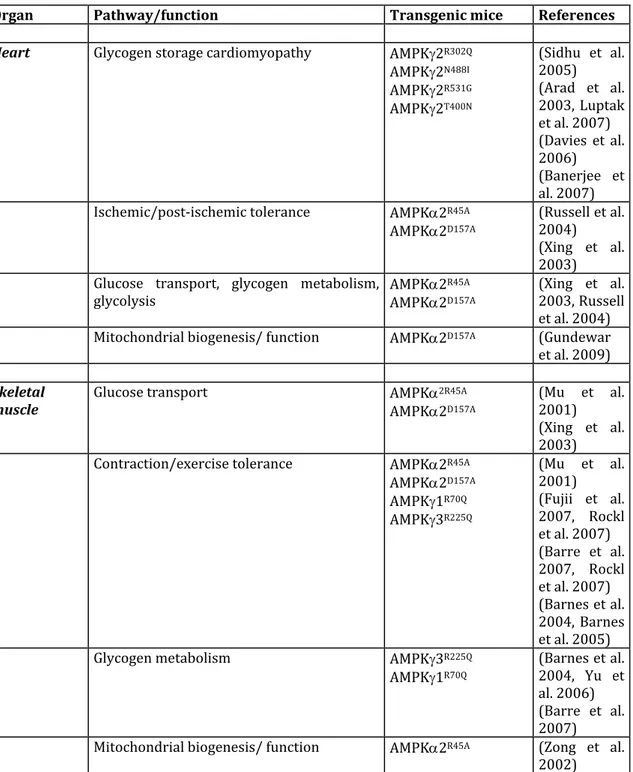
Strong support for the causative nature of the R200Q mutation in the elevated skeletal muscle glycogen content has been provided by transgenic mouse models overexpressing primarily in white skeletal muscle the mouse 3 R200Q mutant form (in these studies, the R200Q missense mutation is designated R225Q as the start methionine codon proposed by Cheung et al. (Cheung et al. 2000) is used) (Table 1). Muscle‐specific transgenic mouse lines overexpressing the AMPK3 R225Q mutant under the control of either the myosin light‐chain or the muscle creatine kinase promoter/enhancer were generated and resulted in significant increases in skeletal muscle glycogen, replicating the pig RN‐ phenotype (Barnes et al. 2004, Yu et al. 2006) (Table 2).
The R200Q missense mutation occurs within the cystathionine ‐synthase domain 1 (CBS1) of the regulatory AMPK3 subunit, which is involved in the binding of the allosteric activator of the kinase, AMP. Originally, there was great controversy over the impact of the R200Q mutation on AMPK activity. It was initially reported that the activity of AMPK was reduced in muscle extracts from RN‐ pigs both in the presence and
absence of AMP (Milan et al. 2000). In resting muscle from AMPK3 R225Q mutant mice, AMPK activity was found to be decreased (Yu et al. 2006) or unaltered (Barnes et al. 2004). However, reduced AMPK activity could reflect the potential feedback inhibition of glycogen overload on AMPK activation (Milan et al. 2000, Jorgensen et al. 2004). Thus, to evaluate the impact of CBS domain mutation, activity of 223R225Q trimer was
measured from heterotrimeric complexes purified from transiently transfected COS cells and resulted in a higher basal AMPK activity and loss of AMP dependence (Barnes et al. 2004). Nevertheless, question remains on the mechanisms regulating AMP‐independent activity of the AMPK3 R225Q mutant (Lindgren et al. 2007), indicating that more detailed functional studies are needed to precisely define the nature of this mutation in the CBS domain of AMPK3. Interestingly, a homologous mutation to the RN‐ mutation
found in pigs has been identified in the human PRKAG3 gene from genetic studies of lean and obese human populations (Costford et al. 2007). Subjects bearing the AMPK3 R225W mutation exhibit increased skeletal muscle glycogen content, indicating conserved AMPK3 function across mammalian species (Costford et al. 2007). The AMPK 3 R225W mutation is associated with increased activity of AMPK in
differentiated muscle cells derived from vastus lateralis biopsies (Costford et al. 2007). Furthermore, it should be noted that introduction of the equivalent mutation R70Q in AMPK1 and R302Q in AMPK2 also caused a marked increase in AMPK activity (Gollob et al. 2001, Hamilton et al. 2001, Yavari et al. 2016). More recently, studies with exercise trained RN‐ pigs have shifted towards the recognition that 3 R200Q mutation is a gain‐
of‐function mutation, which results in hyperaccumulation of glycogen due to increased influx of glucose and, enhanced endurance exercise capacity (Granlund et al. 2010, Granlund et al. 2011). Increased glycogen level is not due to impaired glycogen utilization as glycogen breakdown is similar after exercise in carriers and non‐carriers of the RN‐ mutation (Granlund et al. 2010). These results are consistent with the finding
that pigs carrying the R200Q mutation show increased signaling response of Akt and phosphorylation of its substrate, AS160, a higher capacity for phosphorylation of glucose and, faster muscle glycogen resynthesis after exercise (Granlund et al. 2010, Essen‐Gustavsson et al. 2011, Granlund et al. 2011). However, a slightly reduced basal and contraction‐stimulated rates of glucose uptake was evidenced in transgenic AMPK3 R225Q mutant mice (Barnes et al. 2004, Yu et al. 2006), suggesting that enhanced glycogen accumulation is not due to a single mechanism. The possibility that increases in total glycogen synthase activity contribute to elevated glycogen concentrations cannot be excluded (Yu et al. 2006). Other factors that could influence glycogen synthesis are the difference in the gene regulatory responses that facilitate metabolic adaptations. It has been shown that overexpression of AMPK3 R225Q coordinates the transcription of genes important for glycolytic and oxidative metabolism (Nilsson et al. 2006) and leads to change in the adaptive metabolic response of glycolytic skeletal muscle by inducing mitochondrial biogenesis and function (Garcia‐Roves et al. 2008). Accordingly, transgenic AMPK3 R225Q mutant mice display a greater reliance on lipid oxidation and are protected from high fat diet‐induced insulin resistance in skeletal muscle through increased fat oxidation (Barnes et al. 2004, Barnes et al. 2005). One consequence of increased fatty acid oxidation in AMPK3 R225Q skeletal muscle could be the reduction of the demand for glucose oxidation via the Randle effect (Hue and Taegtmeyer 2009), inducing a glucose‐sparing effect that drives glucose towards glycogen synthesis.
Point mutations in the AMPK2 subunit, encoded by the Prkag2 gene, have been associated with a rare autosomal‐dominant genetic disease of the heart in humans (Blair et al. 2001, Gollob et al. 2001, Gollob et al. 2001). Genetic defects in the Prkag2 gene are characterized by a cardiac glycogen overload, ventricular pre‐excitation (Wolff‐ Parkinson‐White syndrome) and cardiac hypertrophy. One of the first human mutation identified occurred at residue 302, resulting in change of arginine to glutamine (R302Q), which is homologous to the R200Q Prkag3 gene mutation in pigs (Milan et al. 2000, Gollob et al. 2001). Subsequently, identification of additional dominant mutations of PRKAG2 gene has been reported in families coupled with congenital hypertrophic cardiomyopathy and familial pre‐excitation syndrome. However, distinct clinical onset and variability in clinical manifestations exist between the various PRKAG2 mutations (Porto et al. 2016). This phenotypic disparity may be a result of the specific effects of individual mutations on AMPK2 activity and function. To date, missense mutations include G100S (Zhang et al. 2013), R302Q (Gollob et al. 2001, Arad et al. 2002), H383R (Blair et al. 2001), R384T (Akman et al. 2007), T400N (Arad et al. 2002), K485E (Liu et al. 2013), Y487H (Arad et al. 2005), N488I (Arad et al. 2002), E506K (Bayrak et al. 2006), E506Q (Kelly et al. 2009), H530R (Morita et al. 2008), R531G (Gollob et al. 2001), R531Q (Burwinkel et al. 2005), S548P (Laforet et al. 2006) and insertion of an additional leucine residue L351Ins (Blair et al. 2001). These mutations occur very selectively in strategic positions within the CBS motifs or in linker sequences between these motifs, but not in other parts of the AMPK2 subunit. One interesting exception is the G100S mutation mapped into a non‐CBS domain. However, it remains unclear if a mutation outside the CBS domains can indirectly changes the binding ability of CBS domains to AMP and ATP. Collectively, these data strongly suggest a specific connection between AMP and ATP binding and the different PRKAG2 mutations, supporting the notion that dysregulation of AMPK activity (gain‐ or loss‐of‐function) contributes to the development of the cardiomyopathy.
Consistent with a causative role of PRKAG2 in Wolff‐Parkinson‐White syndrome, heterozygous mice overexpressing mutant PRKAG2 alleles (R302Q, T400N, N488I, R531G) under the cardiac‐specific ‐myosin heavy chain (‐MHC) promoter develop pathological cardiac changes resembling the human PRKAG2 cardiomyopathy with significant glycogen accumulation, ventricular preexcitation and cardiac hypertrophy (Arad et al. 2002, Arad et al. 2003, Sidhu et al. 2005, Davies et al. 2006, Banerjee et al.
2007) (Table 2). In addition, transgenic zebrafish with cardiac specific expression of the G100S and R302Q mutations in PRKAG2 exhibit typical features of the human disease with cardiac hypertrophy and increased glycogen storage in the heart (Zhang et al. 2014). The PRKAG2 cardiac phenotype has been attributed to alterations in AMPK activity resulting from the AMPK2 mutations. However, the action of the different PRKAG2 mutations on AMPK activity and sensitivity to AMP appears to be partially different, if not opposite (Daniel and Carling 2002, Scott et al. 2004). Transgenic mouse model with cardiac specific overexpression of the AMPKγ2N448I and AMPK2T400N
mutations have been reported to increase AMPK basal activity (Arad et al. 2003, Banerjee et al. 2007), whereas the AMPKγ2R302Q and AMPKγ2R531G mutations results in
the inhibition of AMPK (Sidhu et al. 2005, Davies et al. 2006). These discrepant findings could be related to a biphasic response of AMPK activity in response to the overexpression of PRKAG2 mutations (Banerjee et al. 2007, Folmes et al. 2009). Alteration in cardiac AMPK activity is possibly due to a feedback inhibition of the kinase activity by glycogen accumulation (Davies et al. 2006, Folmes et al. 2009), masking the consequences of human PRKAG2 mutations on AMPK activity. To confirm that increased AMPK activity is responsible for PRKAG2 cardiomyopathy, transgenic mice overexpressing AMPK γ2N448I and γ2T400N were crossbred with transgenic mice
expressing a dominant negative form of the AMPK2 subunit, AMPK2D157A (Table 1) in
the heart to alter AMPK activity (Ahmad et al. 2005, Banerjee et al. 2007). Compound heterozygous mice have reduced cardiac AMPK activity and minimal cardiac dysfunction, demonstrating that activation of 2‐containing AMPK heterotrimeric complexes rather than 1‐containing complexes are responsible for the cardiac phenotype (Ahmad et al. 2005, Banerjee et al. 2007). Furthermore, studies using a transgenic mouse model expressing a human AMPK2N488I mutant protein under
transcriptional control of a tetracycline‐repressible α‐myosin heavy chain promoter (Table 1) provide evidence that clinical manifestations of PRKAG2 cardiomyopathy are significantly reversed by the suppression of mutant AMPK activity after the onset of the disease (Wolf et al. 2008). This finding suggests that the appropriate pharmacological targeting of AMPK or its downstream effectors may help to improve the phenotypic expression of the disease. Compelling evidence now exists that aberrant increase of basal AMPK activity by PRKAG2 mutations results in global remodeling of the metabolic network in favor of glycogen storage (Zou et al. 2005, Luptak et al. 2007) (Table 2).
Thus, the metabolic consequences of chronic activation of AMPK in the absence of energy deficiency is distinct from those previously reported during stress conditions. In heart carrying a mutant PRKAG2 allele, inappropriate activation of AMPK triggers an increase in both glucose uptake and fatty‐acid oxidation, inducing, via the Randle effect, an inhibition of glucose oxidation leading to the storage of the exceeding glucose into glycogen. The mechanism of increased glucose uptake has been attributed to the upregulation of the sodium‐dependent glucose co‐transporter (SGLT) isoform SGLT1 but not facilitated‐diffusion glucose transporter 1 (GLUT1) or GLUT4 in AMPK γ2T400N
transgenic mice (Banerjee et al. 2010). Confirmation of the role of SGLT1 in the phenotypic features of the PRKAG2 cardiomyopathy has been demonstrated by the knockdown of cardiac SGLT1 in transgenic mice overexpressing the PRKAG2 T400N mutation, which attenuates the cardiomyopathy phenotype (Ramratnam et al. 2014). Due to increased glucose uptake, the transgenic mice overexpressing the AMPK γ2N448I
mutation manifest high intracellular glucose‐6‐phosphate (G6P) levels, which contribute to the allosteric activation of glycogen synthase (GS) and enhanced glycogen synthesis (Luptak et al. 2007). By genetic inhibition of G6P–stimulated glycogen synthase activity, the pathological glycogen storage phenotype was rescued, providing definitive evidence for extensive remodeling of substrate metabolism and the causative role for high intracellular G6P in Prkag2 cardiomyopathy (Kim et al. 2014). Surprisingly, elimination of excessive glycogen accumulation eliminated the ventricular preexcitation but not the cardiac hypertrophy phenotype, indicating that the abnormal cardiac growth is regulated by separate mechanisms. Recent studies indicate that AMPK2 N448I and AMPK
γ2T400N mutations stimulate hypertrophic signaling with activation of the transcription
factors nuclear factor B (NF‐B) and forkhead box O transcription factor (FoxO), and the mammalian target of rapamycin (mTOR) signaling pathway (Banerjee et al. 2010, Kim et al. 2014).
2‐ Genetically modified mouse models
Murine models have been widely used in biomedical research. Extensive similarities in anatomy, physiology and genetics have allowed numerous inferences about human biology to be drawn from mouse models. The recent development of conditional targeting approaches and the availability of numerous genetically modified mouse
models greatly facilitate functional studies. Important progress has been made during the last decade in the understanding of the pathophysiological function of AMPK, partly due to the generation of whole‐body and conditional KO mouse models as well as tissue‐ specific transgenic mice (Table 1). These mouse models have made it possible to decipher the distinct physiopathological functions of the multiple AMPK isoforms and AMPK heterotrimer combinations.
•1‐1‐ AMPK knock‐out mouse models
Generation of whole‐body deletion of each catalytic or regulatory AMPK subunits results in viable mice (Table 1). Of note, a mouse model of AMPK1 deletion using a gene trap approach resulted in severe brain developmental defect leading to postnatal death (Dasgupta and Milbrandt 2009). These mice express a fusion protein containing a AMPK1 N‐terminal fragment (2‐224) fused to ‐galactosidase and may explain the abnormal brain development and not the loss of the AMPK1 subunit (Dzamko et al. 2010). Interestingly, targeted disruption of AMPK subunits is is associated with distinct phenotypic abnormalities (Viollet et al. 2003, Barnes et al. 2004, Jorgensen et al. 2004, Dzamko et al. 2010, Steinberg et al. 2010, Foretz et al. 2011, Dasgupta et al. 2012). These diverse phenotypes could simply reflect isoform‐preferred substrate phosphorylation between the combination of AMPK1‐ and AMPK2‐containing heterotrimeric complexes, but a recent phosphoproteomic approach in cancer cells fails to identify specific targets for AMPK1 or AMPK2 (Schaffer et al. 2015). These data are rather suggestive of distinct tissue‐specific contribution of the different isoforms in the control of AMPK activity and function. This is in line with the description of specific pattern of expression for AMPK, AMPK and AMPK isoforms between tissues (Stapleton et al. 1996, Thornton et al. 1998, Cheung et al. 2000). Indeed, the expression of AMPK1 and AMPK2 isoforms differ in a number of tissues, with AMPK1 highly expressed in liver and weakly expressed in skeletal muscle, whereas the opposite pattern is observed for AMPK2 (Thornton et al. 1998). Studies from AMPK1‐/‐ and AMPKβ2‐/‐ mice have highlighted the relative importance of these two isoforms in the liver and skeletal muscle, respectively (Dzamko et al. 2010, Steinberg et al. 2010). Another striking example of restricted expression of AMPK isoforms come from reports showing exclusive expression of AMPK1, but not AMPK2, in erythrocytes, macrophages and T cells (Tamas et al. 2006, Sag et al. 2008, Foretz et al. 2010). Mice lacking AMPK1 are
anemic and had markedly enlarged spleens (Foretz et al. 2010). Similar phenotypes are observed in mice lacking AMPK1, the only AMPK isoform expressed in murine erythrocytes (Foretz et al. 2011). Studies of AMPK1‐/‐ and AMPK1‐/‐ mice revealed that AMPK is required for erythrocyte homeostasis by regulating erythrocyte membrane elasticity (Foretz et al. 2010, Foretz et al. 2011) and autophagy‐dependent mitochondrial clearance during erythrocyte differentiation (Wang et al. 2010). In addition, with the use of bone marrow‐derived macrophages from AMPK1‐/‐ and AMPK1‐/‐ mice, the regulatory role of AMPK in the macrophagic differentiation of monocytes (Obba et al. 2015) and the polarization of macrophages to an anti‐ inflammatory phenotype (Zhu et al. 2015) has been demonstrated. Support for a critical role of AMPK in the regulation of the pro‐inflammatory/ anti‐inflammatory balance in macrophage has been provided in previous studies using adoptive transfer of AMPK1‐ /‐ bone marrow in control recipient mice (Galic et al. 2011). It has been also established that AMPK is a key determinant of T cell effector responses by controlling T cell metabolism. AMPKα1‐deficient T cells display reduced metabolic plasticity in vitro as well as in vivo during viral and bacterial infections (Blagih et al. 2015). However, with the exception of these studies on erythrocytes, macrophages and T cells, analysis of the consequence of genetic deletion of one catalytic isoform should be done with caution, since compensatory increase in expression and activity of the remaining isoform can mask a particular phenotype. This has been clearly evidenced in primary culture of mouse proximal tubules from AMPK1‐/‐ and AMPK2‐/‐ mice, where adaptive up‐ regulation of the other AMPK isoform fully compensate and can substitute for the other for ameliorating the response to metabolic stress (Lieberthal et al. 2013). Similarly, it has been reported that AMPK1 activity is higher in AMPK2‐/‐ muscle as compared to control muscles, suggesting a compensatory effect (Jorgensen et al. 2004) (see below). In addition, overexpression of AMPKα2 compensated for loss of AMPKα1 and may explain the lack of phenotype in chondrocyte‐specific ablation of AMPK1 (Yang et al. 2016). In attempt to generate full KO of AMPK, AMPK1‐/‐ and AMPK2‐/‐ or AMPK1‐ /‐ and AMPK2‐/‐ mice have been crossed but combined disruption of AMPK isoforms is incombatible with life, indicating that AMPK is required during embryogenesis (Viollet et al. 2009, O'Neill et al. 2011). AMPK 1‐/‐2‐/‐ double knockout embryos die at 10.5 days post‐conception (B. Viollet, unpublished data). The recent development of tissue‐
specific and conditional knockout technology has now allowed the generation of animal models completely lacking AMPK activity in a specific tissue (Table 1). Among the first tissues to be targeted, skeletal muscle‐specific deletion of both AMPK1/AMPK2 and AMPK1/AMPK2 has been obtained by crossing AMPK1fl/fl2fl/fl and AMPKfl/fl2fl/fl
mice with transgenic mice expression the Cre recombinase under the control of the human skeletal actin and muscle creatine kinase promoter, respectively (O'Neill et al. 2011, Lantier et al. 2014).
•1‐2‐ Insight from mouse models into AMPK‐dependent stimulation of glucose uptake in skeletal muscle
Skeletal muscle contraction is associated with a dramatic increase in energy turnover rates that represents a major metabolic challenge. Since the first report on acute skeletal muscle AMPK activation in response to physical exercise in rodents (Winder and Hardie 1996) and later in humans (Chen et al. 2000, Fujii et al. 2000, Wojtaszewski et al. 2000), the role of AMPK in the adaptive changes in skeletal muscle has been a subject of intense research. During exercise, contracting skeletal muscle rapidly increases glucose uptake in an intensity‐dependent manner to sustain the energy demand caused by increased ATP turnover. The idea that AMPK was involved in regulating glucose transport in skeletal muscle was supported by the observations that stimulation of glucose transport was achieved upon AMPK activation by AICAR (Merrill et al. 1997) and later by other pharmacological AMPK agonists such as Ex229/991 (Lai et al. 2014). The importance of AMPK in skeletal muscle glucose uptake has been investigated in genetically modified mouse models, including transgenic mice expressing naturally occurring mutation in the AMPK3 isoform (Table 1). Muscles from knock‐out mice (AMPK2‐/‐, AMPK2‐/‐ and AMPK3‐/‐) or transgenic mice expressing a dominant negative form of AMPK2 in skeletal muscle (AMPK2‐KD and AMPK2i) completely abolished ex vivo AICAR‐ and EX229/991‐stimulated glucose uptake (Mu et al. 2001, Barnes et al. 2004, Jorgensen et al. 2004, Fujii et al. 2005, Steinberg et al. 2010, Lai et al. 2014). From these data, it has been suggested that contraction‐stimulated glucose uptake is dependent on AMPKα2β2γ3, the major heterotrimer activated by exercise in human muscle. However, the role of AMPK in contraction‐stimulated glucose uptake remains controversial. In AMPK2‐/‐, AMPK2‐/‐, AMPK3‐/‐, AMPK2‐KD and AMPK2i mice ex vivo contraction‐stimulated glucose uptake is normal or only moderately reduced (Mu et al.
2001, Barnes et al. 2004, Jorgensen et al. 2004, Fujii et al. 2005, Steinberg et al. 2010). Similarly, the role of AMPK in exercise‐induced skeletal muscle glucose transport in vivo is not clear. While one study reported no alterations in exercise‐induced glucose transport (Maarbjerg et al. 2009), a clear reduction was shown in another study (Lee‐ Young et al. 2009). These discrepancies are probably due to redundancy of signaling coming from residual AMPK activity in these mouse models where only a single AMPK isoform is genetically altered/deleted and is probably sufficient to increase glucose uptake during muscle contractions. AMPK1 subunit is still present in AMPK2‐/‐ and AMPK1 activity can be detected in AMPK2‐KD mice and may sustain alone the coordination of muscle metabolism and adaptation to exercise. To circumvent this problem, conditional muscle‐specific knockout of both AMPK and AMPK subunits have been generated. Deletion of AMPK1 and AMPK2 isoforms inhibited both contraction‐ (ex vivo) and exercise‐ (in vivo) induced glucose transport in skeletal muscle (O'Neill et al. 2011), while the deletion of both AMPK1 and 2 showed reduced contraction‐stimulated glucose uptake in soleus but not EDL muscle (Lantier et al. 2014). Consistent with the idea of functional redundancy between isoforms, it should be noted that mice lacking the AMPK2 subunit specifically in skeletal muscle had normal glucose uptake despite reductions in AMPK activity of more than 90% (O'Neill et al. 2011). Altogether, these findings support the notion that AMPK is necessary to achieve the effects of exercise on muscle glucose transport. The mechanism underlying AMPK‐ dependent contraction‐induced stimulation of glucose uptake has been described to act through the phosphorylation of the Rab GTPase activating protein Tre‐2/BUB2/ cdc 1 domain (TBC1D) 1. TBC1D1 has several common contraction and AICAR responsive phosphorylation sites that are blunted in AMPK2‐/‐, AMPK3‐/‐, AMPK2‐KD and AMPK2i mice (Treebak et al. 2006, Pehmoller et al. 2009, Vichaiwong et al. 2010). This has been further supported by recent findings showing a reduction in TBC1D1 phosphorylation in contracted muscle from skeletal muscle‐specific AMPK12 KO mice concomitantly with decreased glucose transport (O'Neill et al. 2011).
3‐ Genetically modified fly models
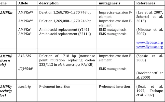
AMPK system is highly conserved between insects and mammals. Drosophila AMPK contains three protein subunits, , and , which are encoded, in contrast to mammalian
AMPK, by single genes named dAMPK, alicorn and loechrig (also known as SNF4A), respectively (Pan and Hardie 2002) (Figure 2), making Drosophila an attractive animal model to study AMPK functions in vivo. However, Drosophila expresses several isoforms for each subunit generated fromalternative splicing and differential transcription initiation (Tschape et al. 2002) and can presumably form a high number of different heterotrimeric complexes. Like mammalian AMPK, drosophila AMPK is allosterically activated by AMP and by treatments that depleted cellular ATP. This is associated with phosphorylation of Thr‐184 within the activation loop of dAMPK and of acetyl‐CoA carboxylase at a homologous site also conserved between mammals and insects (Pan and Hardie 2002). Although mice have been the most commonly used animal model to decipher the biology of AMPK, genetically engineered fruit fly Drosophila melanogaster (Tables 3 and 4) has been particularly important in demonstrating the role of AMPK in the regulation of cell polarity during energy stress and in neuronal survival (Tschape et al. 2002, Lee et al. 2007, Mirouse et al. 2007, Spasic et al. 2008). Identification and generation of mutant AMPK drosophila has been realized by forward genetic screens using ethylmethanesulfonate (EMS) mutagenesis (Medina et al. 2006) or by the P‐ element transposon technology (Rubin and Spradling 1982). Germline transformation of drosophila can be realized via insertion of a single P transposable element or by imprecise mobilization of the P‐elements (excision) to generate null mutants. In addition, creation of transgenic flies using different approaches, including the UAS/Gal4 and "tet‐on" systems (Tower 2000), have been instrumental to complement more traditional genetics.
The Drosophila loechrig (loe) mutant is characterized by degeneration and severe vacuolization (loechrig is the German word for full of holes) in the brain. The mutation in loe is caused by the insertion of a P‐element that affects a neuronal isoform of the AMPKγ subunit (Tschape et al. 2002). Interestingly, among the six different Drosophila AMPK isoforms, this particular protein isoform strongly expressed in the nervous system contains a unique N‐terminus and is the only isoform to rescue the loe phenotype when expressed in neurons, highlighting its specific role for the integrity of the central nervous system. The loe/AMPK mutation affects the regulatory function of AMPK on isoprenoid synthesis via the inhibition of its downstream target hydroxy‐ methylglutaryl (HMG)‐CoA reductase and that changes play a pivotal role in the observed degenerative phenotype (Tschape et al. 2002). The loss of functional neuronal
AMPK upregulates the synthesis of isoprenoids leading to increased prenylation of the small GTPase Rho1, the fly ortholog of vertebrate RhoA, and thereby progressive neurodegeneration (Cook et al. 2012). Upregulation of RhoA signaling pathway induces changes in the actin cytoskeleton through an increase in phosphorylated cofilin and accumulation of F‐actin, resulting in deleterious consequences on neuronal growth and impaired axonal integrity (Cook et al. 2014). Additional evidence for a role of AMPK in neuroprotective processes comes from the study of a P‐element insertion line found to target alicorn (alc), encoding the Drosophila homolog of the regulatory AMPK subunit. Disruption of the alc/AMPK gene causes early‐onset progressive retinal degeneration, characterized by extensive vacuolization and general structural disorganization (Spasic et al. 2008). The mechanism of progressive neuronal death observed as a consequence of loss of AMPK does not involve apoptosis (Tschape et al. 2002, Poels et al. 2012). It has been suggested that AMPK could contribute to the protection of neurons from increased metabolic activity by the regulation of the autophagic process. Accordingly, the loechrig/ SNF4A gene has been identified in a genetic screen to select P‐element insertion that affect autophagy in the larval fat body (Lippai et al. 2008). Surprisingly, in alc mutants, a severe induction of autophagy was reported (Poels et al. 2012). This is most likely a consequence of the absence of the negative regulatory feedback loop mediated by AMPK in condition of excessive autophagic induction (Loffler et al. 2011).
Parkinson's disease is one of the most common neurodegenerative diseases in the aging population. Recently, a Drosophila Parkinson’s disease model was used as an initial system to evaluate the therapeutic potential of a number of candidate compounds (Ng et al. 2012). Epigallocatechin gallate (EGCG), a green tea‐derived catechin, was found to provide the best protection against the loss of dopaminergic neurons and mitochondrial dysfunction, the essential pathological phenotypes of human Parkinson's patients. Importantly, the protective effects of EGCG are abolished when AMPK is knocked down, and loss of AMPK activity exacerbates neuronal loss and associated phenotypes (Ng et al. 2012), suggestive of a pathological role of AMPK in neurodegenerative diseases. In addition, it was demonstrated that genetic activation of AMPK also protects against neuronal loss and reproduces EGCG's protective effects, indicating that targeting AMPK will be useful therapeutically in the treatment of neurodegenerative disorders. Indeed, as mitochondrial dysfunction is currently widely accepted to be a key driver of neurodegeneration, activation of AMPK could preserve neuronal function by preserving
the energy balance and by restoring the clearance of damaged mitochondria via induction of mitophagy through the phosphorylation of the autophagy initiator autophagy‐related gene 1 (Atg1)/ Unc‐51‐like kinase 1 (ULK1).
In agreement, with a role of AMPK in neural maintenance, lethal mutations in AMPK, identified by forward genetic screen with EMS mutagenesis or generated by imprecise excision of P‐elements, confirmed the importance for the kinase in the maintenance of cell integrity (Lee et al. 2007, Swick et al. 2013). AMPK‐null fly embryos showed severe abnormalities in cell polarity and disorganization of epithelial structures and lead to embryonic lethality (Lee et al. 2007). Moreover, AMPK‐null embryos contained defective mitotic divisions with lagging or polyploid chromosomes. It was established that AMPK functions in mitosis and epithelial polarity by targeting myosin II regulatory light chain (MRLC), since phosphomimetic mutant of MRLC rescued the AMPK‐null defects in cell polarity and mitosis (Lee et al. 2007). These findings uncovered a link between energy status and cell structures, revealing new pathophysiological functions for AMPK signaling pathway in the regulation of cellular structures.
Drosophila has been used as a model system to dissect possible roles of AMPK in aging and lifespan determination. Dietary restriction, a reduction in total food intake, has been shown to increase lifespan by modulating nutrient‐sensing pathways in flies as well as in a wide range of organisms including nematodes and mammals (Fontana et al. 2010). However, question remains regarding the importance of cellular energy homeostasis and AMPK signaling as an evolutionary conserved determinant of life span control. For this purpose, to circumvent the lethality caused by AMPK deletion in late larval stages (Lee et al. 2007), the consequences of tissue‐specific knock‐down of AMPK and transgenic expression of AMPK using the inducible GeneSwitch system (Poirier et al. 2008), which provided both temporal and spatial control, have been examined to influence drosophila life span. Different approaches were used including RNAi‐mediated knock‐down of AMPK (Tohyama and Yamaguchi 2010) or AMPK (Johnson et al. 2010) and expression of a dominant negative construct (AMPKK57A), in which the catalytic
domain of the AMPK subunit is inactive (Johnson et al. 2010) (Table 4). Consistent with a causal role for AMPK in regulating energy homeostasis, reduced AMPK signaling leads to hypersensitivity to starvation conditions (Bland et al. 2010, Johnson et al. 2010, Tohyama and Yamaguchi 2010). Interestingly, a similar sensitivity to starvation conditions was observed with a selective loss of AMPK signaling in muscle (Tohyama
and Yamaguchi 2010) and was associated with a reduction in lifespan (Bland et al. 2010, Stenesen et al. 2013), suggesting potential tissue‐specific requirements for AMPK‐ mediated lifespan extension. To study the impact of tissue‐restricted upregulation of AMPK, pro‐longevity effects were investigated in transgenic flies overexpressing AMPK in a subset of metabolic tissues. It was found that localized AMPK activation (e.g., by overexpressing wild‐type AMPK in muscle, fat body, brain or intestinal epithelium) extends lifespan a non‐cell‐autonomous manner (Bland et al. 2010, Stenesen et al. 2013, Ulgherait et al. 2014). Activation of AMPK produces a non‐cell autonomous induction of autophagy due to upregulation of Atg1/ULK1 signaling, which appears to be necessary and sufficient to slows systemic aging (Ulgherait et al. 2014). A recent study demonstrated that dietary application of ‐guanidinopropionic acid (‐GPA), a creatine analog, can extend lifespan through AMPK‐dependent induction of autophagy (Yang et al. 2015). Such findings may provide significant insight for pharmaceutical strategies to manipulate AMPK function in single tissue to prolong lifespan in mammals. 4‐ Genetically modified worm models
The nematode Caenorhabditis elegans has many excellent advantages as an in vivo
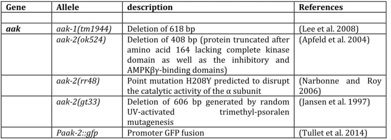
model for detailed molecular analyses and functional genomics. It is a small nematode with a life cycle of 3.5 days and a lifespan of about 2‐3 weeks. In addition, C. elegans displays conserved developmental programs, genetic tractability, and a fully sequenced genome, making an ideal model system to understand biological processes. To identify the biological function of specific genes in C. elegans, different methods have been developed using reverse and forward genetics or transgenesis techniques (Baylis and Vazquez‐Manrique 2011, Boulin and Hobert 2012). C. elegans has many genes which are similar to those of other higher eukaryotes, suggesting many similar functions in cellular and molecular mechanisms. The C. elegans genome encodes the aak‐1 and aak‐2 genes, which are homologs of the catalytic subunits of mammalian AMPK, the aakb‐1 and
aakb‐2 genes, which are homologs of the regulatory subunits of mammalian AMPK,
and the aakg‐1, aakg‐2, aakg3, aakg‐4 and aakg‐5 genes, which are homologs of the regulatory subunits of mammalian AMPK (Apfeld et al. 2004) (Figure 2). Hence, a potential of 20 alternative heterotrimeric complexes can be formed in C. elegans.
During the last decade, C. elegans has been at the forefront as a model system to understand biological processes linking energetics to longevity. Limiting energy
availability through dietary restriction confers lifespan extension in organisms as diverse as yeasts, nematodes, and rodents (Fontana et al. 2010). It has been found that increases in cellular AMP/ATP ratio are associated with age in C. elegans, suggesting a link between genes known to affect lifespan and the control of energy metabolism (Apfeld et al. 2004). Thus, it is not surprising that AMPK has emerged as a key regulator of lifespan determination (Burkewitz et al. 2014). The use of aak‐2 mutants (Table 5) has highlighted the role of AMPK in the regulation of lifespan in response to environmental stress and insulin‐like signaling (Apfeld et al. 2004, Narbonne and Roy 2006). C. elegans lacking aak‐2 failed to extend lifespan in response dietary restriction and low energy conditions (Greer et al. 2007, Schulz et al. 2007, Lee et al. 2008, Narbonne and Roy 2009, Fukuyama et al. 2012). Conversely, overexpression of aak‐2 significantly promotes lifespan extension and mimics dietary restriction in well‐fed wild‐type animals (Apfeld et al. 2004). Similar increase in lifespan is observed in transgenic worms expressing an active form of the AMPK (Mair et al. 2011) or the AMPK (Greer et al. 2007) subunits. C. elegans fed with the AMPK agonist metformin also display increase in lifespan that is dependent on aak‐2 (Onken and Driscoll 2010). The effect of metformin on extension of C. elegans lifespan can be direct by promoting resistance to biguanide toxicity through AMPK activation and indirect by impairing folate metabolism of E. coli, its trophic microbial partner (Cabreiro et al. 2013).
Under condition of energetic stress, the aak‐2 subunit becomes phosphorylated at threonine 243 (Thr243), equivalent to Thr172 in the mammalian ortholog, and primarily takes part in its activity to phosphorylate and inhibits the CREB‐regulated transcriptional coactivator CRTC‐1, a cofactor involved in diverse physiological processes including energy homeostasis (Mair et al. 2011). Modulation of CRTC‐1 phosphorylation status by AMPK activation in the central nervous system is required to locally and cell autonomously promote remodeling of mitochondrial metabolic networks and increase longevity (Burkewitz et al. 2015). Upon reduced AMPK activity, CRTC‐1 modulates AMPK‐mediated longevity cell nonautonomously via regulation of the neurotransmitter/hormone octopamine secretion, which drives mitochondrial fragmentation in distal tissues, and suppresses the effects of AMPK on systemic mitochondrial metabolism and longevity. These findings highlight the dominance of neuronal energy‐sensing mechanisms and neuronal signals on systemic metabolic homeostasis and impact on aging process. This regulatory mechanism is consistent with
the cell‐nonautonomous role for neuronal AMPK in modulating peripheral lipid storage in worms (Cunningham et al. 2014).
Concluding remarks
The AMP‐activated protein kinase (AMPK) system was first discovered 35 years ago. Since that time, knowledge of the diverse physiological functions of AMPK has grown rapidly and continues to evolve. Most certainly, genetically modified mice have become instrumental to the study of AMPK. However, the use of fly and worm model organisms has also played a key role in delineating the physiological role of AMPK signaling pathway and will continue to contribute to the expanding field of AMPK.
Identifying the best animal model to study AMPK function or characterize AMPK agonists/ antagonists is an important consideration and requires a thorough understanding of the advantages and disadvantages of each animal model, as there are important factors to consider. Animal models with comparable AMPK heterotrimeric composition to human cells and tissues are typically relevant models to examine AMPK pathophysiological role and develop tissue‐specific therapeutic interventions. However, although mouse models possess many advantages for biomedical research, it has been reported that the composition of AMPK heterotrimers differ between rodent and human hepatocytes (Stephenne et al. 2011, Wu et al. 2013), addressing a major challenge for future pre‐clinical translational studies.
References
Ahmad, F., M. Arad, N. Musi, H. He, C. Wolf, D. Branco, A. R. Perez‐Atayde, D. Stapleton, D. Bali, Y. Xing, R. Tian, L. J. Goodyear, C. I. Berul, J. S. Ingwall, C. E. Seidman and J. G. Seidman (2005). "Increased alpha2 subunit‐associated AMPK activity and PRKAG2 cardiomyopathy." Circulation 112(20): 3140‐3148.
Akman, H. O., J. N. Sampayo, F. A. Ross, J. W. Scott, G. Wilson, L. Benson, C. Bruno, S. Shanske, D. G. Hardie and S. Dimauro (2007). "Fatal infantile cardiac glycogenosis with phosphorylase kinase deficiency and a mutation in the gamma2‐subunit of AMP‐ activated protein kinase." Pediatr Res 62(4): 499‐504.
Andreelli, F., M. Foretz, C. Knauf, P. D. Cani, C. Perrin, M. A. Iglesias, B. Pillot, A. Bado, F. Tronche, G. Mithieux, S. Vaulont, R. Burcelin and B. Viollet (2006). "Liver adenosine monophosphate‐activated kinase‐alpha2 catalytic subunit is a key target for the control of hepatic glucose production by adiponectin and leptin but not insulin." Endocrinology 147(5): 2432‐2441.
Apfeld, J., G. O'Connor, T. McDonagh, P. S. DiStefano and R. Curtis (2004). "The AMP‐ activated protein kinase AAK‐2 links energy levels and insulin‐like signals to lifespan in C. elegans." Genes & development 18(24): 3004‐3009.
Arad, M., D. W. Benson, A. R. Perez‐Atayde, W. J. McKenna, E. A. Sparks, R. J. Kanter, K. McGarry, J. G. Seidman and C. E. Seidman (2002). "Constitutively active AMP kinase mutations cause glycogen storage disease mimicking hypertrophic cardiomyopathy." The Journal of clinical investigation 109(3): 357‐362.
Arad, M., B. J. Maron, J. M. Gorham, W. H. Johnson, Jr., J. P. Saul, A. R. Perez‐Atayde, P. Spirito, G. B. Wright, R. J. Kanter, C. E. Seidman and J. G. Seidman (2005). "Glycogen storage diseases presenting as hypertrophic cardiomyopathy." N Engl J Med 352(4): 362‐372.
Arad, M., I. P. Moskowitz, V. V. Patel, F. Ahmad, A. R. Perez‐Atayde, D. B. Sawyer, M. Walter, G. H. Li, P. G. Burgon, C. T. Maguire, D. Stapleton, J. P. Schmitt, X. X. Guo, A. Pizard, S. Kupershmidt, D. M. Roden, C. I. Berul, C. E. Seidman and J. G. Seidman (2003).
"Transgenic mice overexpressing mutant PRKAG2 define the cause of Wolff‐Parkinson‐ White syndrome in glycogen storage cardiomyopathy." Circulation 107(22): 2850‐2856. Banerjee, S. K., K. R. McGaffin, X. N. Huang and F. Ahmad (2010). "Activation of cardiac hypertrophic signaling pathways in a transgenic mouse with the human PRKAG2 Thr400Asn mutation." Biochim Biophys Acta 1802(2): 284‐291.
Banerjee, S. K., R. Ramani, S. Saba, J. Rager, R. Tian, M. A. Mathier and F. Ahmad (2007). "A PRKAG2 mutation causes biphasic changes in myocardial AMPK activity and does not protect against ischemia." Biochemical and biophysical research communications 360(2): 381‐387.
Banerjee, S. K., D. W. Wang, R. Alzamora, X. N. Huang, N. M. Pastor‐Soler, K. R. Hallows, K. R. McGaffin and F. Ahmad (2010). "SGLT1, a novel cardiac glucose transporter, mediates increased glucose uptake in PRKAG2 cardiomyopathy." J Mol Cell Cardiol 49(4): 683‐ 692.
Barnes, B. R., Y. C. Long, T. L. Steiler, Y. Leng, D. Galuska, J. F. Wojtaszewski, L. Andersson and J. R. Zierath (2005). "Changes in exercise‐induced gene expression in 5'‐AMP‐ activated protein kinase gamma3‐null and gamma3 R225Q transgenic mice." Diabetes 54(12): 3484‐3489.
Barnes, B. R., S. Marklund, T. L. Steiler, M. Walter, G. Hjalm, V. Amarger, M. Mahlapuu, Y. Leng, C. Johansson, D. Galuska, K. Lindgren, M. Abrink, D. Stapleton, J. R. Zierath and L. Andersson (2004). "The 5'‐AMP‐activated protein kinase gamma3 isoform has a key role in carbohydrate and lipid metabolism in glycolytic skeletal muscle." The Journal of biological chemistry 279(37): 38441‐38447.
Barre, L., C. Richardson, M. F. Hirshman, J. Brozinick, S. Fiering, B. E. Kemp, L. J. Goodyear and L. A. Witters (2007). "Genetic model for the chronic activation of skeletal muscle AMP‐activated protein kinase leads to glycogen accumulation." American journal of physiology 292(3): E802‐811.
Baylis, H. A. and R. P. Vazquez‐Manrique (2011). "Reverse genetic strategies in Caenorhabditis elegans: towards controlled manipulation of the genome." ScientificWorldJournal 11: 1394‐1410.
Bayrak, F., E. Komurcu‐Bayrak, B. Mutlu, G. Kahveci, Y. Basaran and N. Erginel‐Unaltuna (2006). "Ventricular pre‐excitation and cardiac hypertrophy mimicking hypertrophic
cardiomyopathy in a Turkish family with a novel PRKAG2 mutation." Eur J Heart Fail 8(7): 712‐715.
Blagih, J., F. Coulombe, E. E. Vincent, F. Dupuy, G. Galicia‐Vazquez, E. Yurchenko, T. C. Raissi, G. J. van der Windt, B. Viollet, E. L. Pearce, J. Pelletier, C. A. Piccirillo, C. M. Krawczyk, M. Divangahi and R. G. Jones (2015). "The energy sensor AMPK regulates T cell metabolic adaptation and effector responses in vivo." Immunity 42(1): 41‐54.
Blair, E., C. Redwood, H. Ashrafian, M. Oliveira, J. Broxholme, B. Kerr, A. Salmon, I. Ostman‐Smith and H. Watkins (2001). "Mutations in the gamma(2) subunit of AMP‐ activated protein kinase cause familial hypertrophic cardiomyopathy: evidence for the central role of energy compromise in disease pathogenesis." Human molecular genetics 10(11): 1215‐1220. Bland, M. L., R. J. Lee, J. M. Magallanes, J. K. Foskett and M. J. Birnbaum (2010). "AMPK supports growth in Drosophila by regulating muscle activity and nutrient uptake in the gut." Dev Biol 344(1): 293‐303. Boulin, T. and O. Hobert (2012). "From genes to function: the C. elegans genetic toolbox." Wiley Interdiscip Rev Dev Biol 1(1): 114‐137.
Buhl, E. S., N. Jessen, R. Pold, T. Ledet, A. Flyvbjerg, S. B. Pedersen, O. Pedersen, O. Schmitz and S. Lund (2002). "Long‐term AICAR administration reduces metabolic disturbances and lowers blood pressure in rats displaying features of the insulin resistance syndrome." Diabetes 51(7): 2199‐2206.
Burkewitz, K., I. Morantte, H. J. Weir, R. Yeo, Y. Zhang, F. K. Huynh, O. R. Ilkayeva, M. D. Hirschey, A. R. Grant and W. B. Mair (2015). "Neuronal CRTC‐1 governs systemic mitochondrial metabolism and lifespan via a catecholamine signal." Cell 160(5): 842‐ 855.
Burkewitz, K., Y. Zhang and W. B. Mair (2014). "AMPK at the nexus of energetics and aging." Cell Metab 20(1): 10‐25.
Burwinkel, B., J. W. Scott, C. Buhrer, F. K. van Landeghem, G. F. Cox, C. J. Wilson, D. Grahame Hardie and M. W. Kilimann (2005). "Fatal congenital heart glycogenosis caused by a recurrent activating R531Q mutation in the gamma 2‐subunit of AMP‐activated protein kinase (PRKAG2), not by phosphorylase kinase deficiency." Am J Hum Genet 76(6): 1034‐1049.
Cabreiro, F., C. Au, K. Y. Leung, N. Vergara‐Irigaray, H. M. Cocheme, T. Noori, D. Weinkove, E. Schuster, N. D. Greene and D. Gems (2013). "Metformin retards aging in C. elegans by altering microbial folate and methionine metabolism." Cell 153(1): 228‐239. Chen, Z. P., G. K. McConell, B. J. Michell, R. J. Snow, B. J. Canny and B. E. Kemp (2000). "AMPK signaling in contracting human skeletal muscle: acetyl‐CoA carboxylase and NO synthase phosphorylation." American journal of physiology. Endocrinology and metabolism 279(5): E1202‐1206.
Cheung, P. C., I. P. Salt, S. P. Davies, D. G. Hardie and D. Carling (2000). "Characterization of AMP‐activated protein kinase gamma‐subunit isoforms and their role in AMP binding." Biochem J 346 Pt 3: 659‐669.
Ciobanu, D., J. Bastiaansen, M. Malek, J. Helm, J. Woollard, G. Plastow and M. Rothschild (2001). "Evidence for new alleles in the protein kinase adenosine monophosphate‐ activated gamma(3)‐subunit gene associated with low glycogen content in pig skeletal muscle and improved meat quality." Genetics 159(3): 1151‐1162.
Claret, M., M. A. Smith, R. L. Batterham, C. Selman, A. I. Choudhury, L. G. Fryer, M. Clements, H. Al‐Qassab, H. Heffron, A. W. Xu, J. R. Speakman, G. S. Barsh, B. Viollet, S. Vaulont, M. L. Ashford, D. Carling and D. J. Withers (2007). "AMPK is essential for energy homeostasis regulation and glucose‐ sensing by POMC and AgRP neurons " J Clin Invest 117(8): 2325‐2336. Cook, M., B. J. Bolkan and D. Kretzschmar (2014). "Increased actin polymerization and stabilization interferes with neuronal function and survival in the AMPKgamma mutant Loechrig." PLoS One 9(2): e89847. Cook, M., P. Mani, J. S. Wentzell and D. Kretzschmar (2012). "Increased RhoA prenylation in the loechrig (loe) mutant leads to progressive neurodegeneration." PLoS One 7(9): e44440.
Cool, B., B. Zinker, W. Chiou, L. Kifle, N. Cao, M. Perham, R. Dickinson, A. Adler, G. Gagne, R. Iyengar, G. Zhao, K. Marsh, P. Kym, P. Jung, H. S. Camp and E. Frevert (2006). "Identification and characterization of a small molecule AMPK activator that treats key components of type 2 diabetes and the metabolic syndrome." Cell Metab 3(6): 403‐416.
Costford, S. R., N. Kavaslar, N. Ahituv, S. N. Chaudhry, W. S. Schackwitz, R. Dent, L. A. Pennacchio, R. McPherson and M. E. Harper (2007). "Gain‐of‐function R225W mutation in human AMPKgamma(3) causing increased glycogen and decreased triglyceride in skeletal muscle." PLoS One 2(9): e903.
Cunningham, K. A., A. D. Bouagnon, A. G. Barros, L. Lin, L. Malard, M. A. Romano‐Silva and K. Ashrafi (2014). "Loss of a neural AMP‐activated kinase mimics the effects of elevated serotonin on fat, movement, and hormonal secretions." PLoS genetics 10(6): e1004394. Daniel, T. and D. Carling (2002). "Functional analysis of mutations in the gamma 2 subunit of AMP‐activated protein kinase associated with cardiac hypertrophy and Wolff‐ Parkinson‐White syndrome." J Biol Chem 277(52): 51017‐51024.
Dasgupta, B., J. S. Ju, Y. Sasaki, X. Liu, S. R. Jung, K. Higashida, D. Lindquist and J. Milbrandt (2012). "The AMPK beta2 subunit is required for energy homeostasis during metabolic stress." Mol Cell Biol 32(14): 2837‐2848.
Dasgupta, B. and J. Milbrandt (2009). "AMP‐activated protein kinase phosphorylates retinoblastoma protein to control mammalian brain development." Dev Cell 16(2): 256‐ 270.
Davies, J. K., D. J. Wells, K. Liu, H. R. Whitrow, T. D. Daniel, R. Grignani, C. A. Lygate, J. E. Schneider, G. Noel, H. Watkins and D. Carling (2006). "Characterization of the role of gamma2 R531G mutation in AMP‐activated protein kinase in cardiac hypertrophy and Wolff‐Parkinson‐White syndrome." American journal of physiology 290(5): H1942‐ 1951.
Deak, P., M. M. Omar, R. D. Saunders, M. Pal, O. Komonyi, J. Szidonya, P. Maroy, Y. Zhang, M. Ashburner, P. Benos, C. Savakis, I. Siden‐Kiamos, C. Louis, V. N. Bolshakov, F. C. Kafatos, E. Madueno, J. Modolell and D. M. Glover (1997). "P‐element insertion alleles of essential genes on the third chromosome of Drosophila melanogaster: correlation of physical and cytogenetic maps in chromosomal region 86E‐87F." Genetics 147(4): 1697‐ 1722.
Dietzl, G., D. Chen, F. Schnorrer, K. C. Su, Y. Barinova, M. Fellner, B. Gasser, K. Kinsey, S. Oppel, S. Scheiblauer, A. Couto, V. Marra, K. Keleman and B. J. Dickson (2007). "A genome‐wide transgenic RNAi library for conditional gene inactivation in Drosophila." Nature 448(7150): 151‐156.
Dockendorff, T. C., S. E. Robertson, D. L. Faulkner and T. A. Jongens (2000). "Genetic characterization of the 44D‐45B region of the Drosophila melanogaster genome based on an F2 lethal screen." Mol Gen Genet 263(1): 137‐143.
Dzamko, N., B. J. van Denderen, A. L. Hevener, S. B. Jorgensen, J. Honeyman, S. Galic, Z. P. Chen, M. J. Watt, D. J. Campbell, G. R. Steinberg and B. E. Kemp (2010). "AMPK beta1 deletion reduces appetite, preventing obesity and hepatic insulin resistance." The Journal of biological chemistry 285(1): 115‐122.
Enfalt, A. C., K. Lundstrom, A. Karlsson and I. Hansson (1997). "Estimated frequency of the RN‐ allele in Swedish Hampshire pigs and comparison of glycolytic potential, carcass composition, and technological meat quality among Swedish Hampshire, Landrace, and Yorkshire pigs." Journal of animal science 75(11): 2924‐2935.
Essen‐Gustavsson, B., A. Granlund, B. Benziane, M. Jensen‐Waern and A. V. Chibalin (2011). "Muscle glycogen resynthesis, signalling and metabolic responses following acute exercise in exercise‐trained pigs carrying the PRKAG3 mutation." Exp Physiol 96(9): 927‐937.
Estrade, M., S. Ayoub, A. Talmant and G. Monin (1994). "Enzyme activities of glycogen metabolism and mitochondrial characteristics in muscles of RN‐ carrier pigs (Sus scrofa domesticus)." Comparative biochemistry and physiology. Biochemistry and molecular biology 108(3): 295‐301.
Folmes, K. D., A. Y. Chan, D. P. Koonen, T. C. Pulinilkunnil, I. Baczko, B. E. Hunter, S. Thorn, M. F. Allard, R. Roberts, M. H. Gollob, P. E. Light and J. R. Dyck (2009). "Distinct early signaling events resulting from the expression of the PRKAG2 R302Q mutant of AMPK contribute to increased myocardial glycogen." Circ Cardiovasc Genet 2(5): 457‐466. Fontana, L., L. Partridge and V. D. Longo (2010). "Extending healthy life span‐‐from yeast to humans." Science 328(5976): 321‐326.
Foretz, M., S. Guihard, J. Leclerc, V. Fauveau, J. P. Couty, F. Andris, M. Gaudry, F. Andreelli, S. Vaulont and B. Viollet (2010). "Maintenance of red blood cell integrity by AMP‐ activated protein kinase alpha1 catalytic subunit." FEBS Lett 584(16): 3667‐3671. Foretz, M., S. Hebrard, S. Guihard, J. Leclerc, M. Do Cruzeiro, G. Hamard, F. Niedergang, M. Gaudry and B. Viollet (2011). "The AMPKgamma1 subunit plays an essential role in
erythrocyte membrane elasticity, and its genetic inactivation induces splenomegaly and anemia." FASEB J 25(1): 337‐347. Fu, X., J. X. Zhao, M. J. Zhu, M. Foretz, B. Viollet, M. V. Dodson and M. Du (2013). "AMP‐ activated protein kinase alpha1 but not alpha2 catalytic subunit potentiates myogenin expression and myogenesis." Mol Cell Biol 33(22): 4517‐4525. Fu, X., M. Zhu, S. Zhang, M. Foretz, B. Viollet and M. Du (2016). "Obesity Impairs Skeletal Muscle Regeneration Through Inhibition of AMPK." Diabetes 65(1): 188‐200.
Fu, X., M. J. Zhu, M. V. Dodson and M. Du (2015). "AMP‐activated protein kinase stimulates Warburg‐like glycolysis and activation of satellite cells during muscle regeneration." J Biol Chem 290(44): 26445‐26456.
Fujii, N., T. Hayashi, M. F. Hirshman, J. T. Smith, S. A. Habinowski, L. Kaijser, J. Mu, O. Ljungqvist, M. J. Birnbaum, L. A. Witters, A. Thorell and L. J. Goodyear (2000). "Exercise induces isoform‐specific increase in 5'AMP‐activated protein kinase activity in human skeletal muscle." Biochemical and biophysical research communications 273(3): 1150‐ 1155.
Fujii, N., M. F. Hirshman, E. M. Kane, R. C. Ho, L. E. Peter, M. M. Seifert and L. J. Goodyear (2005). "AMP‐activated protein kinase alpha2 activity is not essential for contraction‐ and hyperosmolarity‐induced glucose transport in skeletal muscle." The Journal of biological chemistry 280(47): 39033‐39041.
Fujii, N., M. M. Seifert, E. M. Kane, L. E. Peter, R. C. Ho, S. Winstead, M. F. Hirshman and L. J. Goodyear (2007). "Role of AMP‐activated protein kinase in exercise capacity, whole body glucose homeostasis, and glucose transport in skeletal muscle ‐insight from analysis of a transgenic mouse model." Diabetes Res Clin Pract 77 Suppl 1: S92‐98. Fukuyama, M., K. Sakuma, R. Park, H. Kasuga, R. Nagaya, Y. Atsumi, Y. Shimomura, S. Takahashi, H. Kajiho, A. Rougvie, K. Kontani and T. Katada (2012). "C. elegans AMPKs promote survival and arrest germline development during nutrient stress." Biol Open 1(10): 929‐936.
Fullerton, M. D., S. Galic, K. Marcinko, S. Sikkema, T. Pulinilkunnil, Z. P. Chen, H. M. O'Neill, R. J. Ford, R. Palanivel, M. O'Brien, D. G. Hardie, S. L. Macaulay, J. D. Schertzer, J. R. Dyck, B. J. van Denderen, B. E. Kemp and G. R. Steinberg (2013). "Single phosphorylation
sites in Acc1 and Acc2 regulate lipid homeostasis and the insulin‐sensitizing effects of metformin." Nat Med 19(12): 1649‐1654.
Galic, S., M. D. Fullerton, J. D. Schertzer, S. Sikkema, K. Marcinko, C. R. Walkley, D. Izon, J. Honeyman, Z. P. Chen, B. J. van Denderen, B. E. Kemp and G. R. Steinberg (2011). "Hematopoietic AMPK beta1 reduces mouse adipose tissue macrophage inflammation and insulin resistance in obesity." J Clin Invest 121(12): 4903‐4915.
Garcia‐Roves, P. M., M. E. Osler, M. H. Holmstrom and J. R. Zierath (2008). "Gain‐of‐ function R225Q mutation in AMP‐activated protein kinase gamma3 subunit increases mitochondrial biogenesis in glycolytic skeletal muscle." J Biol Chem 283(51): 35724‐ 35734.
Gollob, M. H., M. S. Green, A. S. Tang, T. Gollob, A. Karibe, A. S. Ali Hassan, F. Ahmad, R. Lozado, G. Shah, L. Fananapazir, L. L. Bachinski and R. Roberts (2001). "Identification of a gene responsible for familial Wolff‐Parkinson‐White syndrome." The New England journal of medicine 344(24): 1823‐1831.
Gollob, M. H., J. J. Seger, T. N. Gollob, T. Tapscott, O. Gonzales, L. Bachinski and R. Roberts (2001). "Novel PRKAG2 mutation responsible for the genetic syndrome of ventricular preexcitation and conduction system disease with childhood onset and absence of cardiac hypertrophy." Circulation 104(25): 3030‐3033.
Gong, H., J. Xie, N. Zhang, L. Yao and Y. Zhang (2011). "MEF2A binding to the Glut4 promoter occurs via an AMPKalpha2‐dependent mechanism." Med Sci Sports Exerc 43(8): 1441‐1450.
Granlund, A., M. Jensen‐Waern and B. Essen‐Gustavsson (2011). "The influence of the PRKAG3 mutation on glycogen, enzyme activities and fibre types in different skeletal muscles of exercise trained pigs." Acta Vet Scand 53: 20.
Granlund, A., O. Kotova, B. Benziane, D. Galuska, M. Jensen‐Waern, A. V. Chibalin and B. Essen‐Gustavsson (2010). "Effects of exercise on muscle glycogen synthesis signalling and enzyme activities in pigs carrying the PRKAG3 mutation." Exp Physiol 95(4): 541‐ 549.
Greer, E. L., D. Dowlatshahi, M. R. Banko, J. Villen, K. Hoang, D. Blanchard, S. P. Gygi and A. Brunet (2007). "An AMPK‐FOXO pathway mediates longevity induced by a novel method of dietary restriction in C. elegans." Current biology : CB 17(19): 1646‐1656.
Guigas, B., L. Bertrand, N. Taleux, M. Foretz, N. Wiernsperger, D. Vertommen, F. Andreelli, B. Viollet and L. Hue (2006). "5‐Aminoimidazole‐4‐Carboxamide‐1‐{beta}‐D‐ Ribofuranoside and Metformin Inhibit Hepatic Glucose Phosphorylation by an AMP‐ Activated Protein Kinase‐Independent Effect on Glucokinase Translocation." Diabetes 55(4): 865‐874.
Gundewar, S., J. W. Calvert, S. Jha, I. Toedt‐Pingel, S. Y. Ji, D. Nunez, A. Ramachandran, M. Anaya‐Cisneros, R. Tian and D. J. Lefer (2009). "Activation of AMP‐activated protein kinase by metformin improves left ventricular function and survival in heart failure." Circ Res 104(3): 403‐411.
Halseth, A. E., N. J. Ensor, T. A. White, S. A. Ross and E. A. Gulve (2002). "Acute and chronic treatment of ob/ob and db/db mice with AICAR decreases blood glucose concentrations." Biochem Biophys Res Commun 294(4): 798‐805.
Hamilton, S. R., D. Stapleton, J. B. O'Donnell, J. T. Kung, S. R. Dalal, B. E. Kemp and L. A. Witters (2001). "An activating mutation in the gamma1 subunit of the AMP‐activated protein kinase." FEBS Lett 500(3): 163‐168.
Hardie, D. G. (2014). "AMP‐activated protein kinase: maintaining energy homeostasis at the cellular and whole‐body levels." Annu Rev Nutr 34: 31‐55.
Hedegaard, J., P. Horn, R. Lametsch, H. Sondergaard Moller, P. Roepstorff, C. Bendixen and E. Bendixen (2004). "UDP‐glucose pyrophosphorylase is upregulated in carriers of the porcine RN‐ mutation in the AMP‐activated protein kinase." Proteomics 4(8): 2448‐ 2454.
Huang, L. S., J. W. Ma, J. Ren, N. S. Ding, Y. M. Guo, H. S. Ai, L. Li, L. H. Zhou and C. Y. Chen (2004). "Genetic variations of the porcine PRKAG3 gene in Chinese indigenous pig breeds." Genet Sel Evol 36(4): 481‐486.
Hue, L. and H. Taegtmeyer (2009). "The Randle cycle revisited: a new head for an old hat." Am J Physiol Endocrinol Metab 297(3): E578‐591.
Jansen, G., E. Hazendonk, K. L. Thijssen and R. H. Plasterk (1997). "Reverse genetics by chemical mutagenesis in Caenorhabditis elegans." Nature genetics 17(1): 119‐121.