This article was downloaded by:[Nyman, Ulf] On: 2 April 2008
Access Details: [subscription number 791897936] Publisher: Informa Healthcare
Informa Ltd Registered in England and Wales Registered Number: 1072954 Registered office: Mortimer House, 37-41 Mortimer Street, London W1T 3JH, UK
Acta Radiologica [Old Series]
Publication details, including instructions for authors and subscription information:
http://www.informaworld.com/smpp/title~content=t789802412
Catheter Replacement of the Needle in Percutaneous
Arteriography: A new technique
Sven Ivar Seldingera
aRoentgen Diagnostic Department, Karolinska Sjukhuset, Stockholm, Sweden
Online Publication Date: 01 May 1953
To cite this Article: Seldinger, Sven Ivar (1953) 'Catheter Replacement of the Needle in Percutaneous Arteriography: A new technique', Acta Radiologica [Old Series], 39:5, 368 - 376
To link to this article: DOI: 10.3109/00016925309136722 URL:http://dx.doi.org/10.3109/00016925309136722
PLEASE SCROLL DOWN FOR ARTICLE
Full terms and conditions of use:http://www.informaworld.com/terms-and-conditions-of-access.pdf
This article maybe used for research, teaching and private study purposes. Any substantial or systematic reproduction, re-distribution, re-selling, loan or sub-licensing, systematic supply or distribution in any form to anyone is expressly forbidden.
The publisher does not give any warranty express or implied or make any representation that the contents will be complete or accurate or up to date. The accuracy of any instructions, formulae and drug doses should be independently verified with primary sources. The publisher shall not be liable for any loss, actions, claims, proceedings, demand or costs or damages whatsoever or howsoever caused arising directly or indirectly in connection with or arising out of the use of this material.
Downloaded By: [Nyman, Ulf] At: 20:30 2 April 2008
FROM THE KOENTBEN DIAQNOSTIC DEPARTMENT (DIRECTOR: PROFESSOR KNUT LIKDBLOM), KAROLINSKA SJUKIiUSET, STOCKHOLM, SWEDEN
.~
CA'l'HE'l'ER IZEI'LACEMENT OF THE NEEDLE 1N
PERCUTANEOUS AltTERlOGItAPHY
A new technique
byS v e n
I v a r S e l d i n g e r
The catheter method of angiography has become more popular in
the past few years,
as
it provides the following advantages over the
method
of
injecting the contrast medium by means of
a
simple needle:
1)
The contrast medium may be injected into
a
vessel a t any level
desired.
2)
Risk of extravascular injection
of
the contrast medium is mini-
mised.
3)
The patient may be placed in any position required.
4)
The catheter may be left in situ without risk while the films are
being developed, thus facilitating re-examination
if necessary.
Until recently, however, the use of the catheter method was restricted
because
of
the lack of
a
suitable flexible thin-walled catheter which
could be used percutaneously. FARINAS,
in 1941, described
a
method
in
which
a
urethral catheter was passed up into the aorta through
a
trocar
inserted in the exposed femoral artery. I n 1947, RADNER
catheterized
the exposed and ligated radial artery and performed vertebral angio-
graphy and later thoracic aortography. Since then, many authors have
catheterized arteries for various purposes, by surgical exposure followed
by ligature
or
resuturing
of
the artery. In 1949,
JONSSONperformed
thoracic aortography after puncture
of
the common carotid artery by
means
of
a
blunt cannula provided with an inner sharp needle.
The
cannula, guided by
a
silver thread,
was
then directed downwards. Later
Ihiefly presented a t the Congress of the Northern Association of Medical Radiol- ogy, Helsinki, June, 1952; submitted for publication, October 28, 1952.
Downloaded By: [Nyman, Ulf] At: 20:30 2 April 2008
CATHETER REPLACEMENT OF THE NEEDLE 1N PERCCTANEOUS ARTERIOGRAPHY
the procedure was abandoned, partly because
it was considered that the cannula might in-
jure the aortic wall. This percutaneous method
might have proved more useful
if
a
technique
for using
a
flexible catheter of adequate lumen
had been available a t the time.
The artery exposure technique of catheter-
ization is time-consuming, troublesome and
may present certain risks. The thin-walled poly-
ethylene tube, however, makes percutaneous
catheterization possible,
as
reported by
PEIRCE
in
1951,
who passed in the tubing through a
large bore needle. This method was suitable
for aortography via the femoral artery. I n the
same year, DONALD,
KESMODEL,
ROLLINS
and
PADDISON,
employing
a
similar technique, cath-
eterized the common carotid artery in cere-
bral angiography
.
The method necessitates the
use
of
a
large bore needle which may make
puncture difficult and limits its use
t o
com-
paratively large arteries, hence
PEIRCE'S
at-
tempts to catheterize the brachial artery were
disappointing. There is also extra damage to
the artery and,
as
the hole in the artery
is
larger than the catheter, haemorrhage after
removal of the needle may be troublesome.
To
prevent bleeding, the needle may be kept in
situ during the investigation; this, however, in-
creases the risk of injury to the patient during
movement.
There is
a
simple method, however, of using
a
catheter the same size as the needle, and
which has been used
at
Karolinska Sjukhuset
since April
1952.
The main principle consists
369
I
I
Fig. 1. The equipment. The stilette is removed and the leader inserted through the needle (left) and the catheter
(right).
in the catheter being introduced o'n
a
flexible
leader through the puncture hole after withdrawal of the puncture
needle. The details are as follows:
Epuipnaent. (Supplied by
A.
B.
Stille- Werner, Stockholm.)
I )
A puncture needle with stilette.
2)
A flexible rounded-end metal leader with increased flexibility
3)
A polyethylene tube, of the same diameter
as
the needle, with
of its distal
3
cm.
an adapter for the attachment of
a
syringe.
Downloaded By: [Nyman, Ulf] At: 20:30 2 April 2008
370 SVEN IVAR SELDINGEH
Q
C
d
f
e
Pig. 2. Diagrani of the technique used. a) The artery punctured. The needle pushed upwards. b) The leader inserted. c) The needle withdrawn and the artery compressed, d) The catheter threaded on to the leader. e) The catheter inserted into the artery.
f ) The leader withdrawn.
The leader should have
a
diameter slightly less than the bore
of
the needle and the catheter,
so
that it is capable
of
passing through
both, and should be
at
least
8-9
ern longer than the latter; on the other
hand it should just
fit the lumen of the catheter (Fig.
1 ) .The tip of the
catheter may be cut before use as shown in Fig.
2.
Technique (see Pig.
2).
a)
After local anaesthesia, the artery is punctured percutaneously
a t a relatively small angle.
After puncture it is best to rotate the needle
180"and push it
a
little
into the artery using the bleeding as
a
guide to ensure that the needle
remains in the artery. Puncture of arteries smaller than the femoral
artery is facilitated by using an inner needle
as
a
guide over which the
outer needle is directed into the artery.
b) The supple tip of the leader is inserted
a
very short distance
into the lumen of the artery through the needle.
Downloaded By: [Nyman, Ulf] At: 20:30 2 April 2008
CATIIETER REI’IIACEMII,ST OF THE NEEDLE I N PERCUTAhEOUP dn’IEBI@GI{AI’IIP 371
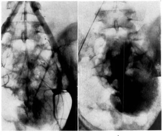
Pig. 3. Hypoplastic lowrr pole of t h e right kidney. I3lood supply from t w o branchesIof
A sinall a l w r a r r t nrtrry. Catheter inscrtetl through t h e right femoral artery with t i p 2 c m
below t h e rerial arteries.
At this nioineiit bleeding should be controlled by pressure on t h e
mtery proximal t o the puncture site, because the diameter of the leader
is srnaller than the hole in the artery.
d)
The catheter is threaded
on
t o the leader; when the tip reaches
the skin the free end of t h e leader must protrude from the catheter.
e)
The catheter and leader are gripped near the skin through which
they are inserted. The catheter enters the artery easily as a n opening
has already been made by the needle. The catheter and leader are pushed
just far enough to ensure t h a t the tip of the former is in the lumen of
the vessel.
f ) The leader is removed and the catheter directed t o the leveI
required, after good arterial bleeding through the catheter has been
obtained. The unsupported catheter is usually pushed up the vessel
without difficulty, but occasionally the leader must be re-introduced
into the catheter in order to support it. The leader should not be passed
beyond the tip of the catheter.
This technique is simpler than appears on paper and after a little
practice should present no difficulties.
It
is important t h a t the leader
passes into the artery easily. JVhen the tip of the catheter enters t h e
artery, the same resistance is often felt as when puncturing is perfarmed
Downloaded By: [Nyman, Ulf] At: 20:30 2 April 2008
372 SVEN IVAR SELDlNGER
a . b.
Fig. 4. Left-sided ectopic kidney in pelvis. (Right kidney absent.) Blood supply by one artery from the iliac bifurcation and one from the left internal iliac artery. Catheter inserted through the right femoral artery with tip a t the bifurcation. a. Arterial phase.
b. Capillary phase.
by means
of
a
needle. However, the resistance is generally but slight
or
may be completely absent. If considerable resistance be encountered,
it
is probable that the tip of the leader
is obstructed and force must
therefore never be applied.
Polyethylene tubing
is
unfortunately not radio-opaque.
For
this
reason, in aortography via the femoral artery, a small amount
of
contrast
medium may be injected and followed by
a
test exposure. This will
show the position
of
the catheter and also the exact situation of the
renal arteries and
of
the iliac bifurcation. When the brachial artery
is
catheterized, the procedure is carried out in the fluoroscopy
room
and
the leader used as a n indicator of position; the catheter is then kept
free from blood
by the injection of saline solution.
Downloaded By: [Nyman, Ulf] At: 20:30 2 April 2008
CATllETER IW,I’LACEMI<NT OF THE N W D L E I N PERCUTAkEOUS ARTEklOGRAPIIY 373
Pig.
r).
(la\c~rnoris nngionias of t h r Fig. 6. Occlusion of the right external iliac1 1 e d -4rterial phasr. Catheter inserted artery. Collaterals from the superior gluteal t o
through the fwioral artery w i t h t i p in the deep femoral artery. Jnner part of the thigh
the popliteal artery. The difficult p n c - supplied from the inferior gluteal artery. Catheter
ture of the popliteal artery was replaced inserted through the left femoral artery with Ily the easily perfornircl catheterization tip a t the bifurcation. G. cr. = superior gluteal
from the ingiiiii:il region. artery. G. mud. = inferior gluteal artery. P. f. : deep femoral artery.
S11tllIlli1ry
of
Inrestigatioiis l’erforined
10 arterial cathetcriz:Ltions have been carried out; of these, 35 were aortographies
via the femoral artery, 3 subclavian arteriographies by means of puncture of the brachial artery in the antecubital fossa, and 2 catheterizations of the femoral artery in a distal tlirection. I n no case was general anaesthesia employed. Injection was made throughout by hand. The contrast medium used was 30 cc of Umbradil in each irijection with a concen- tration of 35 o/o in peripheral arteriographies and 70
yo
in aortographies except in those cases in which compression of the femoral arteries was used, when a 50yo
solution was t.mployed. The tubing used was in all cases No. 200 (internal diameter 1.40 mm, external diameter l.:)n mm) or No. 205 ( 1 . 5 7 mm and 2 . 0 8 mm). The latter seemed to be the op- timal one for aortograpliy. As the thickness of the wall of the needles available is nearlyDownloaded By: [Nyman, Ulf] At: 20:30 2 April 2008
374 SVEN IVAR SELDIXBEK.
Fig. 7. Catheter inserted through the antecubital artery of both sides. The tip in the subclavian artery. (The metal tip is no longer in use.) The left inferior thyroid artery forks into two branches, the termina- tions of the longer and 1owr.r one of which run in a markwl curve dowti- wards and laterally as if around a tumour: examination of a resected part of the left lower lobe of the thyroid showed adenoniatous para-
thyroid tissue in the parenchyma.
the same as that of the catheter, a needle of 2 mm outer diameter is required. If the catheter is 40-45 cm long it permits a faster iiijection of the contrast nittlium than the 12-15 mni needle of 1 niin lumen, used in this department €or translumbar aortography.
25 catheterizations were performed by the author atid 1.5 by four other workers in the department.
In one patient catheterization did not succeed, in spite of 3 atteinpts on the ftmoral
arteries. as sufficient blood-flow through the catheter was not obtained. I n one patient no attempt a t catheterization was made as resistance t o the leadw was encountered. I n one obese patient, introduction of the catheter into the right femoral artery failed. but was carried out without difficulty on the left side. In the other cases the cathetri
was inserted easily a t the first puncture and the investigation resulted in good filiiis excepting in two cases in which the tip of the catheter did not reach the level required. In one of the paticntr, 75 years old, resistance was encountered after 6-7 cm, and in another the deep femoral artery instead of the superficial one. was persistently catheter- ized.
In 6 of the aortographies and in the 3 subclavian arteriographies the catheter was
Downloaded By: [Nyman, Ulf] At: 20:30 2 April 2008
CATLIRTER RISl’LACEMEXT 01?’ T H E KEEDLE I N PERCUTANEOUS ARTENIOQRAI~HP 375
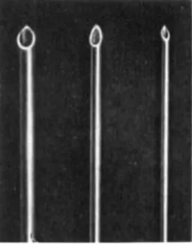
Fig. 8. Saturiil size. The middle needle permits, with the technique described, the insertion of a catheter (in this case No. 203) which requires R needle of the size of that pictured on the Icft, were it to be passed through the lumen of the needle. The same relative advantage exists between the right
:tilt1 niitldle needles.
insert the catheter without it, and the artery wall sometimes contracted around it during its renioval. Furthermore, it was realized that it might damage the arterial wall
as happened in one of the subclal ian arteriographies, so t h a t part of contrast medium was injected extmvascularly.
As regards complications any tendency t o bleed a t the site of puncture was unini- portant and was mostly observed in elderly patients. No haematoma of clinical conse- quence e\ er formed. No thrombosis or any kind of circulatory disturbance in the region of the artery punctured was observed. There was no case of extravascular injection except the one previously mentioned. In the unsuccessful attempts a t catheterization. the leader probably passed through the posterior wall of the artery or its intima via a hole made during puncture. In these cases the needle could not be pushed far enough u p into the artery. In neither did the patient suffer any ill effects. No kinking or rupture of the catheter, or arterial spasin around i t occurred. After local anaesthesia, the patients felt nothing during the manipulations and following the injection of the contrast medium thrre was, with intlividual variations, only the wellknown, rapidly passing discomfort.
In one case the patient was operated o n two weeks after bilateral femoral cathe- terization and both arteries were exposed. Traces of blood under the fascia intlicated the situation, but the exact site of puncture could not be discerned.
Figs. 3-7 form representative illustrations.
Discussion
The advantage of the author’s method of percutaneous cstheteriza-
tion is the smaller size of needle required for
a
given catheter.
As
the
catheter needs
a
certain clearance to enable it t o glide through the bore
of
a needle, the difference is more marked than would appear from the
thickness of the material (Fig.
8).In
other words,
a
larger catheter can be inserted by the same sized
needle.
POISEUILLE’S
law states t h a t when pressure and viscosity are
con-
stant, the rate of flow through narrow tubes is:
inversely proportional t o the length of the tube, and
directly proportional t o the 4th power of the radius of the tube.
This shows the dominant influence of the cross section of the catheter.
Catheter No.
205,
used
here for abdominal aortography, has a n inner
diameter, corresponding t o
a
heart catheter
No.
9-10.
If
a pressure
Downloaded By: [Nyman, Ulf] At: 20:30 2 April 2008
3 T G SVEN I V A R PELDINOER
apparatus were used, the gauge might doubtlessly be diminished con-
siderably,
i.
e. to a size No.
160,
corresponding
t o a
heart catheter No.
8,
and which may be inserted with the help of
a
needle,
1 . 5mm in external
diameter.
Though the extra manipulation with the leader is a disadvantage,
it is very quickly performed. Furthermore, there is
a
little risk that the
leader, when handled unskilfully, will pass through the posterior
wall of
the artery, although,
no
doubt, experience and improved equipment
will eliminate this possible complication and avoid failure.
S U M M A R Y
The author describes a method by which i t is possible, after percutaneous puncture,
to insert a catheter of the same size as the needle used into an artery.
Z U S A M M E N F
.
i
4
S S U N G
Der Verf. beschreibt eine Methode, die es ermoglicht, nach perkutaner Punktion rinen Katheter von derselben Grosse wie die benutzte Nadel in eine Arterie einzufiihren.
R E S U M E
L’auteur decrit une methode qui permet, a p r b ponction percutanee, d’introduire dans une a r t b e un catheter de m&me calibre que l’aiguille utilisde.
L I T E R A T U R E
DONALD, D. C., KESMODEL, K. F., ROLLINS, S. L. and PADDISON, R. M.: An improved
technic for percutaneous cerebral angiography. Arch. Neurol. and Psych. 65 (1951), 508.
FARIGAS, P. L.: A new technique for the arteriographic examination of the abdominal
aorta and its branches. Am. J. Roentgenol. 46 (194l), 641.
JONSSON, G.: Thoracic aortography by means of a cannula inserted percutaneously into the common carotid artery. Acta radiol. 31 (1949), 376.
PEIRCE, E. C.: Percutaneous femoral artery catheterization in man with special reference to aortography. Surg., Gynec. & Obst. 93 (1951), 56.
RADNER, S.: Intracranial angiography via the vertebral artery. Preliminary report of a
new technique. Acta radiol. 28 (1947), 838.