Stem Cell Research
5
0
0
Texte intégral
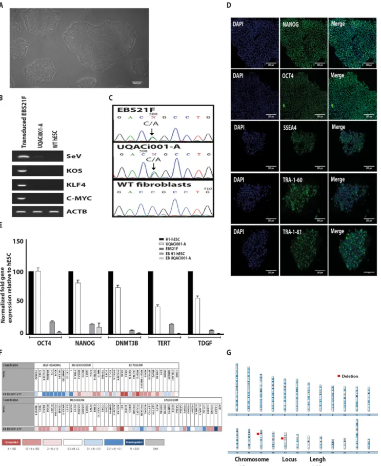
Figure
Documents relatifs