HAL Id: tel-01684253
https://tel.archives-ouvertes.fr/tel-01684253
Submitted on 15 Jan 2018
HAL is a multi-disciplinary open access archive for the deposit and dissemination of sci-entific research documents, whether they are pub-lished or not. The documents may come from teaching and research institutions in France or abroad, or from public or private research centers.
L’archive ouverte pluridisciplinaire HAL, est destinée au dépôt et à la diffusion de documents scientifiques de niveau recherche, publiés ou non, émanant des établissements d’enseignement et de recherche français ou étrangers, des laboratoires publics ou privés.
Generating a new vaccine for protecting poultry from
Newcastle disease and controlling viral shedding
Haijin Liu
To cite this version:
Haijin Liu. Generating a new vaccine for protecting poultry from Newcastle disease and controlling viral shedding. Veterinary medicine and animal Health. Université Montpellier, 2017. English. �NNT : 2017MONTT025�. �tel-01684253�
THÈSE POUR OBTENIR LE GRADE DE DOCTEUR
DE L’UNIVERSITÉ DE MONTPELLIER
En: Biologie-Santé
École doctorale: Sciences Chimiques et Biologiques pour la Santé] Unité de recherche: ASTRE, CIRAD
Présentée par HAIJIN LIU
Le 28 Septembre 2017
Sous la direction de Emmanuel AIbina
Devant le jury composé de
Emmanuel Albina, Docteur, CIRAD Nicolas Eterradossi, Docteur, ANSES
Natàlia Majo Masferrer, Docteur, IRTA CRESA Barcelone Stéphane Bertagnoli, Professeur, Ecole Vétérinaire de Toulouse Yannick Simonin, Docteur, CNRS-Université de Montpellier
Directeur de thèse Rapporteur Rapporteur
Président; Examinateur Examinateur
Génération d’un nouveau vaccin pour protéger les volailles contre
la maladie de Newcastle et l’excrétion virale
(Generating a new vaccine for protecting poultry from Newcastle
disease and controlling viral shedding)
1
Acknowledgement
I very appreciate Chinese government who offers me four years’ scholarship to study in France and Univeristy of Montpellier, CBS2 and CIRAD who provide a great platform for me to open horizons, improve skills and accomplish the thesis.
During processes of my thesis, there are many persons in my unit (Cirad, UMR ASTRE) giving me a help hand. Firstly, I want to thank my supervisor, Emmanuel Albina who spends so many time to guide me, to find funding and cooperation for animal experiments of my thesis, to assist me in thesis writing and publication. I cannot forget you decided to be my supervisor four years ago, which makes my dream come true to study in abroad. Secondly, I am very appreciative for Renata Servan de Almeida. I cannot speak French. Without you, I cannot image how my life will be like in Cirad. Thanks for your patience, your help in registration, suggestion and modification in paper and thesis writing. Thirdly, I deeply appreciate Patricia Gil for your help in doing experiments and advice in writing. You are the first person I met in France. I was moved by your warm welcome. You invited me to your home to celebrate Christmas, which makes me not fell alone in Montpellier. Last but not least, Cécile, Genevieve, Lucia, Valérie, Aurélie, Sylvain, Nadège, Denise, Tiffany, Serafin, Olivier, Philippe and other members of ASTRE unit, please receive my sincere acknowledgement. Without your help, it is impossible for me to finish my thesis so smooth.
I also want to thank persons from other units who give me help in the thesis. Firstly, I quite appreciate Natàlia Majo Masferrer who helps me a lot to do animal experiments, to review my manuscript of thesis and to be one of members of jury. Secondly, my great thanks belong to Patti J. Miller and Bénédicte Lambrecht who kindly afford viruses for my thesis. Furthermore, it is my honor that Nicolas Eterradossi, Yannick Simonin and Stéphane Bertagnoli are members of my jury.
My parents are quite important for me during my study. They don’t be educated in university and even in high school, but they totally believe education is necessary for a person. Thus, they are always doing their best to encourage me to get bachelor and master degrees. Without their support, I don’t think I have chance to pursue PhD degree.
My wife is another quite important person for me. She is like the lighthouse to light roads for my study career. She always encourages me to insist when I want to give up. I never forget
2
she got up so early to practice English with me. In recent 4 years, I spend so little time with her. However, she never complains and always supports me to get PhD degree.
Finally, I also want to thank my parents in law, my little brother and my friends. Without you help, I cannot focus on my PhD study.
3
Contents
Acknowledgements…...1
Summary in French... 4
Introduction………...14
Chapter 1………47
Development and Efficiency Comparison of Newcastle Disease Virus
Reverse Genetics Systems with Different Number of Plasmids………47
Chapter 2………61
Two-Plasmid System to Increase the Rescue Efficiency of Paramyxoviruses
by Reverse Genetics: the example of rescuing Newcastle Disease Virus….61
Chapter 3………82
Newcastle disease virus uses cell-to-cell transmission to maintain persistent
infection, resist superinfection exclusion and enhance co-infection………82
Chapter 4………..114
Generation of a Recombinant Attenuated Vaccine to Protect Chickens from
Newcastle Disease and Block Viral Shedding………...114
General discussion………...134
4
Génération d’un nouveau vaccin pour protéger les volailles contre la
maladie de Newcastle et l’excrétion virale
RESUME EN FRANÇAIS
Par
LIU Haijin
La maladie de Newcastle est, avec l’influenza aviaire, une des deux pestes aviaires impactant fortement la santé des oiseaux d’élevage. Au-delà de l’impact sanitaire, ce sont les conséquences liées aux restrictions des mouvements d’animaux et aux mesures de contrôle de l’infection qui désorganisent les filières et fragilisent l’équilibre économique des éleveurs et des pays, en particulier au sud. La maladie de Newcastle est très largement répandue à l’échelle du globe et si elle semble relativement bien contrôlée au nord par l’association de vaccinations ciblées de populations d’oiseaux domestiques à risques (exemple des pigeons d’élevage en France) et de la surveillance événementielle, elle reste une des premières causes de mortalité dans les élevages au sud, en dépit d’un large usage de vaccins.
La maladie est provoquée par un virus enveloppé à ARN négatif d’environ 80-100 nm, appartenant au genre Avulavirus dans la famille Paramyxoviridae. Le génome viral d’un peu plus de 15 kilobases, code pour 6 protéines structurales (Figure 1). Trois, la nucléoprotéine (N), la phosphoprotéine (P) et la large protéine (L), entrent dans la composition du complexe ribonucléoprotéique (RNP) qui est la structure minimale du virus permettant la réplication du génome et la synthèse des protéines virales. C’est cette RNP qui doit être reconstituée dans le cytoplasme de la cellule pour générer des virus modifiés par génétique inverse. La protéine de membrane (M) sert à la formation de la particule virale par son interaction avec la membrane cellulaire et les protéines de surface du virion. Les protéines de surface, l’hémagglutinine (HN) et la protéine de fusion (F), servent à l’interaction avec un récepteur cellulaire et la fusion des
5
membranes virales et cellulaires pour la libération du génome viral dans le cytoplasme cellulaire.
Figure 1. Représentation schématique de la structure et de la composition du virus de la maladie de Newcastle.
Le génome de 15 186 à 15 198 bases selon les virus, code pour 6 protéines structurales et deux protéines non structurales
Il existe une grande variabilité de pouvoir pathogène chez les oiseaux, liée aux propriétés intrinsèques du virus. Si les déterminants de la virulence semblent multiples et répartis sur l’ensemble du génome, il existe toutefois un mécanisme bien décrit qui conditionne à lui seul le pouvoir pathogène du virus. Il s’agit du clivage de la protéine de fusion. Dans son état natif, la protéine F est un monomère F0 inactif qui pour devenir actif doit être clivé en deux sous-unités F1 et F2 liées par un pont difulfide. Cette activation est réalisée par les enzymes cellulaires qui reconnaissent un motif situé en positions 112-117. Le type d’enzymes capables de faire ce clivage dépend du motif présent. Plus le motif contient des acides aminés basiques pus il est facilement clivé par des enzymes ubiquitaires de type furine. Inversement, les motifs avec peu d’acides aminés basiques sont essentiellement clivés par des enzymes de type trypsine. Or, chez l’animal, ces dernières sont abondantes au niveau des muqueuses alors que les furines sont répandues en profondeur dans les organes et tissus : ce qui explique que les virus à motif basique, très virulents ou dits vélogènes, ont la capacité à envahir les tissus et produire une maladie sévère alors que les virus à motif moins basique, atténués ou lentogènes, seront moins invasifs.
6
A ce jour, il n’existe qu’un seul sérotype du virus de la maladie de Newcastle. Autrement dit, un animal protégé contre un virus l’est contre tous les autres. Cependant, il existe une grande variabilité génétique parmi les virus Newcastle, avec près d’une vingtaine de génotypes décrits (Figure 2). Tous les vaccins courants sont dérivés de génotypes anciens (I, II, III ou IV), identifiés il y a près de 70 ans. Pour autant, la dérive génétique accumulée depuis lors amène à des taux de substitution en nucléotides par site de l’ordre de 20% entre les protéines F des anciens et des nouveaux génotypes. Un tel niveau de variation peut interroger en terme d’efficacité des vaccins basés sur des anciens génotypes pour lutter contre les souches virulentes circulant actuellement. Cette interrogation a été le point de départ de notre travail de thèse. Fin des années 2000, le CIRAD a reçu des informations d’Afrique de l’ouest et de Madagascar tendant à accréditer l’idée que les vaccins actuels ne seraient que partiellement efficaces dans les élevages faisant l’objet d’une vaccination bien conduite. Cette perte d’efficacité se manifestait par l’apparition de signes atténués de maladie de Newcastle chez des volailles régulièrement vaccinées. Pour confirmer que cette observation pouvait être réellement attribuée à une perte d’efficacité du vaccin, le CIRAD en collaboration avec l’ANSES a mis en œuvre un essai expérimental sous condition contrôlée visant à évaluer l’efficacité d’un vaccin actuel contre un nouveau génotype virulent isolé à Madagascar en 2008.
7
Figure 2. Arbre phylogénétique de virus Newcastle établi sur la base de la séquence complète du gène F. Les
virus responsables de la maladie de Newcastle sont principalement regroupés dans la classe II. Les vaccins actuels sont à base de souches dérivées des anciens génotypes I, II, III et IV.
8
L’essai réalisé à l’ANSES a permis de vérifier que le vaccin protégeait toujours cliniquement les animaux mais qu’en revanche il contrôlait beaucoup moins bien la réplication et la ré-excrétion du virus malgache (Figure 3).
Figure 3. Des poulets vaccinés par voie oculonasale et intramusculaire avec deux vaccins commerciaux à base
de souches de génotype II ont été éprouvés soit avec une souche virulente ancienne de génotype IV (Herts), soit avec une souche virulente récente de génotype XI, isolée en collaboration par le CIRAD et le FOFIFA-DRZV. Dans le premier cas, aucune excrétion virale n’est observée après épreuve avec la souche Herts. En revanche, la souche malgache de génotype XI est excrétée plusieurs jours par les animaux vaccinés alors qu’ils ne présentent aucune symptomatologie.
L’objectif initial de cette thèse était de produire un vaccin vivant modifié permettant de contrôler l’excrétion virale de plusieurs génotypes virulents, anciens et récents. Pour ce faire, nous sommes partis d’un vaccin vivant déjà connu et très largement répandu à l’échelle du globe : le vaccin à base de la souche LaSota de génotype II. Puis nous avons envisagé de modifier les antigènes immunoprotecteurs HN et F de cette souche par des antigènes provenant d’une souche plus récente. Notre choix s’est porté sur une souche de Madagascar isolée en 2008 par notre groupe. Cette souche est particulière dans le sens où elle est issue d’un génotype IV ancien et porte des motifs antigéniques sur HN et F, intermédiaires entre génotypes anciens et nouveaux (Maminiaina et al., 2010). Elle présentait donc à nos yeux un profil intéressant pour l’objectif visé.
MG-725/08
9
Dans un premier temps, nous avons adopté la technique de génétique inverse pour générer toute une série de virus modifiés. Cette technique est classiquement basée sur l’utilisation de 4 plasmides permettant de reconstituer une RNP virale dans une cellule sensible à l’infection. Un des plasmides comprends le génome complet du virus. Les trois autres plasmides fournissent la N, la P et la L. Utilisant cette technologie classique, nous n’avons eu aucun problème à générer des virus virulents. En revanche, nous avons été confrontés à des échecs pour générer certains virus atténués modifiés. Pour contourner cet obstacle, nous avons dû innover et développer un système plus efficace. Nous avons développé un système de génétique inverse basé sur 2 plasmides : un contenant le génome complet et un second contenant les trois cassettes exprimant N, P et L. Nous démontrons que ce système est plus efficace que le système conventionnel pour générer des virus atténués (Figure 4, Liu et al., 2017)
Figure 4. Efficacité comparative du système conventionnel de génétique inverse et de celui mis au point dans le
cadre de cette thèse sur neuf virus modifiés (reproduction du tableau 5 de l’article Liu et al., 2017)
Par ailleurs, nous avons comparé différentes associations à 4, 3, 2 ou 1 plasmides en termes d’efficacité en génétique inverse. Nous montrons que c’est le système à 2 plasmides qui est le plus efficace (Figure 5)
10
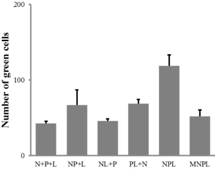
Figure 5. Efficacité comparée de différentes associations de plasmides sur l’expression d’un minigénome EGFP.
N+P+L = 4 plasmides (minigénome, N, P et L) ; NP+L = 3 plasmides (minigénome, NP, L) ; NL+P = 3 plasmides (minigénome, NL, P) ; PL+N = 3 plasmides (minigénome, PL, N) ; NPL = 2 plasmides (minigénome, NPL) ; MNPL = 1 plasmide (minigénome-NPL). Toutes les associations testées font au moins jeu égal avec le système conventionnel à 4 plasmides. Toutefois, c’est le système à 2 plasmides qui détient le meilleur rendement.
Dans un second temps, nous nous sommes intéressés à étudier les propriétés des virus modifiés produits. En particulier, nous avons évalué la capacité de ces virus à établir des infections persistantes in vitro et à générer des co-infections qui pourraient dans l’absolu conduire à des événements de recombinaison indésirables notamment entre souches vaccinales et souches virulentes. Pour faciliter ces études, certains de nos virus d’intérêts ont été équipés d’une cassette d’expression d’un marqueur fluorescent, soit EGFP, soit mCherry. Nous montrons que tous les virus vélogènes ou lentogènes sont capables d’établir des infections persistantes en culture cellulaire in vitro. En revanche, une cellule infectée par un premier virus ne peut être infectée directement par une seconde particule virale, probablement parce que les récepteurs membranaires sont indisponibles : il s’agit d’un mécanisme d’exclusion de la surinfection. Toutefois, nous démontrons que les virus établis de façon persistante sont capables de gagner une autre cellule, infectée ou non, en guidant la formation de connexions cytoplasmiques parfois de longue dimension. Par ce mécanisme, la co-infection devient alors possible (Figure 6).
11
Figure 6. Des cellules BHK21 ont été co-infectées par un virus vélogène fluorescent vert (EGFP) et par un virus
lentogène fluorescent rouge (Cherry). Alors qu’une surinfection directe d’une cellule déjà infectée n’est pas possible, nous observons ici qu’une co-infection a été possible entre deux cellules et que celle-ci s’est opérée par la formation d’une connexion entre cellules à longue distance permettant le trafic des deux virus (fluorescence verte et rouge).
Ces observations permettent d’expliquer pourquoi les analyses de séquences d’isolats de terrain identifient régulièrement des événements de recombinaison entre souches virulentes et souches vaccinales. Notre hypothèse est que par le biais d’infections persistantes chez l’animal, des surinfections par ce mécanisme de connexions intercellulaire distantes pourraient créer les conditions de recombinaisons entre virus.
Dans la dernière partie de ce travail de thèse, nous nous sommes attachés à vérifier si notre prototype vaccinal était capable de contrôler la ré-excrétion après épreuve infectieuse. Le vaccin correspond à la souche LaSota dans laquelle les gènes HN et F ont été remplacés par ceux de la souche Madagascar MG-725 : il s’agit donc d’une souche hybride génotype II – génotype XI. Le gène F de la souche Madagascar ayant à l’origine un motif de site de clivage vélogène, nous l’avons modifié en motif lentogène avant insertion dans le vaccin. Dans un essai en condition contrôlée, des poulets ont été vaccinés soit avec la souche LaSota conventionnelle soit avec notre prototype vaccinal recombinant entre LaSota et MG-725 (rLaSota/M-Fmu-HN). Trois semaines après vaccination, les animaux ont été éprouvés soit avec une souche virulente de génotype II, soit la souche de génotype XI MG-725, soit une souche virulente de génotype VII. Nous montrons que les deux vaccins confèrent le même niveau de protection clinique contre les trois souches virulentes. En revanche, le prototype vaccinal rLaSota/M-Fmu-HN contrôle mieux la réplication des trois souches virulentes. En
12
effet, nous observons sur des poulets sacrifiés trois jours après épreuve que les poulets vaccinés LaSota présentent plus de virus génotype II et génotype XI dans le système respiratoire que des poulets vaccinés rLaSota/M-Fmu-HN (Figure 7). Par ailleurs, le vaccin LaSota a empêché l’excrétion virale dans tous les écouvillons trachéaux et cloacaux de poulets à 3, 5, 7 et 10 jours après infection par la souche de génotype VII. Aucun écouvillon n’a été trouvé positif chez les poulets vaccinés rLaSota/M-Fmu-HN quelle que soit la souche d’épreuve.
Figure 7. Recherche de virus dans les prélèvements de poulets vaccinés par LaSota, rLaSota/M-Fmu-HN ou non
vaccinés et érpovés trois semaines plus tard par un virus de génotype XI (graphe de gauche), de génotype II (graphe du milieu) ou de génotype VII (graphe de droite). On observe plus de prélèvements positifs chez les poulets vaccinés avec LaSota. A noter, qu’aucune présence de virus n’est observée avec l’un ou l’autre des vaccins après épreuve infectieuse avec le génotype VII, génotype actuellement dominant, en particulier en Asie.
Conclusion :
Dans ce travail de thèse, nous proposons un système de génétique inverse du virus de la maladie de Newcastle optimisé pour générer des virus atténués modifiés. Nous pensons que ce système peut également améliorer la génétique inverse d’autres virus à ARN négatif. Grâce à ce système nous avons pu générer divers virus atténués dont certains équipés d’un traceur fluorescent afin de caractériser le comportement de ces virus en culture cellulaire in vitro. Après avoir vérifié que le mécanisme d’exclusion de la surinfection existe bien chez le virus Newcastle, nous identifions une propriété originale de ce virus consistant à diffuser de cellule en cellule par l’établissement de connexions membranaires de longue distance. Par ces connexions, le virus est capable de surinfecter une cellule infectée de manière persistante par un autre virus, ouvrant la voie à de possibles évènements de recombinaison. Un candidat vaccin comprenant l’essentiel du génome de la souche LaSota mais les gènes HN et F d’une
13
souche malgache a été testée in vivo contre trois souches virulentes de génotype II, VII et XI. Cette souche s’est montrée moins réplicative que la souche LaSota d’origine mais tout autant protectrice. Ces propriétés en font un prototype vaccinal pertinent pour les animaux vaccinés et l’environnement.
14
15
Fig. 1.1. Clinical signs and haemorrhagic lesions in chickens infected by virulent NDV. a) Infected chickens;
b) Feces; c) Eyelid; d) Thymus; e) Proventricular glands; f) Spleen; g) Cloacal bursa; h)Intestine (5).
1.
Newcastle disease
Newcastle disease (ND) is caused by velogenic strains of Newcastle disease virus (NDV) and has large economic impacts in the poultry industry due to its high pathogenicity and animal movement bans imposed for the control of the infection (1-3). Chickens infected by virulent NDV can show serious clinical signs like diarrhea, nervous and respiratory illnesses, egg laying reduction, generation of deformed eggs and mortality (4-6). NDV also causes haemorrhagic lesions in the trachea, intestine and proventriculus (5, 7-12).
Newcastle disease was first described in 1926, in Indonesia and one year later in England in Newcastle and then gradually spreads worldwide through bird trade, movement of human and wild bird (13-15). However, an outbreak of this disease was probably observed in 1897 based on Macpherson’s report and this date of first occurrence is more in agreement with the results of molecular dating performed by Yee et al., estimating the time to the most recent common ancestor (TMRCA) of NDV between 1868-1891 (13, 16). Since its description, three main ND panzootics have successively occurred in the word. The first one started in 1926 and vanished in 1960 (1). The second one lasted from 1970 to 1974 and the third one was during 1980s (1). ND outbreaks have been confirmed in many countries up to now (17).
16 2. Newcastle disease virus (NDV)
Birds are the main hosts of NDV with at least 234 susceptible species described (18), but the virus can also infrequently be isolated from other animals like mink and pig (19, 20). NDV belongs to the Avulavirus genus in the Paramyxoviridae family (21). The virion of NDV is enveloped and pleiomorphic, ranging from 100 nm to 500 nm in diameter (22, 23). The size of viral particle relates to copies of the genome packaged into the particles. Usually, each NDV virion only wraps one genome, but some particles can tolerate more than one copy of the genome (24, 25).
The genome of NDV is composed by a non-segmented RNA, negative sense with 15,186, 15,192 or 15198 bases (26-28). The size of the genome of NDV respects the rule of 6: the number of nucleotides in the genome has to be divided by 6 as a consequence of the stochiometric interaction between the viral nucleoprotein and the genome (29).
The genome consists of six gene segments (3’-NP-P-M-F-HN-L-5’) and inserted between the Leader and Trailer regions at the 3’ and 5’ terminus which act as promoters for RNA transcription and replication. Each gene unit, surrounded by a gene start (GS, 3’- UGCCCAUCUU-5’) and gene end (GE, 3’- AAUCUUUUUU-5’) regions, is separated by inter-genic residues (30, 31). These genes encode six structural proteins – Nucleocapsid (NP)
17
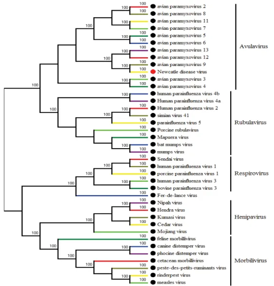
Fig. 1.3. Phylogenetic tree of paramyxovirus was built by MEGA6 based on L genes of differnet
virus from paramyxovirus family.
protein, Phosphoprotein (P), Matrix (M) protein, Fusion (F) protein, Hemagglutinin-Neuraminidase (HN) protein, Large (L) protein, and two nonstructural proteins, V and W due to P gene editing (32, 33). NP protein encapsidates viral RNA to be a template for transcription and replication by RNA-dependent RNA polymerase (RdRP) consisting of P and L protein. The complex containing viral RNA, NP, P and L proteins is named ribonucleoprotein complex (RNP) (34-36). Through the direct interactions with RNP and HN, M protein plays a pivotal role in the viral assembly (37, 38). On the surface of viral envelope, F and HN proteins are in charge of the virus-receptor interactions and virus entry (39-41). Both F and HN are glycoproteins that cause immunity reaction of hosts, notably to generate NDV neutralizing antibodies (42-46).
18 3. Newcastle disease virus life cycle.
NDV life cycle processes start at receptor binding and end at viral budding (15, 47). Fig. 1.4. Virions of NDV and its genome structure (Viral zone: http://viralzone.expasy.org/84?outline=all_by_protein).
19
The NDV HN protein interacts with the cell receptor. HN protein is divided into three regions based on its structure-one is N-terminal transmembrane domain, another is stalk and the last one is C-terminal head (41, 48). On the head of HN, there are two receptor binding sites that recognize sialic acids on the cellular membrane (49-51). After binding to its receptor, HN initiates a series of changes from “Head-down” to “Head-up” conformation followed by the exposure of the F protein stalk region which contains the active fusion sites (52-55). When activated, the F protein insert its fusion peptide into the cell membrane and also start conformational changes from a pre-fusion to post-fusion structure, which close membranes of cell and virus and then fuse them in a pH-dependent manner (56-58).
The RNP is released into cytoplasm when fusion is achieved (56). After releasing, RdRp complex uses encapsidated RNA as the template to transcribe viral RNA in a “stop-start” mode to produce viral proteins from NP to L into the cytoplasm according to a decreasing gradient (59-65). P proteinˈas chaperone, interacts with NP to prevent its oligomerization until the virus genome RNA is encapsidated (66, 67). After post-translational modifications, M protein is transported to the inner layer of cell membraneˈwhile F and HN proteins display on the cell surface (37, 68). RdRp changes its role from transcription to generate viral negative RNA using the positive RNA strand as template, followed by viral RNA and NP protein encapsidation (61, 62). Finally, RNP complexes move to the plasma membrane to be assembled with the other viral proteins under the guidance of M for viral budding (15, 47, 68). Fig. 1.6. HN protein conformation before and after receptor recognition (54).
20 4. Newcastle disease virus virulence
Based on mortality of chicken embryo and clinical signs observed in chickens, NDV is divided into three pathotypes: velogenic, mesogenic (pathogenic strains) and lentogenic (apathogenic strain). Usually, the mean death time (MDT) of embryonated chicken eggs and the intracerebral pathogenicity index (ICPI) in one-day-old chickens are used to assess NDV virulence. Velogenic strains have MDT lower than 60 hours (<60 h) and ICPI around 2.0, while MDT is beyond 90 hours (>90h) and IPCI approaches 0.0 for lentogenic strains (69). The cleavage site of F protein plays a major role for the NDV virulence (31, 70, 71). F protein is synthesized and glycosylated in the endoplasmic reticulum as an inactive F0 precursor shifted to an active F protein after cleavage by cellular proteases resulting into a disulfide-linked F1 and F2 sub-units. F1 and F2 are necessary for viral infection (72, 73). The lentogenic strains usually own a cleavage motif with dual-basic amino acids which can be only cleaved by trypsin-like proteases located in the respiratory and intestinal tract. In contrast, the cleavage site of velogenic strains has multi-basic amino acids which are recognized by ubiquitous furin-like proteases (74). Consequently, virulent strains are fatal for chickens due
21
to systemic infection. Modifications of the cleavage motif of velogenic viruses to that of lentogenic strains significantly attenuate the virus virulence (31). In addition, the replication of lentogenic viruses in chickens, often leads to the emergence of velogenic variants by the accumulation of multi-basic amino-acids in the cleavage site (75-77).
Despite the fact that the F protein cleavage site is considered as the primary determinant of NDV virulence, significant differences in virulence can exist between strains having the same velogenic F cleavage site. In addition, some NDV strain from pigeon or dove (PPMV-1) with muti-basic amino acids can be lentogenic on chickens (78-80). These observations indicate that other factors can be also involved in NDV virulence (81). Replacement of whole F protein of a mesogenic strain by that of a velogenic PPMV-1 strain decrease ICPI from 1.36 to 0.60, even though both F proteins contain polybasic amino acids (82). Exchange or modification of NP, P, L, M or HN proteins between velogenic and lentogenic strains can decrease or increase the pathogenicity of viruses (33-36, 83-88). V protein antagonizes the host interferon response induced by viral infection. Consequently, the inactivation of the expression of V protein decreases the viral virulence while the restoration of its expression results in reversion to virulence (89-92). Interestingly, non-coding inter-genic regions of viral genome can also affect viral pathogenicity (28, 93-95).
5. Newcastle disease virus transmission
22 5.1 Transmission between hosts
Since infected birds excrete NDV in feces and oculo-nasal secretions, NDV transmits mainly horizontally by direct contact between infected and uninfected birds within the same flock (7, 96-98). Furthermore, NDV can transmit to offspring via chicken embryos and is able to transmit from parental to descendant cells in persistently infected (PI) cells (99-103). Even if NDV easily loses its infectivity under exposure to high temperatures and irradiation of sunlight, the high virus load and relative persistence of the virus in birds’ bodily discharges, feathers and other materials can be source of distant spread of the infection by human activities between different flocks (98, 104-107). Contacts between wild and yard birds is another route for viral transmission (108, 109). Some infected exotic birds shed NDV without clinical symptoms and spread virus to other birds located in remote areas (110, 111).
5.2 Transmission between cells
Virus can spreads among cells though two ways. One is the cell-free virus-cell route that requires an initial complete virus life cycle releasing progeny viruses into the cell-medium capable of infecting new susceptible cells. The second transmission route is a direct cell-cell spread without the release of virus particles into the culture medium (112-114). Since cultured cells lack trypsin-like proteases, the F protein cleavage motif with dual-basic amino acids cannot be activated. Consequently, lentogenic strains transmit and amplify much lower than velogenic strains in cultured cells (31, 115). Cells infected by virulent strains release a high number of viral particles outside the cells (115). These suggest that NDV, in favorable conditions, spreads primarily through the route of cell-free virus-cell among cells.
NDV can also take a cell-cell route for spreading, notably through the constitution of cell syncytia (116). Syncytia are resulting from the virus infection that induces the fusion of different cells and forms giant cells containing several nucleuses (55). The formation of syncytium requires the activation of F protein and allows virus transmission to surrounding connecting cells (117).
In addition to syncytia, some viruses use cell connections to spread from one cell to another one (113). These cell connections can be preexisting cell appendices like for instance the axons and neurological synapses used by the measles virus to transmit between neuronal cells (118, 119). In other cases, viral proteins are required to shape cell connections for viral spread. The murine leukemia virus, for example, expresses viral envelope protein to build cell-cell
23
contact for virus transmission (120). Furthermore, due to actin rearrangement, cells can be induced in the production of cell extensions after infection by some viruses, such as respiratory syncytial virus, human metapneumovirus, influenza virus and parainfluenza virus 5 (121-123). However, for NDV and at the beginning of this work, it was still unclear whether the virus could spread via a direct cell-to-cell route.
Fig. 1.10. Model for virus transmission among cells via the cell-to-cell route (121).
Fig. 1.9. Syncytium formed on BHK-21 cells persistently infected with the recombinant NDV – MG_725
24
5.3 Assets of the cell-cell transmission route for the virus.
Since the cell-cell transmission can maintain an infection without the release of viral particles, it assists the virus in escaping the host immune response based on neutralizing antibodies (124). In addition, the cell-cell route can allow the transmission of incomplete viruses, thus saving the time and costs for a full virus cycle to release free particles (125).
Another possibly interesting feature for the virus is the capacity of the cell-cell transmission to circumvent the superinfection exclusion and promote co-infection and evolution by recombination events. Indeed, recombination events have been detected by sequence comparisons in NDV (13, 126). Whether this is resulting from a natural biological process remains to be established and at least requires that the two parental viruses infect the same cell at the same time. However, co-infection is inhibited by the superinfection exclusion due to receptor destroyed or taken up by the first virus infection (127, 128). Since virus can be directly transmitted from infected cells to others though the cell-cell route, virus infection bypasses the step of receptor interaction. Thus, super-infection or co-infection could occur under the condition of cell-cell transmission.
Furthermore, birds can shed NDV for a long time (sometimes up to 3 weeks) after initial infection, even if the host has generated NDV neutralizing antibodies against viral re-infection (7, 129). NDV strains antigenically distinct from the first infecting virus can still successfully infect and replicate in chickens without inducing morbidity (130, 131). If the second strain can spread among cells of the immunized chicken via the cell-cell route, it may encounter a cell already infected by the first virus and then create a condition which may allow NDV recombination.
6. Genotyping and evolution of Newcastle disease viruses.
Based on sequence analyses of the F gene (position 47 to 421), NDV strains have been split into two clades - Class I and II (1, 132). Class I strains are mostly lentogenic viruses isolated from water birds, while Class II strains consists of viruses of different virulence, affecting different avian hosts and being responsible for the ND outbreaks (133). Although the purifying selection has been considered as the main driving force in the evolution of NDV, the evolutionary rate of Class II virus genome is around 10-3 (per site per year) that is similar to other RNA viruses (13, 132). Under this evolution rate, Class II NDV strains have involved into multiple genotypes in the last 100 years. The current genotyping is based on the F gene sequences (134).
25
From 1920s to 1960s, only genotypes I-IV (considered as the old genotypes) and IX were identified and virulent strains from these genotypes were responsible for the first ND panzootics (1). Since 1960s, genotypes V- XVIII (recent genotypes) have been isolated (133). Genotypes V and VI caused the second and the third panzootics, respectively (135).
26
Nowadays, most ND outbreaks, particularly in Asia, are due to genotype VII (133). Besides differences in the F gene sequences, an obvious difference between old and recent genotypes lays in the genome size. The genome of old genotypes has 15,186 bases whereas new genotypes have an insertion of 6 nucleotides into the 5’ un-translated region of NP gene (136). Some recent isolates of genotype VII strains also show another insertion of a stretch of 6 nucleotides into the inter-genic region between HN and L (26).
Among NDV strains, the genotype XI was recently identified in Madagascar Island. This genotype is interesting since it is present only in Madagascar, probably at least from 1992 and is currently responsible for most ND outbreaks in the island even though genotype VII is also present (136). Based on phylogenetic and evolutionary analyses, genotype XI is very distant from currently circulating genotypes and is presumed to have emerged from the old genotype IV that has now vanished from the planet (136). Furthermore, genotype XI viruses has unique amino acid motifs exposed at the globular head of the F and HN proteins, which suggests this genotype has evolved in a specific direction under the pressure of the host immune system in a context of vaccination in domestic poultry with attenuated old-genotype strains (136, 137). 7.Newcastle disease vaccines
Vaccination is currently the most effective mean to control ND (138). Currently, live and inactivated ND vaccines are used around the world. Inactivated vaccines are safe for the animals and environment, but the administration is not suitable for large flocks since it requires intramuscular injections with adjuvant and the cell immunogenicity is weak (139). Live vaccines are much more efficient since they can be easily administrated though spray and drinking water, and also induce stronger immune responses (17). Even if they are produced from highly attenuated strains, they still represent a risk of release into the environment (140). However, live vaccines are widely applied in commercial poultry farms. The first live ND vaccine was licensed in 1950s. Currently, ND vaccines are based on lentogenic viruses from genotype I (V4 and Ulster strains) and also genotype II (LaSota, B1, VG/GA and Clone 30 strains) One live vaccine, was generated from the mesogenic Mukteswar strain (genotype III) but it still has a residual risk in younger birds, causing morbidity, and is only recommended in chickens of at least four-weeks of age. Even if live vaccines contribute a lot to prevent NDV, there are still many ND outbreaks around the world, suggesting that ND vaccination and/or vaccine efficacy still needs to be improved (17).
27 7.1 Increasing vaccine’s thermal stability.
One of the reasons why the ND vaccination may partly fail in the field could be related to the thermal susceptibility of the attenuated strains and the difficulty to maintain a cold chain in tropical and sub-tropical countries. For these reasons, the thermostability of NDV vaccine strains has been improved and then used in licensed live vaccines (141). Derived from the parental V4 strain, the I-2 strain has a better thermostability and is now licensed in Australia (142, 143). Other thermostable NDV strains have been identified showing stable infective titers after heating at 56 ć for one hour (144). Furthermore, one thermostable strain has been proved to 100% protect chicken from virulent challenge (145). In addition, replacing the HN gene of thermolabile Lasota strain by the one of thermostable TS09-C strain (derived from passaging V4 strain in BHK-21 cells at high temperature) increased the virus thermal stability with virus activity maintained up to 16 days at 30ć (146-148). In addition, this modified strain also prevents chicken from ND in in vivo tests.
7.2 Inducing earlier protection in commercial flocks.
After vaccination, chickens need days to mount a full protective immunity to prevent NDV infection (149). In blood, antibodies are detected starting at six days and peaks around 21 days after vaccination (150-152). During this period, vaccinated chickens remain susceptible and can be infected. In order to reduce the period into which chickens are not protected, in ovo vaccination using live vaccines has been proposed (153, 154). The vaccine is directly injected into embryos to allow the immune system to develop a protective immunity in the early days after hatching. The in-ovo vaccination is already in place against Marek's disease in the USA (155, 156). A hurdle to use in ovo vaccination against ND is the residual virulence of attenuated vaccine strains for the embryos. At the moment, several commercialized ND live vaccines can cause morbidity in chicken embryos and also significantly affect hatch ability and survival rate after hatch (157, 158). However, attenuated strains suitable for in ovo vaccine have been developed. One of them is a recombinant NDV strain that keeps a high hatchability and global survival rate after injection into 18-day old chicken embryos. It also protects 100% of chickens against a lethal NDV challenge (158). Furthermore, after in ovo vaccination with 5.7 Log10 EID50 dose, the modified NDV-P1 strain was able to
down-regulate the V protein expression, maintains 90% and 85% hatchability and survival rate, respectively, and also blocks the virulent NDV infection at two-week after hatch (157). Recently, another improved strain named TS09-C not only showed the same capabilities than
28
the NDV-P1 strain, but also prevented histopathologic lesions in vaccinated chickens compared to the parental V4 strain (159). Another possibility to prevent undesirable side effects of in ovo vaccination is to mix the live vaccine and antibodies to obtain antigen-antibody complexes that enhance hatch ability and protect chickens from virulent strain challenge (160).
7.3 Resisting maternal antibody.
Another option to improve vaccination in the field is to circumvent the interference of maternal antibodies on the replication of attenuated vaccine strains (161). Chickens have three isotypes of antibodies, IgY, IgA and IgM. In the chicken plasma, IgY is the predominant antibody that is transferred from hens to chicken embryos via the egg yolk and then to chicks through the embryo (162). Even if the transfer efficiency is approximate 30%, chicks can maintain NDV maternal antibodies for around 30 days (162, 163). Live vaccines delivered into host by nanoparticles are able to escape neutralizing by maternal antibodies (164). Chitosan with very low toxicity is used as the material of nanoparticles carriers and can be administered by multiple routes including oral, intranasal-drop (165, 166). One-day old chicks immunized by attenuated NDV encapsulated in chitosan generate more NDV special IgY and IgA antibodies in serum and show better protection level than those of commercial live vaccine (167). Beyond nanoparticles, some cytokines also can help virus in resisting maternal antibodies. The granulocyte-macrophage colony-stimulating factor (GM-CSF) improves vaccine immunogenicity by recruiting more dendritic and B cells (168). A recombinant lentogenic NDV, expressing GM-CSF, induced higher antibodies in the vaccinees compared to the parent Clone 30 strain, regardless the presence of maternal antibodies before the vaccination (169).
7.4 Matching antigens of vaccine strains with circulating strains.
NDV is described as a single serotype, which suggests that any vaccine strain can protect chickens against any virulent virus. This is currently what is observed in terms of clinical protection (170). However, a clinical protection does not mean that current commercial vaccines are able to totally prevent virus infection and subsequent viral shedding by vaccinated chickens. Extents of viral shedding vary among birds and virulent strains (26, 130, 131, 171). In case that chickens produce enough NDV neutralizing antibodies, protection against viral shedding properly relates to the extent of antigenic community between the vaccine and virulent challenge strains (172). The F protein is the most immunogenic antigen
29 Fig. 1.12. Diversity of F gene of different genotypes (133).
of NDV and is able to induce neutralizing antibodies (45, 46). Consequently, the cross-protection mainly depends on the F gene diversity between strains. In Class II, the diversity of F gene ranges from 8% to 29% among the 18 genotypes currently described (133). To date, all commercial attenuated live vaccines are generated based on old genotypes (I and II) strains (isolated 70 years ago). The F gene diversity between currently circulating virulent strains (genotypes V – VII, XI – XVIII) and vaccine strains is beyond 16% (133). Consequently, current commercial live vaccines cannot prevent current virulent strains from viral shedding even in optimized conditions of vaccination.
It is also suggested that a strong herd immunity can only be achieved if a high proportion of birds (>85%) have a high antibody titre (log2 haemagglutination inhibition titre ≥3) after vaccination (96, 173). The viral shedding allows virulent strains circulating in immunized flocks. When the herd immunity decreases, and circulating strains are distant from the vaccine strain, then ND outbreaks may occur, which may be one of the reasons why farmers complain about vaccine failure in some countries (17). Therefore, generation of antigenically matched and improved vaccines is quite important to inhibit the viral shedding by currently circulating virulent strains and then properly control ND outbreaks.
Several homologous live vaccines made of a recent genotype have been developed. All of them not only fully protect chickens from ND, but also block the viral shedding after lethal challenge with genotype matched strains. According to our knowledge, all these live vaccines
30
were generated from genotype VII strains and their protection ability against viral shedding was only tested using a virulent strain from the same genotypes (115, 174, 175). It is unclear whether those vaccines have efficiency to prevent virus shedding against the most recent circulating NDV genotypes including genotypes XI, XIV, XVII and XVIII. In Madagascar, genotype XI strains have been responsible for outbreaks for at least 25 years (133). Based on F gene, the diversity between genotypes XI and I, II and VII is up to 20%, 23% and 25%, respectively, which suggests that the current commercial living vaccines cannot prevent viral shedding from genotypes XI strains. Similarly, vaccines based on genotype VII strains may experience similar problem, ie they are not able to prevent an excretion of a virulent strain of genotype XI. Consequently, generating a live vaccine based on genotypes XI strain may be important to control ND outbreaks due to virulent genotypes XI viruses. However, all isolated genotypes XI are velogenic strains with five basic amino acids at the cleavage motif of F protein and cannot be directly used as candidate live vaccines (133, 137). Thus, the first step is to attenuate virus by modifying the cleavage motif into a lentogenic-like motif by reverse genetics.
8. Reverse genetics.
Reverse genetics has been used for decades to rescue RNA viruses (176). The systems vary according to the virus genome characters (177). Rescuing positive-sense-non-segment virus is simple, because the genome is an RNA molecule directly translated into proteins by the cell (178, 179). Consequently, a genomic cDNA is sufficient to generate infectious viruses. In contrast, rescuing negative-sense-non-segmented virus (NSNSV), such as NDV, is more complex. The cDNA derived from the genome has to be designed to produce both the viral proteins and the negative-sense RNA virus genome. The basic principle of the technology to be used with negative RNA viruses lies on the generation of a copy of the virus genome and a ribonucleoprotein (RNP) complex, consisting of the viral RNA dependent RNA polymerase or RdRP which is the large (L) protein, and two others protein, the nucleoprotein N and the phosphoprotein P (179). The first NDV strain was rescued in 1999. Since then, a large number of modified strains have been rescued by reverse genetics (31).
8.1 NDV revers genetics system
The conventional system consists of four plasmids and one eukaryotic cell line. The four plasmids are consist of a plasmid with the full viral genome in a positive orientation (pFull-genome) and three plasmids pN, pP and pL which encode NP, P and L, respectively
31
(31). Due to the size of NDV genome, around 15 Kb, the generation of the pFull-genome plasmid remains tricky. So far, there are three methods used for the generation of this plasmid (180). The first one is based on the use of restriction enzymes (RE). The viral full genome is amplified in different fragments by RT-PCR, which are successively assembled with RE between the promoter and terminator of the virus into a plasmid backbone (31). The disadvantage of this strategy is the need of selecting unique RE sites that are not already present in the virus genome. Some viral genomes have to be modified to generate unique RE sites for cloning and multi-cloning steps and this requires intensive and time-consuming laboratory work. However, its high success rate and reliable operation, RE method remains the most popular way for the construction of a pFull-genome plasmid. The second approach depends on In-Fusion PCR technology (181). Using this method, the viral genome is usually divided into three fragments by RT-PCR. There are 20 bp overlapping regions between those fragments based on genome order, which are ligated based on these 20 bp overlap regions into competent cells to get the pFull-genome plasmid. Even though this method does not require any modifications on the viral genome and saves time, the success rate of getting a correct pFull-genome is low due to recurrent rearrangements occurring in competent cells (182, 183). The third approach is based on the generation of a full DNA copy of the viral genome by chemical synthesis (180). This copy is then cloned into a plasmid via RE. This technology is more expensive and must be repeated for each strain. In contrast to the generation of a pFull-genome plasmid, the construction of pN, pP and pL is much easier. NP, P and L gene are amplified from viral RNA by RT-PCR and then cloned by RE into the plasmid, between the promoter and terminator. For the final rescue of the virus, the four plasmids generated are co-transfected into eukaryotic cells with different weight ratios.
32 8.2 Application of NDV reverse genetics
Reverse genetics allow modifying viral genomes and generating recombinant viruses. Therefore, it has been widely used to explore NDV virulence related factors (184, 185). By reverse genetics, the F protein cleavage site, V protein, L protein and RNP complex, have been confirmed as playing roles in NDV virulence (31, 36, 91). From this information, velogenic strains were attenuated by reverse genetics to produce antigenic matching ND vaccines (115). In addition, the NDV genome can afford an insertion of exogenous sequence with a size higher than 3kb (25). Via reverse genetic, recombinant NDV strains have been generated to serve as vectors to express foreign antigens. Thus, multivalent vaccines were produced like rNDV-influenza or NDV-human immunodeficiency virus (186-189). Since some NDV strains exhibit oncolytic activity, modified viruses with improved anti-tumoral capacities have been produced by reverse genetics with a long-term objective to be used in anti-cancer therapy (190, 191).
8.3 Improvement of NDV reverse genetics
8.3.1 Improvement of promoter from T7 to CMV.
In the first approach for NDV reverse genetics, the full genome of NDV was assembled directly downstream to the T7 promoter that is specifically recognized by T7 RNA polymerase (T7poly). In order to increase the transcription efficiency, three G nucleotides are added before the 3’-end promoter of the viral full genome, which results in the extension of the genome size by three additional nucleotides at the 3’-end of the genome (31). Those extra nucleotides can be eliminated by the hammerhead ribozyme which is inserted before the viral full genome in the plasmids. Even if this hammerhead ribozyme does not improve the rescuing efficiency for NDV, it has been shown to enhance the rescuing virus titer for other paramyxoviruses (192, 193). The use of the T7 promote requires the expression of T7 polymerase into the eukaryotic cells used for the virus rescue. To do so, the T7 polymerase has to be provided by other expression systems-a recombinant virus, plasmid transfected cells or T7 gene transgenic cells (31, 194, 195). This expression system has made more complex the reverse genetics and has probably impacted the throughput of the technology. Therefore, alternative systems to express the virus genome have been developed. Alternative promoters, like the promoter of human cytomegalovirus (CMV), have been successfully proposed (196). Under the CMV promoter, several NDVs have been rescued, including lentogenic and velogenic strains (195, 197, 198).
33 8.3.2 Improving the rescuing efficiency of NDV
A high rescuing efficiency system is necessary as long as the NDV reverse genetics is broadly used in many fields. However, since its first development in the 90’s, there has not been a generally acknowledged improvement of NDV reverse genetics with obvious higher rescuing yield. Furthermore, the number of plasmids to be used in reverse genetics varies according to the virus. For example, reverse genetics developed for classical swine fever virus only uses one plasmid, while for the influenza virus it may consist of up to twelve (179, 199). The key point for successful rescuing of the virus is the efficient co-transfection of the plasmids into the same cell. However, the co-transfection of different plasmids with different sizes into the same cell is really tricky. Therefore, the decrease of the number of plasmids to be used is supposed to enhance the efficacy of reverse genetics. For the influenza virus, the reduction of plasmids from 12 to 8, 8 to 3, or 3 to 1 has resulted in an enhancement in terms of the rescued virus titer and the overall rescuing yield (199-201). Furthermore, decreasing plasmids number from 10 to 4 in the reverse genetics of orthoreovirus also increased the titer of the rescued virus of about 2 Log10 (202). Decreasing the number of plasmids was also useful for generating a plant negative-strand RNA virus (203). In that case, the 2-plasmid strategy increases the rescuing efficiency from 5.3 to 12.4% compared to the conventional 4-plasmid system. Before we started this study, reverse genetics system included four plasmids, which had failed in rescuing some strains (204).
References
1. Miller PJ, Decanini EL, Afonso CL. 2010. Newcastle disease: evolution of genotypes and the related diagnostic challenges. Infect Genet Evol 10:26-35.
2. Nagai Y, Yoshida T, Hamaguchi M, Naruse H, Iinuma M, Maeno K, Matsumoto T. 1980. The pathogenicity of Newcastle disease virus isolated from migrating and domestic ducks and the susceptibility of the viral glycoproteins to proteolytic cleavage. Microbiol Immunol 24:173-177. 3. Alexander DJ. 1988. Newcastle disease diagnosis, p 147-160, Newcastle disease. Springer.
4. Biancifiori F, Fioroni A. 1983. An occurrence of Newcastle disease in pigeons: virological and serological studies on the isolates. Comp Immunol Microbiol Infect Dis 6:247-252.
5. Desingu P, Singh S, Dhama K, Kumar OV, Malik Y, Singh R. 2017. Clinicopathological characterization of experimental infection in chickens with sub-genotype VIIi Newcastle disease virus isolated from peafowl. Microb Pathog 105:8-12.
6. Liu M, Qu Y, Wang F, Liu S, Sun H. 2015. Genotypic and pathotypic characterization of Newcastle disease virus isolated from racing pigeons in China. Poult Sci 94:1476-1482.
34
7. Guo H, Liu X, Xu Y, Han Z, Shao Y, Kong X, Liu S. 2014. A comparative study of pigeons and chickens experimentally infected with PPMV-1 to determine antigenic relationships between PPMV-1 and NDV strains. Vet Microbiol 168:88-97.
8. Ecco R, Susta L, Afonso CL, Miller PJ, Brown C. 2011. Neurological lesions in chickens experimentally infected with virulent Newcastle disease virus isolates. Avian Pathol 40:145-152. 9. Xu X, Yin R, Qian J, Sun Y, Wang C, Ding C, Yu S, Hu S, Liu X, Cong Y. 2017. Identification and
pathotypical analysis of a novel VIk sub-genotype Newcastle disease virus obtained from pigeon in China. Virus Res.
10. Desingu PA, Singh SD, Dhama K, Vinodhkumar OR, Barathidasan R, Malik YS, Singh R, Singh RK. 2016. Molecular characterization, isolation, pathology and pathotyping of peafowl (Pavo cristatus) origin Newcastle disease virus isolates recovered from disease outbreaks in three states of India. Avian Pathol 45:674-682.
11. Jaganathan S, Ooi PT, Phang LY, Allaudin ZNB, Yip LS, Choo PY, Lim BK, Lemiere S, Audonnet J-C. 2015. Observation of risk factors, clinical manifestations and genetic characterization of recent Newcastle Disease Virus outbreak in West Malaysia. BMC Vet Res 11:219.
12. Diel DG, Susta L, Garcia SC, Killian ML, Brown CC, Miller PJ, Afonso CL. 2012. Complete genome and clinicopathological characterization of a virulent Newcastle disease virus isolate from South America. J Clin Microbiol 50:378-387.
13. Chong YL, Padhi A, Hudson PJ, Poss M. 2010. The effect of vaccination on the evolution and population dynamics of avian paramyxovirus-1. PLoS Pathog 6:e1000872.
14. Edwards J. 1928. A new fowl disease. Ann Rep Inst Vet Sci, Muktheswar:14-18.
15. Ganar K, Das M, Sinha S, Kumar S. 2014. Newcastle disease virus: current status and our understanding. Virus Res 184:71-81.
16. Macpherson L. 1956. Some observations on the epizootiology of Newcastle disease. Can J Comp Med Vet Sci 20:155.
17. Dimitrov KM, Afonso CL, Yu Q, Miller PJ. 2016. Newcastle disease vaccines—A solved problem or a continuous challenge? Vet Microbiol.
18. Kaleta EF, Baldauf C. 1988. Newcastle disease in free-living and pet birds, p 197-246, Newcastle disease. Springer.
19. Zhao P, Sun L, Sun X, Li S, Zhang W, Pulscher LA, Chai H, Xing M. 2017. Newcastle disease virus from domestic mink, China, 2014. Vet Microbiol 198:104-107.
20. Chen S, Hao H, Wang X, Du E, Liu H, Yang T, Liu Y, Fu X, Zhang P, Yang Z. 2013. Genomic characterisation of a lentogenic Newcastle disease virus strain HX01 isolated from sick pigs in China. Virus Genes 46:264-270.
21. Amarasinghe GK, Bào Y, Basler CF, Bavari S, Beer M, Bejerman N, Blasdell KR, Bochnowski A, Briese T, Bukreyev A. 2017. Taxonomy of the order Mononegavirales: update 2017. Arch Virol:1-12. 22. Alexander D, Calnek B, Barnes H, Beard C, Reid W, Yoder Jr H. 1991. Newcastle disease and other
paramyxovirus infections, chapt. 19. Diseases of poultry, 9th ed Iowa State University Press, Ames, IA. 23. DiNapoli JM, Yang L, Suguitan A, Elankumaran S, Dorward DW, Murphy BR, Samal SK, Collins PL,
35
respiratory tract induces a high titer of serum neutralizing antibodies against highly pathogenic avian influenza virus. J Virol 81:11560-11568.
24. Goff PH, Gao Q, Palese P. 2012. A majority of infectious Newcastle disease virus particles contain a single genome, while a minority contain multiple genomes. J Virol 86:10852-10856.
25. Gao Q, Park M-S, Palese P. 2008. Expression of transgenes from Newcastle disease virus with a segmented genome. J Virol 82:2692-2698.
26. Samuel A, Nayak B, Paldurai A, Xiao S, Aplogan GL, Awoume KA, Webby RJ, Ducatez MF, Collins PL, Samal SK. 2013. Phylogenetic and pathotypic characterization of Newcastle disease viruses circulating in west Africa and efficacy of a current vaccine. J Clin Microbiol 51:771-781.
27. Czeglédi A, Ujvári D, Somogyi E, Wehmann E, Werner O, Lomniczi B. 2006. Third genome size category of avian paramyxovirus serotype 1 (Newcastle disease virus) and evolutionary implications. Virus Res 120:36-48.
28. Paldurai A, Xiao S, Kim S-H, Kumar S, Nayak B, Samal S, Collins PL, Samal SK. 2014. Effects of naturally occurring six-and twelve-nucleotide inserts on Newcastle disease virus replication and pathogenesis. PLoS One 9:e103951.
29. Phillips R, Samson A, Emmerson P. 1998. Nucleotide sequence of the 5′-terminus of Newcastle disease virus and assembly of the complete genomic sequence: agreement with the “rule of six”. Arch Virol 143:1993-2002.
30. de Leeuw O, Peeters B. 1999. Complete nucleotide sequence of Newcastle disease virus: evidence for the existence of a new genus within the subfamily Paramyxovirinae. J Gen Virol 80:131-136.
31. Peeters BP, de Leeuw OS, Koch G, Gielkens AL. 1999. Rescue of Newcastle disease virus from cloned cDNA: evidence that cleavability of the fusion protein is a major determinant for virulence. J Virol 73:5001-5009.
32. Steward M, Vipond IB, Millar NS, Emmerson PT. 1993. RNA editing in Newcastle disease virus. J Gen Virol 74:2539-2547.
33. Paldurai A, Kim S-H, Nayak B, Xiao S, Shive H, Collins PL, Samal SK. 2014. Evaluation of the contributions of individual viral genes to newcastle disease virus virulence and pathogenesis. J Virol 88:8579-8596.
34. Rout SN, Samal SK. 2008. The large polymerase protein is associated with the virulence of Newcastle disease virus. J Virol 82:7828-7836.
35. Yu X-h, Cheng J-l, Xue J, Jin J-h, Song Y, Zhao J, Zhang G-z. 2017. Roles of the polymerase-associated protein genes in Newcastle disease virus virulence. Front Microbiol 8.
36. Dortmans J, Rottier P, Koch G, Peeters B. 2010. The viral replication complex is associated with the virulence of Newcastle disease virus. J Virol 84:10113-10120.
37. Battisti AJ, Meng G, Winkler DC, McGinnes LW, Plevka P, Steven AC, Morrison TG, Rossmann MG. 2012. Structure and assembly of a paramyxovirus matrix protein. Proc Natl Acad Sci U S A 109:13996-14000.
38. Pantua HD, McGinnes LW, Peeples ME, Morrison TG. 2006. Requirements for the assembly and release of Newcastle disease virus-like particles. J Virol 80:11062-11073.
36
39. Swanson K, Wen X, Leser GP, Paterson RG, Lamb RA, Jardetzky TS. 2010. Structure of the Newcastle disease virus F protein in the post-fusion conformation. Virology 402:372-379.
40. Chen L, Gorman JJ, McKimm-Breschkin J, Lawrence LJ, Tulloch PA, Smith BJ, Colman PM, Lawrence MC. 2001. The structure of the fusion glycoprotein of Newcastle disease virus suggests a novel paradigm for the molecular mechanism of membrane fusion. Structure 9:255-266.
41. Yuan P, Swanson KA, Leser GP, Paterson RG, Lamb RA, Jardetzky TS. 2011. Structure of the Newcastle disease virus hemagglutinin-neuraminidase (HN) ectodomain reveals a four-helix bundle stalk. Proc Natl Acad Sci U S A 108:14920-14925.
42. Boursnell M, Green P, Samson A, Campbell J, Deuter A, Peters R, Millar N, Emmerson P, Binns M. 1990. A recombinant fowlpox virus expressing the hemagglutinin-neuraminidase gene of Newcastle disease virus (NDV) protects chickens against challenge NDV. Virology 178:297-300.
43. Cosset F-l, Bouquet J-F, Drynda A, Chebloune Y, Rey-Senelonge A, Kohen G, Nigon VM, Desmettre P, Verdier G. 1991. Newcastle disease virus (NDV) vaccine based on immunization with avian cells expressing the NDV hemagglutinin-beuraminidase glycoprotein. Virology 185:862-866.
44. Karaca K, Sharma JM, Winslow BJ, Junker DE, Reddy S, Cochran M, McMillen J. 1998. Recombinant fowlpox viruses coexpressing chicken type I IFN and Newcastle disease virus HN and F genes: influence of IFN on protective efficacy and humoral responses of chickens following in ovo or post-hatch administration of recombinant viruses. Vaccine 16:1496-1503.
45. Meulemans G, Letellier C, Gonze M, Carlier M, Burny A. 1988. Newcastle disease virus f glycoprotein expressed from a recombinant vaccinia virus vector protects chickens against liveϋvirus challenge. Avian Pathol 17:821-827.
46. Kumar S, Nayak B, Collins PL, Samal SK. 2011. Evaluation of the Newcastle disease virus F and HN proteins in protective immunity by using a recombinant avian paramyxovirus type 3 vector in chickens. J Virol 85:6521-6534.
47. Takimoto T, Portner A. 2004. Molecular mechanism of paramyxovirus budding. Virus Res 106:133-145.
48. Crennell S, Takimoto T, Portner A, Taylor G. 2000. Crystal structure of the multifunctional paramyxovirus hemagglutinin-neuraminidase. Nat Struct Mol Biol 7:1068-1074.
49. Porotto M, Fornabaio M, Greengard O, Murrell MT, Kellogg GE, Moscona A. 2006. Paramyxovirus receptor-binding molecules: engagement of one site on the hemagglutinin-neuraminidase protein modulates activity at the second site. J Virol 80:1204-1213.
50. Connaris H, Takimoto T, Russell R, Crennell S, Moustafa I, Portner A, Taylor G. 2002. Probing the sialic acid binding site of the hemagglutinin-neuraminidase of Newcastle disease virus: identification of key amino acids involved in cell binding, catalysis, and fusion. J Virol 76:1816-1824.
51. Zaitsev V, von Itzstein M, Groves D, Kiefel M, Takimoto T, Portner A, Taylor G. 2004. Second sialic acid binding site in Newcastle disease virus hemagglutinin-neuraminidase: implications for fusion. J Virol 78:3733-3741.
52. Brindley MA, Suter R, Schestak I, Kiss G, Wright ER, Plemper RK. 2013. A stabilized headless measles virus attachment protein stalk efficiently triggers membrane fusion. J Virol 87:11693-11703.