RESEARCH OUTPUTS / RÉSULTATS DE RECHERCHE
Author(s) - Auteur(s) :
Publication date - Date de publication :
Permanent link - Permalien :
Rights / License - Licence de droit d’auteur :
Dépôt Institutionnel - Portail de la Recherche
researchportal.unamur.be
University of NamurIn vitro reconstruction of epidermis from primary Darier's disease keratinocytes replicates the histopathological phenotype
Lambert de Rouvroit, Catherine; Charlier, Céline; Lederer, Damien; De Glas, Valérie; De Vuyst, Evelyne; Dargent, Jean Louis; Grammatico, Paola; Binni, Francesco; Rousseau, Catherine; Hennecker, Jean Luc; Nikkels, Arjen F.; Poumay, Yves
Published in:
Journal of Dermatological Science DOI:
10.1016/j.jdermsci.2013.04.016
Publication date: 2013
Document Version
Early version, also known as pre-print
Link to publication
Citation for pulished version (HARVARD):
Lambert de Rouvroit, C, Charlier, C, Lederer, D, De Glas, V, De Vuyst, E, Dargent, JL, Grammatico, P, Binni, F, Rousseau, C, Hennecker, JL, Nikkels, AF & Poumay, Y 2013, 'In vitro reconstruction of epidermis from primary Darier's disease keratinocytes replicates the histopathological phenotype', Journal of Dermatological Science, vol. 71, no. 2, pp. 138-140. https://doi.org/10.1016/j.jdermsci.2013.04.016
General rights
Copyright and moral rights for the publications made accessible in the public portal are retained by the authors and/or other copyright owners and it is a condition of accessing publications that users recognise and abide by the legal requirements associated with these rights. • Users may download and print one copy of any publication from the public portal for the purpose of private study or research. • You may not further distribute the material or use it for any profit-making activity or commercial gain
• You may freely distribute the URL identifying the publication in the public portal ?
Take down policy
If you believe that this document breaches copyright please contact us providing details, and we will remove access to the work immediately and investigate your claim.
Letter to the editor:
In vitro reconstruction of epidermis from primary Darier’s disease
keratinocytes replicates the histopathological phenotype
Catherine Lambert de Rouvroit 1, Céline Charlier 1, Damien Lederer 2, Valérie De Glas 1, Evelyne
De Vuyst1, Jean-Louis Dargent 2, Paola Grammatico 3, Francesco Binni 3,4, Catherine Rousseau 5,
Jean-Luc Hennecker 5, Arjen F. Nikkels 6, and Yves Poumay1
1 Research Unit for Molecular Physiology (URPHYM), Cell and Tissue Laboratory, NARILIS, University of Namur, Namur, Belgium.
2 Institute of Pathology and Genetics, Gosselies, Belgium
3 Medical Genetics, Molecular Medicine Department, “Sapienza” University of Rome, San Camillo Forlanini Hospital, Rome, Italy
4 Medical Genetics, Experimental Medicine Department, “Sapienza” University of Rome, Rome, Italy
5 Dermatology & Pediatrics, Clinique Notre-Dame de Grâce, Gosselies, Belgium 6 Department of Dermatology, University Medical Center of Liège, Liège, Belgium
Corresponding author: Prof. Yves Poumay, Research Unit for Molecular Physiology
(URPHYM), Cell and Tissue Laboratory, NARILIS, University of Namur (FUNDP), 61 Rue de Bruxelles, B-5000 Namur, Belgium. Phone : +32 81 72 42 57 Fax: +32 81 72 42 61
E-mail: yves.poumay@fundp.ac.be
Funding : Pierre Fabre Dermo-Cosmétique, Toulouse; FNRS and FRFC: contract grants
numbers: 1.5.033.06F and 2.4.522.10F to YP.
The authors have no conflict of interest to declare.
Darier disease (DD) is a rare dominant human disorder characterized by warty papules and plaques in seborrheic areas of the skin, which may cause a severe discomfort. Lesions are
exacerbated by UV exposure, heat and sweating, and subject to secondary infection. Treatments often remain unsatisfactory. Onset usually occurs during adolescence, with highly variable expressivity. Histological findings in the epidermis reveal suprabasal acantholysis and two
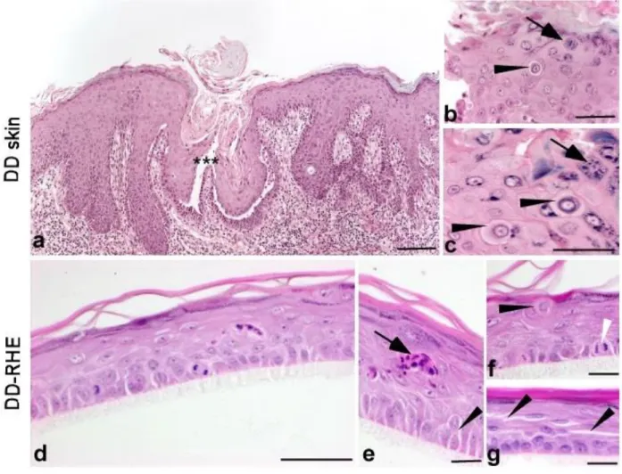
abnormal types of keratinocytes. Corps ronds refer to cells with small pycnotic nuclei, perinuclear clear halo and eosinophilic cytoplasm, while grains are compressed cells with elongated nuclei preferentially found in granular and cornified layers (Figure 1a-c).
DD arises from mutations in the ATP2A2 gene encoding SERCA2, a pump transporting Ca2+ from the cytosol to the lumen of endoplasmic reticulum (ER) [1]. Numerous mutations have been described with no clear genotype/phenotype correlation [2]. At the cellular level, ATP2A2 mutations lead to depletion of Ca2+ ER stores and aggregation of mutant proteins, both contributing to
permanent ER stress and apoptosis of certain keratinocytes [3-5] . Moreover, DD keratinocytes show defective trafficking of desmosomal proteins which could explain the acantholytic phenotype, and abnormal differentiation with altered labelling patterns of involucrin, keratin 14 and keratin 10 (reviewed in [6]). Studies using monolayers of keratinocytes cultured from DD patients or normal keratinocytes transduced with lentiviral vectors expressing mutant SERCA2 proteins have
contributed to unravel the molecular basis of DD [4,5]. Nevertheless, a model tool of DD displaying stratification and complete epidermal differentiation is needed for functional studies and testing new treatments . Recently, efforts have being undertaken in order to create three-dimensional models mimicking various dermatological conditions in vitro. For example, keratinocytes derived from patients suffering from epidermolytic ichthyosis are able to reconstitute an epidermis closely resembling the native skin phenotype resulting that results from mutations in genes encoding keratins 1 and 10 [7]. A variety of models for multifactorial diseases such as atopic dermatitis and
psoriasis have been obtained by altering either the environment of the reconstructed tissue or its gene expression profile [8-12].
In this study, we have used epidermis reconstruction [13] from primary keratinocytes cultured from a DD lesion in order to reproduce in vitro the phenotype of DD epidermis.
Keratinocytes were obtained from a surgical resection of a congenital DD lesion of the scalp from a 7-months-old baby. Diagnosis was based on histopathological examination and positive family history. In accordance with the standards of the Institute for Pathology and Genetics ethics committee, parental consent was obtained for DNA analysis and keratinocyte culture.
Sequencing of the SERCA2 gene on DNA extracted from peripheral blood lymphocytes revealed the presence of a previously described missense mutation Thr357Lys [1]. The same mutation was found in blood DNA from the father affected by a mild form of the disease. When cultured as primary monolayers, DD keratinocytes were undistinguishable from normal cells with respect to the growth rate and visual aspect by phase contrast microscopy (data not shown).
Sequencing of SERCA2 exon 8 in DNA extracted from cultured keratinocytes revealed heterozygosity for the Thr357Lys mutation. After amplification, DD keratinocytes were seeded at high density on polycarbonate filters in culture medium containing 1,5 mM Ca2+. After 24 hours,
cells were exposed to the air-liquid interface in order to reconstruct human epidermis (RHE) in
vitro, according to our method [13]. After 11 days of culture, a stratified epidermis was obtained,
exhibiting abnormal features strikingly similar to those observed in DD lesions (Figure 1 d-g). The intercellular spaces were markedly enlarged in all suprabasal layers, suggestive of acantholysis. Dyskeratotic keratinocytes with the typical appearance of corps ronds and grains were observed. In addition, as compared to normal RHE, DD-RHE revealed an increased number of mitoses in the basal layer. This histopathological phenotype was found consistently in all tissues reconstructed with these keratinocytes.
The DD-RHE was further characterized using immunodetection of several maturation markers and compared to DD lesional skin from the patient and to control samples (Figure 2). As expected, healthy skin and RHE obtained from normal keratinocytes exhibited keratin 14
expression restricted to the basal layer [13]. In contrast, keratin 14 was abnormally expressed in several suprabasal layers in DD-RHE, similarly to the pattern reported in lesional DD skin [4] and observed in our sample. The distribution of keratin 10 covers all suprabasal layers in normal skin and RHE [13]. Conversely, at least two layers of cells were unlabeled at basal side of DD-RHE, resembling the picture reported in lesional DD skin [4] and observed in this study. The distribution of the late differentiation marker involucrin, normally restricted to the upper spinous and granular layers in healthy skin, was also studied. In normal RHE, involucrin labelling appears sometimes in deeper layers than in vivo (Figure 2) but is never observed in the basal layer [13].In DD-RHE, the distribution of involucrin is markedly abnormal, with cytoplasmic labelling over the whole
thickness of the reconstructed tissue, reminiscent of lesional DD skin, where the expression of involucrin is also extended towards deeper layers than in normal skin (Figure 2), although the labelling is not observed in the basal layer. Premature expression of involucrin in lower spinous layers and even in basal layer is a common feature of DD epidermis [14].
Our work demonstrates that RHE can be obtained from keratinocytes isolated from lesional skin of DD patient. The phenotype of the resulting DD-RHE presents striking similarities with histological characteristics of DD lesions in vivo, namely acantholysis and presence of dyskeratotic keratinocytes. Moreover, the abnormal differentiation process described in lesional epidermis is also observed in the DD-RHE after 11 days of culture.
Undoubtfully, this work provides a new model to better investigate the onset of the cellular defects which result from defective SERCA2 function and progressively alter the process of
As a first step, the response of the DD-RHE to the topical or systemic application of retinoids, commonly used as treatments for DD, will be characterized.
Important issues remain to be addressed. Actually, the patient behind this study suffers from an atypical congenital form of the disease. Reconstruction of DD-RHE will be repeated using biopsies from unrelated DD cases carrying different mutations. Comparison of the phenotypes of the resulting RHE may help to clarify the genotype/phenotype correlation, which remains elusive [2, 3, 5].
The achievement of a culture model of DD epidermis attests the feasibility of reproducing a pathological condition in vitro for research or therapeutic perspectives.
Acknowledgements
References
[1] Sakuntabhai A, Ruiz-Perez V, Carter S, Jacobsen N, Burge S, Monk S, Smith M, Munro CS, O'Donovan M, Craddock N, Kucherlapati R, Rees JL, Owen M, Lathrop GM, Monaco AP, Strachan T, Hovnanian A. (1999) Mutations in ATP2A2, encoding a Ca2+ pump, cause Darier disease. Nat Genet 21:271-7.
[2] Ringpfeil F, Raus A, DiGiovanna JJ, Korge B, Harth W, Mazzanti C, Uitto J, Bale SJ, Richard G. (2001)Darier disease--novel mutations in ATP2A2 and genotype-phenotype correlation. Exp
Dermatol 10:19-27.
[3] Ahn W, Lee MG, Kim KH, Muallem S. (2003) Multiple effects of SERCA2b mutations associated with Darier's disease. J Biol Chem 278:20795-801.
[4] Leinonen PT, Myllylä RM, Hägg PM, Tuukkanen J, Koivunen J, Peltonen S, Oikarinen A, Korkiamäki T, Peltonen J. (2005) Keratinocytes cultured from patients with Hailey-Hailey disease and Darier disease display distinct patterns of calcium regulation. Br J Dermatol 153:113-7.
[5] Wang Y, Bruce AT, Tu C, Ma K, Zeng L, Zheng P, Liu Y, Liu Y.(2011) Protein aggregation of SERCA2 mutants associated with Darier disease elicits ER stress and apoptosis in keratinocytes. J
Cell Sci 124:3568-80.
[6] Savignac M, Edir A, Simon M, Hovnanian A. (2011) Darier disease : a disease model of impaired calcium homeostasis in the skin. Biochim Biophys Acta1813:1111-7.
[7] Chamcheu JC, Pihl-Lundin I, Mouyobo CE, Gester T, Virtanen M, Moustakas A, Navsaria H, Vahlquist A, Törmä H. (2011) Immortalized keratinocytes derived from patients with epidermolytic ichthyosis reproduce the disease phenotype: a useful in vitro model for testing new treatments. Br J
[8] Barker CL, McHale MT, Gillies AK, Waller J, Pearce DM, Osborne J, Hutchinson PE, Smith GM, Pringle JH. (2004). The development and characterization of an in vitro model of psoriasis. J
Invest Dermatol 123:892-901.
[9] Engelhart K, El Hindi T, Biesalski H-K, Pfitzner I. (2005) In vitro reproduction of clinical hallmarks of eczematous dermatitis in organotypic skin models. Arch Dermatol Res 297:1-9.
[10] Mildner M, Jin J, Eckhart L, Kezic S, Gruber F, Barresi C, Stremnitzer C, Buchberger M, Mlitz V, Ballaun C, Sterniczky B, Födinger D, Tschachler E. (2010) Knockdown of filaggrin impairs diffusion barrier function and increases UV sensitivity in a human skin model. J Invest
Dermatol 130:2286-94
[11] Kamsteeg M, Bergers M, de Boer R, Zeeuwen PL, Hato SV, Schalkwijk J, Tjabringa GS.(2011) Type 2 helper T-cell cytokines induce morphologic and molecular characteristics of atopic dermatitis in human skin equivalent. Am J Pathol. 178:2091-9.
[12] Bernard FX, Morel F, Camus M, Pedretti N, Barrault C, Garnier J, Lecron JC. (2012)
Keratinocytes under Fire of Proinflammatory Cytokines: Bona Fide Innate Immune Cells Involved in the Physiopathology of Chronic Atopic Dermatitis and Psoriasis. J Allergy 2012:718725
[13] Frankart A, Malaisse J, De Vuyst E, Minner F, Lambert de Rouvroit C and Poumay Y. (2012) Epidermal morphogenesis during progressive in vitro 3D reconstruction at the air-liquid interface.
Exp Dermatol 21:871-5.
[14] Kassar S, Charfeddine C, Zribi H, Tounsi-Kettiti H, Bchetnia M, Jerbi E, Cassio D, Mokni M, Abdelhak S, Ben Osman A, Boubaker S. (2008) Immunohistological study of involucrin expression in Darier’s disease skin J Cutan Pathol 35:635-40.
Figure Legends:
Figure 1. Histopathological features of Darier’s disease are conserved after epidermis reconstruction with primary keratinocytes derived from lesional skin
(a) Section of Darier’s disease (DD) lesion from the patient, showing epidermal hyperplasia, parakeratosis, and suprabasal clefting (asterisks) due to acantholysis (scale bar : 200µm). (b,c) Higher magnification showing dyskeratosis with corps ronds (black arrowheads) and grains (arrows) (scale bars: 50 µm). (d-g) Perpendicular sections of RHE cultured from primary DD keratinocytes for 11 days at air-liquid interface. Similarities to Darier native epidermis include the presence of grains (d, arrow in e), corps ronds (d, black arrowhead in f), and enlargement of intercellular spaces (acantholysis) (d,asterisks in e and g). Frequent mitoses are seen in the basal layer (d, white arrowhead in f). Hematoxylin-eosin stain (scale bars: 50 µm).
Figure 2. Immunohistochemical staining for keratin 14, keratin 10 and involucrin shows
altered distribution in DD-RHE, reflecting a similar disease-linked differentiation defect both
in vitro and in vivo
Keratin 14 labeling (detected with antibody from Santa Cruz, CA, USA; dilution 1:50) is restricted to the basal layer in normal epidermis and RHE. In DD lesional skin as well as in DD-RHE, keratin 14 is detected in basal and suprabasal layers. Keratin 10 labeling (detected with antibody from Dako, Glostrup, Denmark; dilution 1:100) is present in all layers except the basal layer in normal epidermis and RHE. In contrast, in DD skin and DD-RHE, at least the two deepest cell layers are devoid of keratin 10 labeling. Involucrin immunoreactivity (detected with antibody from
Sigma-Aldrich, Saint Louis, Oregon, USA; dilution 1:1000) is expressed in the stratum granulosum in normal skin and RHE. In lesional skin, involucrin labeling appears in more basally located keratinocytes. In DD-RHE, involucrin labeling is expressed through all cell layers of the tissue, including the basal layer. Immunohistochemical (peroxidase)-hematoxylin stain (scale bar: 50µm).