HAL Id: hal-02074059
https://hal.inria.fr/hal-02074059
Submitted on 20 Mar 2019
HAL is a multi-disciplinary open access
archive for the deposit and dissemination of
sci-entific research documents, whether they are
pub-L’archive ouverte pluridisciplinaire HAL, est
destinée au dépôt et à la diffusion de documents
scientifiques de niveau recherche, publiés ou non,
Estimation of Axon Conduction Delay, Conduction
Speed, and Diameter from Information Flow using
Diffusion MRI and MEG
Samuel Deslauriers-Gauthier, Rachid Deriche
To cite this version:
Samuel Deslauriers-Gauthier, Rachid Deriche. Estimation of Axon Conduction Delay, Conduction
Speed, and Diameter from Information Flow using Diffusion MRI and MEG. ISMRM 2019 - 27th
Annual Meeting of International Society for Magnetic Resonance in Medicine, May 2019, Montreal,
Canada. �hal-02074059�
Estimation of Axon Conduction Delay,
Conduction Speed, and Diameter from
Information Flow using Diffusion MRI and MEG
Samuel Deslauriers-Gauthier
1and Rachid Deriche
11Inria Sophia Antipolis M´editerran´ee, Universit´e Cˆote d’Azur, France
1
Introduction
The different lengths and conduction velocities of axons connecting cortical re-gions of the brain yield information transmission delays which are believed to be fundamental to brain dynamics (Caminiti et al., 2013). While early work on axon conduction velocity was based on ex vivo measurements (Hursh,1939), more recent work makes use of a combination of diffusion Magnetic Resonance Imaging (MRI) tractography and electroencephalography (EEG) to estimate axon conduction velocity in vivo (Horowitz et al.,2015). An essential interme-diary step in this later strategy is to estimate the inter hemispheric transfer time (IHTT) using EEG. The IHTT is estimated by measuring the latency between the peaks or by computing the lag to maximum correlation on contra lateral electrodes (Saron and Davidson,1989). These approaches do not take the sub-jects anatomy into account and, due to the limited number of electrodes used, only partially leverage the information provided by EEG.
In previous work (Deslauriers-Gauthier et al.,2017), we proposed a method, named Connectivity Informed Maximum Entropy on the Mean (CIMEM), to estimate information flow in the white matter of the brain. CIMEM is built around a Bayesian network which represents the cortical regions of the brain and their connections, observed using diffusion MRI tractography. This Bayesian network is used to constrain the EEG inverse problem and estimate which white matter connections are used to transfer information between cortical regions. In our previous work, CIMEM was used to infer the information flow in the white matter by assuming a constant conduction velocity for all connections. In this context, the conduction speed, and thus the delays, were inputs used to help constrain the problem. Here, we instead assume that the connection used to transfer information across the hemispheres is known, due the design of the acquisition paradigm, but that its conduction velocity must be estimated.
2
Methods
Our estimation of the axon diameter is based on the relation between axon diameter d and conduction speed v given by v = 6d (Hursh, 1939). Assum-ing a constant diameter along axons yields a relation between the information conduction delay s and the axon diameter which is d = `/(6s) where ` is the axon length in meters. Here, we propose to estimate the conduction delay us-ing CIMEM and the axon length usus-ing diffusion MRI. Results illustratus-ing the ability of CIMEM to estimated delays on simulated data were previous pub-lished. Briefly, CIMEM estimates delays by building a Bayesian network using the structural connectivity estimated using diffusion MRI. For each connection of the model and at every time instant, a variable is added to the model with two possible state: active (0) or inactive (1). MEG signals are then used as evidence into this network to compute the posterior probability of a connection being ac-tive at a particular time. Let Z(Ci,n= 1) be the posterior probability that the
ithconnection is active at the nth time point obtained by solving the CIMEM problem described inDeslauriers-Gauthier et al.(2017). We define the connec-tivity power of the ith connection as Γ
s(Ci = 1) = N−1P N −1
n=0 Z(Ci,n= 1)2
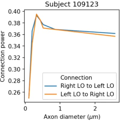
for a given delay s. The connectivity power is then computed for a series of delays and the estimated delay for a given connection is the one that maximizes Γd(Ci). The rationale is that the CIMEM model will only be able to use the
connection to explain the EEG measurements if the selected delay is correct. The axon diameter in the splenium of the corpus callosum was estimated for four subjects of the Human Connectome Project. The MEG data was format-ted using MNE-HCP1 processing was performed using MNE-python (Gramfort
et al., 2013,2014). The MEG epochs were created by selecting a -0.1 to 0.250 second window around the apearance of a visual cue in the motor task. They were then averaged to produce a evoked potential, which was lowpass filtered at 50 Hz and resampled at 100 Hz.
Describe diffusion MRI processing.
Our estimate of the axon diameter is given by d = `/(6s) where ` is the con-nection length in meters and s is the conduction delay in seconds. In CIMEM, a Bayesian network is built using the structural connectivity estimated using diffusion MRI. For each connection of the model and at every time instant, a variable is added to the model with two possible state: active (0) or inac-tive (1). MEG signals are then used as evidence into this network to com-pute the posterior probability of a connection being active at a particular time. Let Z(Ci,n = 1) be the posterior probability that the ith connection is active
at the nth time point obtained by solving the CIMEM problem described in
Deslauriers-Gauthier et al.(2017). We define the connectivity power of the ith
connection as Γs(Ci = 1) = N−1P N −1
n=0 Z(Ci,n= 1)2 for a given delay s. The
connectivity power is then computed for a series of delays and the estimated delay for a given connection is the one that maximizes Γd(Ci). The rationale is
that the CIMEM model will only be able to use the connection to explain the
EEG measurements if the selected delay is correct.
Figure 1: Axon diameter as a function of the internodal distance. Taken from Hursh(1939).
3
Conclusion
Acknowledgements
This work has received funding from the European Research Council (ERC)under the European Union’s Horizon 2020 research and innovation program(ERC Ad-vanced Grant agreement No 694665 : CoBCoM - Computational BrainConnec-tivity Mapping).
Data were provided by the Human Connectome Project (HCP), WU-Minn Consortium (Principal Investigators: David Van Essen and Kamil Ugurbil; 1U54MH091657) funded by the 16 NIH Institutes and Centers that support the NIH Blueprint for Neuroscience Research; and by the McDonnell Center for Systems Neuroscience at Washington University.
References
R. Caminiti, F. Carducci, C. Piervincenzi, A. Battaglia-Mayer, G. Confalone, F. Visco-Comandini, P. Pantano, and Innocenti G. M. Diameter, length, speed, and conduction delay of callosal axons in macaque monkeys and hu-mans: Comparing data from histology and magnetic resonance imaging dif-fusion tractography. The Journal of Neuroscience, 33(36):14501–14511, 2013. S. Deslauriers-Gauthier, J. M. Lina, R. Butler, P. M. Bernier, K. Whittingstall, R. Deriche, and M. Descoteaux. Inference and Visualization of Information Flow in the Visual Pathway using dMRI and EEG. In MICCAI 2017 Medical Image Computing and Computer Assisted Intervention, 2017.
A. Gramfort, M. Luessi, E. Larson, D. Engemann, D. Strohmeier, C. Brodbeck, R. Goj, M. Jas, T. Brooks, L. Parkkonen, and M. H¨am¨al¨ainen. MEG and EEG data analysis with MNE-Python. Frontiers in Neuroscience, 7, 2013. A. Gramfort, M. Luessi, E. Larson, D. Engemann, D. Strohmeier, C. Brodbeck,
L. Parkkonen, and M. H¨am¨al¨ainen. MNE software for processing MEG and EEG data. NeuroImage, 86:446–460, 2014.
A. Horowitz, D. Barazany, I. Tavor, M. Bernstein, G. Yovel, and Y. Assaf. In vivo correlation between axon diameter and conduction velocity in the human brain. Brain Struct Funct, 220:1777–1788, 2015.
J. B. Hursh. Conduction velocity and diameter of nerve fibers. American Journal of Physiology, 127(1):131–139, 1939.
C. D. Saron and R. J. Davidson. Visual evoked potential measures of interhemi-spheric transfer time in humans. Behavioral Neuroscience, 103(5):1115–11138, 1989.