Urol Res (1997) 25 [Suppl 1]: S 7-S 12 © Springer-Verlag 1997
T. S u l s e r • W . J o c h u m • P. U . H e i t z • D . H a u r i
Histomorphological changes after neodymium: YAG laser-coagulation
of the human prostate with the Side Focus fiber
Effect of power setting and time
A b s t r a c t The objective of our study was to determine optimal treatment parameters and appropriate methods of examination for neodymium:yttrium-aluminum-garnet (Nd:YAG) high-power laser coagulation of the human pros- tate in relation to power setting and time. Transurethral free-beam laser coagulation was performed with the Side- Focus side-firing laser fiber in ten patients prior to planned radical surgery, of whom six underwent transperitoneal la- paroscopic lymphadenectomy and laser coagulation 4 - 9 days prior to open surgery. Depth and volume of coagu- lated prostatic tissue were measured at power setting/time combinations of 40 W/90 s and 60 W/60 s, respectively, while holding total energy delivery constant. Microscopic examination in the early phase showed that epithelial cells had become loose from the basal-cell membrane. By 4-9 days there was evidence of conspicuous squamous epithe- lial metaplasia with a high proliferation rate as a sign o f r e - epithelialization. Using the Side Focus side-firing laser fi- ber, both treatment modalities showed comparable volume coagulation. In contrast there was a significant difference between those prostates removed at 4 - 9 days and those re- moved at 60-210 min after laser coagulation. We conclude that laser-induced changes in the human prostate are con- clusively discernible only after 4 days.
K e y w o r d s P r o s t a t e • Benign prostatic hyperplasia • Lasers - Laser surgery • Histomorphology
Introduction
Benign prostatic hyperplasia (BPH), which is a common disease affecting men, leads to micturition difficulties in
T. Sulser ([]) • D. Hauri
Department of Urology, University Hospital Zurich, CH-8091 Zurich, Switzerland
W. Jochum. R U. Heitz
Department of Pathology, Institute of Clinical Pathology, University Hospital Zurich, CH-8091 Zurich, Switzerland
50% of 50-year-old men with the disease. At present two to four out of every ten males over 60 years of age are op- erated on for BPH [3]. For many years standard treatment has been transurethral resection of the prostate (TURP) and occasionally in the case of large adenomas open prostatec- tomy. Both treatments, although they have proved effec- tive, are associated with a significant morbidity rate [8, 20]. Therefore minimally invasive procedures for the treatment of BPH are currently under debate among urologists. Vis- ual laser ablation of the prostate (VLAP) using neody- mium:yttrium-aluminum-garnet (Nd:YAG) laser is the main procedure in use at present. Many publications have considered the question of laser dosimetry and the extent of laser-induced coagulation using various fibers of differ- ent treatment modalities [2, 9-14, 17, 21]. So far the im- pact and extension of visual laser ablation in humans have rarely been subject to examination. In 1993 Bolton and Co- stello [4] first presented histological results from human prostate after Nd:YAG laser ablation. In the following years further studies were published dealing in particular with the extension of the laser-induced changes and the estab- lishment of fiber-specific optimal treatment modalities for some special light guides with respect to time and energy used and method of application [5, 16, 18, 19, 23, 26, 29]. In this study we examined the histomorphological changes in human prostates following laser application with a specific fiber as well as carefully evaluated the ex- tent of coagulation in relation to the power setting and time used. Based on an empiricial optimal energy delivery of 3600 J for each setting [2, 9-11, 14], we assessed the ef- fect of the energy (40 W/90 s vs 60 W/60 s) and time used on morphological changes.
Materials and methods
From September 1994 to June 1995 a group of ten patients with a mean age of 60.8 (range 55-70 years) was accepted for the study af- ter their written personal consent was obtained and approval obtained from the Ethics Committee. A transperitoneal laparoscopic lymphad- enectomy was performed in six patients with bioptically verified
$ 8
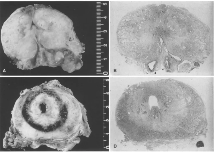
Fig. 1A-D Macroscopic view of human prostate after VLAP pro- cedure (horizontal section, native and H & E-stained) at 210 rain (A, B) and at 4 days (C, D) following a 40-W/90-s power setting
adenocarcinoma of the prostate, which was identified from the en- larged staging, after laser treatment and with the patient under the same anesthetic. Following histological examination of lymphade- nectomy six patients underwent radical prostatectomy 4-9 days lat- er. Another three patients in whom carcinoma of the prostate was lo- cated were primarily treated with radical cancer surgery. Another pa- tient with urothelial carcinoma underwent radical cystoprostatecto- my. In the latter four patients laser treatment was applied immedi- ately prior to the planned surgery and with the patient under the same anesthetic. The specimen was taken at 60-210 min following laser treatment.
We used a 100-W cw-Nd:YAG laser (Dornier Medilas 4100) for energy delivery and the high-energy-density SideFocus fiber (Dorni- er), 600 #m in diameter, as the light guide. Laser treatment took place with the patient under insufflation anesthetic in the dorsal lithotomy position. It was performed under direct vision using a 23-Fr. flow- laser cystoscope (Circon, ACMI) under continuous irrigation with a new fiber for the settings of 40 W at 90 s and 60 W at 60 s, respec- tively. It was aimed close to the verumontanum in the 2, 4, 8 and 10 o'clock positions and with a total energy delivery of 14400 J.
After fixation in phosphate-buffered formalin (PBS, 10%), the prostatovesiculectomy specimens were sectioned horizontally into slides 5 mm in width, macroscopically classified and recorded nu- merically. Subsequently the organ slices were embedded in paraffin and the cut surface of the sections stained with hematoxylin and eo- sin and examined under a light microscope. Laser-induced changes in the organ were analyzed as to their quality and extension.
Results
R a d i c a l p r o s t a t o v e s i c u l e c t o m y was p e r f o r m e d at an inter- val of 1 h to 9 days f o l l o w i n g transurethral laser ablation.
Early c h a n g e s
O n gross e x a m i n a t i o n acutely harvested h u m a n prostates s h o w e d a zone of c o a g u l a t i o n necrosis e x t e n d i n g from the urethral l u m e n b u t with no sharp and w e l l - d e f i n e d margin. No clear h e m o r r h a g i c ring was observed in t]aese s p e c i m e n s r e m o v e d at an early stage, with only slight p e e l i n g a n d s w e l l i n g visible (Fig. 1A, B). M i c r o s c o p i c e x a m i n a t i o n showed that epithelial cells of the prostate glands had loos-
Fig. 2A-H Histology of earlier (A, E) and later (B, C, E, F, G, H) VLAP-induced changes in human prostate: Desquamation of dam- aged epithelial cells into glandular lumen (A, x 160), margin of peri- urethral necrotic zone with extensive hemorrhage (B, x40), granu- lar and histiocytic inflammation infiltration (C, x260), squamous epithelial metaplasia of glandular epithelium (D, x70), myxoid swelling and partial loss of stromal cell detail (E, × 160), coagula- tion necrosis of epithelial as well as strornal cell elements (F, × 125), pseudocystic resorption of cell dentritus (G, ×70), marked prolife- rative activity of the regenerating glandular epithelium (I-I, x 160, Mib-1 immunohistochemistry)
S10
ened from the basal-cell membrane and showed desquama- tion in the glandular lumina (Fig. 2A). Solidified chroma- tinic nuclei and vacuolated cytoplasm were noted. Stroma was found to be edematous and numerous stromal cells lacked nuclei (Fig. 2E). With both treatment modalities the laser application sites could not be identified either by car- bonization or by tissue disruption.
Delayed changes 20- v 15 Y: 1 0 E __= 0 > 0 O By 4 days after laser application a demarcated spherical lesion was identified periurethrally with a rim of hemor-
o rhage surrounding it (Fig. 1C, D). Microscopic exam- ination revealed coagulative necrosis of epithelial and stromal cells with a nearly complete loss of cell and tissue Fig. 3 detail. Cell nuclei were rare (Fig. 2F). Peripheral to the necrotic area, ectatic and in part thrombosed vessels with homogeneous eosinophilic walls alternating with zonal hemorrhage were prominent (Fig. 2B). Subsequently neu- trophil granulocytes and macrophages were responsible for the gradual clearing away of detritus to the point of leav- ing behind absorbent cavities of small size (Fig. 2G). In the prostate tissue adjacent to the necrotic region, there was "8" evidence of a conspicuous squamous epithelial metaplasia _6 of the glandular epithelium (Fig. 2D). The proliferative ra- tio of this epithelium was 5 0 - 8 0 % as seen in the immuno- -r- histochemical staining for the proliferation-associated
antigen Ki-67 (Fig. 2H). E.,
The discernible changes at 6 or 9 days following laser ablation were the same as those at 4 days. Particularly not- .u able, however, were the absorbent and regenerative pro- cesses which had occurred by the later date. For both ap- plied treatment settings no differences were noted in the quality of the morphological findings.
A confluent, periurethral necrotic area of variable di- mensions was found in all six cases independent of the power setting at which prostate specimens were taken between 4 and 9 days after laser treatment. Necrotic areas showed a homogeneous character, thus not influencing ex- tension by the variable tissue structure. Lesions were lim- ited to the transition zones and they were confined to the organ in all specimens (Fig. 1A, B).
The dimension of the necrotic zones showed no major differences between modalities (Fig. 3). In the histologi- cal examination the necrosis showed a diameter of between
2.5 and 3.3 cm transversely after application of 40 W and Patient 60 W, respectively, with a depth of between 2.5 and 3.1 cm.
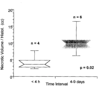
The coagualated volume for the 40-W group was between 6.2 and 16.4 cm 3 and between 8.4 and 11 cm 3 for the 60-W group, respectively. Both the largest and smallest co- 1 agulated volumes were found in the 40-W group (Table 1). 2 3 Macroscopically laser-induced changes were not distinctly 4 demarcated in specimens taken at an early stage 5 60-210 rain after laser treatment (Fig. 1C, D). Microscopic 6 changes were recorded in four cases, taking into consider- 7 ation that the obtained values were significantly lower than 8 those in specimens taken 4 - 9 days following laser irradi- 9 10 ation (Fig. 4). n = 5 n = 5 , ~ L ~ i , ~ ... p = 0 . 5 2 40 W P o w e r ~ m g"e":n- 60 W
Necrotic volume after VLAP on two different initial power settings. There is no significant difference
20- 15 10 n = 4 n = 6
5_
>
<
p = 0.02 I < 4 h T i m e Interval 4-9 daysFig. 4 Significant difference in necrotic volume of the removed prostate at two time intervals after VLAP
Table 1 Histological results of the four-quadrant laser procedure (2, 4, 8, 10 o'clock) with both modatities (40 W//90 s vs 60 W/ 60 s) with a total energy delivery of 14 400 J for each setting. (t trans- verse, a axial, s sagittal)
Modality Interval Necrotic Coagulated extension volume (txsxa) (cm) (txsxaxTc/6) (cm 3) 40 W/90 s 4 days 40 W/90 s 7 days 40 W/90 s 9 days 60 W/60 s 7 days 60 W/60 s 7 days 60 W/60 s 8 days 40 W/90 s 210 min 40 W/90 s 120 min 60 W/60 s 180 min 60 W/60 s 60 rain 3.3 x3.2x3.0 16.5 2.5 x3.0x2.5 9.8 2.7x2.2x2.0 6.2 3.1 x2.6x2.0 8.4 2.5 x2.5x3.0 9.8 2.8 x2.7x2.8 ll 2.7 × 1.7 x 1.5 3.6 1.5x 1.5x2.0 2.3 2.4x2.2x2.8 7.7 2.2x2.0x2.l 4.8
Discussion
Since the first report by Johnson's et al. [10] on visual la- ser application for the treatment of BPH, further experi- mental animal reports and dosimetry studies have been published [2, 9, 13, 14, 19, 21, 23]. So far data from clin- ical trials have shown very promising results [1, 6, 7, 15, 22]. Apart from optimal laser energy delivery and laser treatment time there are the different physical properties of the diverse light guides to be taken into consideration [25, 27, 30]. Not many more than a dozen fibers are cur- rently available, and a number of reliable studies have looked at their impact on human tissue [13, 16, 18, 19, 23, 26, 29]; however, we have only three 3 light guides at our disposal (Urolase, Prolase II and Ultraline). In this paper we present the results of thermal coagulation in human prostates following laser treatment using a SideFocus fi- ber with subsequent systematic histopatholgoical exam- ination. In addition to comparing the two procedure modes, we also evaluated laser-induced changes in relation to treatment time.
Visual prostate laser application in four quadrants inde- pendent of power setting produces a confluent periureth- ral necrotic area of variable extent. Upon examination there were no substantial differences between irradiation power used as to the extent of necrotic zone; on the contrary we found the largest and the smallest necrotic volumes fol- lowing 40 W irradiation. The reasons for the different ex- tensions are a matter for debate: the variable tissue struc- ture of the prostate from person to person leads to variable thermal conductivity and thermal storage including vari- able dissipation of heat through the vascular system. More- over, the changing qualities of the light guides during treat- ment must be taken into account [25, 27, 30]. Although no differences was found in necrotic volume between both la- ser treatment modalities, from our experience we suggest using the 40-W power setting for the SideFocus fiber; at this setting there is little danger of the fiber being dam- aged, and inadvertent carbonization effects can thus be avoided.
Laser treatment with the SideFocus fiber produces ther- mocoagulation as reported by several authors [4, 13, 16, 19, 23, 26]. The maximum extension in the transverse plane (1.65 cm in radius) is nearly identical to that (l.75 cm) noted by Shanberg et al. [26] upon irradiation with the Pro- lase II fiber [26]. To lase the lateral lobe, Shanberg et al. used a dose higher than 50 000 J, targeting the whole sur- face extensively. Thus he achieved satisfactory ablation. We, however, used only a dose of 7200 J delivered at two spots for each lateral lobe. There is some doubt whether by increasing the applied energy up to more than 7200 J for each lobe, the depth of penetration and necrotic vol- ume increased significantly. Dosimetry studies by Kabalin et al. [13, 16, 19] show that from a certain energy dose on- ward the tissue effect plateaus irrespective of the fiber type. This is probably due to the thermic and optical qualities of the tissue (heat storage, thermal transfer, light scatter) be- ing altered through thermocoagulation. Moreover, we have
S l l to take into account that with high-energy doses over a long period the above-noted damage to the fiber top appears in- creasingly likely to occur [27, 30].
For thermoregulation effects to become evident a cer- tain period of time is needed, but we require further details as to its length. The specimens taken between 1 and 3.5 h showed no macroscopically laser-induced lesions. Micro- scopic examination revealed distinctly smaller areas of dis- cernible necrosis than in the group of specimens taken 4 - 9 days following treatment (Table 1). This confirms ear- lier statements by other authors that a period longer than only several hours is required for laser-induced changes to turn into discernible demarcated lesions [28]. The earliest interval for this to occur in our study was 4 days. With this specimen (Fig. 1A, B) the periurethral necrotic zone had reached its final state following laser treatment. Therefore we suggest that laser-induced changes in human prostates are not conclusively discernible until 1 or 2 days after treat- ment at the earliest.
Laser radiation of the prostate histologically showed a coagulative necrosis of epithelial and stromal cells with ectatic and in part thrombosed vessels peripheral to ne- crotic zones. A homogeneous necrotic area not influenc- ing extension by a variable tissue structure was present in all specimens. Furthermore, the lesions were limited to the transition zone and were confined to the organ in all spec- imens. Gradual clearing of detritus by neutrophil granu- locytes and macrophages commenced and led to resorptive cavities of small size. Conspicuous squamous epithelial metaplasia of the glandular epithelium with a high prolife- ration rate was sign of re-epithelialization was present as soon as 4 days after laser application with the resorptive and regenerative faculties in those specimens removed at 6 or 9 days being particularly notable, in accordance with the findings of other authors [5, 24].
We conclude that laser treatment with the SideFocus fi- ber in a four-quadrant procedure is a safe way of produc- ing a spherical zone of periurethral coagulative necrosis. Although there were no differences between both power modalities and the fibers used, we recommend a 40-W/ 90-s combination to give slow heating and thus avoid car- bonization and vaporization at the application site in the urethra. For further dosimetry studies in human prostates we suggest that evaluation of the laser-induced coagula- tive necrotic zone be carried out after an interval of only 4 days.
References
1. Anson K, Nawrocki J, Buckley J, Fowler C, Kirby R, Lawrence W, Paterson P, Watson G (1995) A multicenter, randomized, prospective study of endoscopic laser ablation versus transureth- ral resection of the prostate; Urology 46:305
2. Assimos DG, McCullough DL, WoodruffRD, Harrison LH, Hart LJ, Li WJ (1991) Canine transurethral laser-induced prostatec- tomy. Endourology 5:145
3. Barry MJ (1990) Epidemiology and natural history of benign prostatic hyperplasia. Urot Clin North Am 17:495
S 12
4. Bolton DM, Costello AJ (1993) Histological study of Nd:YAG laser energy on prostatic adenoma as demonstrated in the intact prostate gland. Br J Urol 71:757
5. Costello AJ, Bolton DM, Ellis D, Crowe H (1994) Histopatho- logical changes in human prostatic adenoma following neody- mium: YAG laser ablation therapy. J Urol 152:1526
6. Costello AJ, Lusaya DG, Crowe H (1995) Transurethral laser ablation of the prostate - long-term results. World J Urol 13:119 7. Cowles RS, Kabalin JN, Childs S, Lepor H, Dixon C, Stein B, Zabbo A (1995) A prospective randomized comparison of trans - urethral resection to visual laser ablation of the prostate for treat- ment of benign prostatic hyperplasia. Urology 46:155
8. Doll HA, Black NA, McPherson K, Flood AB, Williams GB, Smith JC (1992) Mortality, morbidity and complications follow- ing transurethral resection of the prostate for benign prostatic hypertrophy. J Urol 147:1566
9. Gill HS, Kabalin JN, Mikus PW (1994) Characterization of tis- sue effects produced by the ProLase II lateral-firing neodymi- um:YAG laser fiber in the canine prostate. Lasers Surg Med
15:185
10. Johnson DE, Levinson AK, Greskovich FJ, Cromeens DM, Ro JY, Costello AJ, Wishnow KI (1991) Transurethral laser prosa- tectomy using a right angle laser delivery system. SPIE Proc 1421:36
11. Johnson DE, Price RE, Cromeens DM (1992) Pathologic chang- es occurring in the prostate following transurethral laser prosta- tectomy. Lasers Surg Med 12:254
12. Kabalin JN (1993) Laser prostectomy performed with a right an- gle firing neodymium:YAG laser fiber at 40 Watts power set- ting. J Urol 150:95
13. Kabalin JN (1995) Laser dosimetry studies in the human pros- tate. In: Marberger M (ed) Application of newer forms of ther- apeutic energy in urology. Isis Medical Media, Oxford, p 143 14. Kabalin JN, Gill HS (1994) Dosimetry studies utilizing the Uro-
lase right angle firing neodymium:YAG laser fiber. Lasers Surg Med 14:145
15. Kabalin JN, Doll S (1996) Neodymium:YAG laser coagulation prostatectomy: 3 years of experience with 227 patients. J Urol
155:181
16. Kabalin JN, Sellers R, Bite G (t995) Neodymium:yttrium-alu- minium-garnet laser dosimetry for the Prolase II side-firing de- livery system in the human prostate. Urology 45:248
17. Kabalin JN, Gill HS, Gunars B (1995) Laser prostatectomy per- formed with a right-angle firing neodymium:YAG laser fiber at 60 Watts power setting. J Urol 153:1502
18. Kabalin JN, Gong M, Issa M, Sellers R (1995) Insight into mech- anism of neodymium:yttrium-aluminium-garnet laser prostatec-
tomy utilizing the high-power contact-free beam technique. Urology 45:421
19. Kabalin JN, Terris MK, Mancianti ML, Fajardo LF (1996) Do- simetry studies utilizing the Urolase right-angle firing neody- mium:YAG laser fiber in the human prostate. Lasers Surg Med 18:72
20. Mebust WK, Holtgrewe HL, Cockett ATK, Peters PC (1989) Transurethral prostatectomy - immediate and postoperative complications. A cooperative study of 13 participating institu- tions evaluating 3885 patients. J Urol 141:243
21. Motamedi M, Torres JH, Orihuela E, Pow-Sang M, Cowan DF, Warren MM (1995) Laser photocoagulation of prostate: Influ- ence of dosimetry. Lasers Surg Med 17:49
22. Norris JP, Norris DM, Lee RD, Rubenstein MA (1993) Visual laser ablation of the prostate: clinical experience in 108 patients. J Urol 150:1612
23. Orihuela E, Motamedi M, Pow-Sang M, LaHaye M, Cowan DF, Warren MM (1995) Histopathological evaluation of laser ther- mocoagulation in the human prostate: Optimization of laser ir- radiation for benign prostatic hyperplasia. J Urol 153:1531 24. Pow-Sang M, Orihuela E, Motamedi M, Torres J, Adesokan A,
Cowan D, Warren MM (1996) Healing response of the canine prostate to Nd:YAG laser radiation. Prostate 28:287
25. Protsenko D, Torres JH, Chakrabarti P, Bell B, Orihuela E, Mot- amedi M (1996) Optical characterization and coagulation per- formance of side-emitting fiber delivery systems for laser ther- apy of benign prostatic hyperplasia: A comparative study. Urol- ogy 47:845
26. Shanberg AM, Lee IS, Tansey LA, Sawyer DE, Rodgers LW, Ahlcring T (1994) Depth of neodymium:yttrium-aluminium- garnet laser in the human prostate at various dosimetry. Urolo- gy 43:809
27. Slaa ET, Van Ettergem AF, Van't Hof CA, Debruyene FMJ, De 1 a Rosette JJMCH (1995) Durability of laser fibers. World J Urol 13:83
28. Stein BS (1994) Laser-tissue interaction. In: Smith JA Jr, Stein BS, Benson RC (eds) Lasers in urologic surgery, 3rd edn. Mos- by-Year Book, Chicago, p 10
29. Sulser T, Jochum W, Huch B0ni RA, Briner J, Krestin GP, Hau- ri D (1995) Morphologische und MRT-Ver~inderungen nach vi- sueller Laserablation der Prostata. Akt Urol 26:109
30. van Swol CFP, Slaa ET, Verdaasdonk RM, De La Rosette JJMCH, Boon TA (1996) Variation in output power of laser pros- tatectomy fibers: A need for power measurements. Urology 47:672