ARTICLE
Polymorphisms in Toll-like receptor 4 (
TLR4) are associated
with protection against leprosy
P.-Y. Bochud&D. Sinsimer&A. Aderem&M. R. Siddiqui&
P. Saunderson&S. Britton&I. Abraham&A. Tadesse Argaw&
M. Janer&T. R. Hawn&G. Kaplan
Received: 6 January 2009 / Accepted: 31 March 2009 / Published online: 9 May 2009
# Springer-Verlag 2009
Abstract Accumulating evidence suggests that polymor-phisms in Toll-like receptors (TLRs) influence the patho-genesis of mycobacterial infections, including leprosy, a disease whose manifestations depend on host immune responses. Polymorphisms in TLR2 are associated with an increased risk of reversal reaction, but not susceptibility to leprosy itself. We examined whether polymorphisms in TLR4 are associated with susceptibility to leprosy in a cohort of 441 Ethiopian leprosy patients and 197 healthy controls. We found that two single nucleotide polymor-phisms (SNPs) in TLR4 (896G>A [D299G] and 1196C>T [T399I]) were associated with a protective effect against the
disease. The 896GG, GA and AA genotypes were found in 91.7, 7.8 and 0.5% of leprosy cases versus 79.9, 19.1 and 1.0% of controls, respectively (odds ratio [OR]=0.34, 95% confidence interval [CI] 0.20–0.57, P<0.001, additive model). Similarly, the 1196CC, CT and TT genotypes were found in 98.1, 1.9 and 0% of leprosy cases versus 91.8, 7.7 and 0.5% of controls, respectively (OR =0.16, 95% CI 0.06-–.40, P<0.001, dominant model). We found that Mycobacterium leprae stimulation of monocytes partially inhibited their subsequent response to lipopolysaccharide (LPS) stimulation. Our data suggest that TLR4 polymor-phisms are associated with susceptibility to leprosy and that P.-Y. Bochud
:
A. Aderem:
M. Janer:
T. R. HawnInstitute for Systems Biology, Seattle, WA, USA
A. Aderem
:
T. R. HawnDepartment of Medicine, University of Washington, Seattle, WA, USA
D. Sinsimer
:
M. R. Siddiqui:
G. KaplanLaboratory of Mycobacterial Immunity and Pathogenesis, Public Health Research Institute Center
at the University of Medicine and Dentistry of New Jersey, Newark, NJ, USA
P. Saunderson
All Africa Leprosy Rehabilitation and Training, Addis Ababa, Ethiopia
S. Britton
:
I. Abraham:
A. Tadesse Argaw Armauer Hansen Research Institute, Addis Ababa, EthiopiaD. Sinsimer
Graduate School of Biomedical Sciences,
University of Medicine and Dentistry of New Jersey, Newark, NJ, USA
Present Address: P.-Y. Bochud (*)
Service of Infectious Diseases, Department of Medicine, Institute of Microbiology, University Hospital
and University of Lausanne, 1011 Lausanne, Switzerland e-mail: [email protected] Present Address:
M. R. Siddiqui
Centre for Infections, Health Protection Agency, London, UK
Present Address: P. Saunderson
American Leprosy Missions, Greenville, SC, USA Present Address: S. Britton
Department of Medicine, Karolinska Institute, Stockholm, Sweden
Present Address: A. Tadesse Argaw
Mount Sinai School of Medicine, New York, NY, USA
this effect may be mediated at the cellular level by the modulation of TLR4 signalling by M. leprae.
Introduction
Leprosy remains an important public health concern in developing countries, with an estimated 250,000 new patients
each year [1]. Infection with Mycobacterium leprae is
characterised by a polarised spectrum of clinical manifesta-tions that correlate with the level and type of cell-mediated
immunity [2, 3]. At one end of the spectrum, tuberculoid
leprosy patients have localised lesions that contain few bacilli
and a Th1 T-cell-mediated cellular immune response [3–5]. At
the opposite end, lepromatous leprosy patients have dissemi-nated infection with extensive lesions containing numerous intra-cellular bacilli and a Th2 T-cell-mediated cellular immune response. This wide range of clinical and immunological presentations makes leprosy a prototypical disease to under-stand how polymorphisms in innate immunity genes influence adaptive immune responses. Furthermore, insights into leprosy pathogenesis may help elucidate mechanisms of protective immunity to other mycobacteria such as M. tuberculosis, a leading cause of morbidity and mortality worldwide.
Toll-like receptors (TLRs) constitute a family of trans-membrane proteins that play an important role in initiating the host immune response to various pathogens, including
the pathogenesis of mycobacterial infections [6–8]. The
interaction of specific mycobacterial agonists with TLRs leads to the production of inflammatory mediators that are critical for the activation of innate and adaptive immune
responses [9,10]. Among the members of the TLR family,
TLR2 (as a heterodimer with TLR1 or TLR6), TLR4 and TLR9 have been shown to interact with mycobacterial
agonists [9–11]. A number of studies have established a
role for the TLR2-mediated recognition of M. tuberculosis,
M. leprae and other non-tuberculous mycobacteria [10,12].
In vitro data also supported a role for the TLR4-mediated recognition of an M. tuberculosis ligand that was
stimula-tory only when isolated from live organisms [13].
For decades, host genetic factors have been known to play an important role in leprosy. Genome-wide studies have identified several loci that are associated with the disease or
its clinical presentation (reviewed in [14, 15]). In addition,
studies have reported associations between leprosy and/or leprosy type and polymorphisms in numerous candidate genes,
including the tumour necrosis factor α (TNF-α) gene, the
interleukin-10 gene, the transporter associated with antigen processing 1 and 2 (TAP1 and TAP2) genes, the natural resistance associated macrophage protein 1 (NRAMP1) gene
and the vitamin D receptor gene (reviewed in [14,16–18]).
Human genetic studies suggest that polymorphisms in
TLRs influence susceptibility to a variety of infections [6,19].
We recently demonstrated that a microsatellite polymorphism in TLR2 was significantly associated with the occurrence of
reversal reaction among leprosy patients [20]. However, this
polymorphism did not strongly influence susceptibility to leprosy itself, nor leprosy type. Therefore, we hypothesised that polymorphisms in TLRs other than TLR2 may influence susceptibility to the disease. Two TLR4 polymorphisms (896G>A [D299G] and 1196C>T [T399I]) are emerging as major variants in TLR genes that influence human suscep-tibility to a number of infectious diseases, including
tuberculosis [21] and invasive aspergillosis [22,23]. In the
present study, we investigated the role of TLR4 polymor-phisms in susceptibility to leprosy and its clinical manifes-tations in an Ethiopian cohort.
Methods Human subjects
The enrolment procedures and clinical definitions have been
described previously [20, 24]. Briefly, 441 leprosy patients
were drawn from the All Africa Leprosy Rehabilitation and Training (ALERT) Multi-Drug Therapy (MDT) Field
Eval-uation Study (AMFES) [24] and 197 unrelated, healthy
controls were collected from the local population. Self-reported ethnicity included three major ethnic groups (Oromo, Amhara and Gurage). Leprosy types were estab-lished on clinical grounds according to the simplified Ridley/ Jopling classification, which adds the rarely occurring borderline borderline (BB) patients to the borderline
lepro-matous (BL) patients [25]. Twenty-five additional patients
were classified as multibacillary (MB) or paucibacillary (PB) according to the WHO classification. Leprosy reactions were reported only in a subgroup of 216 patients and diagnosed as
reported elsewhere [26]. Informed consent was obtained from
each study participant. The study was approved by human subject review boards from the Armauer Hansen Research Institute (Addis Ababa, Ethiopia), the Rockefeller University (New York, NY), the Public Health Research Institute (Newark, NJ), the University of Washington (Seattle, WA) and the Western Institutional Review Board (Olympia, WA). DNA sequencing and genotyping
Genomic DNA was extracted from whole blood using
standard protocols [20]. To determine whether there were
any polymorphisms that are unique to Ethiopians, we sequenced the coding region of 28 individuals (9 controls and 19 cases). The polymerase chain reaction (PCR) products were sequenced with Big Dye Terminator v3.0 and analysed on an ABI PRISM 3730 capillary sequencer (Applied Biosystems). Sequences were aligned and
ana-lysed with the programs PHRED/PHRAP and CONSED
[27]. Genotyping among the whole cohort was carried out
by the MassARRAY™ technique (Sequenom, San Diego,
CA) using a chip-based matrix-assisted laser desorption/ ionisation time-of-flight mass spectrometer as previously
described [28]. Single nucleotide polymorphism (SNP)
selection included four SNPs, three previously reported
SNPs (896G>A, 1196C>T and 1530G>T [Q510H] [6,29])
and a previously unreported SNP that was discovered by sequencing (1976A>G [M658G]).
Statistics
Statistical analyses were performed using Stata 9 (StataCorp, College Station, TX). All SNPs were tested for Hardy-Weinberg equilibrium using the genhw program. Pairwise linkage disequilibrium was assessed using the pwld program. Haplotypes were inferred separately for each ethnic group using the expectation-maximisation algo-rithm implemented in the DECIPHER program (S.A.G.
E.) [30]. Haplotypes with frequencies <1.5% were
grouped together. The association of TLR4 SNPs with susceptibility to leprosy and leprosy type was first assessed in a multivariate logistic regression model (general model) that did not assume any particular mode of inheritance and accounted for the presence of each genotype versus the wild type genotype (0/1 and 1/1, each versus 0/0). For statistically significant associations, we performed likelihood ratio tests for three different models versus the general model. Each of these three models assumed one of the following modes of inheritance: dominant (comparing the presence of one or two copies of the minor allele versus no copies), recessive (comparing the presence of two copies of the minor allele versus no or one copies) and additive (no, one or two copies of the minor allele were coded 0, 1 and 2, respectively, assuming greater effect with increased copy number of the minor allele). The best fitting model was assumed in the final presentation of the results. The association of TLR4 haplotypes with susceptibil-ity to leprosy was calculated in a logistic regression model including all haplotypes (each coded 0, 1 or 2 for the presence of no, one or two copies of the haplotype), using haplotype 1 as a reference. All analyses were adjusted for age groups, sex and ethnicity (when applicable). Since the sample size was limited, especially for rare SNPs, the statistical analyses were initially performed in the whole population. The consistency of significant results was then verified within each ethnic group.
Bacterial strains
M. leprae strain Thai-53 was provided by Dr. James Krahenbuhl of Lousiana State University and was propagated
and collected from the mouse footpad model as described
previously [31]. M. leprae was irradiated as described
previously [31]. Since M. leprae grows optimally at 30–
33°C, which is suboptimal for cell culture, heat-killed M. leprae were used as described previously. Heat-killed and viable M. leprae were compared previously and were shown to induce similar relative cytokine response from infected
monocytes [32,33, and G. Kaplan, unpublished data].
Human monocyte and peripheral blood mononuclear cell stimulations
Human peripheral blood monocytes and peripheral blood mononuclear cells (PBMCs) were isolated from healthy donors (New Jersey Blood Center, East Orange, NJ) by
Ficoll-Paque separation and plated at a density of 3×10⁶
PBMCs per well in a 24-well tissue culture plate. For the preparation of monocytes, isolated PBMCs were resus-pended in RPMI 1640 and supplemented with 1% human AB serum (Gemini Bioproducts, Calabasas, CA), allowed to adhere for 2 h and then washed to remove non-adherent cells. Both adherent monocytes and PBMCs were cultured in RPMI 1640 with 20% human serum prior to stimulation at a multiplicity of infection (MOI) of 5:1 with heat-killed M. leprae (ML) or stimulation with ultrapure lipopolysac-charide (LPS; 100 ng/mL, InvivoGen, San Diego, CA). Supernatants were collected at 48 h post-stimulation and analysed by Luminex for the presence of multiple cytokines (Bio-Rad, Hercules, CA).
Results
The study included 441 leprosy cases and 197 controls
(Table 1). The majority of participants were males (71.6%
of cases and 73.5% of controls). Self-reported ethnicities were Oromo (43.8% of cases and 46.7% of controls), Amhara (28.6% of cases and 22.3% of controls) and Gurage (25.4% of cases and 25.9% of controls). Age categories were not equally represented among cases and controls (mean age 39.0 ± 15.1 years in cases versus 29.1 ± 12.4 years in controls, P < 0.001). A total of 298 cases (68.1%) were grouped into the lepromatous pole, includ-ing 199 borderline lepromatous (BL), 81 lepromatous lepromatous (LL) and 18 multibacillary (MB, WHO classification). Among the 138 cases grouped into the tuberculoid pole (31.9%), 128 were borderline tuberculoid (BT), three polar tuberculoid (TT) and seven paucibacil-lary (PB, WHO classification). Among the 441 patients, 216 had complete data on leprosy complications: neuritis occurred in 133 patients (61.6%), reversal reaction in 66 (30.6%) and erythema nodosum leprosum (ENL) in 17 (7.9%).
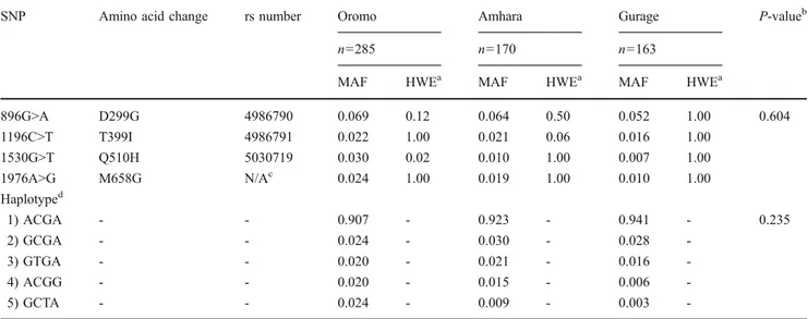
The TLR4 SNPs were at Hardy-Weinberg equilibrium and their frequencies were similar among the three ethnic groups, except for 1530G>T among Oromo (P= 0.02)
(Table2). While both 896G>A and 1196C>T are in strong
linkage disequilibrium among Caucasians (minor allele
frequency ~7%, R2>0.95), we found that this was not the
case in the present study (R2=0.32) and that 896A was
~3-fold less frequent than 1196T, as expected in an African
population (Table 3) [34]. To investigate whether
poly-morphisms in TLR4 were associated with susceptibility to leprosy, we compared the frequencies of TLR4 SNPs among leprosy cases and controls, adjusting for sex, age
groups and ethnicity (Table4). Both TLR4 SNPs 896G>A
and 1196C>T were less frequent among leprosy cases than controls. The 896GG, GA and AA genotypes were found in 91.7, 7.8 and 0.5% of leprosy cases versus 79.9, 19.1 and 1.0% of controls, respectively (odds ratio [OR]=0.34, 95% confidence interval [CI] 0.20–0.57, P<0.001, additive
model, Table5). These results were still significant when
the three different ethnic groups were analysed separately (OR=0.43, 95% CI 0.21-–.89, P=0.02 in Oromo; OR= 0.34, 95% CI 0.13-0–94, P=0.04 in Amhara and OR=0.18, 95% CI 0.05–0.65, P=0.008 in Gurage). Similarly, the 1196CC, CT and TT genotypes were found in 98.1, 1.9 and 0% of leprosy cases versus 91.8, 7.7 and 0.5% of controls, respectively (OR = 0.16, 95% CI 0.06–0.40, P<0.001, dominant model, comparing the presence of one or two copies of the minor allele versus no copies). Again, these results were consistent among the three different ethnic groups, although the significance level was not reached in the Gurage population (P=0.02 in Oromo; P=0.04 in Amhara and P=0.3 in Gurage, two-sided Fisher’s exact test). A third TLR4 SNP (1530G>T) also tended to be less frequent among leprosy cases than controls (1530GG, GT and TT genotypes were found in 98.0, 1.8 and 0.0% of leprosy cases versus 93.6, 5.8 and 0.6% of controls, respectively, OR=0.38, 95% CI 0.14–1.01, P=0.05, dom-inant model). However, the assessment of this third SNP Table 1 Baseline characteristics of leprosy cases and control subjects
Characteristics Total leprosy cases Controls P-valuea
n (%) n (%) Number of cases 441 (100.0) 197 (100.0) Male gender 302 (71.6) 144 (73.5) 0.700 Ethnicityb Oromo 193 (43.8) 92 (46.7) 0.337 Amhara 126 (28.6) 44 (22.3) Gurage 112 (25.4) 51 (25.9) Age groups 0–19 41 (9.9) 43 (22.2) <0.001 20–39 159 (38.5) 109 (56.2) 40–59 168 (40.7) 37 (19.1) ≥60 45 (10.9) 5 (2.6) Leprosy typec Lepromatous lepromatous (LL) 81 (19.7) Borderline lepromatous (BL) 199 (48.4) Borderline tuberculoid (BT) 128 (31.1) Tuberculoid (TT) 3 (0.7) Leprosy complicationsd Neuritis 133 (61.6) Reversal reaction 66 (30.6)
Erythema nodosum leprosum 17 (7.9)
aDouble-sided Fisher’s exact test for the distribution of characteristics among all cases versus controls b
Twenty patients had other (5 cases and 9 controls) or missing (5 cases and 1 control) ethnicity
c
Leprosy type was missing in five patients; 25 patients classified according the WHO as multibacillary (MB, 18 cases) or paucibacillary (PB, 7 cases) are not shown. Borderline borderline patients were classified together with BL (seeMethods section). A total of 298 patients were lepromatous (81 LL + 199 BL + 18 MB) and 138 were tuberculoid (128 BT + 3 TT + 7 PB, see Table4)
d
Complications data were available in 216 patients only
Note that the characteristics of patients and controls have already been reported [20]. They are presented here to provide the reader with an overview of the study population.
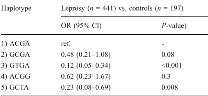
among the different ethnic groups was limited by the small sample size. In the analyses by haplotypes, all haplotypes that contained the 896A, the 1196C and/or the 1530T alleles were associated or tended to be associated with protection against leprosy (OR =0.48, 95% CI 0.21–1.08, P=0.08 for
haplotype 2, OR=0.12, 95% CI 0.05–0.34 for haplotype 3
and OR=0.23, 95% CI 0.08–0.69, P=0.008 for haplotype
5, compared to haplotype 1, Table 6). Together, these
results suggested that TLR4 polymorphisms are associated with susceptibility to leprosy.
Then, we compared frequencies of the TLR4 SNPs among leprosy patients with lepromatous (LL and BL) and tuberculoid (BT and TT) leprosy, adjusting for sex, age
groups and ethnicity (Table4). There was an association at
the limit of the significant level between the 1196C>T SNP and lepromatous leprosy. The 1196CC, CT and TT genotypes were found in 98.9, 1.1 and 0% of lepromatous leprosy patients versus 96.2, 3.8 and 0% of tuberculoid
patients, respectively (OR=0.23, 95% CI 0.05–0.99, P=
0.05, dominant model). No significant association was found when lepromatous lepromatous patients were com-pared with the other leprosy patients. Together, these results suggested that TLR4 SNPs are not strongly associated (if at all) with the type of leprosy.
Next, we investigated whether TLR4 polymorphisms were associated with the occurrence of leprosy complica-tions. No association was found between TLR4 SNPs and neuritis. The 1530T allele was more frequent in patients with reversal reaction (2/15 [13%] in patients with reversal reaction versus 3/177 [1.7%] in those without reversal reaction, P=0.05, Fisher’s exact test) and ENL (4/58 [6.9%] in patients with ENL versus 1/133 [0.7%] in those without ENL, P=0.03). However, the interpretation of these results is limited by the small sample size, which prevented stratification into separate ethnic groups and the use of multivariable models.
Table 2 Allelic and haplotypic frequencies of Toll-like receptor 4 (TLR4) single nucleotide polymorphisms (SNPs) in the three Ethiopian populations
SNP Amino acid change rs number Oromo Amhara Gurage P-valueb
n=285 n=170 n=163
MAF HWEa MAF HWEa MAF HWEa
896G>A D299G 4986790 0.069 0.12 0.064 0.50 0.052 1.00 0.604 1196C>T T399I 4986791 0.022 1.00 0.021 0.06 0.016 1.00 1530G>T Q510H 5030719 0.030 0.02 0.010 1.00 0.007 1.00 1976A>G M658G N/Ac 0.024 1.00 0.019 1.00 0.010 1.00 Haplotyped 1) ACGA - - 0.907 - 0.923 - 0.941 - 0.235 2) GCGA - - 0.024 - 0.030 - 0.028 -3) GTGA - - 0.020 - 0.021 - 0.016 -4) ACGG - - 0.020 - 0.015 - 0.006 -5) GCTA - - 0.024 - 0.009 - 0.003
-MAF = minor allele frequency; HWE = Hardy-Weinberg equilibrium test
a
Hardy-Weinberg equilibrium was calculated for each locus and the P-value is reported. A P-value of <0.05 indicates that the alleles are not in HWE
bDouble-sided Fisher’s exact test for the overall distribution of alleles/haplotypes among the three ethnic groups c
SNP 1976A>G has not been previously reported
d
Haplotypes with an MAF <1.5% in the whole population are not shown
TLR4 SNPs were not in linkage disequilibrium with any of the TLR2 polymorphisms that have been previously analysed in this population [20]
Table 3 Linkage disequilibrium between the TLR4 SNPs in all ethnic groups SNPs 896G>A 1196C>T 1530G>T 1976A>G 896G>A 0.06 1196C>T 0.32 0.02 1530G>T 0.26 0.00 0.02 1976A>G 0.00 0.00 0.00 0.02 Pairwise linkage disequilibrium (LD) were calculated with the pwld program developed in Stata 9 (StataCorp, College Station, TX). Off-diagonal elements are estimates of R2, assuming
Hardy-Weinberg equilibrium. The diagonal elements are minor allele frequencies (in italics). LD between 896G>A and 1196C>T was similar among the three ethnic groups (R2 = 0.34 in Oromo, R2 = 0.32 in Amhara and R2 = 0.27 in Gurage)
Finally, we examined the cellular mechanism of how M. leprae modulates TLR4-mediated immune responses. We previously demonstrated that irradiated M. leprae or its cell wall extracts stimulate TNF-α secretion through TLR2, but
not TLR4, in bone marrow-derived macrophages [35]. In
HEK293 cells transfected with an NF-κB luciferase construct, we also found that TLR2, but not TLR4, mediates signalling in response to stimulation with irradiated M.
leprae or its cell wall extracts (data not shown and [35]).
Together, these results suggested that M. leprae does not directly stimulate an immune response through TLR4, which is consistent with the absence of LPS in its cell wall. We hypothesised that M. leprae modulates TLR4-mediated immune responses by regulating shared compo-nents of the downstream TLR signalling pathway. To test this hypothesis, we isolated and stimulated human mono-cytes with LPS and M. leprae and measured cytokine production in culture supernatants. In contrast to LPS, M.
leprae did not induce IL-1β, IL-6 or IL-12p70 in
mono-cytes (Fig. 1A, B, C). To examine whether M. leprae
inhibits or merely fails to activate cytokine production, we examined cytokine production with mixed stimulations. In monocytes that were concurrently co-stimulated with M. leprae and LPS, cytokine production was similar to that of LPS alone. In contrast, when cells were pre-stimulated with M. leprae for 5 or 24 h and then stimulated with LPS,
IL-1β production was lower in comparison to stimulation with
LPS alone (Fig.1A). A similar trend was observed for IL-6
production after 24 h of M. leprae pre-stimulation, but the
differences were not statistically significant (Fig. 1B). In
contrast to 6 and 1β, there was no difference in IL-12p70 production among the different conditions tested
(Fig. 1C). As a control, we repeated the experiment in the
opposite stimulation order (LPS followed by M. leprae) and found no difference in cytokine levels. Next, we examined whether M. leprae modulated LPS-induced signalling in PBMCs. Similar to monocytes, we found that the pre-stimulation of PBMCs with M. leprae for 24 h led to decreased LPS-induced secretion of IL-6 in comparison to
stimulation with LPS alone (Fig. 1E). IL-1β levels were
also lower in the PBMCs that were pre-treated with M. leprae for 24 h, although the difference was not statistically
significant (Fig.1D). Similar to monocytes, no differences
in IL-6 or IL-1β levels were observed when the cells were stimulated with M. leprae after LPS. There was no difference among the different stimulation conditions when comparing levels of IL-12p70 or several T cell cytokines
(IFN-γ, IL-4 or IL-17). Together, these results suggest that
M. leprae inhibits the LPS induction of IL-1β and IL-6 secretion from monocytes. Furthermore, this effect was selective and not found for T-cell cytokines or other monocyte cytokines, such as IL-12p70.
Table 4 Association of TLR4 SNPs with leprosy and leprosy type SNP Leprosy n=441 Controls n=197 P-value Lepromatous, n=298 Tuberculoid, n=138 P-value Lepromatous Lepromatous, n=81 Other, n=355 P-value n (%) n (%) n (%) n (%) n (%) n (%) 896G>A
G/G 375 (91.7) 155 (79.9) ref. 256 (91.8) 119 (91.5) ref. 73 (91.3) 302 (91.8) ref. G/A 32 (7.8) 37 (19.1) <0.001 22 (7.9) 10 (7.7) 0.95 7 (8.8) 25 (7.6) 0.86
A/A 2 (0.5) 2 (1.0) 0.08 1 (0.4) 1 (0.8) - 0 (0.0) 2 (0.6)
-1196T>C
T/T 407 (98.1) 179 (91.8) ref. 282 (98.9) 125 (96.2) ref. 78 (98.7) 329 (97.9) ref. T/C 8 (1.9) 15 (7.7) <0.001 3 (1.1) 5 (3.8) 0.05 1 (1.3) 7 (2.1) 0.58
C/C 0 (0.0) 1 (0.5) - 0 (0.0) 0 (0.0) - 0 (0.0) 0 (0.0)
-1530G>T
G/G 391 (98.0) 162 (93.6) ref. 271 (98.2) 120 (97.6) ref. 75 (98.7) 316 (97.8) ref. G/T 7 (1.8) 10 (5.8) 0.06 5 (1.8) 2 (1.6) 0.72 1 (1.3) 6 (1.9) 0.98
T/T 1 (0.3) 1 (0.6) 0.56 0 (0.0) 1 (0.8) - 0 (0.0) 1 (0.3)
-1976A>G
A/A 386 (96.5) 164 (94.8) ref. 267 (95.7) 119 (98.3) ref. 73 (94.8) 313 (96.9) ref. A/G 14 (3.5) 9 (5.2) 0.64 12 (4.3) 2 (1.7) 0.29 4 (5.2) 10 (3.1) 0.62 P-values are calculated in a logistic regression model that did not assume any particular mode of inheritance and accounted for the presence of each genotype (0/1 and 1/1) versus the wild type genotype (0/0), adjusted for age group, sex and ethnicity (general model). For statistically significant associations, we performed a likelihood ratio tests for three different models (dominant, recessive and additive) versus the general model. The best fitting models are shown in Table5. The numbers of individuals with assessable genotypes may differ slightly for the different SNPs
T able 5 Association of TLR4 SNPs with leprosy in three ethnic groups All ethnicities Oromo Amhara Gurage Leprosy , n = 441 Controls, n= 197 Leprosy , n = 193 Controls, n=9 2 Leprosy , n = 126 Controls, n=4 4 Leprosy , n =1 1 2 Controls, n=5 1 SNP n (%) n (%) n (%) n (%) n (%) n (%) n (%) n (%) 896G>A G/G 375 (91.7) 155 (79.9) 162 (90.5) 72 (80.9) 108 (90.8) 35 (79.5) 97 (94.2) 41 (80.4) G/A 32 (7.8) 37 (19.1) 15 (8.4) 16 (18.0) 1 1 (9.2) 8 (18.2) 6 (5.8) 10 (19.6) A/A 2 (0.5) 2 (1.0) 2 (1.1) 1 (1.1) 0 (0.0) 1 (2.3) 0 (0.0) 0 (0.0) Add. model OR = 0.34 (0.20 –0.57), P < 0.001 OR = 0.43 (0.21 –0.89), P = 0.02 OR = 0.34 (0.13 –0.94), P = 0.04 OR = 0.18 (0.05 –0.65), P = 0.008 1 196T>C T/T 407 (98.1) 179 (91.8) 175 (97.8) 82 (91.1) 121 (98.4) 40 (90.9) 103 (98.1) 48 (94.1) T/C 8 (1.9) 15 (7.7) 4 (2.2) 8 (8.9) 2 (1.6) 3 (6.8) 2 (1.9) 3 (5.9) C/C 0 (0.0) 1 (0.5) 0 (0.0) 0 (0.0) 0 (0.0) 1 (2.3) 0 (0.0) 0 (0.0) Dom. model a OR = 0.15 (0.06 –0.39), P < 0.001 (P = 0.02) b (P = 0.04) b (P = 0.3) b 1530G>T G/G 391 (98.0) 162 (93.6) 167 (97.1) 72 (90.0) 1 1 5 (99.1) 37 (94.9) 101 (98.1) 45 (100.0) G/T 7 (1.8) 10 (5.8) 4 (2.3) 7 (8.8) 1 (0.9) 2 (5.1) 2 (1.9) 0 (0.0) T/T 1 (0.3) 1 (0.6) 1 (0.6) 1 (1.3) 0 (0.0) 0 (0.0) 0 (0.0) 0 (0.0) Dom. model OR = 0.38 (0.14 –1.01), P = 0.05 (P = 0.03) b (P = 0.2) b -W e performed a likelihood ratio test for three dif ferent models (dominant, recessive and additive) versus the general model. The best fitting models are shown. a Indicates that the best fitting models could not be determined by likelihood ratio testing, since homozygous cases were missing in certain groups; th e dominant model is shown bFor small groups, rare allele carriers (0/1 or 1/1) were compared to non-carriers (0/0) using a two-sided Fisher ’s exact test. Numbers of individuals with assessable genotypes may dif fer slightly for the dif ferent SNPs. The ef fect of TLR4 896G>A and 1 196T>C were independent from the presence of TLR2 polymorphisms previously associated with reversal reaction [ 54 ] (not shown)
Discussion
In this paper, we showed that two SNPs in TLR4 (896G>A [D299G] and 1196C>T [T399I]) were associated with protection against leprosy in an Ethiopian cohort of leprosy patients and healthy controls. We also demonstrated that M. leprae downregulates TLR4-mediated cytokine production in monocytes.
Recent studies have also found associations of TLR SNPs and leprosy susceptibility. An SNP in TLR1 (T1805G
[I602S]) was associated with M. leprae infection [36–38].
Another study reported that a TLR2 SNP (C2029T [R677W]) located in a critical region of the Toll-interleukin receptor (TIR) domain was associated with lepromatous leprosy in Table 6 Association of TLR4 haplotypes with leprosy
Haplotype Leprosy (n = 441) vs. controls (n = 197) OR (95% CI) P-value) 1) ACGA ref. -2) GCGA 0.48 (0.21–1.08) 0.08 3) GTGA 0.12 (0.05–0.34) <0.001 4) ACGG 0.62 (0.23–1.67) 0.3 5) GCTA 0.23 (0.08–0.69) 0.008
P-values are calculated in a logistic regression model including all haplotypes (each coded 0, 1 or 2, for the presence of no, one or two copies of the haplotype), using haplotype 1 as a reference. The results are adjusted for age group, sex and ethnicity
Fig. 1 Monocyte and peripheral blood mononuclear cell (PBMC) cytokine response to mixed stimulation with Mycobacterium leprae and lipopolysaccharide (LPS). Human peripheral blood monocytes and PBMCs were isolated and infected at a multiplicity of infection (MOI) of 5:1 with heat-killed M. leprae (ML), LPS (100 ng/mL), M. leprae and LPS simultaneously, or pre-stimulation with either M. leprae or LPS for 5 or 24 h, followed by treatment with the other
stimulus. Supernatants were collected at 48 h post-stimulation and analysed by Luminex for the presence of multiple cytokines. The results are the means ± standard error of three experiments (donors) performed in duplicate, with the exceptions of IL-1β production from monocytes (five donors) and IL-6 production from monocytes (four donors). A two-tailed paired t-test was used for statistical analysis (*P< 0.05, **P<0.005, #P<0.001, in comparison to LPS alone)
Korea [39]. While an amino-acid substitution at this location completely abolished TLR2 signalling in transfected cells
[35], subsequent studies revealed that this supposedly
functional SNP was, in fact, located in a highly homologous pseudogene region located 23 kB upstream of the TLR2 gene
[40]. More recently, we showed that a microsatellite
poly-morphism located in TLR2 significantly increased the risk of
leprosy reversal reaction [20]. Allelic length variations in the
microsatellite may influence promoter activity and modify
TLR2 function through alteration of its expression level [41].
However, the TLR2 microsatellite polymorphism associated with leprosy reversal reaction did not strongly influence susceptibility to leprosy itself.
In the present study, we examined whether polymorphisms in TLR4 influenced susceptibility to or the clinical character-istics of leprosy in an Ethiopian cohort. We found that the minor alleles of two TLR4 SNPs (896G>A [D299G] and 1196C>T [T399I]) were associated with a reduced risk of developing leprosy. Such a protective effect is in contrast with most studies. In fact, TLR4 896A and 1196T were frequently associated with increased susceptibility to patho-gens, including Gram-negative bacteria, Brucella species, respiratory syncytial virus (RSV), Plasmodium falciparum
and Candida albicans (reviewed in [6]).
Our data support the hypothesis that, in the case of specific infections, dysfunctional TLR SNPs may reduce, instead of increase, susceptibility to infections. This hypothesis is also supported by previous observations: the dysfunctional allele of TLR1 1805G (602S) exerted a
protective effect against leprosy [37]; the dysfunctional
2258G/A (R753Q) was shown to increase the risk of
tuberculosis [42] but to protect against Lyme borreliosis
[43]; finally, as it is the case for leprosy, TLR4 896A and
1196T were shown to protect from Legionnaire’s disease
[44]. The reason why a specific TLR allele exerts a protective
effect against some infections and an increased risk for others is unknown. These differences reflect the complexity of human immune responses to infectious agents and may result from co-evolving selective pressures in hosts and pathogens. TLR4 896G>A and 1196C>T have been intensively exam-ined in functional and genetic epidemiological studies. 896A was associated with LPS hypo-responsiveness after the in
vivo inhalation of endotoxin [23, 45]. In vitro cellular
investigations also suggested that 299G and/or 399I was
unable to mediate LPS signalling in some [23,46] but not all
cell types [47–49]. These seemingly contradictory findings
may be explained by the use of small sample sizes, which resulted in differences with borderline statistical significance; comparison of different cell types; use of different doses and types of LPS; and measurement of different cytokines and inflammatory markers. Taken together, these studies suggest that SNPs 896G>A and 1196C>T partially influence inflammatory pathways under some experimental conditions.
While TLR2 has been clearly implicated in the innate immune response to M. leprae, the role of TLR4 is currently unknown. We previously found that irradiated M. leprae or its cell wall extracts stimulated TNF-α production through
TLR2, but not TLR4 [35]. If live M. tuberculosis signals
through TLR4, then it is plausible that M. leprae may do so
similarly [13]. Alternatively, TLR4 may modulate
inflam-matory processes that influence leprosy disease manifes-tations but are not attributable to direct stimulation by M. leprae. Consistent with this hypothesis, we found that the stimulation of monocytes with M. leprae inhibited their subsequent response to TLR4 stimulation with LPS. There are several possible mechanisms of this inhibitory effect, including tolerance, a well-described phenomenon where prior exposure of cells to TLR ligands results in a transient hypo-responsive state that is refractory to additional
stimulation [50]. Several mechanisms have been proposed
for tolerance, including the alteration of expression levels of negative and positive regulators of the TLR signalling pathway. For example, IRAK-M is a negative regulator of the TLR signalling pathway that is downstream of both TLR2 and TLR4 and is involved in endotoxin tolerance
[51]. M. leprae may alter the expression of molecules such
as IRAK-M through TLR2 stimulation and, ultimately, modulate LPS/TLR4 responsiveness through such a shared signalling intermediate. An alternative mechanism could be from direct inhibitory effects of an M. leprae molecule on the TLR signalling pathway. There are several precedents for this mechanism, including YopJ inhibition of MAPK
signalling by Yersinia pestis [52] and the recently described
TIR domain containing proteins (Tcps) in Escherichia coli that bind MyD88 and inhibit TLR-induced signalling
pathways [53]. Future studies will address these possible
mechanisms of M. leprae inhibition of TLR4 signalling. The alteration of monocyte responses to M. leprae by TLR4 polymorphisms via these different mechanisms could modulate the growth of bacilli, the nature and degree of the innate immune response, and the subsequent adaptive immune response. TLRs are known to influence Th1–Th2 pathway development in mice and such immunomodulatory
effects could operate in humans as well [7]. TLR4
poly-morphisms may modulate several of these immunologic effects and, thereby, influence overall susceptibility to leprosy. Future studies will address which cellular immune responses are modulated by TLR4 polymorphisms
This study was limited by the low frequency of the minor alleles of TLR4. This issue was of particular concern for the analyses of leprosy types (lepromatous versus tuberculoid), as well as for leprosy complications, for which it prevented stratification into separate ethnic groups and the use of multivariable models. However, the analyses of susceptibility to leprosy itself were performed in larger sample sizes. In this case, initial analyses were performed
for the whole population, including the three major ethnic groups. In order to limit the risk of population stratification, we also performed the analyses in the three different groups separately. Overall, most observations were consistent among the three different ethnic groups.
In summary, our data show that polymorphisms in TLR4 are associated with susceptibility to leprosy in Ethiopian patients. It suggests that polymorphisms in individual TLRs may influence different clinical manifestations of the disease. Acknowledgements This work was supported by grants from the Swiss National Science Foundation (81LA-65462 to PYB), the Swiss Foundation for Medical-Biological Grants (1121 to PYB), the Leenaards Foundation (to PYB) and the National Institute of Health (AI25032 to AA, AI22616 and AI 54361 to GK). The AMFES project in Ethiopia was funded by the International Federation of Anti-Leprosy Associations (ILEP) Members through Netherlands Anti-Leprosy Relief (NLR). We thank Sarah Li for technical assistance with genotyping and Abraham Aseffa for his help and advice in bringing the manuscript to press.
The authors have no conflict of interest in the present manuscript.
References
1. [No authors listed] (2007) Global leprosy situation, 2007. Wkly Epidemiol Rec 82:225–232
2. Ridley DS, Jopling WH (1966) Classification of leprosy according to immunity. A five-group system. Int J Lepr Other Mycobact Dis 34:255–273
3. Scollard DM, Adams LB, Gillis TP et al (2006) The continuing challenges of leprosy. Clin Microbiol Rev 19:338–381. doi:10.1128/ CMR.19.2.338-381.2006
4. Yamamura M, Uyemura K, Deans RJ et al (1991) Defining protective responses to pathogens: cytokine profiles in leprosy lesions. Science 254:277–279. doi:10.1126/science.1925582
5. Salgame P, Abrams JS, Clayberger C et al (1991) Differing lymphokine profiles of functional subsets of human CD4 and CD8 T cell clones. Science 254:279–282. doi:10.1126/science.1681588
6. Bochud PY, Bochud M, Telenti A et al (2007) Innate immuno-genetics: a tool for exploring new frontiers of host defence. Lancet Infect Dis 7:531–542. doi:10.1016/S1473-3099(07)70185-8
7. Iwasaki A, Medzhitov R (2004) Toll-like receptor control of the adaptive immune responses. Nat Immunol 5:987–995. doi:10.1038/ ni1112
8. Akira S, Uematsu S, Takeuchi O (2006) Pathogen recognition and innate immunity. Cell 124:783–801. doi:10.1016/j.cell.2006.02.015
9. Quesniaux V, Fremond C, Jacobs M et al (2004) Toll-like receptor pathways in the immune responses to mycobacteria. Microbes Infect 6:946–959. doi:10.1016/j.micinf.2004.04.016
10. Jo EK, Yang CS, Choi CH et al (2007) Intracellular signalling cascades regulating innate immune responses to Mycobacteria: branching out from Toll-like receptors. Cell Microbiol 9:1087– 1098. doi:10.1111/j.1462-5822.2007.00914.x
11. Heldwein KA, Fenton MJ (2002) The role of Toll-like receptors in immunity against mycobacterial infection. Microbes Infect 4:937– 944. doi:10.1016/S1286-4579(02)01611-8
12. Krutzik SR, Ochoa MT, Sieling PA et al (2003) Activation and regulation of Toll-like receptors 2 and 1 in human leprosy. Nat Med 9:525–532. doi:10.1038/nm864
13. Means TK, Wang S, Lien E et al (1999) Human toll-like receptors mediate cellular activation by Mycobacterium tuberculosis. J Immunol 163:3920–3927
14. Alcaïs A, Mira M, Casanova JL et al (2005) Genetic dissection of immunity in leprosy. Curr Opin Immunol 17:44–48. doi:10.1016/ j.coi.2004.11.006
15. Alcaïs A, Alter A, Antoni G et al (2007) Stepwise replication identifies a low-producing lymphotoxin-alpha allele as a major risk factor for early-onset leprosy. Nat Genet 39:517–522.
doi:10.1038/ng2000
16. Santos AR, Suffys PN, Vanderborght PR et al (2002) Role of tumor necrosis factor-alpha and interleukin-10 promoter gene polymorphisms in leprosy. J Infect Dis 186:1687–1691.
doi:10.1086/345366
17. Moraes MO, Pacheco AG, Schonkeren JJ et al (2004) Interleukin-10 promoter single-nucleotide polymorphisms as markers for disease susceptibility and disease severity in leprosy. Genes Immun 5:592–595. doi:10.1038/sj.gene.6364122
18. Mira MT (2006) Genetic host resistance and susceptibility to leprosy. Microbes Infect 8:1124–1131. doi:10.1016/j.micinf.2005.10.024
19. Misch EA, Hawn TR (2008) Toll-like receptor polymorphisms and susceptibility to human disease. Clin Sci (Lond) 114:347– 360. doi:10.1042/CS20070214
20. Bochud PY, Hawn TR, Siddiqui MR et al (2008) Toll-like receptor 2 (TLR2) polymorphisms are associated with reversal reaction in leprosy. J Infect Dis 197:253–261. doi:10.1086/524688
21. Ferwerda B, Kibiki GS, Netea MG et al (2007) The toll-like receptor 4 Asp299Gly variant and tuberculosis susceptibility in HIV-infected patients in Tanzania. AIDS 21:1375–1377.
doi:10.1097/QAD.0b013e32814e6b2d
22. Bochud PY, Chien JW, Marr KA et al (2008) Toll-like receptor 4 polymorphisms and aspergillosis in stem-cell transplantation. N Engl J Med 359:1766–1777. doi:10.1056/NEJMoa0802629
23. Arbour NC, Lorenz E, Schutte BC et al (2000) TLR4 mutations are associated with endotoxin hyporesponsiveness in humans. Nat Genet 25:187–191. doi:10.1038/76048
24. Saunderson P, Gebre S, Desta K et al (2000) The ALERT MDT Field Evaluation Study (AMFES): a descriptive study of leprosy in Ethiopia. Patients, methods and baseline characteristics. Lepr Rev 71:273–284 25. Jopling WH (1981) A practical classification of leprosy for field
workers. Lepr Rev 52:273
26. Saunderson P, Gebre S, Byass P (2000) Reversal reactions in the skin lesions of AMFES patients: incidence and risk factors. Lepr Rev 71:309–317
27. Gordon D, Abajian C, Green P (1998) Consed: a graphical tool for sequence finishing. Genome Res 8:195–202
28. Hawn TR, Dunstan SJ, Thwaites GE et al (2006) A polymorphism in Toll-interleukin 1 receptor domain containing adaptor protein is associated with susceptibility to meningeal tuberculosis. J Infect Dis 194:1127–1134. doi:10.1086/507907
29. Turvey SE, Hawn TR (2006) Towards subtlety: understanding the role of Toll-like receptor signaling in susceptibility to human infections. Clin Immunol 120:1–9. doi:10.1016/j.clim.2006.02.003
30. S.A.G.E. Statistical Analysis for Genetic Epidemiology. Case Western Reserve University. 5.0 edn, 2005
31. Lahiri R, Randhawa B, Krahenbuhl J (2005) Application of a viability-staining method for Mycobacterium leprae derived from the athymic (nu/nu) mouse foot pad. J Med Microbiol 54:235– 242. doi:10.1099/jmm.0.45700-0
32. Suzuki K, Fukutomi Y, Matsuoka M et al (1993) Differential production of interleukin 1 (IL-1), IL-6, tumor necrosis factor, and IL-1 receptor antagonist by human monocytes stimulated with Mycobacterium leprae and M. bovis BCG. Int J Lepr Other Mycobact Dis 61:609–618
33. Murray RA, Siddiqui MR, Mendillo M et al (2007) Mycobacte-rium leprae inhibits dendritic cell activation and maturation. J Immunol 178:338–344
34. Ferwerda B, McCall MB, Alonso S et al (2007) TLR4 poly-morphisms, infectious diseases, and evolutionary pressure during
migration of modern humans. Proc Natl Acad Sci USA 104:16645–16650. doi:10.1073/pnas.0704828104
35. Bochud PY, Hawn TR, Aderem A (2003) Cutting edge: a Toll-like receptor 2 polymorphism that is associated with lepromatous leprosy is unable to mediate mycobacterial signaling. J Immunol 170:3451–3454
36. Misch EA, Macdonald M, Ranjit C et al (2008) Human TLR1 deficiency is associated with impaired mycobacterial signaling and protection from leprosy reversal reaction. PLoS Negl Trop Dis 2:e231. doi:10.1371/journal.pntd.0000231
37. Johnson CM, Lyle EA, Omueti KO et al (2007) Cutting edge: A common polymorphism impairs cell surface trafficking and functional responses of TLR1 but protects against leprosy. J Immunol 178:7520–7524
38. Hawn TR, Misch EA, Dunstan SJ et al (2007) A common human TLR1 polymorphism regulates the innate immune response to lipopeptides. Eur J Immunol 37:2280–2289
39. Kang TJ, Chae GT (2001) Detection of Toll-like receptor 2 (TLR2) mutation in the lepromatous leprosy patients. FEMS Immunol Med Microbiol 31:53–58. doi:10.1111/j.1574-695X.2001.tb01586.x
40. Malhotra D, Relhan V, Reddy BS et al (2005) TLR2 Arg677Trp polymorphism in leprosy: revisited. Hum Genet 116:413–415.
doi:10.1007/s00439-004-1249-9
41. Yim JJ, Ding L, Schäffer AA et al (2004) A microsatellite polymorphism in intron 2 of human Toll-like receptor 2 gene: functional implications and racial differences. FEMS Immunol Med Microbiol 40:163–169. doi:10.1016/S0928-8244(03)00342-0
42. Ogus AC, Yoldas B, Ozdemir T et al (2004) The Arg753GLn polymorphism of the human toll-like receptor 2 gene in tuberculosis disease. Eur Respir J 23:219–223. doi:10.1183/ 09031936.03.00061703
43. Schröder NW, Diterich I, Zinke A et al (2005) Heterozygous Arg753Gln polymorphism of human TLR-2 impairs immune activation by Borrelia burgdorferi and protects from late stage Lyme disease. J Immunol 175:2534–2540
44. Hawn TR, Verbon A, Janer M et al (2005) Toll-like receptor 4 polymorphisms are associated with resistance to Legionnaires’ disease. Proc Natl Acad Sci USA 102:2487–2489. doi:10.1073/ pnas.0409831102
45. Michel O, LeVan TD, Stern D et al (2003) Systemic responsive-ness to lipopolysaccharide and polymorphisms in the toll-like receptor 4 gene in human beings. J Allergy Clin Immunol 112:923–929. doi:10.1016/j.jaci.2003.05.001
46. Fagerås Böttcher M, Hmani-Aifa M, Lindström A et al (2004) A TLR4 polymorphism is associated with asthma and reduced lipopolysaccharide-induced interleukin-12(p70) responses in Swedish children. J Allergy Clin Immunol 114:561–567.
doi:10.1016/j.jaci.2004.04.050
47. Erridge C, Stewart J, Poxton IR (2003) Monocytes heterozygous for the Asp299Gly and Thr399Ile mutations in the Toll-like receptor 4 gene show no deficit in lipopolysaccharide signalling. J Exp Med 197:1787–1791. doi:10.1084/jem.20022078
48. von Aulock S, Schröder NW, Gueinzius K et al (2003) Heterozygous toll-like receptor 4 polymorphism does not influ-ence lipopolysaccharide-induced cytokine release in human whole blood. J Infect Dis 188:938–943. doi:10.1086/378095
49. Paulus SC, Hirschfeld AF, Victor RE et al (2007) Common human Toll-like receptor 4 polymorphisms—role in susceptibility to respiratory syncytial virus infection and functional immunological relevance. Clin Immunol 123:252–277
50. Medvedev AE, Sabroe I, Hasday JD et al (2006) Tolerance to microbial TLR ligands: molecular mechanisms and relevance to disease. J Endotoxin Res 12:133–150. doi:10.1179/096805106X102255
51. Kobayashi K, Hernandez LD, Galán JE et al (2002) IRAK-M is a negative regulator of Toll-like receptor signaling. Cell 110:191– 202. doi:10.1016/S0092-8674(02)00827-9
52. Viboud GI, Bliska JB (2005) Yersinia outer proteins: role in modulation of host cell signaling responses and pathogenesis. Annu Rev Microbiol 59:69–89. doi:10.1146/annurev.micro.59. 030804.121320
53. Cirl C, Wieser A, Yadav M et al (2008) Subversion of Toll-like receptor signaling by a unique family of bacterial Toll/interleukin-1 receptor domain-containing proteins. Nat Med Toll/interleukin-14:399–406.
doi:10.1038/nm1734
54. Bochud PY, Hawn TR, Siddiqui MR et al (2008) Toll-like receptor 2 (TLR2) polymorphisms are associated with reversal reaction in leprosy. J Infect Dis 197:253–261. doi:10.1086/ 524688