HAL Id: cea-02999253
https://hal-cea.archives-ouvertes.fr/cea-02999253
Submitted on 10 Nov 2020
HAL is a multi-disciplinary open access
archive for the deposit and dissemination of
sci-entific research documents, whether they are
pub-lished or not. The documents may come from
teaching and research institutions in France or
abroad, or from public or private research centers.
L’archive ouverte pluridisciplinaire HAL, est
destinée au dépôt et à la diffusion de documents
scientifiques de niveau recherche, publiés ou non,
émanant des établissements d’enseignement et de
recherche français ou étrangers, des laboratoires
publics ou privés.
Molecular Simulations of Dodecyl-β-maltoside Micelles
in Water: Influence of the Headgroup Conformation and
Force Field Parameters
Stéphane Abel, François-Yves Dupradeau, E. Prabhu Raman, Alexander
Mackerell, Jr., Massimo Marchi
To cite this version:
Stéphane Abel, François-Yves Dupradeau, E. Prabhu Raman, Alexander Mackerell, Jr., Massimo
Marchi. Molecular Simulations of Dodecyl-β-maltoside Micelles in Water: Influence of the Headgroup
Conformation and Force Field Parameters. Journal of Physical Chemistry B, American Chemical
Society, 2011, 115 (3), pp.487-499. �10.1021/jp109545v�. �cea-02999253�
Molecular Simulations of Dodecyl-
β-maltoside Micelles in Water: Influence of the
Headgroup Conformation and Force Field Parameters
Ste´phane Abel,*,†Franc¸ois-Yves Dupradeau,‡ E. Prabhu Raman,§
Alexander D. MacKerell, Jr.,§and Massimo Marchi†
Commissariat a` l’Energie Atomique, DSV/iBiTEC-S/SB2SM/LBMS & CNRS URA 2096, Centre d’Etudes,
Saclay, F-91191 Gif-sur-YVette Cedex, France, CNRS URA2096, F-91191, Gif-sur-YVette Cedex, France, Laboratoire des glucides, UFR de Pharmacie & CNRS UMR 6219, UniVersite´ de Picardie - Jules Verne, Amiens, France, and Department of Pharmaceutical Sciences, School of Pharmacy, UniVersity of Maryland, Baltimore, Maryland, United States
ReceiVed: October 5, 2010; ReVised Manuscript ReceiVed: NoVember 21, 2010
This paper deals with the development and validation of new potential parameter sets, based on the CHARMM36 and GLYCAM06 force fields, to simulate micelles of the two anomeric forms (R and β) of N-dodecyl-β-maltoside (C12G2), a surfactant widely used in the extraction and purification of membrane proteins.
In this context, properties such as size, shape, internal structure, and hydration of the C12G2anomer micelles
were thoroughly investigated by molecular dynamics simulations and the results compared with experiments. Additional simulations were also performed with the older CHARMM22 force field for carbohydrates (Kuttel, M.; et al. J. Comput. Chem. 2002, 23, 1236-1243). We find that our CHARMM and GLYCAM parameter sets yield similar results in the case of properties related to the micelle structure but differ for other properties such as the headgroup conformation or the micelle hydration. In agreement with experiments, our results show that for all model potentials the β-C12G2micelles have a more pronounced ellipsoidal shape than those
containing R anomers. The computed radius of gyration is 20.2 and 25.4 Å for the R- and β-anomer micelles, respectively. Finally, we show that depending on the potential the water translational diffusion of the interfacial water is 7-11.5 times slower than that of bulk water due to the entrapment of the water in the micelle crevices. This retardation is independent of the headgroup in R- or β-anomers.
I. Introduction
Glycolipids (GLs) are glycosyl derivatives of lipids that belong to a large family of molecules known as glycoconju-gates.1 From a chemical point of view, GLs designate any
molecule with surfactant properties containing a carbohydrate headgroup (with one or more monosaccharide units) attached to a lipophilic tail. In particular, alkyl-glycosides are simple glycolipid molecules obtained, for instance, by condensation such as “Fischer glycosidation” of a sugar with a fatty alcohol.2
Their tension-active properties depend on the length of the alkyl chain.3,4 Because they are highly effective, ecologic, and
nontoxic, this class of surfactants has a wide range of applica-tions in food, cosmetic, and pharmaceutical industries.5-7Similar
to other surfactants, GLs can form different mesophases in water, such as micelles, lamellae, vesicles, etc., depending on the experimental conditions (see, for instance, refs 3 and 8-15). During the last decades, alkyl-glucosides have also attracted a great deal of interest in the context of membrane protein extraction16,17 given that they have a low critical micellar
concentration (cmc) around 10-3-10-4mol · L-1(see Table 2 of ref 7 for data and references), form large aggregates, and keep the protein structure and its activity intact.18,19In particular,
alkyl-glucosides have been successfully used for solubilization of large membrane protein complexes, such as rhodopsin,20
cytochrome c oxidase,21,22and protein channel.23,24
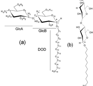
Among these detergents, N-dodecyl-β-maltoside (C12G2)
(Figure 1) is one of the most used alkyl-glucosides in membrane protein extraction experiments.17Two anomers, R- and β-, exist
for C12G2due to two possible connectivities of the dodecane
chain (DOD) and of the anomeric carbon (C1′) linking DOD to * Corresponding author. E-mail: stephane.abel@cea.fr.
†DSV/iBiTEC-S/SB2SM/LBMS & CNRS URA2096. ‡Universite´ de Picardie - Jules Verne.
§University of Maryland.
Figure 1. The R- (a) and β-anomers (b) of C12G2surfactants with the
atom numbering scheme used in the paper. The anomeric center C1′ of
the molecule is underlined with a star.
10.1021/jp109545v 2011 American Chemical Society Published on Web 12/30/2010
Downloaded via CEA SACLAY on November 10, 2020 at 18:01:27 (UTC).
the maltose head. The β-isomer is in a linear conformation, whereas the R-isomer is in a right-angle bent formed between the maltose headgroup and the alkyl tail. The R-anomer is less soluble than the β-anomer7,25 and is not commonly used in
membrane protein studies, albeit with some notable exceptions. For instance, in the bacterial E. coli Na+/H+antiporter, it was reported recently24that the R-C
12G2surfactant is needed to obtain
a crystallized protein in its native state. To explain this feature, it is generally assumed that the orientation of the maltose head in the surfactant leads to differences in sterical constraints (i.e., due to different packing parameters) and, possibly, in hydration behaviors. SANS and SAXS scattering experiments have shown that the R-anomer of C12G2forms a small quasi-spherical micelle,
while the β-conformation forms large oblate aggregates.13,26
In the past few years, molecular dynamics simulations (MD) have been extensively used to study the structure and hydration of mono- and disaccharides, in aqueous solutions (e.g., refs 27-40). MD with alkyl-glycosides (such as alkyl-β-O-glucoside (C8Glc1) or alkyl-β-O-galactoside (C8Gal)) have also been
performed in the past, involving bilayers41-43or micelles44-47
or interacting with a plant protein.48In the studies of Bogusz et
al.44and Chong et al.,47the influence of the surfactant headgroup
conformation on the micelle structure and hydration has been thoroughly examined. It was reported that despite the confor-mational changes in the headgroup, several properties remain nearly unchanged for the aggregates, in particular micelle shape, surfactant tail length, and conformation. In contrast, other properties directly related to the interaction between the solvent and the headgroup (i.e., solvent accessible surface area, head-group cluster structure, number of isolated water molecules at the micelle surface) are significantly modified by the stereo-chemistry of the carbohydrate head.44,47It was also noted that
the sugar counterpart is responsible for strong perturbations of the water structure at the micelle surface and it affects the formation of a large hydrogen bond (HB) network at the micelle surface between water and the headgroups, and within the headgroups themselves (inter- and intra-HBs, respectively).41,47,49
This HB network may also explain the good thermal stability of the surfactants49and cryoprotective effects.36,50
Despite their crucial role in membrane protein extractions, C12G2 micelles have rarely been studied by computational
methods, and to our knowledge, only a single MD study has been performed on these systems in the past.51That investigation
was mainly focused on the hydration properties of the micelles and used a thermodynamical approach to examine the micel-lization behavior of surfactants with different chemical nature, and was compared with experiments. To simplify the calcula-tions, the simulations were carried out starting from a spherical aggregate of 45 β-C12G2. This aggregation number is far from
that of 130, commonly accepted in the literature.13,14,26Moreover,
the authors used the OPLS-AA force field not fully optimized for glycolipid molecules.52In absence of optimized parameters
for the two C12G2anomers for the widely used CHARMM and
GLYCAM06 force fields, we have developed two sets of parameters compatible with these force fields for condensed-phase MD simulation studies. We expect that these new parameters will allow us to examine the influence of the headgroup stereochemistry in C12G2 micelle structures under
experimental conditions. Our endeavor included the calculations of new RESP atomic charges for the current GLYCAM06 potential and an optimization of the dihedral angles for the acetal linkage between the maltose headgroup and the alkyl chain for both of the R- and β-anomers compatible with the CHARMM36 force field (see next sections). These new CHARMM and
GLYCAM parameters (named GLYCAM06 and CHARMM-Opt, respectively) have been tested and validated by performing different MD simulations of two realistic models of C12G2
micelles in explicit water. To compare our results with previous MD studies of glycolipid micelles,44-46we have also performed
two simulations with the old CHARMM22 parameters taken from Kuttel et al.53and Reiling et al.,54named throughout the
paper with the acronym CHARMM-K.
This paper is organized as follows: in the next sections, we describe the procedures followed to derive RESP atomic charges as well as the optimization of the dihedral angles for the acetal linkage for the R- and β-anomers for the GLYCAM and CHARMM force fields. This is followed by sections covering MD simulations, the results, as well as the interpretations of these results.
II. Methods
1. RESP Charge Derivation for the r- and β-Anomers
of C12G2 for GLYCAM. RESP charge derivation for C12G2
GL for the GLYCAM force field was carried out using standard methods.55-57 For this purpose, we have followed a similar
approach to that described by Gouin et al.39Three molecules,
i.e., methyl R-D-glucopyranoside (AMG), methyl β-D-glucopy-ranoside (BMG), and 1-dodecanol (OH-DOD), were involved in charge derivation. For each glucose unit, two rotamers were selected for the ω (i.e., represented by the O6C6C5C4 and
O6C6C5O5rotational angles) in the gauche-gauche (gg, ω )
-60°) and gauche-trans (gt, ω ) +60°) conformations (see Figure S1 in the Supporting Information) considering that they are the most populated in solution.58 Optimized geometries
presenting intramolecular HBs were excluded from charge derivation to avoid the overpolarization effect.56 As a
conse-quence, geometry optimization was carried out with dihedral angle constraints. The HO4′O4′C4H4′ dihedral angle of the
glucosides AMG and BMG was constrained to 180°, whereas the HO3-O3-C3-H3and HO2-O2-C2-H2dihedrals of AMG
and BMG were constrained to 180°, respectively. A single conformation for 1-dodecanol was considered in charge deriva-tion, and the selected geometry was optimized in its extended conformation (i.e., “all-trans dihedrals”). Indeed, it has been previously shown that the surfactant alkyl chain adopts mainly this conformation in the micelle core (see, for instance, refs 44, 59, and 60). Frequencies were calculated for all the molecules, and transition state structures were excluded from the charge derivation procedure. Geometry optimization, fre-quency calculation, and molecular electrostatic potential (MEP) computation were carried out using the Gaussian 03 quantum mechanics (QM) package in the gas phase,61 whereas charge
fitting was performed using the RESP program.55,56The
HF/6-31G** level of theory was used in the geometry optimization and frequency calculations,62while MEP computation was based
on HF/6-31G*, leading to implicit polarization required in condensed phase simulations when using the nonadditive AMBER force field model.55,56The CHELPG algorithm was
used to compute the grid of points involved in MEP computa-tion.63The molecular orientation of each optimized geometry
was controlled using the rigid-body reorientation algorithm implemented in the RED program.63Four molecular orientations
for each optimized geometry of AMG and BMG (based on the glucose atom names C1C3C5, C5C3C1, C2C4O5, and O5C4C2; see
Figure S1 in the Supporting Information) and for the alkyl chain (based on the 1-dodecanol atom names C1C2C3, C3C2C1, C1C3C5,
and C5C3C1; see Figure S1 in the Supporting Information) were
fitting procedure to yield reproducible atomic charges. Charge fitting was performed using a single RESP stage with a hyperbolic constraint value of 0.01. Intramolecular charge constraints between the methyl and the C4 hydroxyl groups of AMG and BMG and intermolecular charge constraints between the methyl group of each methyl glucoside and the hydroxyl group of OH-DOD (Figure S1, Supporting Information) were set to a value of zero during charge fitting, allowing the definition of the molecular fragments and force field libraries required to build C12G2GL. Each hydrogen atom bound to an
sp3 carbon was also constrained to a target value of zero during charge fitting to ensure a compatibility with the GLYCAM force field.64 The charge derivation procedure was automatically
carried out using version IV of the R.E.D. program.63 RESP
charges for the R- and β-anomers of C12G2are reported in the
Supporting Information, and are freely available from the R.E.D.D.B. server (http://q4md-forcefieldtools.org/REDDB/)65
with the accession code “F-72”.
2. Optimization of the Dihedral Angle Potentials for the
Acetal Linkage in r- andβ-C12G2for CHARMM. Ethoxy
tetrahydropyran (Et-THP) was used as a model compound to parametrize the acetal linkage in C12G2GL. R- and β-Substituted
anomers were involved in the study (Figure S2, Supporting Information). Tetrahydropyran (THP) parameters were those previously developed in the context of hexopyranoses.66 The
bond, angle, dihedral, Lennard-Jones, and partial charge pa-rameters involving the acetal group were transferred by analogy to methoxy-THP (Met-THP) parameters developed previously67
and existing linear ether parameters (Listing S1, Supporting Information).68The optimization of these transferred parameters
was undertaken as follows. QM MP2/cc-pVTZ//MP2/6-31G* Φ/Ψ scans for both R- and β-anomers were performed (Φ ) OR-C1-OE-C6, Ψ ) C1-OE-C6-C7) at 15° increments, thus
giving rise to 576 conformations as target data. Figure S3 in the Supporting Information shows that the transferred parameters serve as a good initial guess, as they reproduce the QM energy surface satisfactorily. The dihedral parameters involving the exocyclic heavy atoms were directly fitted using the MCSA automated dihedral fitting procedure69 to the QM Φ/Ψ data,
resulting in a marked improvement in the root-mean-square error (RMSE) from 2.01 (1.23) to 0.81 (0.83) kcal/mol for both the R- and β-anomer. Figure S3 in the Supporting Information shows that the fit parameters reproduce well the QM Φ/Ψ potential energy surface for both anomers. Additionally, Figure S4 in the Supporting Information shows that the fitted param-eters reproduce the 1-D QM energy as a function of Ψ (Φ is kept constant at (60° for the R- and β-anomer) much better than the transferred ones. Unconstrained MP2/6-31G* optimiza-tion of the molecules revealed the QM global minima for the R-anomer at values (61.5°, 175.1°) and for the β-anomer at values of (-63.2°, -172°). These QM minima are correctly reproduced by our optimized dihedral parameters, as seen in Table S2 in the Supporting Information. We also notice that they also well reproduce the energy difference between the global minima of the two anomers, ∆ER-β (∆ER-β ) -1.52
kcal/mol for QM and ∆ER-β) -1.85 kcal/mol for MM).
The transferred partial charge on the ether oxygen atom (OE)
was validated by performing water pair interaction energy calculations as performed in previous studies.66This consists
of optimizing the solute/water interaction distance at the HF/ 6-31G* level of theory, with constraints on all other degrees of freedom. Table S3 in the Supporting Information shows that our parameters reproduce well the scaled QM interaction energies and adjusted distances,66thus validating the transfer
of charges. These new sets of parameters are provided in the Supporting Information in the CHARMM readable format (Listing S1, Supporting Information).
3. Simulation Methods. To be consistent with the force
fields developed in this study, we used an “all-atom” approach to model all micellar systems. RESP charges derived in section II.1 were involved in GLYCAM-based MD simulations with the bonded and nonbonded parameters taken from the GLYCAM06 force field version f.64 The 1-4 van der Waals
and electrostatic scaling factors were set to 1.0, in agreement with the developer recommendations.64 In the case of the
CHARMM-K simulations, we used two sets of parameters to model the sugar headgroup and the exocyclic atom connecting the maltose head and the alkyl chain. For the maltose head, we used the parameters of Kuttel et al.53 (a revision of the
CHARMM22 force field for sugars of Ha et al.)70and for the
acetal atom those of Reiling et al.,54 which is assigned to an
ether oxygen atom type with a partial charge of -0.30 e. These two sets of parameters were previously employed by Bogusz et al.44,45 and Konidala et al.46 in MD simulations of C
8G1
micelles in water. For the CHARMM-Opt simulations, we used the new set of parameters for hexopyranose66 with optimized
parameters for the ΦH/ΨH glycosidic dihedral angles which
significantly improves the CHARMM force field for simulations of polysaccharides.67For the connection between the maltose
head and the alkyl chain, we adopted the optimized parameter set for ethers by Lee et al.71combined with the optimized torsion
parameters developed for Et-THP described in the previous section II.2. For all MDs performed with the CHARMM force fields, the alkyl chain of the surfactant was modeled with parameters developed by Klauda et al. for alkanes.72 This
contrasts with previous investigations44,45 and the work of
Konidala et al.46where for alkyl chain the dihedral parameters
described in ref 73 were used. Finally, the TIP3P water model73
was adopted to model the solvent for all the simulations.
4. Construction of the Micelles and Simulation Tech-niques. In this paper, we made the choice of using two
preassembled micelles with 75 and 132 monomers of R- and β-C12G2, respectively. For the three different force fields studied
in this work, the same approach was used to construct the six micelles and an identical protocol was applied in MD simula-tions. The aggregation numbers (Ndet) were obtained by Dupuy
et al.13by fitting the scattering curves from SAXS and SANS
experiments at a temperature of 297 K and a concentration range between 20 and 100 mM for monodispersed micelles. We should emphasize that the Ndetvalues may depend on the experimental
methods and on the concentration. For example, the Ndetvalue
for the β-C12G2micelle used here is higher than the value of
98 found by Rosevear et al.74 using the gel filtration method,
the value of 82-120 obtained by SAXS measurements26at 310
K, and the value of 111 ( 10 derived by time resolved fluorescence quenching (TRFQ) spectroscopy.75On the other
hand, our Ndet value is well within the findings of other
experiments, e.g., Ndet ) 125 ( 10 was reported using other
TRFQ results at 4.89-19.6 mM and 289-333 K,14 whereas
Ndet) 135-145 was found by Lipfert et al.76by SAXS at 298
K. In the case of the R-C12G2micelle, only one aggregation
number has been reported in the literature, Ndet) 75.13
The initial configurations of the two aggregates were con-structed using the Packmol program.77 This program created
initial condition for MD by packing in the sphere NdetC12G2
molecules in their extended conformation, with the first carbon atom (C7) of the alkyl chain, near the headgroup at∼17 Å from
(∼14.0 Å) of the DOD chain in an extended conformation obtained with the DS Visualizer v2.0 (Accelrys Inc., San Diego, CA) modeling program. In this way, the number of “steric clashes” for the terminal ethyl groups of the C12G2alkyl chain
is reduced. To remove other inter- and intramolecular overlaps, additional stages of conjugate gradient minimization followed by MD equilibration in a vacuum with a small time step were carried out. Following this step, the headgroups were random-ized by running MD simulations of the aggregates at 450 K for 300 ps, with the surfactant tails kept fixed to their minimized conformation. From this point on, in all cases, the systems were solvated by adding TIP3P water at standard density in a truncated octahedron cell, corresponding to a simple cubic primitive cell unit with parameters a ) b ) c and R ) β ) γ ) 109.472°. Given the two different Ndet values used in our
study, we chose a ) 85 Å and a ) 95 Å for the simulations of the R- and β-anomers, respectively. These box sizes were chosen to ensure that all the molecular systems were within the experimental L1phase (i.e,<45 % in weight concentration),75
and that there was a sufficient distance (∼15 Å) between the micelle surface and the edge of the box to minimize interactions with periodic images.78As shown in Table 1, where we provide
the composition of the two simulated systems, the simulated concentrations of the detergent are near 0.3 and 0.40 M for the R- and β-C12G2anomers, respectively. This is higher than the
surfactant concentration used in the experimental study of Dupuy et al.13(0.02-0.04 mol · L-1) and higher than the critical micelle
concentration (cmc) for the R- and β-C12G2(∼1.5 × 10⁻⁴and
∼2.0 × 10⁻⁴mol · L-1, respectively)13,75at a temperature of 298
K. Indeed, simulations near the cmc would require about 2000 times more water in the simulation system, leading to a dramatic increase of the calculation time. Furthermore, in order to equilibrate the solvent molecules, the systems were run for 300 ps at 450 K, with the ensemble of the micelle atoms fixed to their initial positions. Then, the constrained micelle atoms were released and the system slowly heated from 0 to 297 K in 300 ps as described in ref 79. Finally, the resulting conformations were simulated in the NPT ensemble (T ) 297 K and P ) 0.1 MPa) for 14 ns after discarding the initial 300 ps. MD snapshots were saved every 240 fs for subsequent analysis.
5. Molecular Dynamics. For all the simulations and analysis
performed in this study, serial and parallel versions of the ORAC code were used.80To simulate in the NPT ensemble, ORAC
uses a method based on the extended system approach by adding extra (virtual) coordinates and momenta in the system to control the temperature and the pressure.81-85To integrate the equation
of motion, a five-time-step r-RESPA integration scheme with a Liouvillean separation in three nonbonded shells was used.86
The procedure combines the smooth particle mesh Ewald
(SPME) method to handle electrostatic interactions87 and the
method of constraints to keep the covalent bonds involving hydrogens at their equilibrium length.88The SPME parameters
were chosen to maintain a relative error of less than 0.1% on the electrostatic interactions for all the micelles. For this purpose, a convergence parameter of R ) 0.43 Å-1, a fifth-order B-spline, and a 96 grid points in each Cartesian direction to take care of the SPME charge interpolation for each simulated system was used.
III. Results
First, we consider the general structure of the R- and β-C12G2
aggregates at different simulation times: tsim≈ 0 fs, tsim) 7.0
ns, and tsim) 14.0 ns of the production period. Figure 2 provides
three snapshots for each type of C12G2micelle taken from the
CHARMM-Opt simulations; these results are representative of the other simulations described in this paper. Visual inspection of the figure shows that, at the beginning of the run, the micelles have rougher surfaces with some glycolipid protrusions (see Figure 2a and d). Thus, the micelles present large solvent-exposed surfaces where the alkyl chains of the surfactant are exposed to the solvent (see below). However, after typically 2-3 ns of production, Figure 2b-e, the surfactants arrange themselves to form compact aggregates where a major part of the alkyl tails are buried and the surface is covered with the glycolipid headgroups. These pictures also show that the R- and
β-C12G2micelles are not perfectly spherical and that the β-C12G2
micelle is more ellipsoidal than the R-one (see below). This finding has been observed in the different MD simulations performed in this study, regardless of the force field. Finally, in contrast with previous simulations46,47of octyl-β-glucoside
micelles, the integrity of the micelles studied with the GLYCAM and CHARMM force fields is maintained all along our simula-tion time window (14 ns), and no surfactant molecules escaped from the micelle. This is consistent with previous experimental results obtained for alkyl-maltoside micelles where it was found that the monomer exchange time between the micelle and the solvent is in the order of 0.1-1.2 µs.89
A. Size of the Micelles. To examine the stability and to
measure the dimensions of the micelle in the six runs, we have computed their instantaneous radii of gyration, Rg, over the
course of the simulations (Figure 3) using the following expression:
where miis the mass of atom i of the micelle at the distance ri
from the center of mass rcm. For all the micelles studied here,
the Rgvalues are stable after typically∼3 ns of production. As
indicated previously, during the beginning of the productive run (i.e., between∼0 and 3 ns time period), local arrangements and compaction of the surfactants on the micelle surfaces are observed. This feature was also observed in previous glycolipid micelle simulations.47
The instantaneous Rgvalues for the R-C12G2micelle in the
three runs stabilizes after∼3 ns around ∼20-21 Å until the end of the trajectory. The average Rgvalues,〈Rg〉, of the R-C12G2
micelles, computed from the last 11 ns of CHARMM-K, CHARMM-Opt, and GLYCAM06 runs (20.5 ( 0.1, 20.2 ( 0.1, and 20.0 ( 0.1 Å, respectively) are close to the Rg
experimental value (18.6 ( 0.6 Å13) calculated from the semiaxis
lengths of the micelle aM, bM, and cMand the expression Rg2)
TABLE 1: Parameters for Micelle Simulationsa
system R-C12G2 β-C12G2 NC12G2 75 132 NH2O 13771 18389 Natm 47388 65859 mC12G2/mTot 13.4 16.9 T (K) 297 297 tsim(ns) 14.0 14.0 F (g/cm3) 1.02 ( 0.01 1.03 ( 0.01 aN
C12G2, NH2O, and Natm are the numbers of
N-dodecyl-β-D-maltopyranoside monomers (C12G2), water, and atoms
composing the simulated systems. mC12G2/mTot is the weight
concentration (in %) of C12G2 in each system. tsimis the simulation
time (the 300 ps equilibration period was excluded from the analysis). Rg 2)
∑
i mi(ri- rcm) 2/
∑
i mi (1)(aM2+ bM2+ cM2)/5 for an ellipsoid with a uniform density.79
In the case of the β-C12G2micelles, Rgreached stable values
after∼2.5-3 ns depending on the force field used. In contrast to the R-C12G2 simulations, the three force fields lead to
significant differences (up to 1.0 Å) for the instantaneous and averaged Rg, especially for the two CHARMM force field
versions. For β-C12G2, the〈Rg〉’s are equal to 26.4 ( 0.1, 25.4
( 0.1, and 25.1 ( 0.1 Å, for the aggregates simulated with the CHARMM-K, CHARMM-Opt, and GLYCAM06, respectively. The〈Rg〉’s obtained with the CHARMM-Opt and GLYCAM06
parameters are closer to the experimental value of 23.3 ( 0.6 Å.13
B. Shape of the Micelles. The micelle shape can be
characterized by computing the instantaneous ellipsoidal axis ratio between the micelle major (aM) and minor (cM) semiaxis
lengths, or aM/cM, obtained from the inertia tensors (see ref 79
for details). As shown in Figure 4, the six micelles present a
stable ellipsoidal shape (i.e., aM/cM> 1.0) after typically ∼4.0
ns. For the R-C12G2 micelles, their shapes do not depend
significantly on the force field. This contrasts with the behaviors of the β-C12G2micelles where the ratio aM/cMshows much larger
fluctuations. To be consistent with the calculation of Rg, the
initial 3 ns of each trajectory were discarded in the calculation of aM/cM. The average values of the ratio,〈aM/cM〉, are reported
in the seventh column of Table 2 for the different micelles studied. In our calculation, both R- and β-C12G2are ellipsoidal,
but R-C12G2is the closest to the shape of a sphere. In the case
of β-C12G2aggregates, the instantaneous aM/cMvalues stabilize
around∼1.40 at the end of the simulations independently of the considered force field and the〈aM/cM〉 values are close for
the three micelles: 1.38 ( 0.03, 1.43 ( 0.01, and 1.38 ( 0.04 for CHARMM-K, CHARMM-Opt, and GLYCAM06, respec-tively. In contrast, β-C12G2micelles have a pronounced oblate
shape (aM> bM≈ cM), which agrees well with the experimental Figure 2. Representative MD snapshots of the R- (a-c) and β-anomers (d-f) of C12G2micelles simulated with the CHARMM-Opt force field at
t≈ 240 fs (a, d), t ≈ 7.0 ns (b, e), and t ≈14 ns (c, f) of the production period. Oxygen and hydrogen are in red and white. Carbons in GlcA, GlcB,
and the alkyl chain are blue, cyan, and gray, respectively. Water molecules are removed for visual clarity. Graphics were produced with the PyMOL program.122
Figure 3. Time evolution of the radius of gyration, Rg, for R- (a) and
β-anomers (b) of C12G2micelles.
Figure 4. Time evolution of the major and minor semiaxis of inertia ratio for R- (a) and β-anomers (b) of C12G2micelles.
observations.13,26,75However, we must emphasize that the〈a M/
cM〉 values for the β-C12G2micelles remain, depending on the
force field, lower than the estimated experimental values (i.e.,
aM/cM) 1.70 from Dupuy et al.).13
The instantaneous length values of the semiaxis of each micelle hydrophobic core (i.e., aHC, bHC, and cHC) have also been
computed by including only the contributions from the surfactant hydrophobic chain in the calculations of the inertia tensors. The average values of the three semiaxes (〈aHC〉, 〈bHC〉, and 〈cHC〉)
are reported in the 8th, 9th, and 10th columns of Table 2. We find that the average major and minor semiaxis lengths 〈aHC〉
and〈cHC〉 are in relative agreement with experimental values
for R-C12G2and β-C12G2(i.e.,〈aHC〉 ) 〈cHC〉 ) 18.6 ( 1.0 Å
and 〈aHC〉 ) 28.2 ( 1.0 Å and 〈cHC〉 ) 14.1 ( 1.0 Å,
respectively).13The average thickness of the polar outer layer,
or 〈lpl〉, computed by subtracting the semiaxis length of the
hydrophobic core from those of the whole micelle (〈aM〉, 〈bM〉,
and〈cM〉) does not change significantly with the force field and
is 6.7 ( 0.3 Å for the R-anomer of the C12G2and increases up
to 7.3 ( 0.4 Å for the β-anomer. Due to the partial folding of the maltose head, these values are 39.1 and 33.4% smaller than the calculated length of the maltose in its extended configuration (∼11.0 Å). The larger 〈lpl〉 value for β-C12G2is consistent with
the fact that the β-anomer is linear and extends in the solvent, whereas the R-anomer is more folded, and hence constrained on the micelle surface. In all cases, the〈lpl〉 values are within 1
Å of the experimental values estimated from SAXS and SANS experiments, i.e., 5.4 ( 0.1 and 6.2 ( 0.1 Å for R-C12G2and
β-C12G2, respectively.13,76
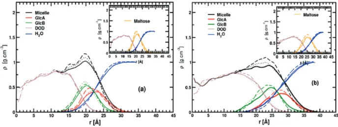
C. Density Profiles of the Micelles. To compare the spatial
extent of the most relevant atomic group components of the
micelles, we have computed their average radial mass density profiles F(r) as a function of the distance, r, of the group from the center of mass (COM) of the micelle.79We discuss here
the F(r) values obtained for the entire micelle (Micelle), the maltose headgroup (Maltose), the dodecane tail (DOD), the two maltose glucose rings (GlcA and GlcB), and the hydration water (H2O). As previously discussed,79 the nonspherical nature of
the micelles will affect the F(r) value to a certain extent, causing the broadening of these functions. F(r) values, averaged over the last 11 ns of each trajectory, are shown in Figure 5. The hydrophobic core F(r) profiles extend from 0 to∼19.0 Å from the micelle COM, and present similar shapes regardless of the force field. For the hydrophilic maltose head, the F(r)’s present a strong peak with density maxima at 21.0 ( 0.5 and 25.0 ( 0.5 Å for the R- and β-C12G2micelles, respectively.
These values are close to those obtained previously for 〈Rg〉.
We notice that the position of the main peaks for the GlcA and GlcB density profiles depends more strongly on the force field than that of the other groups studied here. Considering all the results, GlcA F(r)’s extend from 15.0 to 25.0 Å and from 18.0 to 28.0 Å for R- and β-C12G2 micelles, respectively. Larger
values, of 1-2 Å, are found for all GlcB F(r) values. The water density profile in Figure 5 shows that water molecules deeply penetrate into the micelle headgroup and solvate the maltose heads to a different degree with a preference for the outermost glucose ring (GlcA). We can also emphasize that water shares significant contact with the micelle hydrophobic core, as seen by the intercepts of the water and DOD F(r) curves. Finally, the water F(r) curves reach their bulk density value (∼1.0 g/cm3)
near the edge of the box at 35 Å (R-C12G2) and 38 Å (β-C12G2)
from the micelle COM.
TABLE 2: Average Dimensions and Shapes of the Six Micellesa
force field micelle 〈Rg〉 〈aM〉 〈bM〉 〈cM〉 〈aM/cM〉 〈aHC〉 〈bHC〉 〈cHC〉 〈lpl〉
CHARMM-K R-C12G2 20.5 28.4 25.6 23.8 1.20 22.1 19.1 16.5 6.7 β-C12G2 26.4 38.8 34.7 28.1 1.38 33.1 27.3 20.0 7.2 CHARMM-Opt R-C12G2 20.2 27.8 26.1 24.3 1.14 21.5 19.1 16.7 7.0 β-C12G2 25.4 38.1 33.0 26.5 1.43 30.6 24.8 19.3 7.7 GLYCAM06 R-C12G2 20.0 28.4 25.5 23.1 1.23 22.3 18.6 16.5 6.5 β-C12G2 25.2 37.2 32.0 30.1 1.38 30.1 24.4 19.2 7.2
a〈...〉 stands for the ensemble average. Values with M and HC subscripts were computed by including all the micelle atoms and those of the
hydrophobic core, respectively. The radii of gyration and the semi-axis lengths were computed using the inertia tensor (e.g., see ref 79) and the main text for details.〈lpl〉 is the average polar layer thickness of the micelle in Å. The statistical errors (maximum errors) are always lower than
0.1, 0.8, and 0.3 Å for Rg, semi-axis lengths, and polar layer thickness, respectively.
Figure 5. Average radial density profiles with respect to the center of mass (r ) 0 Å) for simulations performed with CHARMM-K (continuous line), CHARMM-Opt (dotted lines), and GLYCAM06 force fields (dashed line) for R- (a) and β-anomers (b) of C12G2micelles. In the inset, the
radial profiles for the micelle hydrophobic core (maroon), the maltose headgroup (orange), and water (blue) are plotted. A 0.5 Å bin width was used for both figures.
D. Hydration of the Headgroup. As discussed in the
previous section, radial mass density profiles show significant interaction between water and the C12G2surfactant headgroups.
To gain insight on this aspect, we provide in Table 3 some surface area (SA) properties of the micelles computed from the last 11 ns of the simulations. We first compare the instantaneous value of the SA per surfactant headgroup (SAHGC12G2) with the
results of Dupuy et al.13 obtained from SAXS and SANS
experiments assuming that the R- and β-micelles are spherical. As described by these authors SAHGC12G2, the surface of the
hydrophobic core of a sphere of radius RjHCwas calculated. The
average ellipsoid semiaxes of the micelle hydrophobic core 〈aHC〉, 〈bHC〉, and 〈cHC〉 were used to calculate RjHC with the
expression RjHC ) (〈a
HC〉〈bHC〉〈cHC〉)1/3. The average values of
SAHGC12G2(〈SAHGC12G2〉) are reported in the third column of Table 3.
We found that the〈SAHGC12G2〉 values are in the range 60.3-61.1
and 55.5-65.1 Å2for the C
12G2R- and β-anomers, respectively.
The values obtained with CHARMM-Opt and GLYCAM06 agree well with the experimental values (58 and 52 Å2for the
C12G2R- and β-anomers, respectively), whereas the CHARMM-K
results are further away.
In the fourth and fifth columns of Table 3 are reported the average surface areas (SA) for the whole micelles〈SAvC12G2〉 and
〈SAeC12G2〉, respectively. The SAvC12G2values were obtained from
the Voronoi construction90 by adding up the surface areas of
each Voronoi polyhedron shared between all surfactant atoms and water.91 Instead, SA
e
C12G2 is the ellipsoidal surface of the
micelle obtained from the average semiaxis lengths (〈aM〉, 〈bM〉,
and〈cM〉) computed in section III.B. Comparison of the two
values confirms that the micelle interfaces are corrugated (as also shown in Figure 1). Indeed, the surface rugosity factors fs
) SAvC12G2/SAeC12G2 were calculated between 1.7 and 2.1 and
between 1.64 and 2.0 for the R- and β-anomers, respectively. In the sixth column of Table 3, we also provide the average C12G2surface alkyl chain ratio of the micelles,〈ftail〉, in contact
with the solvent. The 〈ftail〉 values were calculated with the
SAvC12G2and the surface area of the alkyl chain atoms, SAvC12.
〈ftail〉 values for all the micelles decrease by ∼1.0% when the
surfactant head changes from the R- to the β-anomer and are calculated between 9.8 and 11.1% and between 9.3 and 10% for the R- and β-anomers, respectively. The lower〈ftail〉 values
of GLYCAM06 indicate that for those micelles the surfactant alkyl chains of the micelle are more protected from the solvent than those obtained in the simulations with the CHARMM force fields. In comparison, the〈ftail〉 values reported in this work are
lower than those found by Stephenson et al.51for a micelle with
45 β-C12G2(∼17.0%), and also lower than those for micelles
with 27 octyl-β-galactose47 (∼30%) and 92 octyl-β-glucose
monomers (20%).46 The latter is probably due to the smaller
size of the headgroup in those micelles, which shields less the micelle hydrophobic core from water.
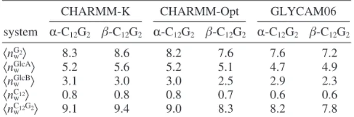
The values of the average number of water molecules,〈nw〉,
in the first solvation shell of each surfactant, or hydration number, were also computed during the last 11 ns of each run. They are presented in Table 4. For each configuration, the hydration water molecules were selected using a simple cutoff radius criterion, as described in ref 92. Briefly, a water molecule is considered near the micelle surface if its distance with any of the detergent atoms is less than Rcut) f(Rw+ RC12G2), where
Rwand RC12G2are the force field van der Waals radii of the water
and of the C12G2 atoms, respectively. f is a parameter set
arbitrarily to 1.1 in our calculations.92Five hydration numbers
were calculated here: for the whole C12G2molecule (〈nwC12G2〉),
for the headgroup (〈nwG2〉), for the GlcA (〈nwGlcA〉) and GlcB
(〈nwGlcB〉) units, and for the alkyl chain (〈nwC12〉). For all 〈nw〉’s,
higher values are found for the simulations obtained with CHARMM-K, whereas the 〈nw〉’s of CHARMM-Opt and
GLYCAM06 simulations are close to one another. The〈nwG2〉
values for CHARMM-Opt and GLYCAM06 are computed around 8.2 and 7.2, respectively, and compare well with the estimation of〈nwC12G2〉 obtained from SANS, SAXS (8.0),13or
TRFQ experiments for β-C12G2 micelles (7.9).75 The bent
conformation of the R-C12G2slightly increases the headgroup
hydration by 0.4-0.6 waters for the micelles simulated with the CHARMM-Opt and GLYCAM06 parameters, whereas for CHARMM-K the 〈nwG2〉 decreases by 0.3 units. For maltose
molecules in water, hydration numbers between 5.7 and 22.6 are estimated by experimental studies, whereas values within 6.5-14.5 are obtained from computer simulations (see Table 3 of ref 33 and references therein). The validity of these results depends on the experimental techniques or the force field considered. Comparison of the hydration of the glucose rings GlcA and GlcB shows that the number of water molecules around GlcB, connected to the alkyl chain, is, on average,∼2.6 units lower than that for GlcA. This is independent of the force field employed and the conformation of the headgroup. Finally, the low values for〈nwC12〉 (<1.0 water) confirm that the micelle
hydrophobic core only has sporadic contacts with the solvent consistently with the〈ftail〉 value calculated in this work.
To further examine the headgroup hydration, in Table 5, we provide the average number of nearest waters for each maltose-oxygen, or 〈nOx-Ow〉, obtained from the computed radial pair
density functions (RDFs) of the maltose-oxygen (O1-10) and
water-oxygen (Ow) atoms. To obtain〈nOx-Ow〉 the RDF functions
were integrated until the first minimum at r≈ 3.5 Å after the first peak. The〈nOx-Ow〉 results indicate that water molecules
solvate primarily the hydroxyl oxygen atoms (O1, O2, O3, O4,
O6, O8, O9, and O11) of the maltose headgroup. This is consistent
TABLE 3: Surface Properties of the Micellesa
force field micelle 〈SAHGC12G2〉 〈SAVC12G2〉 〈SAeC12G2〉 〈ftail〉
CHARMM-K R-C12G2 61.1 19425.0 9244.5 11.0 β-C12G2 65.1 33237.6 16581.5 10.0 CHARMM-Opt R-C12G2 60.4 17625.0 9013.2 10.9 β-C12G2 56.8 29422.8 15894.6 9.3 GLYCAM06 R-C12G2 60.3 15675.0 9146.6 9.8 β-C12G2 55.5 25726.8 15666.0 9.3
a〈...〉 stands for the ensemble average. SA
HG
C12G2 (in Å2) is the
average surface area per headgroup computed using the micelle oil core radius. SAvC12G2 and SAeC12G2 are the average micelle surfaces
computed with the Voronoi polyhedron,90assuming an ellipsoid-like
geometry for the micelle.〈ftail〉 is the average surface fraction shared
between the water and the C12G2 alkyl chain. The statistical errors
(maximum errors) are always lower than 3.0 and 0.2% for the surfaces and〈ftail〉 values.
TABLE 4: Hydration Numbersa
CHARMM-K CHARMM-Opt GLYCAM06 system R-C12G2 β-C12G2 R-C12G2 β-C12G2 R-C12G2 β-C12G2 〈nwG2〉 8.3 8.6 8.2 7.6 7.6 7.2 〈nwGlcA〉 5.2 5.6 5.2 5.1 4.7 4.9 〈nwGlcB〉 3.1 3.0 3.0 2.5 2.9 2.3 〈nwC12〉 0.8 0.8 0.8 0.7 0.6 0.6 〈nwC12G2〉 9.1 9.4 9.0 8.3 8.2 7.8
a〈...〉 stands for the ensemble average. 〈n
w G2〉, 〈n w GlcA〉, 〈n w GlcB〉, 〈n w C12〉,
and〈nwC12G2〉 give the average number of water molecules at 4.0 Å of
the maltose head, the outermost and innermost glucose rings, and the alkyl chain, respectively. The statistical errors (maximum errors) are always lower than 0.1%.
with previous simulation studies performed on other glycolipid micelles.44,46,47The number of water molecules in the first shell
of these atoms is found to be between 1.9 and 3.7 as a function of the oxygen localization on the headgroup. These values vary little with the force field employed and the surfactant conforma-tion. In general, the higher〈nOx-Ow〉 values are obtained for the
oxygen atoms O3(2.9-3.4) and O6(3.2-2.4), the most the
atoms are exposed to the solvent in GlcA. As an example, the hydroxyl oxygen O11in GlcB has about one water molecule
in its first shell that the equivalent hydroxyl oxygen atom O6in
GlcA. The hydration of the ring oxygen atoms O5and O10of
GlcA and GlcB does not change much (∼0.3 water) when the surfactant conformation changes from the R- to β-anomer. Finally, for the oxygen atoms involved in the glycosidic bond (O1) and the link between the headgroup and alkyl chain (O7),
low〈nOx-Ow〉 values (<0.8 and <0.5) are found, indicating that
these oxygens are shielded from the solvent. This result is also consistent with another study,46 where a〈n
Ox-Ow〉 value close
to 0.2-0.3 for O7was observed.
Considering the strong interaction between the C12G2
head-group atoms and water, it is clear that the headhead-group oxygen atoms will form an extended network of hydrogen bonds with water molecules and with donor groups of the detergent themselves.93 The average number of HBs between water
molecules and the maltose headgroup (WHB) and within the surfactant headgroups (HHB) were computed for the last 11 ns of the simulations, using the following geometrical criterion: a HB was considered if the oxygen-oxygen distance equals or is smaller than 3.5 Å and the angle O-H · · · O was between 120 and 180°.94 These values are reported in Tables S4, S5,
and S6 of the Supporting Information. We observed that each R-C12G2 molecule presents 0.5-1.0 more WHB than the
β-anomer. These values vary only slightly with the force fields,
with CHARMM-K having the largest number of HBs in R-C12G2
followed by CHARMM-Opt and GLYCAM06. This result is in agreement with small variations of hydration water in the first shell of the C12G2headgroup and the decrease of〈nOx-Ow〉
reported in the previous paragraph. We found that each C12G2
headgroup makes, on average,∼1.5 times more in the establish-ment of HB as an acceptor (∼7.0) than as a donor (∼5). This is consistent with previous computer simulation studies of C8G1
and C8Gal in water.47 As also expected, the total number of
WHBs obtained in this work is lower than the total WHBs found for maltose in TIP3P water where the number of WHBs is∼15.9
and 22.4.27,32Further analysis of the individual HB donors and
acceptors shows that the O3 and O6 (GlcA) and O8 and O9
(GlcB) hydroxyl oxygens make about one additional HB with water than the other oxygens. For these same oxygens, the number of WHBs decreases by ∼0.2 when the headgroup changes from the R- to the β-anomer. Finally, due to the strong entanglement of the maltose head at the micelle surface and to the low hydration of some of the hydroxyl oxygen atoms, it is expected that a large network of inter- and intra-headgroup donor/acceptor HBs (INHBs and INTHBs, respectively) will be favored. Several inter-headgroup HBs have been computed (among these previously examined by Chong et al.47) and are
reported in the fifth to ninth columns in Tables S4, S5, and S6 in the Supporting Information. The number of INHBs is found to be much lower than the number of solvent-headgroup HBs. On average, there are ∼0.10 and ∼0.25 HBs per lipid for structures obtained from the CHARMM and GLYCAM force field simulations, respectively. The number of INHBs is greater in β-C12G2 micelles and is mostly due to the O6-H · · · O4,
O8-H · · · O10, and O8-H · · · O9HBs. Furthermore, three other
INTHBs were previously observed (O6H6· · · O5 and
O11H11· · · O10 in the first and second glucose rings and
O2H2· · · O9 between GlcA and GlcB).27,28,94 These HBs are
correlated with the rotational freedom of the ω torsion angles and the R(1f4) glycosidic linkage flexibility. Our calculations indicate that these HB types exist during all of the simulations. In particular, O2H2· · · O9 is present during approximately
70-80% of the simulation times, whereas the two other INTHBs only∼30-40% of the time.
E. Conformation of the Surfactant. To examine in more
detail the hydration differences of the micelles, we have studied the surfactant conformation by computing the main peak of the average end-to-end distance probability distributions P(r). Specifically, we have computed dC12and dG2by collecting from
the trajectories the distances between the atoms C7and C19of
the alkyl chain and between the O4and O12of the maltose head,
respectively. Since the dC12 and dG2end-to-end distances are
strongly related to the conformations of the alkyl tail and headgroup of the surfactant, respectively, we have examined the conformation of several characteristic torsion angles of the tail (such as all the CCCC dihedral angles, the first C7C8C9C10,
and last C15C16C17C18dihedrals),44the torsion angles involved
in the connection of the alkyl chain and the maltose head (i.e., O7C7C8C9,44O10C1′O7C7,27,95and C1′O7C7C828,96), and ω1 and
TABLE 5: Nearest Neighbors for the Maltose Oxygensa
CHARMM-K CHARMM-Opt GLYCAM06
〈nOx-Ow〉 R-C12G2 β-C12G2 ∆Rfβ R-C12G2 β-C12G2 ∆Rfβ R-C12G2 β-C12G2 ∆Rfβ O1-Ow 0.7 0.8 0.1 0.5 0.5 0.0 0.5 0.4 -0.1 O2-Ow 2.3 2.7 0.4 2.5 2.3 -0.2 2.1 2.6 0.5 O3-Ow 3.6 3.7 0.1 2.9 3.2 0.3 3.0 3.4 0.4 O4-Ow 2.0 2.6 0.6 2.3 2.6 0.3 1.9 2.4 0.5 O5-Ow 1.1 1.3 0.2 1.3 1.0 -0.3 1.3 1.0 -0.3 O6-Ow 3.1 2.5 -0.6 2.6 2.3 -0.3 3.2 2.4 -0.8 total GlcA 12.8 13.6 0.8 12.1 11.9 -0.2 12.0 12.2 0.2 O7-Ow 0.2 0.5 0.3 0.3 0.3 0.0 0.2 0.4 0.2 O8-Ow 2.4 1.9 -0.5 1.9 1.6 -0.3 1.7 1.5 -0.2 O9-Ow 2.4 2.0 -0.4 2.2 1.8 -0.4 2.2 1.6 -0.6 O10-Ow 0.8 0.9 0.1 0.7 0.6 -0.1 0.6 0.8 0.2 O11-Ow 2.0 1.7 -0.3 1.7 1.6 -0.1 1.8 1.3 -0.5 total GlcB 7.8 7.0 -0.8 6.8 5.9 -0.9 6.5 5.6 -0.9
aThe micelle oxygen atoms are labeled as follows: O
1and O7are the acetalic oxygens, O5and O10are the maltose ring oxygens, and all the
remaining are hydroxyl oxygens (see Figure 1). Also, ∆β-Ris the difference in the average number of nearest neighbors between the β- and
R-anomers. The values have been obtained by integrating the Ox-Owpair correlation functions g(r) up to the first minimum at around r≈ 3.5
ω2 angles (i.e., O6C6C5O595 and O11C6C5O1064) by computing
the corresponding normalized dihedral angle distribution P(φ). These functions were computed and averaged over the last 11 ns of each run. The relative gauche+- (pg+-) and trans (pt)
populations were also extracted and are presented in Table 6.
P(r) functions for the two CHARMM simulations (not shown
here) display similar shapes with a strong peak at dC12≈ 12.3
Å for all the micelles regardless of the surfactant headgroup anomer. This is ∼10.9% smaller than the value (∼14.0 Å) calculated for an extended dodecane chain with the modeling program Discovery Studio Visualizer. In contrast to the CHARMM results, the alkyl tail P(r) functions for the two micelles simulated with GLYCAM06 parameters present two peaks at dC12≈ 13.0 Å and dC12≈ 13.8 Å (figures not shown),
indicating a coexistence of two populations for the alkyl chain length. In the literature, a smaller value for dC12is strongly related
to the partial folding of the hydrophobic chain in the micelle core and existence of “gauche defaults”.97,98In this context, we
found that the CCCC dihedral angles of the alkyl chain are mostly in their trans state with relative populations ptclose to
72.0 and 84.6% for the CHARMM and GLYCAM06 force fields, respectively. These results are consistent with previous simulations of micelles containing surfactant with a dodecane chain (see, for instance, refs 59, 99, and 100). As for the CHARMM simulations, the peaks in P(r) peaks do not change much (<1%) when going from an axial (R-) to an equatorial (β-) conformation of the surfactant headgroup (see Table 6). Further investigation shows that the outermost and innermost CCCC dihedral angles (i.e., C7C8C9C10and C15C16C17C18) have
a larger gauche+- population, pg+- ≈ 27%, than the others
dihedrals in the alkyl chain, pg
+-≈ 15%. Similar results were found in previous MD simulations of octyl-β-glucoside mi-celles,44where the lower steric conflicts found at the extremities
of the alkyl chains decrease the gauche conformation of the chain. Finally, we find only small differences, less than 12% in
pt, in the CCCC angles between the CHARMM and
GLYCAM06 simulations. Similar behaviors were obtained in simulations of LDAO micelles simulated with the CHARMM27 and AMBER94 force fields, pt ≈ 71.0% and pt ≈ 82.8%,
respectively.101
In our simulations, the O7C7C8C9dihedral angle remain in a
trans state, in agreement with previous simulations of
octyl-β-glucoside micelles.44Nevertheless, we found that
CHARMM-Opt has a pg+-value 25% smaller than that of CHARMM-K,
closer to GLYCAM06, and in agreement with QM/MM calcula-tions carried for the R- and β-anomers of maltose.67 For the
O10C1′O7C7(Φ) dihedral angle, we observed that, for all the
simulations, this angle is in gauche+(>∼90%) for the R- and
β-anomers. Also, our results show that for all the micelles the
Ψ dihedral angle, C1′O7C7C8, is always found in a trans state.
Compared to CHARMM-K, in CHARMM-Opt, the ptvalue of
this angle decreases by ∼13 units to 18% depending on the anomer, and is closer to the values obtained for the GLYCAM force field.
In the last two rows of Table 6, we have listed for all the simulations the average relative population of the two ω dihedral angles in the reducing (GlcA) (ω1) O6C6C5O5) and
nonreduc-ing (GlcB) (ω2) O11C6′C5′O10) glucose units of the maltose
head. In glucopyranose, due to rotational freedom of the ω dihedral angle, three stable conformers exist, defined by the position of the OH oxygen relative to oxygen ring atoms (here, O5and O10), termed gauche-gauche (gg), gauche-trans (gt),
and trans-gauche (tg) which correspond to ω ) -60, -60, and 180°.102-104Computational results on maltose have shown
that ω has a strong preference for gauche values.58,105This is
due to steric repulsions between the hydroxyl groups in position 4 and 6, described as the “gauche effect”.106
In contrast to maltose in vacuum, where the ω angle adopts preferentially a gt conformation, for maltose in solution, MD simulations performed in condensed phase28,37and experimental
studies107have shown that the gg conformation is favored. Our
results for CHARMM-K and CHARMM-Opt show a different behavior for ω, for which the conformation gt is favored with respect to gg (gt/gg ≈ 60:40). For GlcB, we find that these angles are mainly in the gg conformation except for the
β-anomer where ω2has highly similar gg and gt populations.
These results can be explained by the incomplete hydration of the maltose headgroup in the detergent and the existence of inter-maltose HBs.
Turning our attention to the maltose head linkage conforma-tion, the P(r) functions (not shown here) present peaks at dG2
≈ 8.8 Å (R-anomer) and dG2≈ 9.8 Å (β-anomer). These values
change slightly (0.2 Å) with the potential employed and are smaller than the length for a maltose molecule in an elongated conformation (10.3 Å, with ΦH) ΨH) 0.0°). In Figure 6, we
have plotted the distribution maps of the P(ΦH,ΨH) glycosidic
dihedral angle pair in population percentage for all simulations. We find that, except for the R-C12G2micelle simulated with
the new CHARMM potential, all of the P(ΦH,ΨH) present a
compact elliptic shape with a maximum point near (-40°, -25°) ( 10°. In the former system, instead, two areas are sampled, around (-25°; -20°) and (-65°; -50°), for 70% and 30% of the total simulation time, respectively. It is difficult to validate these results, as no experimental data exist in the literature for
P(ΦH,ΨH) of C12G2 micelles and these results differ for a
maltose molecule in water where experimental (such as NMR32,38,108,109 or optical rotation110) and theoretical studies
TABLE 6: Trans and Gauche Populationsa
CHARMM-K CHARMM-Opt GLYCAM06
micelle R-C12G2 β-C12G2 R-C12G2 β-C12G2 R-C12G2 β-C12G2 CCCC 71.8, 28.2a 72.0, 28.0a 71.4, 28.6a 72.1, 27.9a 84.6, 15.4a 84.7, 15.3a C7C8C9C10 67.4, 32.6a 70.9, 28.1a 66.7, 33.3a 67.7, 32.3a 82.0, 18.0a 80.0, 20.0a C15C16C17C18 73.3, 26.7a 75.4, 24.6a 70.8, 29.2a 68.5, 31.5a 81.0, 19.0a 82.8, 17.2a O7C7C8C9 80.5, 20.5a 80.6, 20.4a 54.4, 46.6a 53.3, 47.7a 61.8, 38.2a 63.0, 37.0a O10C1′O7C7 2.3, 97.7b 0.1, 99.9c 0.5, 99.5b 4.9, 95.1c 2.4, 97.6b 6.7, 93.3c C1′O7C7C8 90.1, 8.9a 89.1, 10.9a 71.7, 28.3a 76.4, 23.6a 81.7, 18.3a 78.1, 11.9a O6C6C5O5(ω1) 59.0b, 43.7c 59.3b, 39.4c 60.9b35.0c 51.6b, 39.3c 51.7b, 44.7c 50.9b, 44.5c O11C6C5O10(ω2) 24.5c, 75.3c 37.0b, 54.4c 36.7b, 60.0c 29.9b, 26.7c 30.9b, 65.9c 37.1b, 53.9c
aDihedral angles between -180 and -120°, between -120 and +120°, and between +120 and +180° are defined as gauche+
(pg+), trans
(pt), and gauche
-(pg
-), respectively. The exponents a, b, and c are foragauche
+-,bgauche+
, andcgauche
-, respectively. The statistical errors (maximum errors) are always lower than 0.3%. See the main text for details.
(such as QM38,111,112and MD28,32,37,69,113) predicted two peaks
for P(ΦH,ΨH) around (-50°; -30°) ( 20° and (-40°; -30°)
( 20°.
F. Water Dynamics at the Micelle Surface. To examine
the dynamic behavior of water molecules around the micelles, we have computed the mean square displacement (MSD) of the translational diffusion〈|r(t) - r(0)|2〉 for water molecules at
the micelle surface. Water molecules included in this calculation were at a distance of less than 4.0 Å from any detergent molecule at a given time of the trajectory (details about the calculation are available in ref 47). For the different micelles studied in this work, the MSD functions are shown in Figure 7 and the translational diffusion parameters are reported in Table 7. MSD functions are compared with the results obtained from a NPT simulation (T ) 297 K and P ) 0.1 MPa) for 1000
TIP3 water molecules simulated in a cubic box for 1 ns. As shown previously for water molecules near protein surfaces,114,115
direct micelles,116,117or reverse micelles,79,118 the water MSD
presents a subdiffusive regime and can be fitted with a power law (i.e.,〈|r(t) - r(0)|2〉 ∝ tθ) rather than a linear dependence in
time as observed in bulk water. This behavior is assumed to be the consequence of the geometric/energetic disorder found at the micelle and protein surfaces.114For all micelles, the fit to a
power law leads to similar values for θ, around 0.34 and 0.40. These values are comparable to the θ values found previously by one of us (M.M.) for water around the C12E6micelle surface
(0.3 and 0.4 at T ) 283 and 318 K, respectively).117 In this
micellar system, it has been shown that the water interacts with the long hydrophilic ethylene oxide (EO)15chain through a large
hydrogen bond network.119For a subdiffusive regime, it is useful
to define a rough estimate of the water residence time, τw, as
the time needed by a water molecule to cover a distance of 9 Å2, i.e., corresponding to a path spanning a water molecular
diameter.120The ratios between τ
wand the residence time of
the bulk water values (τwb) 2.7 ps) are reported in the fourth
and fifth rows in Table 7. We find that the τwvalues change
significantly with the force field employed but not with the surfactant headgroup conformation. τwvalues are calculated for
all the simulations between 18.9 and 30.5 ps. In the GLYCAM06 simulations, water dynamic retardation, or τwb/ Figure 6. Distribution map of the ΦH/ΨHglycosidic dihedral angle pairs in population percentage for R- (a) and β-anomers (b) of C12G2micelles
for the CHARMM-K (a and d), CHARMM-Opt (b and e), and GLYCAM06 (c and f) force fields, respectively. The grid interval is 5.0°, and the contour lines are spaced every 0.2%.
Figure 7. Interfacial water mean square displacement (MSD) as a function of time for R- (continuous line) and β-anomers (dotted line). The black dashed line gives the distance for computing the residence time τw(see the main text).
TABLE 7: Translational Diffusion of the Hydration Watersa
CHARMM-K CHARMM-Opt GLYCAM06 system R-C12G2 β-C12G2 R-C12G2 β-C12G2 R-C12G2 β-C12G2
θ 0.39 0.40 0.34 0.36 0.38 0.35
τw(ps) 18.9 18.3 24.8 24.8 27.2 30.5
τw/τwb 6.7 6.5 8.5 8.5 11.7 11.3
a
θ is the dispersion regime parameter obtained by fitting the
〈|r(t) - r(0)|2〉 function to tθ. τ
wis the water residence time, defined
as the time (in ps) for a water to cover a distance equal to its own diameter (i.e., 3 Å), and τw/τwb is the retardation or the ratio
τw, is∼11.3 for the R-anomer and 11.7 for the β-anomer. The
differences obtained for water diffusion is probably the result of the headgroup conformation variations and the number of trapped water molecules at the micelle interface for the six micelles.
To the best of our knowledge, no experimental data on water dynamics are available for C12G2 micelles. Notwithstanding,
Chong et al.47have computed the residence time of water near
each headgroup oxygen atom for micelles of octyl-glucose (C8G1) and galactose (C8Gal). The authors found that τw
maximum values are between 41.0-164.0 ps (C8G1) and
28.4-88.0 ps (C8Gal) depending on the localization of the
hydroxyl group in the headgroup and the cutoff employed. The largest τwvalues are found for water molecules trapped within
the crevices and clefts at the micelle surface. Finally, the τw
values obtained here largely exceed the average residence time found by MD for water in the first shell of maltose (∼11.6 ps) in water31or in QENS experiments at 320 K (∼3.4 ps).121
IV. Conclusion
In this paper, we have presented the development of two new potential models, based on the CHARMM36 and GLYCAM06 force fields, to be used in the molecular modeling of the R- and
β-anomers of C12G2GL. These surfactants are widely employed
to extract and solubilize membrane proteins. To validate these potentials, we have investigated the structure of significant C12G2
micelles by MD simulations. In particular, we have studied the structural properties of the micelles with the two anomeric forms, and described the dynamical properties of water mol-ecules at the micelle/water interface. The results obtained for the new potentials were also compared with those obtained with older parameters developed for carbohydrate for CHARMM22. We found that the three force fields studied here lead to different results particularly for the properties related to the headgroup conformation and micelle hydration. In particular, our calculations show that the R- and β-C12G2micelles have
a 〈Rg〉 value close to 20.2 and 25.4 Å, respectively. The
computed〈Rg〉’s agree reasonably well with SAXS and SANS
experiments13when the micelles are simulated with the new
parameters developed for CHARMM36, CHARMM, and GLYCAM06.
Concerning the global shape of the micelles, we observed no significant differences between the three force fields and the different micelles studied. The R-C12G2micelles have an average
major-to-minor semiaxis ratio〈aM/cM〉 close to 1.20, whereas
the β-C12G2micelles have a more pronounced oblate shape with
〈aM/cM〉 values around 1.38-1.43, in qualitative agreement with
published experimental observations.13,26,75The computed radial
density profiles indicate that water penetrates deeply at the micelle headgroup and hydrates preferentially the outermost glucose ring (GlcA). Due to the difference in the surfactant headgroup conformations, we also observed slight differences in the headgroup hydration. Consistent with previous simulations carried out with other glycolipid systems,44,46,47 we found that
the micelle/water interfaces are highly corrugated with rugosity factor values around 1.6-2.0. The average solvent surface areas for the R- and β-C12G2anomers simulated with
CHARMM-Opt and GLYCAM06 are found to be around 60.6 and 55.5 Å2
per molecule, respectively, which are close to experimental values.13
Concerning micelle hydration, we found that the alkyl chains of C12G2are in all cases sturdily protected from the solvent
since∼10 % of the micelle hydrophobic core is in contact with water. Due to the strong entanglement of the maltose head at
the micelle surface, within each micelle, we observed a large hydrogen bond network consisting of a complex combination of inter- and intra-HBs between adjacent headgroups. HBs between water and headgroups do not depend on the headgroup conformation, as we found highly similar patterns for headgroup/ water HBs for both R- and β-anomers (with only on average ∼0.6 units of difference).
Finally, investigations of the dynamic behavior of the hydrating water for all the micelles show that the water translational diffusion is strongly retarded with respect to the bulk by a factor of 7.0-11.7, depending on the potential. Interestingly, the headgroup conformation does not affect the water diffusion for the three force fields examined here. In particular, water diffusion results near the micelle interface are similar for CHARMM-Opt and GLYCAM06 (∼8.5 and ∼11.7 smaller than for the bulk, respectively) and are∼1.5 times larger than the diffusion values obtained in CHARMM-K-based simulations
In conclusion, the molecular modeling and simulation results reported in this paper are a first and necessary step to improve our atomic level knowledge of the C12G2micelles. In addition,
because our potential parameters are consistent with two major protein force fields (CHARMM and AMBER), they offer a solid starting point for further studies on the interaction and aggrega-tion of membrane proteins with C12G2detergents.
Acknowledgment. This work was, in part, granted access to
the HPC resources of CCRT/CINES under the allocation 2009-t2009076076 made by GENCI (Grand Equipement National de Calcul Intensif) and financial support from the NIH (GM070855) to A.D.M.
Supporting Information Available: Force field parameters,
dihedral parameters for R- and β-anomer molecules, and additional figures and tables. This material is available free of charge via the Internet at http://pubs.acs.org.
References and Notes
(1) Wiegandt, H. Glycolipids; Elsevier Science Ltd: Amsterdam, The Netherlands, 1985.
(2) Fischer, E. Ber. Dtsch. Chem. Ges. 1893, 26 (3), 2400–2412. (3) Koeltzow, D.; Urefer, A. J. Am. Oil Chem. Soc. 1984, 61, 1651– 1655.
(4) Yakimchuk, O. D.; Kotomin, A. A.; Petel skii, M. B.; Naumov, V. N. Russ. J. Appl. Chem. 2004, 77, 2001–2005.
(5) Matsumura, S.; Imai, K.; Yoshikawa, S.; Kawada, K.; Uchibor, T. J. Am. Oil Chem. Soc. 1990, 67, 996–1001.
(6) von Rybinski, W.; Hill, K. Angew. Chem., Int. Ed. 1998, 37, 1328– 1345.
(7) Balzer, D.; Lu¨ders, H. Nonionic Surfactants: Alkyl Polyglucosides; CRC Press: 2000; Vol. 91.
(8) Shinoda, K.; Yamanaka, T.; Kinoshita, K. J. Phys. Chem. 1959,
63, 648–650.
(9) Nilsson, P. G.; Lindman, B. J. Phys. Chem. 1982, 86, 271–279. (10) D’Aprano, A.; Giordano, R.; Janelli, M. P.; Magazu, S.; Maisano, G.; Sesta, B. J. Mol. Struct. 1996, 383, 177–182.
(11) Nilsson, F.; Soderman, O.; Johansson, I. Langmuir 1996, 12, 902– 908.
(12) Zhang, L.; Somasundaran, P.; Maltesh, C. Langmuir 1996, 12, 2371–2373.
(13) Dupuy, C.; Auvray, X.; Petipas, C.; Rico-Lattes, I.; Lattes, A.
Langmuir 1997, 13, 3965–3967.
(14) Aoudia, M.; Zana, R. J. Colloid Interface Sci. 1998, 206, 158– 167.
(15) Nilsson, F.; So¨derman, O.; Johansson, I. J. Colloid Interface Sci. 1998, 203, 131–139.
(16) Walian, P.; Cross, T.; Jap, B. Genome Biol. 2004, 5, 215. (17) Raman, P.; Cherezov, V.; Caffrey, M. Cell. Mol. Life Sci. 2006,
63, 36–51.
(18) le Maire, M.; Champeil, P.; Moller, J. V. Biochim. Biophys. Acta,
Biomembr. 2000, 1508, 86–111.