HAL Id: inserm-02971633
https://www.hal.inserm.fr/inserm-02971633
Submitted on 19 Oct 2020
HAL is a multi-disciplinary open access
archive for the deposit and dissemination of
sci-entific research documents, whether they are
pub-lished or not. The documents may come from
teaching and research institutions in France or
abroad, or from public or private research centers.
L’archive ouverte pluridisciplinaire HAL, est
destinée au dépôt et à la diffusion de documents
scientifiques de niveau recherche, publiés ou non,
émanant des établissements d’enseignement et de
recherche français ou étrangers, des laboratoires
publics ou privés.
Multi-omics analysis delineates the distinct functions of
sub-cellular acetyl-CoA pools in Toxoplasma gondii
Joachim Kloehn, Rebecca Oppenheim, Ghizal Siddiqui, Pieter-Jan de Bock,
Sunil Kumar Dogga, Yohann Couté, Mohamed-Ali Hakimi, Darren Creek,
Dominique Soldati-Favre
To cite this version:
Joachim Kloehn, Rebecca Oppenheim, Ghizal Siddiqui, Pieter-Jan de Bock, Sunil Kumar Dogga, et al..
Multi-omics analysis delineates the distinct functions of sub-cellular acetyl-CoA pools in Toxoplasma
gondii. BMC Biology, BioMed Central, 2020, 18 (1), pp.67. �10.1186/s12915-020-00791-7�.
�inserm-02971633�
R E S E A R C H A R T I C L E
Open Access
Multi-omics analysis delineates the distinct
functions of sub-cellular acetyl-CoA pools
in Toxoplasma gondii
Joachim Kloehn
1†, Rebecca D. Oppenheim
1†, Ghizal Siddiqui
2, Pieter-Jan De Bock
3, Sunil Kumar Dogga
1,
Yohann Coute
3, Mohamed-Ali Hakimi
4, Darren J. Creek
2and Dominique Soldati-Favre
1*Abstract
Background: Acetyl-CoA is a key molecule in all organisms, implicated in several metabolic pathways as well as in transcriptional regulation and post-translational modification. The human pathogen Toxoplasma gondii possesses at least four enzymes which generate acetyl-CoA in the nucleo-cytosol (acetyl-CoA synthetase (ACS); ATP citrate lyase (ACL)), mitochondrion (branched-chainα-keto acid dehydrogenase-complex (BCKDH)) and apicoplast (pyruvate dehydrogenase complex (PDH)). Given the diverse functions of acetyl-CoA, we know very little about the role of sub-cellular acetyl-CoA pools in parasite physiology.
Results: To assess the importance and functions of sub-cellular acetyl-CoA-pools, we measured the acetylome, transcriptome, proteome and metabolome of parasites lacking ACL/ACS or BCKDH. We demonstrate that ACL/ACS constitute a synthetic lethal pair. Loss of both enzymes causes a halt in fatty acid elongation, hypo-acetylation of nucleo-cytosolic and secretory proteins and broad changes in gene expression. In contrast, loss of BCKDH results in an altered TCA cycle, hypo-acetylation of mitochondrial proteins and few specific changes in gene expression. We provide evidence that changes in the acetylome, transcriptome and proteome of cells lacking BCKDH enable the metabolic adaptations and thus the survival of these parasites.
Conclusions: Using multi-omics and molecular tools, we obtain a global and integrative picture of the role of distinct acetyl-CoA pools in T. gondii physiology. Cytosolic acetyl-CoA is essential and is required for the synthesis of parasite-specific fatty acids. In contrast, loss of mitochondrial acetyl-CoA can be compensated for through
metabolic adaptations implemented at the transcriptional, translational and post-translational level.
Keywords: Toxoplasma gondii, Acetyl-CoA, Branched-chainα-keto acid dehydrogenase-complex (BCKDH), ATP citrate lyase (ACL), Acetyl-CoA synthetase (ACS), Acetylome, Multi-omics, Metabolism, Phosphoenolpyruvate carboxykinase (PEPCK), Formate/nitrite transporter (FNT)
© The Author(s). 2020 Open Access This article is licensed under a Creative Commons Attribution 4.0 International License, which permits use, sharing, adaptation, distribution and reproduction in any medium or format, as long as you give appropriate credit to the original author(s) and the source, provide a link to the Creative Commons licence, and indicate if changes were made. The images or other third party material in this article are included in the article's Creative Commons licence, unless indicated otherwise in a credit line to the material. If material is not included in the article's Creative Commons licence and your intended use is not permitted by statutory regulation or exceeds the permitted use, you will need to obtain permission directly from the copyright holder. To view a copy of this licence, visithttp://creativecommons.org/licenses/by/4.0/. The Creative Commons Public Domain Dedication waiver (http://creativecommons.org/publicdomain/zero/1.0/) applies to the data made available in this article, unless otherwise stated in a credit line to the data.
* Correspondence:Dominique.Soldati-Favre@unige.ch
†Joachim Kloehn and Rebecca D. Oppenheim contributed equally to this
work.
1Department of Microbiology and Molecular Medicine, CMU, University of
Geneva, Rue Michel-Servet 1, 1211 Geneva, Switzerland Full list of author information is available at the end of the article
Background
The phylum Apicomplexa groups a range of obligate intracellular parasites including Plasmodium spp., Crypto-sporidium parvum and Toxoplasma gondii, the causative agents of malaria, gastrointestinal disease and toxoplasmo-sis, respectively. The metabolism of these pathogens is an area of intense research, aiming to identify new drug tar-gets and develop inhibitors with new chemotypes to over-come the limitations of current drugs, such as high cost, toxicity and emerging resistance [1,2].
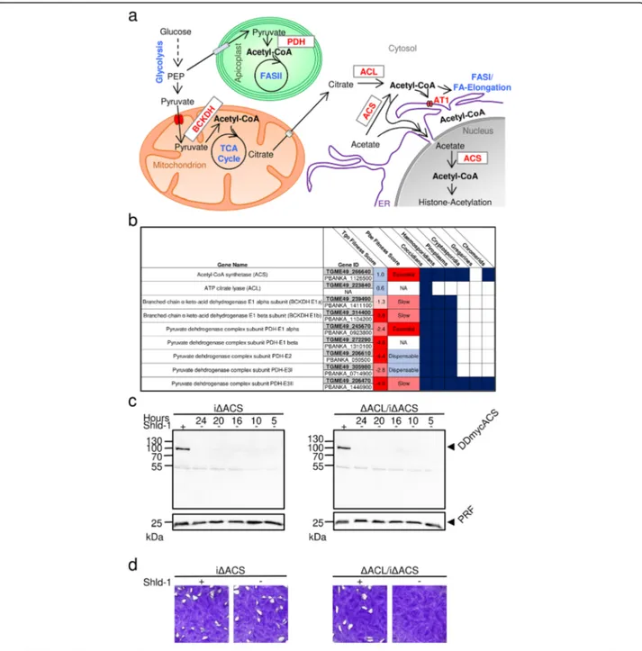
T. gondii harbours several metabolically active sub-cellular compartments including the cytosol, nucleus, endoplasmic reticulum (ER), Golgi apparatus, mitochon-drion and apicoplast, a relict plastid-like organelle derived from secondary endosymbiosis and possibly peroxisomes, which may form in the oocyst/sporozoite stage [3–5]. Acetyl-CoA is a hub metabolite with distinct and crucial functions in each of these compartments, involved in sev-eral anabolic and catabolic pathways and crucial for the acetylation of histones as well as non-histone proteins [6– 9]. Due to its amphiphilic nature and high molecular weight, acetyl-CoA cannot freely cross the membranes and must be produced within, or actively transported into, the compartments which rely on acetyl-CoA (Fig.1a) [9]. In T. gondii, the pyruvate dehydrogenase complex (PDH) converts pyruvate to acetyl-CoA in the apicoplast [12]. In the mitochondrion, the branched-chain α-keto acid dehydrogenase-complex (BCKDH) replaces the function of PDH to generate acetyl-CoA from pyruvate [13]. Two complementary routes generate acetyl-CoA in the cytosol and nucleus: the acetyl-CoA synthetase (ACS) produces acetyl-CoA from acetate, and the ATP citrate lyase (ACL) converts citrate to acetyl-CoA [14]. A putative acetyl-CoA transporter (AT-1) likely enables the import of cytosolic acetyl-CoA into the ER [14,15]. If, and during which life cycle stages, acetyl-CoA is generated byβ-oxidation in T. gondiiis unclear [5].
The high negative scores in a recent fitness screen of T. gondiimetabolic genes indicate fitness-conferring roles of PDH and BCKDH (Fig.1b) [16]. In contrast, the low posi-tive scores of ACS and ACL indicate dispensability, con-sistent with a previous study which demonstrated that ACS and ACL are synthetic lethal [14]. Apart from cocci-dians, other apicomplexans, including the malaria para-sites, lack ACL and thus rely solely on ACS to generate acetyl-CoA in the nucleo-cytosol (Fig. 1b). Consequently, ACS is predicted to be essential in Plasmodium as indi-cated by genome-wide fitness screens and is considered as a promising drug target [10,17,18].
While previous studies focused on determining the extent of protein acetylation in apicomplexans and identified dif-ferences between parasite stages or strains [19–22], little is known about the specific roles of distinct acetyl-CoA pools and how these impact parasite physiology. Here, we
combined multi-omics analysis with molecular tools to re-veal the diverse functions of acetyl-CoA in T. gondii.
Results
Loss of nucleo-cytosolic acetyl-CoA production is
detrimental and causes morphological defects in T. gondii
We have previously postulated that both ACL and ACS contribute to the generation of acetyl-CoA in the cytosol and nucleus and constitute a synthetic lethal pair in T. gondii [14]. To elucidate the role of ACL and ACS, we generated an inducible conditional knock-down of ACS in RH parasites (iΔACS) as well as in parasites in which acl was deleted by double-homologous recombination (ΔACL/iΔACS). For the conditional knock-down, a de-stabilisation domain (DD) was fused to a myc-tag at the N-terminus of ACS in the endogenous acs gene locus by CRISPR/Cas9-mediated genome editing (Additional file1: Figure S1a). The DD allows for rapid proteasome deg-radation in the absence of the protective ligand Shield-1 (Shld-1) [23]. Integration of the DDmyc in the acs locus and replacement of the acl open reading frame (ORF) with the hypoxanthine-xanthine-guanine phosphoribosyl transferase (HXGPRT) resistance cassette (Add-itional file1: Figure S1a, b) were validated by PCR ana-lysis of genomic DNA (Additional file 1: Figure S1c). Expression and effective regulation of the DDmyc-ACS fusion protein in iΔACS and ΔACL/iΔACS parasites were confirmed by western blot (Fig.1c).
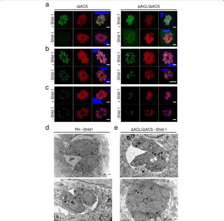
In plaque assays, which assess several lytic cycles, iΔACS or ΔACL parasites showed no defect compared to RH parasites grown in human foreskin fibroblasts (HFFs) over 1 week (Fig.1d). In contrast,ΔACL/iΔACS parasites failed to form plaques after 7 days in the ab-sence of Shld-1 (Fig. 1d), confirming our previous pre-diction of synthetic lethality [14]. Immunofluorescence assays (IFAs) revealed that iΔACS and ΔACL parasites were morphologically normal, whileΔACL/iΔACS para-sites presented a severe impairment in cell division with a loss of pellicle integrity as seen by clear alteration of staining of a pellicle marker, the gliding-associated pro-tein 45 (GAP45) at 24 h after Shld-1 removal (Fig. 2a). Additionally, staining of the mitochondrion and apico-plast appeared diffuse in ΔACL/iΔACS parasites, sug-gesting a loss of integrity of both organelles (Fig. 2b, c). Similarly, electron microscopy examination of ΔACL/ iΔACS highlighted extreme morphological defects 24 h after Shld-1 removal (Fig. 2d, e). Some dividing ΔACL/ iΔACS parasites presented loss of the basal structure (Fig. 2e, top panel), or were entirely amorphic, with the contents of multiple fused parasites enclosed by a joined plasma membrane (Fig. 2e, bottom panel). Altogether, these results support our previous observation that ACL and ACS constitute a synthetic lethal pair, depletion of which causes arrest of parasite growth.
Fig. 1 ACS and ACL are essential to produce acetyl-CoA in the cytosol and nucleus. a Schematic representation of the metabolic pathways in T. gondii for acetyl-CoA production and transport into the cellular compartments where it is required: the apicoplast, mitochondrion, cytosol, nucleus and the endoplasmic reticulum (ER). Metabolic pathways are highlighted in blue and enzymes in red, and metabolites are depicted in black. BCKDH, branched-chainα-keto acid dehydrogenase-complex; PDC, pyruvate dehydrogenase complex; FA, fatty acid; FASII, type II FA synthase, ACL, ATP citrate lyase; ACS, acetyl-CoA synthetase; AT1, acetyl-CoA transporter; ER, endoplasmic reticulum; TCA, tricarboxylic acid. b Table highlighting the essentiality [10,11] of acetyl-CoA-generating enzymes in T. gondii (Tgo) and Plasmodium berghei (Pbe) and their conservation across different apicomplexans. c Immuno-blot of total protein lysates from iΔACS (left panel) and ΔACL/iΔACS parasites (right panel) for which Shield-1 (Shld-1) was removed at several time points prior to egress to test protein regulation. Western blots were probed using α-myc antibody to detect the myc-tag of DD-ACS, and α-profilin (PRF) was used as a loading control. d Plaque assays were performed by inoculating human foreskin fibroblast (HFF) monolayers with either iΔACS, or ΔACL/iΔACS parasite strains and left to grow in the presence (+) or absence (−) of Shld-1 over a period of 7 days. Plaques were revealed by crystal violet staining of infected HFF monolayers
Lack of ACL/ACS alters the T. gondii metabolome including disruption of FA elongation
AnalysingΔACL/iΔACS parasites by IFA and western blot allowed us to conclude that 16 h of Shld-1 removal was suf-ficient to deplete ACS in the ΔACL/iΔACS strain, while parasites showed no growth defect or morphological
abnormalities at this relatively early time point of ACS de-pletion and remained viable. In the following analyses, iΔACS and ΔACL/iΔACS refer to parasites depleted of ACS by removal of Shld-1 for 16 h. To obtain a global pic-ture of the impact of the loss of nucleo-cytosolic acetyl-CoA on parasite metabolism, we performed untargeted
Fig. 2 Loss of ACS and ACL is associated with amorphic cells and loss of organelle integrity. Immunofluorescence assays (IFAs) of intracellular iΔACS or ΔACL/iΔACS parasites grown in the presence (+) or absence (−) of Shield-1 (Shld-1) for 24 h (a–c). IFAs were fixed and stained with α-gliding-associated protein 45 (α-GAP45, red) to show pellicles of the parasites and 4′,6 diamidin-2-phenylindol (DAPI, blue) to stain the nuclei and either,myc (green) to detect ACS (a) or with the monoclonal antibody 5F4 (F1B ATPase, green) marking the mitochondrion (b) or α-apicoplast-associated thioredoxin family protein 1 (α-Atrx1, green) staining the apicoplast (c) (scale bars, 5 μm). Electron micrographs of intracellular RH (d) orΔACL/iΔACS (e) grown in the absence of Shld-1 for 24 h (scale bars, 2 μm). ACL, ATP citrate lyase; ACS,
acetyl-CoA synthetase
metabolomics using liquid chromatography-mass spec-trometry (LC-MS). The full dataset is available in a data re-pository [24]. Over 850 putative metabolites were detected, and the relative abundance of all metabolites was compared to that of RH parasites (Additional file2: Table S1). We fo-cused our analysis on putative metabolites which changed more than 2-fold in abundance (p value < 0.05) and identi-fied 40 putative metabolites as significantly perturbed, with 19 displaying decreased levels, while 21 were increased in abundance in ΔACL/iΔACS parasites (Fig. 3a, Add-itional file2: Table S1). While the loss of nucleo-cytosolic acetyl-CoA affected several metabolic pathways, many clus-tered into lipid/fatty acyl- and peptide metabolism (Add-itional file 2: Table S1, Additional file 3: Figure S2a). Enzymes producing or consuming the affected metabolites were almost exclusively predicted to localise to the cytosol or ER (Additional file2: Table S1). Knock-out of ACL or knock-down of ACS alone had only minor impact with 14 or 1 putative metabolite changing, respectively (Add-itional file 2: Table S1). However, in many cases, loss of ACL alone led to a modest, non-significant drop in levels of certain metabolites, while the lack of both enzymes (ΔACL/ iΔACS) aggravated the phenotype, indicating that the two enzymes have partially redundant functions in metabolism (Fig.3b, Additional file2: Table S1). Purine metabolites (ad-enine, inosine monophosphate (IMP), xanthine) were found increased in parasites lacking ACL and are likely associated with the HXGPRT resistance gene (Additional file3: Figure S2a). Acetyl-CoA-levels decreased in ΔACL and ΔACL/ iΔACS parasites to about 60% compared to RH cells, al-though the drop was not statistically significant (Fig. 3b). This indicates that the major pools of acetyl-CoA are in the mitochondrion and apicoplast, unaffected by the loss of ACS and ACL, consistent with the dramatic drop in total acetyl-CoA inΔBCKDH parasites [13].
Major changes were observed in the abundance of monounsaturated very long-chain FAs (FA C26:1, C28:1) by untargeted LC-MS analysis (Fig. 3b), consistent with acetyl-CoA being required for the elongation of FAs on the cytosolic site of the ER [25]. This defect in FA elong-ation inΔACL/iΔACS parasites was further scrutinised by semi-targeted profiling of FAs by gas chromatography-MS (GC-MS) (Fig.3c). Using GC-MS, we found that the rela-tive levels of five detected FAs were significantly altered between RH and ΔACL/iΔACS parasites. While C18:0 and C20:4 were slightly increased in ΔACL/iΔACS para-sites, FA C18:1, C20:1 and C26:1 were significantly de-creased, with FA C20:1 and C26:1 displaying a dramatic 2-fold and 4-2-fold drop, respectively, while FA C28:1 was not detected by GC-MS (Fig. 3c). Importantly, most FAs are abundant in the host cells (HFFs) and can be salvaged by T. gondii. In particular, C18:0 and C20:4 make up a higher proportion of total FAs in the host compared to T. gondii [16]. Instead, C18:1 is of lower relative abundance in the
host compared to T. gondii, and FAs C20:1, C26:1 and C28:1 are of very low abundance or absent in HFFs [16,
25]. We conclude that T. gondiiΔACL/iΔACS compen-sate for the halt in FA elongation through increased uptake of unsaturated long-chain FAs from the host. The differences in FA abundances in HFFs compared to T. gondii[16] lead to the altered FA composition ofΔACL/ iΔACS parasites. Additionally, we confirmed the defect in FA elongation by labelling with U-13C-glucose or U-13 C-acetate for 16 h simultaneous to the ACS downregulation followed by GC-MS analysis. 13C-labelling in FA C26:1 was significantly decreased inΔACL/iΔACS parasites con-firming the specific loss of FA elongation in these parasites (Fig. 3d, Additional file 3: Figure S2b). In contrast, the abundance as well as 13C-labelling in myristate (C14:0) was similar or increased in ΔACL/iΔACS compared to RH parasites, indicating that cells are metabolically active and that de novo FA synthesis in the apicoplast is not af-fected (Fig.3d, Additional file 3: Figure S2b). Possibly re-lated to the reduced levels of long and very long monounsaturated FAs, abundance of the lipid phosphati-dylserine (PS 38:2) was also found more than 2-fold re-duced in ΔACL/iΔACS parasites by LC-MS analysis (Fig.3b). This may be a consequence of the altered abun-dance of C18:1 and C20:1, both of which were found to be significantly reduced in ΔACL/iΔACS by semi-targeted GC-MS FA profiling, although the FA composition of the affected PS38:2 was not further scrutinised using tandem MS.
While the synthesis of monounsaturated very long chain FAs depends on acetyl-CoA as a substrate, the drop in levels of other metabolites such as D -fructose-1,6-bisphosphate (F1,6-BP) is unexpected and may result from the altered acetylation status of enzymes in the pathway (Fig.3b).
Additionally, we performed U-13C-glucose and U-13 C-acetate labelling followed by LC-MS analysis, to compare the utilisation of these metabolites between RH andΔACL/ iΔACS parasites (Additional file 4: Figure S3a). We ob-served no differences in glucose utilisation when monitor-ing glycolytic and TCA cycle intermediates. While U-13 C-glucose labelling resulted in rapid and extensive labelling of glycolytic and TCA cycle intermediates, U-13C-acetate re-sulted in no or very little labelling of central carbon metab-olites apart from citrate (Additional file 4: Figure S3b,c). Incubation in U-13C-acetate resulted in 50% 13C-labelling in the citrate of RH, which was reduced more than 3-fold in iΔACS and ΔACL/iΔACS parasites (Additional file 4: Figure S3c). The lack of/low levels of13C-labelling in other TCA cycle intermediates indicate that the labelling ob-served in citrate is not in the mitochondrial pool but rather in the cytosolic or a putative apicoplast pool. We propose that a second citrate synthase 2, for which the localisation is yet unknown [26], condenses acetyl-CoA and oxaloace-tate (OAA) to form citrate in the cytosol utilising
acetyl-Fig. 3 (See legend on next page.)
CoA generated by ACS which is derived from U-13 C-acet-ate. We also observed a significant decrease in labelling from U-13C-acetate in some lipid species (Additional file4: Figure S3d-f) including PS38:2 (Additional file 4: Figure S3f) which was also 2-fold reduced in abundance inΔACL/ iΔACS parasites (Fig. 3b). Overall, our metabolomic ana-lyses demonstrate that lack of ACL/ACS results in several changes in the metabolism, most notably the loss of long and very long monounsaturated FAs, which have previously been demonstrated to be essential for T. gondii [25].
Lack of ACL/ACS or BCKDH results in hypo-acetylation of cytosolic and mitochondrial proteins, respectively
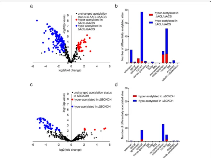
To determine the role of cytosolic and mitochondrial acetyl-CoA in protein acetylation, we characterised the acetylome of parasites lacking ACL and ACS or BCKDH, the complex implicated in the production of acetyl-CoA in the mitochondrion [13]. Generation of a parasite line lacking the BCKDH subunit E1 (ΔBCKDH) was de-scribed previously [13]. Western blot analysis using α-acetyl-lysine antibodies revealed widespread Nε-lysine-acetylation in T. gondii RH,ΔACL/iΔACS and ΔBCKDH parasites (Additional file 5: Figure S4a,b). Quantitative MS-based proteomic analyses comparing RH and ΔACL/iΔACS parasites allowed us to confidently quan-tify 404 acetylated sites on 269 proteins. Out of these, 182 sites (45%) belonging to 137 proteins were found to be differentially acetylated in ΔACL/iΔACS parasites compared to RH (Additional file6: Table S2). Most sites (142) were hypo-acetylated while 40 were hyper-acetylated in ΔACL/iΔACS parasites (Fig.4a). The pre-dominant hypo-acetylation (78% of differentially acety-lated sites) is consistent with the expected reduction of acetyl-CoA. The complete dataset is available in a data repository [28].
To determine the sub-cellular localisation of differen-tially acetylated proteins, we predicted their putative lo-calisation based on the hyperplexed Lolo-calisation of
Organelle Proteins by Isotopic Tagging (hyperLOPIT) data available under https://proteome.shinyapps.io/toxo-lopittzex/ and on ToxoDB (https://toxodb.org) [27]. For simplification, we merged some of the sub-cellular com-partments (19S proteasome/20S proteasome/40S ribo-some/60S ribosome/cytosol into cytosol; nucleolus/ nucleus – chromatin/nucleus non-chromatin into nu-cleus and mitochondrion membranes/mitochondrion soluble into mitochondrion etc.). Differentially acetylated sites were found within proteins of several sub-cellular compartments; however, most hypo-acetylated residues (77%) were of proteins within the cytosol and nucleus, consistent with the localisation of ACS and ACL (Fig. 4b). To account for the different number of pro-teins within the distinct sub-cellular compartments, we determined the number of differentially acetylated proteins relative to the total number of proteins identified within the compartment, which confirmed that hypo-acetylated teins were enriched in the cytosol (12% of all cytosolic pro-teins hypo-acetylated) and nucleus (4.5% of all nuclear proteins hypo-acetylated) (Additional file 5: Figure S4c). Surprisingly, the few sites which were differentially acety-lated on proteins in the apicoplast or mitochondrion in ΔACL/iΔACS parasites were predominantly hyper-acetylated (Fig. 4b). The affected hyper-acetylated apico-plast proteins are the PDH, the enoyl-acyl carrier protein reductase (ENR) and a putative chaperone (Additional file6: Table S2), while the affected mitochondrial proteins are pu-tative chaperones, heat-shock proteins and TCA cycle en-zymes (Additional file6: Table S2). Perhaps, these proteins are hyper-acetylated as part of a stress response to avert the phenotype caused by loss of ACS and ACL. However, the effect of acetylation of these proteins is unknown, and hence, the functional consequences of their hyper-acetylation remain unclear. While ΔACL/iΔACS parasites showed deformation at 24 h of Shld-1 removal, we argue that the observed hypo-acetylation observed in ΔACL/ iΔACS parasites at 16 h of Shld-1 removal is a direct result
(See figure on previous page.)
Fig. 3 Loss of ACS and ACL causes changes in the T. gondii metabolome including a halt in FA elongation. a Volcano plot highlighting changes in metabolite levels ofΔACL/iΔACS compared to RH parasites. Unchanged metabolites are displayed in black, while significantly increased or decreased metabolites (≥ 2-fold change, p < 0.05) in ΔACL/iΔACS are displayed in red and blue, respectively. Statistically significant differences were assessed using a t-test comparing triplicates ofΔACL/iΔACS parasites to triplicates of RH in the absence of Shield-1. b Relative abundance of selected metabolites compared to levels in RH-Shield-1 (dashed line), which are not significantly altered (acetyl-CoA, citrate), significantly decreased (D-fructose 1,6 bisphosphate, FAs C26:1 and C28:1, phosphatidylserine—PS38:2) or significantly increased (adenine) upon loss of ACS and ACL (ΔACL/iΔACS). Error bars represent the standard deviation between replicates (n = 3). Statistical significance was assessed using a t-test and is indicated (*p < 0.05). c GC-MS measurement of FA abundances normalised to cholesterol levels. Error bars represent the standard deviation between replicates (n = 6). Statistical significance was assessed by a t-test and is indicated (**p < 0.005; ***p < 0.0001). d Relative abundance and
13
C-labelling in myristate (C14:0) and FA C26:1, following incubation in medium containing U-13C-glucose or U-13C-acetate for 16 h during simultaneous ACS depletion. Error bars represent the standard deviation between replicates. Top error bars represent the standard variation in abundance, and lower error bars represent the standard deviation in labelling between replicates (n = 6 for abundance measurement, n = 3 for labelling analysis). Statistical significance for abundances as indicated in c. Statistical significance was tested using a t-test. Differences in C14:0 labelling between RHΔACL/iΔACS and are non-significant (n.s.). Statistically significant differences in U-13C-acetate (p < 0.05) and U-13 C-glucose-labelling of C26:1 between RH andΔACL/iΔACS are indicated (*p < 0.05; ***p < 0.0001), respectively. ACL, ATP citrate lyase; ACS, acetyl-CoA synthetase; FA, fatty acid; GC-MS, gas chromatography-mass spectrometry; Glc, glucose; Ac, acetate
of the loss of acetyl-CoA in the cytosol, rather than a gen-eral death phenotype. This is supported by the fact that hypo-acetylation is predominantly observed in the affected cytosol, while other compartments show unaltered acetyl-ation or even hyper-acetylacetyl-ation of proteins (Fig. 4b). Evi-dence that ΔACL/iΔACS parasites are equally viable and metabolically active as RH parasites at 16 h of Shld-1 re-moval can also be inferred from the above described meta-bolomic analyses: e.g. ΔACL/iΔACS parasites show equal rates of FA de novo synthesis (see FA C14:0 levels and la-belling in Fig. 3d) as well as equal levels and labelling of
most central carbon metabolites (see Additional file 2: Table S1 and Additional file4: Figure S3a).
As for ΔACL/iΔACS parasites, we probed the acety-lome of parasites lacking BCKDH using MS-based quan-titative analyses. After stringent filtering, we identified 483 acetylation sites distributed over 300 proteins in RH and ΔBCKDH parasites (Additional file 7: Table S3). Fifty-six lysine residues belonging to 45 different pro-teins were found to be differentially acetylated in para-sites lacking BCKDH compared to RH, with only 6 para-sites displaying hyper-acetylation and 50 sites being
hypo-Fig. 4 Loss of ACL/ACS or BCKDH results in predominant hypo-acetylation of nucleo-cytosolic and mitochondrial proteins, respectively. a Volcano plot highlighting differentially acetylated sites ofΔACL/iΔACS compared to RH parasites. Statistically significant differences between parasite lines were determined as outlined in the‘Material and methods’ section. Unchanged acetylation sites are displayed in black, while sites which are
significantly hyper- or hypo-acetylated inΔACL/iΔACS (≥ 2-fold, n = 3, limma p < 0.01) are displayed in red and blue, respectively. b Hyper- and hypo-acetylated sites inΔACL/iΔACS parasites were sorted according to their sub-cellular localisation using the hyperplexed Localisation of Organelle Proteins by Isotopic Tagging (hyperLOPIT) data available underhttps://proteome.shinyapps.io/toxolopittzex/[27]. c Volcano plot highlighting the differentially acetylated sites inΔBCKDH compared to RH parasites. Statistically significant differences were determined as outlined in the‘Material and methods’ section. Unchanged acetylation sites are displayed in black, while sites which are significantly (≥ 2-fold,
n = 3, limma p < 0.01) hyper- or hypo-acetylated inΔBCKDH are displayed in red and blue, respectively. d Hyper- and hypo-acetylated sites in ΔBCKDH parasites were sorted according to their sub-cellular localisation using hyperLOPIT data available underhttps://proteome.shinyapps.io/ toxolopittzex/[27]. BCKDH, branched-chainα-keto acid dehydrogenase-complex; ACL, ATP citrate lyase; ACS, acetyl-CoA synthetase; PM, plasma membrane; IMC, inner membrane complex; ER, endoplasmic reticulum
acetylated (Fig. 4c). Using the hyper-LOPIT data, we identified that most hypo-acetylated residues (52%) belonged to proteins residing within the mitochondrion, consistent with a decreased production of acetyl-CoA within this compartment due to loss of BCKDH catalytic activity (Fig.4d, Additional file5: Figure S4d). Taken to-gether, these findings reveal that the lack of ACL/ACS or BCKDH results in predominant hypo-acetylation of numerous proteins within the respective compartment.
Cytosolic and mitochondrial acetyl-CoA are required for extensive acetylation of glycolytic and TCA cycle enzymes, respectively
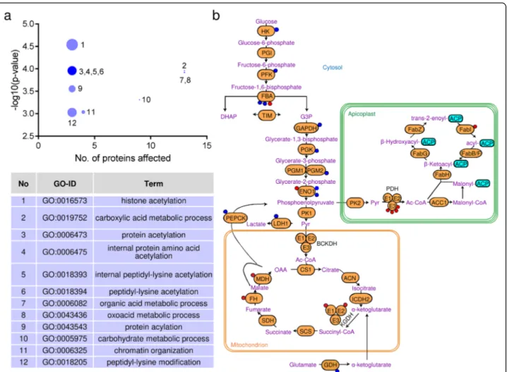
To better understand the consequences of the differen-tial acetylation inΔACL/iΔACS and ΔBCKDH parasites, we aimed to identify the affected biological processes by performing Gene Ontology (GO) enrichment using the R-package topGO [29]. Comparing the differentially acetylated proteins to the entire genome/proteome of T. gondii, we established that the affected proteins cluster into 12 significantly enriched (p < 0.001) biological pro-cesses including histone acetylation (33-fold enrich-ment), chromatin organisation (9-fold enrichenrich-ment), protein acetylation (19-fold enrichment) and carbohy-drate metabolic processes (4-fold enrichment) (Fig. 5a). To avoid bias towards proteins which were identified as acetylated in this study, we performed the GO enrich-ment analysis of differentially acetylated proteins against the relatively small subset of 269 acetylated proteins as background (Additional file5: Figure S4e). This analysis revealed the affected proteins to be significantly enriched (p < 0.05) in 2 biological processes, namely chromatin organisation and cellular protein metabolic processes. The latter also includes protein modification and is thus consistent with the enrichment of histone and protein acetylation identified in the analysis against the total genome/proteome.
Specifically, the proteins differentially acetylated in ΔACL/iΔACS parasites included several histones (H2Bb, H2Bv, H4, H2AZ) as well as histone-modifying enzymes (histone arginine methyltransferase PRMT1, histone ly-sine acetyltransferase MYST-A/B, GCN5-A/B, histone acetyltransferase subunit nua4 protein) (Additional file6: Table S2) [30–33]. Furthermore, we observed consider-able hypo-acetylation of the apicomplexan Apetala 2 (AP2) transcription factors AP2XII-4, AP2VIIa-7, AP2IX-7 and AP2IX-5 [34]. Interestingly, two of the dif-ferentially acetylated transcription factors, AP2XII-4 and AP2IX-7, have previously been shown to interact with GCN5-B, which is essential for replication [33]. Taken together, these changes in acetylation of histones, histone-modifying enzymes and transcription factors are expected to affect gene expression broadly. Cobbold et al. have previously reported extensive acetylation of
AP2 DNA binding proteins in P. falciparum, identifying 16 out of 28 AP2 factors to be acetylated in one or more positions [19]. T. gondii possesses 67 different AP2 tran-scription factors [34], 9 of which we identified to be acetylated in one or more positions (Additional file 6: Table S2). AP2XII-4 displayed extensive acetylation in 6 different sites, 3 of which were hypo-acetylated in ΔACL/iΔACS parasites, while 2 were unchanged and one site was hyper-acetylated (Additional file 6: Table S2). In contrast, 2 acetylation sites were identified for its homologue in P. falciparum, PF3D7_0516800 [19]. Jef-fers and Sullivan have previously identified 5 AP2 domain-containing proteins to be acetylated in T. gondii and similarly identified AP2XII-4 to be extensively acety-lated in 4 sites [20] compared to 6 sites we report here. A recent ground-breaking study identified the micro-rchidia (MORC) protein as a key regulator of T. gondii development by repressing sexual commitment [35]. In-triguingly, the authors found that MORC functions in a complex with AP2 transcription factors and recruits the histone deacetylase HDAC3 to repress genes which trigger sexual differentiation [35]. Thus, nucleo-cytosolic acetyl-CoA is essential for the regulation of T. gondii de-velopment by altering histone accessibility and presum-ably by modifying the activities and interactions of AP2 transcription factors through extensive acetylation.
Besides the hypo-acetylation of nucleo-cytosolic pro-teins, we also identified hypo-acetylation of four rhoptry proteins (ROP12, ROP17, ROP40 and RON2) and two dense granule proteins (GRA1 and GRA2) (Add-itional file 6: Table S2), indicating that acetylation of these secretory proteins relies on cytosolic acetyl-CoA, likely imported into the ER by AT-1 [14]. ROPs and GRAs are effector proteins secreted into the host cells during parasite invasion to hijack host cellular functions and to establish and modify the parasitophorous vacuole [36]. Jeffers and Sullivan similarly identified acetylation of ROP 17 and RON2 and proposed additionally acetyl-ation of ROP8 and RON4 but did not detect acetylacetyl-ation of ROP12 [20]. If or how acetylation effects the function of these proteins unique to apicomplexan parasites re-mains unclear. Nevertheless, hypo-acetylation of these secretory proteins in ΔACL/iΔACS parasites provides the first evidence that their acetylation relies on the cytosolic acetyl-CoA pool.
Next, we analysed the affected metabolic pathways by comparing the differentially acetylated proteins against the entire genome/proteome of T. gondii and using the metabolic pathway enrichment tool on ToxoDB (https:// toxodb.org/) based on the Kyoto Encyclopedia of Genes and Genomes (KEGG) database as the source. The high-est metabolic pathway enrichment was found for pro-teins functioning in glycolysis/gluconeogenesis (9-fold). Most glycolytic/gluconeogenic enzymes were
hypo-acetylated in ΔACL/iΔACS parasites (Fig.5b). Although enzymes implicated in glycolysis are extensively acety-lated in other organisms [37–39], the effects that acetyl-ation has on enzyme activity varies and depends on the organism, enzyme, site and context [37–41]. In the me-tabolome analysis, we observed no differences in glyco-lytic flux, i.e. no changes in levels and labelling of most glycolytic intermediates (Additional file 2: Table S1, Additional file4: Figure S3a), with the exception of a sig-nificant reduction in levels of F1,6-BP in ΔACL/iΔACS
parasites (Fig. 3b). Strikingly, the enzymes producing and consuming F1,6-BP, phosphofructokinase (PFK) and F1,6-BP aldolase (FBA), respectively, were both differen-tially acetylated inΔACL/iΔACS parasites. The differen-tial acetylation may impact on the enzyme’s activity and may cause reduced synthesis or increased consumption of F1,6-BP.
As described above for ΔACL/iΔACS parasites, we performed the GO enrichment analysis of biological pro-cesses and metabolic pathways of proteins differentially
Fig. 5 Loss of ACL/ACS causes extensive hypo-acetylation of glycolytic enzymes. a Proteins identified as differentially acetylated inΔACL/iΔACS parasites were analysed using the GO enrichment R-package topGo to identify the enrichment in biological processes against the total T. gondii genome/proteome. The bubble sizes are proportional to the fold enrichment, ranging from 33-fold (histone acetylation, 1) to 3-fold (organic acid metabolic process, 7). Statistically significant enrichment was assessed by Fisher’s exact test (p value < 0.001). b Scheme highlighting the distribution of differentially acetylated sites on metabolic enzymes ofΔACL/iΔACS parasites. Displayed pathways include the glycolysis/
gluconeogenesis, the TCA cycle and the apicoplast resident FASII. Coloured circles on the enzymes represent sites which are either hyper- (red) or hypo-acetylated (blue) inΔACL/iΔACS. ACL, ATP citrate lyase; ACS, acetyl-CoA synthetase; GO, Gene Ontology; TCA, tricarboxylic acid; FASII, fatty acid synthase II; HK, hexokinase; PGI, phosphoglucose isomerase; PFK, phosphofructokinase; FBA, fructose bis-phosphate aldolase; TIM,
triosephosphate isomerase; GAPDH, glyceraldehyde 3-phosphate dehydrogenase; PGK, phosphoglycerate kinase; PGM, phosphoglycerate mutase; ENO, enolase; PK, pyruvate kinase; LDH, lactate dehydrogenase; PEPCK, phosphoenolpyruvate carboxykinase-1; BCKDH, branched-chainα-keto acid dehydrogenase-complex; E1-E3, BCKDH subunits; CS, citrate synthase; ACN, aconitase; ICDH, isocitrate dehydrogenase; KGDH,α-ketoglutarate dehydrogenase; E1-E3, KGDH subunits; SCS, succinyl coenzyme A synthetase; GDH, glutamate dehydrogenase; SDH, succinate dehydrogenase; FH, fumarate hydratase; MDH, malate dehydrogenase; PDH, pyruvate dehydrogenase complex; E1-E3, PDH subunits; Ac-CoA, acetyl-CoA; ACP, acyl carrier protein; Fab, FAS II subunits; ACP, acyl carrier protein; ACC, acetyl-CoA carboxylase; Pyr, pyruvate; OAA, oxaloacetate; DHAP,
dihydroxyacetone phosphate; G3P, glyceraldehyde-3-phosphate
acetylated in ΔBCKDH parasites. Differentially acety-lated proteins in ΔBCKDH parasites were compared to the total genome/proteome using the R-package topGO [29], which identified affected proteins to be significantly enriched (p < 0.001) in 10 biological processes, most of which are related to the TCA cycle and respiration, in-cluding the top hits with over 30-fold enrichment (tri-carboxylic acid metabolic processes, tri(tri-carboxylic acid cycle and citrate metabolic processes) (Fig.6a). In order to avoid bias towards acetylated proteins, differentially acetylated proteins were probed against the background of the small subset of acetylated proteins. Using this ap-proach, 6 biological processes were identified as signifi-cantly enriched (p < 0.05) all of which related to the TCA cycle and respiration and 5 of which were also identified in the enrichment analysis against the entire genome/proteome (Additional file5: Figure S4f). As ex-pected, analysis of proteins which were differentially acetylated in ΔBCKDH parasites revealed major enrich-ment in the TCA cycle when using the metabolic path-way enrichment tool on ToxoDB (https://toxodb.org/) based on the KEGG database as the source, returned the TCA cycle as top-hit (24-fold enrichment). Seven out of eight TCA cycle enzymes were hypo-acetylated in ΔBCKDH parasites compared to RH cells (Fig. 6b). As for glycolytic enzymes, the role of TCA cycle enzyme acetylation varies and has not been defined for T. gondii enzymes [41,43].
One of the most significantly hypo-acetylated proteins in ΔBCKDH cells was lactate dehydrogenase (LDH1) (Additional file7: Table S3). Acetylation in lysine K5 has been reported to negatively regulate LDH activity in mam-malian cells [44]. Although we observed hypo-acetylation of a different lysine (K218) inΔBCKDH parasites, it may
similarly increase LDH activity, as lactate production is markedly increased inΔBCKDH parasites [13].
Furthermore, we revealed a previously unpublished acetylation site in the gluconeogenic enzyme phospho-enolpyruvate carboxykinase (PEPCK-1) (K116) in addition to the previously identified acetylation sites K223, K231 and K591 of T. gondii PEPCK-1 [20] (Additional file 7: Table S3). Interestingly, lysine residue K223 was found to be significantly hypo-acetylated in ΔBCKDH parasites (Additional file 7: Table S3, Fig. 6b). We previously re-ported constitutive activation of the gluconeogenesis path-way inΔBCKDH parasites [13], leading us to hypothesise that the activation of gluconeogenesis inΔBCKDH para-sites may be attributed to the changed acetylation status of PEPCK-1 (Additional file8: Figure S5a).
The PEPCK-1 acetylation status is not solely responsible for regulating gluconeogenesis in T. gondii
In T. gondii, gluconeogenesis is tightly regulated and in-active under glucose replete conditions [45,46] but essen-tial in glucose-limiting conditions [42, 47]. T. gondii possess two PEPCKs, with PEPCK-1 (TGME49_289650) being the active enzyme in tachyzoites [42]. We identified two in-frame translation starts for PEPCK-1 (Add-itional file 8: Figure S5b). While the long isoform of PEPCK-1 localises to the mitochondrion (Additional file8: Figure S5c), a second ATG, 285 bases downstream of the first predicted translational start, leads to a shorter iso-form with cytosolic localisation (Additional file 8: Figure S5c). Crucially, a 3-Ty-epitope tag at the C-terminal end of the endogenous gene locus (Additional file 8: Figure S5d,e) revealed cytosolic localisation of the endogenous PEPCK-1 (Fig. 6c), contrasting previous reports, which have proposed a mitochondrial localisation [42]. To test
(See figure on previous page.)
Fig. 6 Loss of BCKDH causes extensive hypo-acetylation of TCA cycle enzymes and the gluconeogenic enzyme PEPCK-1. a Proteins identified as differentially acetylated inΔBCKDH parasites were analysed using the GO enrichment R-package topGo to identify enrichment in biological processes against the total T. gondii genome/proteome. The bubble sizes are proportional to the fold enrichment of the respective pathway, ranging from 36-fold (GO:0072350—tricarboxylic acid metabolic process) to 4-fold (GO:0044281—small molecule metabolic process). Statistically significant enrichment was assessed by Fisher’s exact test (p value < 0.001). b Scheme highlighting the distribution of differentially acetylated sites on metabolic enzymes ofΔBCKDH parasites. Displayed pathways include the glycolysis/gluconeogenesis, the TCA cycle and the apicoplast resident FASII. Coloured circles on the enzymes represent sites which are either hyper- (red) or hypo-acetylated (blue) inΔBCKDH. c Endogenous PEPCK-1 presents a nucleo-cytosolic localisation by immunofluorescence assay (IFA) after C-terminal tagging by knock-in of the endogenous PEPCK-1 locus both in RH parasites. PEPCK-1-3Ty was detected usingα-Ty (green) while α-GAP45 (red) was used as a pellicle marker and 4′,6 diamidin-2-phenylindol (DAPI, blue) to stain the nuclei (scale bars in b and c, 5μm). d Schematic representation of PEPCK and its identified acetylation sites. Lysine at position K223 was changed to glutamine (K223Q acetylation mimetics) or arginine (K223R, de-acetylation mimetics). e The ability of different PEPCK-1 acetylation mimetics to grow in a medium lacking glucose was tested in an intracellular growth assay. Error bars represent the standard deviation between 3 independent infections. Per infection, > 100 vacuoles were counted. f Constitutive activation of gluconeogenesis was assessed in PEPCK-1 acetylation mimetics by growing cells in a medium containing U-13C-glutamine in the presence of unlabelled glucose and measuring13C-labelling in glycolytic intermediates using GC-MS (shown here, glucose-6-phosphate). Uptake and utilisation of U-13C-glutamine were confirmed by measuring13C-labelling in the TCA cycle by-product aspartate. Error bars represent the standard deviation between replicates (n = 3). Throughout the figure, PEPCK refers to PEPCK-1, the active enzyme in tachyzoites [42]. GO, Gene Ontology; TCA, tricarboxylic acid; FASII, fatty acid synthase II; PEPCK, phosphoenolpyruvate carboxykinase-1; BCKDH, branched-chainα-keto acid
dehydrogenase-complex; GC-MS, gas chromatography-mass spectrometry; TCA, tricarboxylic acid; other abbreviations, see Fig.5
whether the constitutive activation of gluconeogenesis in ΔBCKDH parasites [13] is due to hypo-acetylation of PEPCK-1 K223, we complemented ΔPEPCK-1 parasites with wild-type PEPCK-1 or versions of PEPCK-1 mimick-ing acetylation (glutamine) or de-acetylation (arginine) of lysine at position 223 (K223Q, K223R) (Fig. 6d). The pepck-1 locus was deleted by double homologous recom-bination (Additional file8: Figure S5f). Its deletion and in-sertion of the chloramphenicol acetyltransferase (CAT) resistance cassette were confirmed by genomic PCR (Additional file 8: Figure S5g). Expression of the acetyl-ation mimetic PEPCK-1 constructs was confirmed by IFA (Additional file8: Figure S5h).
To investigate whether gluconeogenesis was constitutively active or inactive depending on the PEPCK-1 acetylation status, ΔPEPCK-1 strains complemented with PEPCK-1 acetylation mimetics (K223Q, K223R) were assessed in a growth assay in glucose-depleted medium (Fig. 6e). The acetylation/de-acetylation mimetics of PEPCK-1 (K223Q/ K223R) grew equally well and comparable to parasites com-plemented with WT-PEPCK-1 in glucose-depleted medium (Fig.6e). Only parasites lacking PEPCK-1 showed a growth defect in the glucose-depleted medium. These findings indi-cate that acetylation of PEPCK-1 (K223Q) alone is not suffi-cient to de-activate gluconeogenesis. To assess the constitutive activation of gluconeogenesis, PEPCK-1 acetyl-ation mimetic parasites were incubated in a medium con-taining unlabelled glucose and U-13C-glutamine followed by profiling of polar metabolites by GC-MS (Fig.6f). Labelled carbons from U-13C-glutamine were not incorporated into glycolytic/gluconeogenic intermediates, such as glucose-6-phospate, in the presence of unlabelled glucose (Fig.6f), as was previously observed in ΔBCKDH parasites [13], highlighting that activation of gluconeogenesis is not trig-gered by de-acetylation of PEPCK-1 K223 (K223R) alone. Hence, the impact of acetylation on the function of en-zymes in T. gondii central carbon metabolism remains un-clear. Lastly, we assessed whether gluconeogenesis was an important adaptation in parasites lacking BCKDH. As de-scribed above for RH, pepck-1 was deleted in parasites lacking BCKDH (Additional file8: Figure S5i). Strikingly, ΔBCKDH/ΔPEPCK-1 parasites showed no aggravation of their growth phenotype compared to parasites lacking ei-ther BCKDH or PEPCK-1 as assessed by plaque assay and intracellular growth assay in the presence and absence of glucose (Additional file8: Figure S5j-l). These results high-light that the activation of gluconeogenesis in ΔBCKDH parasites is a‘side effect’ rather than a crucial adaptation mechanism.
Lack of ACL/ACS or BCKDH alters the transcriptome and proteome of T. gondii
For a global assessment of the impact caused by the deple-tion of cytosolic acetyl-CoA on gene expression inΔACL/
iΔACS parasites, we performed a combination of expres-sion profiling by ribonucleic acid sequencing (RNA-seq) and quantitative proteomics analyses on freshly egressed extracellular tachyzoites. The complete datasets are avail-able in data repositories [28,48]. We focused our analysis on transcripts and proteins that were changed ≥ 2-fold in their level (p value < 0.01) inΔACL/iΔACS parasites com-pared to RH. RNA-seq analysis allowed the identification of 453 differentially regulated transcripts out of 7250; 377 were found to be upregulated while 76 were downregu-lated (Fig.7a, Additional file9: Table S4). The predomin-ant upregulation of transcripts in parasites depleted in nucleo-cytosolic acetyl-CoA was surprising, considering that histone de-acetylation is associated with a general downregulation of transcription [7]. As anticipated, the HXGPRT resistance cassette transcript belonged to the top upregulated hits while ACL was amongst the top hits of downregulated transcripts, validating the obtained re-sults. Transcripts or proteins influenced directly by the ex-perimental procedure were excluded from further analysis. Of the differentially acetylated transcripts, 284 encoded hypothetical proteins, hindering interpretation of the results. The remaining transcripts encoded for pro-teins of diverse functions and included several T. gondii-specific proteins such as surface antigen (SAG)-related proteins (16 transcripts) and Toxoplasma gondii family (A, B,C) proteins, a group of uncharacterised T. gondii-spe-cific proteins (14 transcripts). GO enrichment against the entire genome/proteome of T. gondii using the biological process enrichment tool on ToxoDB (https://toxodb.org/) revealed no significant enrichment of the affected tran-scripts in biological processes.
MS-based proteomics allowed identification and relative quantification of 2773 T. gondii proteins, out of which 74 were found to be differentially expressed inΔACL/iΔACS parasites compared to RH (Fig. 7a, Additional file 10: Table S5). In contrast to the transcriptome, most proteins [49] were downregulated in ΔACL/iΔACS parasites (Fig.7a, Additional file10: Table S5). Amongst the down-regulated proteins were ACS and ACL, while the HXGPRT selection cassette was amongst the top hits of upregulated proteins, validating the obtained data (Add-itional file10: Table S5). The altered proteins have highly variable functions and include 26 hypothetical proteins, hindering the interpretation of the results.
Myosin J (MyoJ), a myosin motor mediating constric-tion of the basal pole during parasite division [50], was found to be downregulated over 2-fold (p value < 0.01) at the protein level in ΔACL/iΔACS strain compared to RH (Additional file10: Table S5). Loss of MyoJ has been shown to be associated with a loss of fitness in T. gondii, resulting in asynchronous division and the loss of con-nection between daughter cells [50]. Hence, its downreg-ulation may contribute to the severe morphological
defects of ΔACL/iΔACS parasites described above (Fig.2).
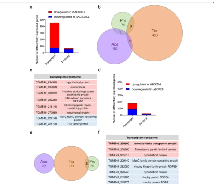
A relatively small subset of 8 genes was affected simul-taneously at the transcriptome and proteome level, with 2 genes being downregulated at the RNA transcript and protein level, while 6 were upregulated at both levels (Fig. 7b, c). This relatively small overlap between the
datasets highlights the diverse and complex conse-quences of cytosolic acetyl-CoA depletion.
Similarly, expression profiling by RNA-seq and quanti-tative proteomics was performed on freshly egressed ΔBCKDH tachyzoites to obtain a more holistic assess-ment of the consequences of mitochondrial acetyl-CoA depletion. Transcriptomic analysis revealed that a
Fig. 7 Loss of ACL/ACS or BCKDH alters the T. gondii gene expression. a Graph showing the number of RNA transcripts and proteins which are ≥ 2-fold up- (red) or downregulated (blue) in ΔACL/iΔACS parasites compared to RH (n = 3; limma p < 0.01). Statistical significance was determined as outlined in the‘Material and methods’ section. b Venn diagram highlighting the overlap between differentially expressed RNA
transcripts (Tra), proteins (Pro) and differentially acetylated proteins (Ace) which present a≥ 2-fold change in ΔACL/iΔACS parasites compared to RH parasites (all p < 0.01). c Table highlighting genes which were found to be significantly up- (red background) and downregulated (blue background) at the transcriptome and proteome level inΔACL/iΔACS parasites. d Graph highlighting the number of RNA transcripts and proteins which are≥ 2-fold up- (red) or downregulated (blue) in ΔBCKDH parasites compared to RH (n = 3; limma p < 0.01). Statistical significance was determined as outlined in the‘Material and methods’ section. e Venn diagram highlighting the overlap between differentially regulated
acetylation sites (Ace), RNA transcripts (Tra) and proteins (Pro), which present a > 2-fold change (p < 0.01) inΔBCKDH parasites compared to RH parasites. f Table highlighting genes which were found to be significantly up- (red background) and downregulated (blue background) at the transcriptome and proteome level inΔBCKDH parasites. BCKDH, branched-chain α-keto acid dehydrogenase-complex; ACL, ATP citrate lyase; ACS, acetyl-CoA synthetase; RNA, ribonucleic acid
relatively small subset of transcripts (175 out of 6926) was differentially expressed (≥ 2-fold change, p value < 0.01) in ΔBCKDH compared to RH parasites (Fig. 7d, Additional file 11: Table S6) with 90 transcripts being down- and 85 being upregulated. Upregulation of the HXGPRT resistance cassette transcripts and downregula-tion of BCKDH subunit E1 transcripts provided confi-dence in the results. Of the differentially expressed transcripts, 100 encode hypothetical proteins hindering the interpretation of the results. GO enrichment analysis using the biological process enrichment tool on ToxoDB (https://toxodb.org/) against the total T. gondii genome/ proteome indicated a significant enrichment of differen-tially expressed transcripts in cell differentiation (p < 0.05) although only 2 transcripts were affected (Additional file11: Table S6). Many transcripts encode T. gondii-specific pro-teins such as SAG-related propro-teins (12 transcripts) and Toxoplasma gondii family proteins (6 transcripts) (Additional file11: Table S6).
MS-based proteomics allowed identification and rela-tive quantification of 2242 T. gondii proteins. As for the transcriptome, a relatively small subset of proteins [30] was found to be differentially expressed (≥ 2-fold change, p value < 0.01) in ΔBCKDH parasites, out of which 16 were downregulated in the mutant parasites while 13 were upregulated (Fig.7d, Additional file12: Table S7). Downregulation of BCKDH-E1 and upregulation of HXGPRT validated the obtained data (Additional file12: Table S7). The differentially expressed proteins included 8 hypothetical proteins and proteins of diverse functions. GO term enrichment analysis of biological processes (limited to GO slim terms) against the entire genome/ proteome of T. gondii using the tool on ToxoDB indi-cated significant enrichment (p < 0.05) in carbohydrate metabolic processes and cellular protein modification processes, with 3 and 4 proteins affected, respectively (Additional file 12: Table S7). Comparison of the differ-ent datasets revealed no overlap of the altered acetylome with either the transcriptome or proteome. However, 8 genes were differentially expressed simultaneously at the transcript and protein level (Fig.7e, f), 3 of which were significantly upregulated at the transcript and protein level, while 5 were downregulated at both levels, includ-ing 3 rhoptry proteins, which are secreted durinclud-ing para-site invasion [36] (Fig.7f).
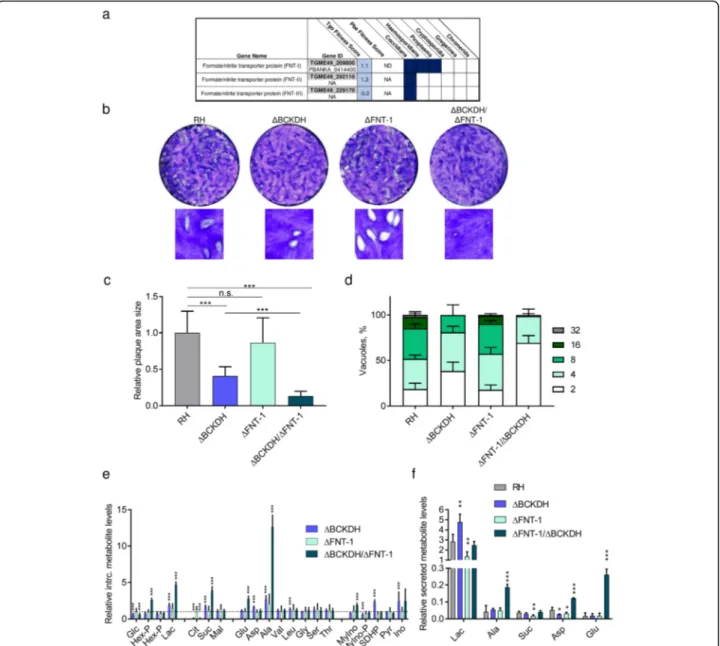
One gene, for which expression was upregulated over 2-fold at the transcript and protein levels inΔBCKDH para-sites, belonged to the formate/nitrite transporter (FNT) family (TGME49_209800, FNT-1) (Fig.7f). In coccidians, three members of this protein family catalyse the transport of monocarboxylate metabolites such as lactate (Fig. 8a) [51]. In contrast, haemosporidians and piroplasms express a single FNT, which has been shown to be essential in Plasmodiumand is the target of several antimalarials [52–
54]. T. gondii parasites lacking BCKDH have previously been shown to have increased levels of intracellular pyru-vate and lactate due to the disruption of the link between glycolysis and the TCA cycle [13]. Under these circum-stances, lactate secretion may be increasingly important to maintain metabolic homeostasis.
FNT-1 is dispensable in RH but becomes fitness-conferring inΔBCKDH parasites
To assess whether the upregulation of FNT-1 is import-ant for the metabolic adaptation in ΔBCKDH cells, we deleted fnt-1 in RH and ΔBCKDH parasites. Using CRISPR-Cas9, we replaced the fnt-1 locus with a dihy-drofolate reductase-thymidylate synthase (dhfr-ts) resist-ance cassette (Additional file13: Figure S6a). Integration of the resistance cassette and the absence of the original gene locus were confirmed by PCR in the single- and double-knock-out (KO) (Additional file 13: Figure S6b). Assessment of the fitness of the different strains by plaque assay confirmed that ΔBCKDH parasites present a modest but significant reduction in plaque size (Fig.8b, c), as previously reported [13]. In contrast,ΔFNT-1 par-asites formed plaques of normal sizes comparable to RH cells, while ΔBCKDH/ΔFNT-1 parasites formed very small plaques, which were considerably smaller than those of ΔBCKDH cells (Fig. 8b,c). An intracellular growth assay confirmed the normal development of ΔFNT-1 cells and the aggravated phenotype of ΔBCKDH/ΔFNT-1 compared to ΔBCKDH cells (Fig.8d). An overview of the growth and fitness defects of the various strains analysed in this study is provided (Add-itional file13: Figure S6c).
To characterise the defect in the metabolism of ΔBCKDH/ΔFNT-1 parasites, intracellular metabolites involved in central carbon metabolism were profiled by GC-MS (Fig.8e). Interestingly, none of the detected me-tabolites was significantly altered in ΔFNT-1 parasites compared to RH. Instead, ΔBCKDH parasites displayed significant changes such as increased lactate and mark-edly reduced citrate consistent with our previous ana-lysis [13]. ΔBCKDH/ΔFNT-1 presented an aggravation of the metabolic phenotype observed inΔBCKDH para-sites (Fig.8e). The observed 5-fold increase in intracellu-lar lactate in ΔBCKDH/ΔFNT-1 (compared to 2-fold in ΔBCKDH) likely impacts on the parasite fitness [51, 53,
55]. To specifically test whetherΔFNT-1 and ΔBCKDH/ ΔFNT-1 parasites displayed defective lactate secretion, we measured the levels of metabolites secreted into the medium by the different T. gondii strains (Fig. 8f). Se-creted metabolites detected in the medium included lac-tate, alanine, succinate, aspartate and glutamate, consistent with previous studies [46].ΔBCKDH cells dis-played the highest secretion of lactate, whileΔFNT-1 se-creted the lowest levels of lactate (Fig. 8f). ΔBCKDH/
ΔFNT-1 exported lactate at similar levels as RH but se-creted markedly higher levels of alanine, aspartate and glutamate (Fig. 8f). Our findings highlight that lactate is exported in cells devoid of ΔFNT-1, consistent with the
presence of other lactate transporters FNT-2 and FNT-3 [51]. However, secretion of lactate is significantly de-creased in ΔFNT-1 compared to RH cells and in ΔBCKDH/ΔFNT-1 compared to ΔBCKDH parasites,
Fig. 8 FNT-1 is dispensable in wild-type T. gondii but becomes highly fitness-conferring inΔBCKDH parasites. a Table highlighting the conservation of formate/nitrite transporters (FNTs) across apicomplexans and their fitness score from a recent screen fitness screen of metabolic genes. b Plaque assay, testing the fitness during the lytic cycle of RH parasites and mutant parasites lacking BCKDH, FNT-1 or both. c
Quantification of plaque area size comparing different strains: RH,ΔBCKDH, ΔFNT-1 and ΔBCKDH/ΔFNT-1 double-KO parasites. Error bars represent the standard deviation between 3 independent infections. Per infection, the areas of > 20 plaques were quantified, and statistically significant differences were determined by a t-test comparing the mutants as indicated (n.s., non-significant; ***p < 0.001). d Intracellular growth assay of RH and mutant parasites over 36 h. Error bars represent the standard deviation between 3 independent infections. Per infection, > 100 vacuoles were counted. e, f Metabolomic analysis of T. gondii RH and mutant parasites (ΔBCKDH, ΔFNT-1 and ΔBCKDH/ΔFNT-1). Levels of intracellular metabolites relative to levels in RH (dashed line) (e) and of metabolites secreted into the medium by purified parasites normalised to valine, an essential amino acid present in the culture medium at 0.8 mM (f). Error bars in e and f represent the standard deviation between replicates (n = 4). Statistically significant differences between each mutant and RH were assessed using a t-test and are indicated (*p < 0.05; **p < 0.005; ***p < 0.001). BCKDH, branched-chainα-keto acid dehydrogenase-complex; Glc, glucose; Hex-P, hexose-phosphate; Lac, lactate; Cit, citrate; Suc, succinate; Mal, malate; Glu, glutamate; Asp, aspartate; Ala, alanine; Val, valine; Leu, leucine; Gly, glycine; Ser, serine; Thr, threonine; MyIno, myo-inositol; MyIno-P, myo-inositol-phosphate; SDHP, sedoheptulose-7-phosphate; Pyr, pyrimidine; Ino, inosine
confirming a prominent role of FNT-1 in efficient lactate secretion. Unable to fuel pyruvate after conversion to acetyl-CoA into the TCA cycle and secreting lactate ineffi-ciently, these cells accumulate toxic intracellular levels of lactate and alanine. Our data highlight the metabolic flexi-bility of T. gondii to adapt to obstructions at the transcrip-tional, translational and post-translational levels.
Discussion
Enzymes generating acetyl-CoA, as well as enzymes in-volved in modulating histone acetylation (histone acety-lases and deacetyacety-lases), have been proposed as drug targets in apicomplexan parasites [17,32,56] and play a crucial role in T. gondii development [35]. However, we know little about how acetylation regulates gene expres-sion and enzyme activities and how acetyl-CoA contrib-utes to the metabolism in these pathogens. We reveal here that cytosolic acetyl-CoA affects the levels of vari-ous transcripts and proteins. Additionally, we demon-strate that it is required for the elongation of FAs. T. gondii synthesises monounsaturated long and very long-chain FAs (FA C20:1, C26:1, C28:1), which are of low abundance or absent in the host cell, making the FA elongation pathway in T. gondii essential [25]. While a previous study reported that depletion of ACS impacts on the FA elongation pathway in T. gondii, the crucial mono-unsaturated very long-chain FAs (FA C26:1, C28:1) were not detected in the study by Dubois and colleagues, and the phenotype was very modest given the compensatory effect of ACL [57]. FAs generated by FASII as well as their derivatives generated through the elongation pathway have a fundamental role for the completion of T. gondii cytokinesis and pellicle formation between the emerging daughter cells [49]. Hence, inhibition of the FA elongation pathway may partially contribute to the described amorphic phenotype of cells devoid of ACS and ACL.
In contrast to the essential pool of nucleo-cytosolic acetyl-CoA, loss of mitochondrial acetyl-CoA can be toler-ated by T. gondii at a modest fitness cost [13]. We reveal here that lack of BCKDH causes relatively few specific changes in the acetylome, transcriptome and proteome, some of which are crucial to enable these parasites to tol-erate obstruction of the link between glycolysis and the TCA cycle. MacRae et al. have proposed that the portion of pyruvate entering the TCA cycle in T. gondii is about 20% under regular culture conditions [46], while the ma-jority is secreted as lactate, through FNT-1 or FNT-2, the two lactate transporters expressed in tachyzoites [51]. In contrast, Plasmodium parasites, which rely on glycolysis during their intraerythrocytic development, express a sin-gle FNT, depletion/inhibition of which leads to parasite death, due to the toxic accumulation of lactate [52,53]. In T. gondii, a genome-wide fitness screen using CRISPR-Cas 9 reported low positive fitness indices for FNT-1 (1.10)
and FNT-2 (1.31), suggesting that both genes are individu-ally dispensable [11]. Indeed, we were able to deplete FNT-1 in tachyzoites without causing any fitness defect. Importantly, these parasites continued to secrete lactate, highlighting that FNT-2 secretes lactate sufficiently under normal culture conditions, compensating for the loss of FNT-1. However, depletion of FNT-1 in cells devoid of BCKDH resulted in a severe additional fitness defect, and double-KO parasites accumulated very high levels of intra-cellular lactate. We propose that the identified hypo-acetylation of LDH1, as well as the overexpression of FNT-1 in cells lacking BCKDH, is part of a coping mech-anism which enables increased reliance on glycolysis in the absence of a functional TCA cycle. These findings provide insights into how parasites adapt their metabolism in response to genetic or pharmacological obstructions.
Conclusions
This study combines molecular tools and multi-omics to provide a uniquely integrative and global picture of the diverse roles of acetyl-CoA in Toxoplasma gondii physi-ology. We demonstrate that loss of nucleo-cytosolic acetyl-CoA results in hypo-acetylation of histones and non-histone proteins causing broad changes in gene ex-pression. Further, we show that the absence of cytosolic acetyl-CoA results in a halt in FA elongation, disabling the synthesis of parasite-specific long-chain monoun-saturated FAs which cannot be salvaged from the host.
In contrast to the cytosolic acetyl-CoA pool, loss of mitochondrial acetyl-CoA can be tolerated due to an al-tered central carbon metabolism [13]. How protozoan parasites remodel their metabolism to adapt to varying environments or to obstructions through genetic alter-ations or drug treatments is poorly understood. We demonstrate here that loss of mitochondrial acetyl-CoA results in the hypo-acetylation of mitochondrial and other proteins as well as in altered gene expression. We provide evidence that these changes at the transcrip-tional, translational and post-translational levels serve to adapt the metabolism and cope with the lack of mito-chondrial acetyl-CoA. These findings provide unprece-dented insights into the plasticity and regulation of the metabolism of T. gondii.
Material and methods
Genetic and cell biology approaches T. gondii culture
All T. gondii strains are derived from RH in which KU80 has been deleted [58]. Parasites were grown in confluent HFFs and maintained in Dulbecco’s modified Eagle medium (DMEM, Life Technology, Invitrogen) supple-mented with 5% foetal calf serum, 2 mM L-glutamine, 25μg/ml gentamicin and where indicated with 0.5 μM Shld-1 [23] in a humidified incubator at 37 °C and 5%
CO2. Specific media for stable isotope labelling experi-ments are described below.
Cloning of DNA constructs
Amplifications of DNA fragments for cloning were per-formed with either the LA Taq (TaKaRa) or the Q5 (New England Biolabs) polymerases, and the primers used for each reaction are listed in Additional file 14: Table S8. Correct integration of the different constructs into the genome of the various strains was determined by genomic PCR using the GoTaq Green Master Mix (Promega) and the primers listed in Additional file14: Table S8.
Preparation of T. gondii genomic DNA
Genomic DNA was extracted from extracellular tachy-zoites using the Wizard SV genomic DNA purification system (Promega).
Inducible knock-down of ACS
To direct the insertion of this PCR product, a specific guide RNA (gRNA) vector targeting the ATG start codon of acs (TGME49_266640) was generated using the Q5 site-directed mutagenesis kit (New England Biolabs) and the vector pSAG1::CAS9-GFP-U6::sgUPRT as a template [59]. The UPRT-targeting gRNA was replaced by an acs-specific gRNA using the primer pair 1-2 listed in Add-itional file14: Table S8. A PCR fragment was amplified of the DD, fused to a myc-tag (DDmyc) with 5′ and 3′ hom-ology sequences to the start ATG of ACS using primers P7/P9 as shown in Additional file 1: Figure S1a,c. (KOD polymerase, Novagen) and pTub8DDmycROM4 as tem-plate [60]. Ten micrograms of the gRNA plasmid, together with the precipitated KOD PCR product, was transfected, and parasites were grown in the presence of Shld-1. Parasites expressing the Cas9-GFP were sorted by fluorescence-activated cell sorting (FACS) and cloned into 96-well plates using a cell sorter (MoFlo Astrios, Beckman Coulter). Clones were analysed by IFA to confirm the ex-pression of the DDmycACS fusion protein and grown in the presence or absence of Shld-1 to evaluate the regula-tion of the protein.
Knock-in construct for epitope tagging at the endogenous locus of pepck-1
A genomic DNA fragment of the C-terminal part of pepck-1(TGME49_289650) was amplified by PCR using the pri-mer pair 3-4 listed in Additional file14: Table S8, digested with the restriction enzymes KpnI/SbfI and cloned into the pTUB8MIC13-3Ty-HX [61], using the KpnI and NsiI sites. Prior to transfection, the plasmid was linearised in the mid-dle of the cloned genomic DNA fragment using the NcoI site. For knock-in insertion of these vectors into the RH and ΔBCKDH strains, the hxgprt cassette was substituted with a dhfr-ts cassette using the two SacII sites.
Generation of PEPCK-1 second copy-expressing plasmids and acetylation mimetics
pTub8-PEPCK-1-L-3Ty and pTub8-PEPCK-1-S-3Ty plas-mids expressing a second copy of both long and short iso-forms of PEPCK-1, respectively, were generated by amplifying the cDNA of pepck-1 using the primers 9-4 (long) or primers 4-10 listed in Additional file14: Table S8, digested with the restriction enzymes EcoRI and SbfI and cloned into the KpnI and NsiI sites of pTub8-APHN21-3Ty-HXGPRT [62]. To complementΔPEPCK-1 with either a wild-type copy of PEPCK-1, acetylation mi-metics or de-acetylation mimi-metics of PEPCK-1, 5’UPRT-DHFR-pTub8-PEPCK-1-4myc-3’UPRT was first gener-ated. 5’UPRT-DHFR-pTub8-PEPCK-1-4myc-3’UPRT was generated by amplifying the cDNA of pepck-1 using the primers 4-10 listed in Additional file14: Table S8, digested with the restriction enzymes EcoRI and SbfI and cloned into the of the pUPRT-pTub8-4myc-3’UPRT plasmid. A dhfr-ts cassette was amplified by PCR using primers 11-12, digested by ApaI, sub-cloned into pUPRT-pTub8-PEPCK-1-4myc-3’UPRT and predigested by ApaI. Lysines K223, K231 and K591 were mutated to either arginine (de-acetylation mimetic) or glutamine (acetylation mi-metic) using the Q5 site-directed mutagenesis kit (New England Biolabs), primer pairs listed in Additional file14: Table S8 and 4myc-3’UPRT as a template. 4myc-K3R-3’UPRT and 5’UPRT-DHFR-pTub8-PEPCK-1-4myc-K3Q-3’UPRT correspond to second copy expressing vectors where PEPCK-1 lysines K223, K231 and K591 were all sequentially mutated to either arginine or glutam-ine respectively. Prior to transfection, all PEPCK-1 com-plementation plasmids were linearised by digestion with NotI-HF and AvrII. Linearised plasmids were co-transfected with 5μg pSAG1::CAS9-GFP-U6::sgUPRT [59]. Transgenic parasites were selected with pyrimeth-amine and cloned in 96-well plates. Expression of PEPCK-1 was validated by IFA.
KO strains
The KO of ACL (TGME49_223840) and BCKDH-E1a (TGME49_239490) were previously described [13,14]. To generate the KO of PEPCK-1 (TGME49_289650), a plas-mid (pTub-CAT-PEPCK-1-ko) was generated and around 1.5 kb of the 5′ and 3′ flanking regions of PEPCK-1 were amplified using primer pairs 5-6 and 7-8, respectively. The 5′ flanking region was then cloned between KpnI and HindIII restriction sites of the pTub5-CAT and the 3′ flanking region between the BamHI and NotI sites. The plasmid was cut with KpnI and NotI prior to transfection. The KO of FNT-1 (TGME49_209800) was generated as follows: 2gRNA plasmid for the FNT-1 KO was generated as described previously [16] with primers as described in Additional file14: Table S8. Forward and reverse primers