HAL Id: hal-01500373
https://hal.archives-ouvertes.fr/hal-01500373
Submitted on 3 Apr 2017HAL is a multi-disciplinary open access archive for the deposit and dissemination of sci-entific research documents, whether they are pub-lished or not. The documents may come from teaching and research institutions in France or abroad, or from public or private research centers.
L’archive ouverte pluridisciplinaire HAL, est destinée au dépôt et à la diffusion de documents scientifiques de niveau recherche, publiés ou non, émanant des établissements d’enseignement et de recherche français ou étrangers, des laboratoires publics ou privés.
Lymph Node Stroma Dynamics and Approaches for
Their Visualization
Rebecca Gentek, Marc Bajenoff
To cite this version:
Rebecca Gentek, Marc Bajenoff. Lymph Node Stroma Dynamics and Approaches for Their Visu-alization. Trends in Immunology, Elsevier, 2017, 38, pp.236 - 247. �10.1016/j.it.2017.01.005�. �hal-01500373�
Lymph node stroma dynamics and approaches for their visualization
Rebecca Gentek1 and Marc Bajénoff1*
1 Aix Marseille Univ, CNRS, INSERM, CIML, Marseille, France *Correspondence: [email protected]
Abstract
Lymphoid stromal cells are best known as the architectural cells of lymphoid organs. For decades, they have been considered as inert elements of the immune system but this view has changed dramatically in recent years, when it was discovered that they are endowed with critical immuno-regulatory functions. It is now accepted that without them, the adaptive immune response would be compromised, if not abrogated entirely. Here, we review the function of the major lymphoid stromal cell types, the way they remodel upon inflammation, discuss the available tools to track their behavior and introduce several methodological approaches that we believe will help improving our knowledge of these pivotal cell types.
Lymphoid stromal cells: versatile and re-configurable 3D immunological networks
Secondary lymphoid organs such as lymph nodes (LNs) are organs in which adaptive immune responses develop. They are composed of 95% motile leukocytes and 5% sessile stromal cells [1]. In 1984, Nossal wrote ‘‘A readership consisting of primarily anatomists has every right to question the favorite sport of research workers in cell immunology. This is to take a lymphoid tissue and totally destroy its beautiful and elaborately designed architecture to obtain simple cell suspension of lymphocytes, which are then asked to do more or less all the jobs of the original anatomic masterpiece’’. In recent years, it was discovered that lymphoid stromal cells are not merely passive architectural cells. Instead, these cells are endowed with immuno-regulatory functions. Within lymphoid organs, various stromal cell subsets create dense three-dimensional (3D) cellular networks that: (i) produce lymphocyte survival signals, (ii) generate ‘roads’ on which lymphocytes migrate, (iii) assemble a rigid backbone of fibers that transports the lymph throughout the LN and (iv) continuously provide the nutrients, soluble factors, antigens as well as the various immune cells required for ‘immunological surveillance’ and the development of adaptive immune responses [1-3] (Figure 1 and table 1). Thus, despite their relatively low abundance, lymphoid stromal cells are key regulators of the immune system. Without them, LNs would not exist and adaptive immunity would be highly compromised. Therefore, understanding the immunobiology of these stromal cells is key to our full comprehension of the immune system.
Unlike most organs, LNs are not rigid structures with a fixed size. Instead, they rapidly and transiently enlarge (up to 10 fold) during an immune response, a process that takes only a few days. This remodeling is crucial not only to meet the increased metabolic needs of the developing immune response, but also to generate new microenvironments mandatory for the proper maturation of this response (e.g germinal centers or medullary cords). This is quite a challenging accomplishment, particularly when considering that some LN stromal cells such as blood endothelial cells need to expand without compromising their sealed tubular structure.
Recent studies have sought to understand the cellular and molecular details of this remodeling. These reports have revealed an impressive level of complexity that regulates LN remodeling and subsequent return to homeostasis. Here, we highlight the work that helped shaping our current understanding of this transient remodeling as well as the recent technological improvements to visualize this process.
Function and remodeling of the main lymphoid stromal cell subsets Fibroblastic Reticular Cells (FRCs)
FRCs are contractile myofibroblasts that form the major mesenchymal stromal cell network of the T cell zone. Through secretion of chemokines such as CCL21 and CCL19, they delineate the boundaries of the T cell zone and physically support lymphocyte migration into that zone [4, 5]. At steady state, FRCs secrete IL-7 and B-Cell Activating factor (BAFF), the main survival factors for T and B cells, respectively [6, 7]. Sure enough, FRC network ablation results in marked alterations in LNs, with significant reduction in organ size, weight and cellularity [6, 8]. FRCs produce and ensheat the conduit system, a network of tiny pipes that transport the lymph throughout the T cell zone [9, 10]. FRCs are also able to induce peripheral tolerance through presentation of peripheral tissue-restricted antigens [11]. Upon inflammation, LNs rapidly enlarge to accommodate the massive influx of naive leukocytes. This initial swelling phase is triggered by the engagement of podoplanin expressed on FRCs with its ligand CLEC-2 present on migrating dendritic cells (DCs). Podoplanin signaling in FRCs induces their relaxation, hence decreasing the tension of the network and the concomitant swelling of the LN[12, 13]. This stretching phase is followed by a late phase of FRC expansion regulated by the engagement of lymphocyte- and DC-derived LIGHT and LTαβ (lymphotoxinαβ) on FRCs [14-16]. Revealing an additional function of FRCs, it was shown more recently that FRCs regulate intestinal inflammation via their production of the cytokine IL-15 and its ability to control group 1 innate lymphoid cells (ILCs) in Peyer's patches and mesenteric LNs [17].
Follicular Dendritic Cells (FDCs) and Marginal Reticular Cells (MRCs)
FDCs and MRCs are the main mesenchymal cell populations of primary B cell follicles. At steady state, FDCs occupy the center of the B cell follicles. FDCs extend multiple long centrifugal processes that secrete the B cell follicle homing chemokine CXCL13 and constitute a cellular scaffold for B cell migration [4, 18-20]. MRCs are located in the outer follicle, just below the subcapsular sinus. They are thought to be the adult counterparts of the lymphoid tissue organizer (LTo) cells, but their exact functions remain elusive in quiescent LNs [21]. During immune responses, FDCs act as antigen-presenting and -retaining cells that remodel the primary follicular network into germinal centers (GCs), a specialized structure in which B cells
proliferate, undergo somatic hypermutation, and carry out class switching [22-24]. Recent reports indicate that the additional FDCs generated during GC formation can originate from both, perivascular mesenchymal cells and the differentiation of MRCs[25, 26].
Blood Endothelial Cells (BECs)
The blood vasculature of the LN is composed of capillary blood endothelial cells (cBEC) and high endothelial venules (HEV). Capillaries are small blood vessels in charge of providing nutrients and oxygen to the surrounding cells while HEVs are the entry doors of blood lymphocytes into the LN parenchyma [27-29]. During an immune response, LN expansion relies on the transient remodeling of its vasculature [30-32]. The remodeling of the LN vasculature is controlled by multiple cell types and is divided into sequential, overlapping phases. In the first days, the blood vasculature undergoes rapid proliferative growth that is initially dependent on IL-1β secreting CD11c+ cells and their ability to stimulate vascular endothelial growth factor (VEGF) by the FRC network located in the T cell zone of the LN[14, 32]. This initiation phase is followed by a T cell-and B cell-dependent expansion phase and ends with the re-establishment of quiescence of the LN[33].
Lymphatic Endothelial Cells (LECs)
LECs assemble to form the afferent, efferent and cortical lymphatics of the LN. Afferent lymphatics convey tissue-derived lymph to the LN where they discharge their content into the subcapsular sinus. Cortical lymphatic sinuses located in the LN parenchyma constitute the exit doors for lymphocytes. They discharge their content into the efferent lymphatics situated in the medulla [34-36]. The primary function of lymphatics is to continuously project the immunological status of a peripheral tissue to its draining LN and to bring efferent LN cells back into the circulation. Occlusion of the afferent lymph flow to the LN severely decreases the ability of HEVs to recruit blood circulating lymphocytes, suggesting that an unknown lymphatic-derived signal is continuously delivered to the HEVs [37]. Additional functions of LECs such as antigen presentation have been described [38]. During an immune response, lymphatics expand to accommodate the growth of the LN in a process known as lymphangiogenesis. B lymphocytes orchestrate this expansion via secretion of VEGF-A that in return supports increased migration of DCs from the periphery to the LN[39].
Studying LN stromal cells: a technical challenge
Stromal cells regulate the immune system on several levels. Yet, our understanding of their biology is rather limited. This limitation of knowledge largely results from technical challenges inherent to the isolation and the culture of LN stromal cells, the inability to model the complexity of LN organization in vitro and the paucity of animal models dedicated to their study. Stromal cells are attached to each other, forming interconnected 3D networks. The 3D nature of all stromal cell networks is key to their functional properties: BECs and LECs assemble in vessels while FDCs and FRCs form complex 3D meshworks. Stromal cell networks display a highly organized structure in which the density, the diameter, the length and the angles of the branches or vessels are precisely defined to perform their functions [8]. Mimicking this complex anatomy is currently impossible in vitro. In addition, stromal cells such as FRCs rapidly de-differentiate in culture and progressively lose their ability to produce chemokines. Therefore, we believe that the dynamics and behavior of stromal cells can only be fully understood from studying them in situ, in their natural micro-environment. To meet this aim, dedicated animal models are required. Stromal cell immunology is a recent field that does not have a large collection of mouse models comparable to the one that has been engineered to study T or B cell responses. Several strains of reporter mice expressing fluorescent reporters and/or Cre recombinase under the control of « stromal cell » promoters have been generated (Table 2). Unfortunately, however, most of these models are not specific and label multiple lymphoid stromal cell types and/or also mark hematopoietic cells. As an example, Lyve-1 Cre mice label LECs, but also a fraction of lymphocytes and myeloid cells while Complement Receptor-2 (CR2) Cre reporter mice label FDCs and B cells[25, 40, 41]. Moreover, with rare exceptions, most of the Cre reporter mice used to study LN stromal cells also affect their counterparts in non-lymphoid organs, preventing the use of classical deletion tools such as the conditional Rosa 26-Diphteria Toxin Receptor (DTR) mouse (Table 2). These models have been critical in unravelling some of the most important functions of LN stromal cells. For example, the presence of FRCs in the T cell zone has been reported decades ago, but their in vivo function remained elusive since then [9, 42]. In 2013, the group of B. Ludewig engineered a bacterial artificial chromosome (BAC)-transgenic mouse model that utilizes the Ccl19 promoter to target the Cre recombinase specifically to FRC in the LNs of adult mice [43]. Taking advantage of this mouse, two groups generated Ccl19-Cre x Rosa26-diphtheria toxin receptor (DTR) mice for conditional ablation of FRCs in vivo. FRCs were rapidly and
efficiently depleted in these mice upon diphtheria toxin administration. Depletion of FRCs in Ccl19-Cre x Rosa26-DTR mice appeared selective, as other stromal cell populations were spared. Using this model, FRC ablation markedly altered T cell homeostasis and compartmentalization, causing profound defects in the activation, migration, expansion and effector function of viral antigen-specific T cells [6, 8]. To further complicate matters, LEC and BEC derive from a common ancestor [44], as do all mesenchymal LN stromal cells [21]. Therefore, reporter mice that express Cre recombinase in these precursors also label their progeny, precluding specificity of targeting. While this does not represent a key issue for imaging studies, it does induce putative caveats when these mice are used to conditionally delete « floxed » genes to assess their functions in LN development or remodeling. Conditional Cre expressing lines such as Cre ERT2 or Tet on/off models partially overcome some issues related to constitutive Cre expressing lines by allowing a temporal activation of Cre in a given subset of stromal cells upon injection of Tamoxifen or Doxycycline.
Stromal cell heterogeneity: understanding the LN stroma at the single cell level
As summarized above, FDCs, FRCs, MRCs, LECs and BECs are considered the major LN stromal cell populations. Nonetheless, additional subsets have been described, including CXCL12-producing follicular stromal cells, integrin α7-expressing pericytes and Versatile stromal cells (VSCs)[29, 45, 46] (Table 1 and 2). It is currently unknown if all these lymphoid stromal cell subsets represent true distinct populations or different anatomical flavors of a single mesenchymal stromal cell. Despite having provided critical insights into their biology, most past studies have been restricted to the analysis of stromal cells at the population level. Thus, we critically lack information on the behaviors of individual stromal cells. Why is this important? During an infection, CD4 T cells originating from a single clone differentiate into several subsets (TFh, Treg, Th17, Th1…) that collectively mount a protective immune response [47]. We currently do not know if stromal cells display a similar heterogeneity within their subsets. Flow cytometry represents an appealing technique to analyze the phenotype and proliferative history of single cells. While the isolation of LN stromal cell populations based on surface markers and/or transgene expression is routinely performed, it should be mentioned that stromal cells represent the most difficult cell types to extract from LNs. As a result, their recovery yields are ranging from good (LEC and BEC) to poor (FRC) and even extremely poor (MRC, FDC)[25, 48, 49]. This technical caveat should be kept in mind when extrapolating the
results obtained from such low cell numbers to the entire population of a given stromal cell type. Moreover, flow cytometry experiments using BrdU and EdU incorporation measurements are classically employed to monitor the proliferative history of stromal cells [50, 51]. However, this approach fails to differentiate a cell that has divided once from a single cell that has divided e.g. twenty times. In summary, analyzing stromal cells at the population levels using flow cytometry bears inherent limitations that might prevent the community from (a) identifying putative proliferative stem cells, (b) unravelling precursor-product relationships amongst stromal subsets and (c) gathering critical anatomical details that would help explaining the process of interest.
Inspiration from neurosciences: multicolor lineage tracing models
Unlike most immunologists who work on cells that can be adoptively transferred or replaced by bone marrow grafting, neurobiologists face the same basic challenges as immunologists studying lymphoid stromal cells: neurons assemble in various, complex and intermingled 3D networks. It is impossible to graft, remove or replace these networks in vivo. Neuroscientists had thus no choice but to generate new models to study neurons in their natural, complex 3D environment in vivo. The recent development of multicolor fate mapping systems based on the Brainbow approach has improved the existing lineage tracing systems and created new tools to study cell dynamics in situ [52-54]. The Brainbow strategy enables combinatorial expression of three or four fluorescent proteins in a stochastic manner. Using incompatible lox variants, these fluorescent proteins (FPs) can be expressed by stochastic recombination using Cre-mediated inversion (Figure 2). When three transgenes of a Brainbow construct expressing three (Brainbow-1.0) or four (Brainbow-2.1) “FPs” are introduced into a mouse, independent recombination of those transgene copies can generate six or ten distinct color combinations. The use of multiple colors within one cell population allows for a shift in the types of questions that can be asked using standard reporter models. Labeling strategies often use a given promoter to drive one-color expression for all members of that particular cell type, which distinguishes that cell type from others. While this strategy is ideal to investigate the behavior of cells at the population level, it cannot reveal the behavior of single cells within that very population. The Brainbow approach distinguishes amonglike cells and is ideal for following individual cells over time and space. As the Brainbow colors are inheritable, an initial pool of progenitor cells that is labeled in specific colors produces labeled progeny
that reflect their cellular lineage, hence allowing the establishment of parentage relationship between different cell types.
Multicolor lineage tracing models to study stromal cells
Far from being fancy tools, Brainbow models have allowed seminal discoveries in the fields of developmental, cancer and stem cell biology in several species (zebrafish, drosophila and mouse) [52-55]. How about immunology? Brainbow models are retrospective tools that allow the reconstruction of the proliferative and migratory history of an individual cell and its progeny by measuring the color, location and size of clones at a given time. By definition, this system only applies to cell types in which the progeny of a labeled cell does not further migrate (Figure 3). In summary, Brainbow models are not adapted to study motile lymphocytes but very well suited to investigate the dynamics of lymphoid stromal cells both during the development of LNs and their inflammation induced remodeling.
Using this approach, two studies investigated the dynamics of stromal cells during an immune response. In the first one, the ontogeny and dynamics of murine LN FDCs were studied. Because FDCs are very difficult to extract from the LNs of untreated animals, this work combined the Brainbow approach with multiple Cre lines to study FDCs in situ [25]. By inducing Cre recombination during development or at steady state, this work revealed that LN FDC networks arise from the clonal expansion and differentiation of MRCs. This study also showed that during an immune response, neither the recruitment of circulating progenitors nor the division of local mature FDCs significantly contributes to the accumulation of FDCs in GCs. Rather, the evidence suggested that newly generated FDCs also arise from the proliferation and differentiation of MRCs, thus unraveling a first critical function of this poorly defined stromal cell population.
In a more recent study, the behavior of BECs during LN expansion and subsequent return to homeostasis was tracked using such multicolor fluorescent fate mapping models [56]. Because conventional tissue sections are too thin to unravel the complexity of vasculature trees, a novel technique for obtaining thick, but optically transparent LN tissue samples was developed, enabling high-resolution imaging and reconstruction of the LN vascular tree with high resolution. Applying this technique, it was reported that LN vasculature expansion relies on the sequential assembly of endothelial cell proliferative units. This segmented growth seemed to be sustained by the clonal proliferation of HEV cells that behaved as local progenitors to
create capillaries and HEV neovessels at the periphery of the LN. This work also suggested that the return to homeostasis was accompanied by the stochastic death of pre-existing and neo-synthesized LN endothelial cells.
Thus, as for the fields of neurosciences, development and stem cells, multicolor fate-mapping studies emerge as promising tools to decipher the complex dynamics of stromal cells.
Limitations and Future directions Better mouse models
Stromal cell immunobiology is a relatively young field that heavily relies on animal models. Paradoxically, however, suitable models are critically lacking. As an example, specific genetic deletion or fate mapping of lymphoid stromal cell subsets is currently impossible in the mouse (Table 2). How can we improve that? Precisely defining the roles of specific cell types is a challenging task within an entire organism. To this aim, biologists have engineered an important repository of genetically-encoded mouse models in which a reporter gene is inserted under the control of a given promoter. However, targeting these tools with adequate specificity remains challenging: most cell types are best defined by the intersection of two or more genetic features such as active promoter elements. Thus, a key challenge of broad significance is to increase the specificity of cell-type targeting. To target immune cells more specifically, several groups have combined two genes; neither of which alone were specific for the cell types of interest. As an example, transgenic mice that carry a BAC encoding a DTR– mCherry fusion protein (DTR-mCherry) preceded by a loxP-flanked transcriptional Stop element under the control of the Csf1r promoter (Csf1rLsL-DTR mice) were generated and crossed with mice that express Cre recombinase under the control of LysM (LysmCre mice). LysM-expressing cells in LysmCre x Csf1rLsL-DTR mice delete the Stop element, which permits transcription of DTR-mCherry specifically in LysM and Csf1r double-positive cells (i.e. macrophages and monocytes), but not in conventional DCs [57]. To further improve the precision of selective cell targeting, several alternative, complimentary approaches were developed by the neuroscience field. These Intersectional methods are based on two to three genes or other cell features and exploit distinct recombinases [58-60]. As an example, intersection of Cre and Flp (Flippase) lines driven by two marker genes target more restricted GABAergic subpopulations [61]. We believe that these strategies could be used to achieve
better targeting and deletion of LN stromal cell subsets in which single genes are routinely shared by many cell types.
Better imaging tools
Although such improved mouse models would allow for more selective targeting ofthe LN stroma, they remain inherently difficult to analyze. As stromal cells associate in large 3D networks, improving imaging techniques to visualize stromal cell networks in situ is also key to the field. Conventional confocal/fluorescence imaging studies achieve a cellular resolution when imaging 8-30µm thick tissue sections. However, these samples are too thin to visualize the complex 3D stromal cell networks and obtain full appreciation of their organization. Macroscopic imaging techniques such as Optical Projection Tomography (OPT) allow the reconstruction of the vascular tree but lack the resolution to image its individual cellular components [31]. Light sheet fluorescence microscopy (LSFM) functions as a non-destructive microtome and microscope that uses a plane of light to optically section and view whole tissues. This method is well suited for imaging deep within transparent tissues or within whole living organisms. However, LSFM currently lacks the submicron resolution of confocal microscopy and is therefore not very well suited to examine the fine details of highly dendritic stromal cell networks such as FRCs or FDCs [62]. Why is it so important to reconstruct an entire LN when studying LN stromal cells? Ontogenic studies of LN development and multicolor fluorescent lineage tracing studies suggest that all the LN mesenchymal cell subsets derive from a pool of LTo cells. According to this model, few LTo cells extensively divide to generate the entire mesenchymal network of the LN. Evaluating the extent of this proliferation, the location and the competition of the various clones are key to our understanding of LN development. Similar reasoning applies to the LN remodeling that occurs after an infection. As the extensive proliferation of few cells can in principle generate gigantic clonal populations, only the reconstruction of an entire LN bears the potential to reveal the « global picture » of these two phenomena.
The scattering of light in heterogeneous tissues remained a limiting factor, preventing researchers from achieving high-resolution 3D renderings of thick tissue. The source of the scattering is a diverse set of cellular constituents including ribosomes, nuclei, lipid droplets, and components of the cytoskeleton and extracellular matrix. Recently, several procedures have been developed to obtain optically transparent tissue samples for imaging, which,
importantly, are also compatible with imaging endogenous fluorescent proteins [63-67]. Together with advances in data acquisition and storage capacity, these improved clearing techniques become increasingly used to gain anatomical details of fundamental biological processes. However, these approaches are time- and resources-consuming: imaging an entire LN typically requires several hours, and so does the post-acquisition treatment on powerful custom-made work stations equipped with expensive imaging software. It is to be hoped that better clearing techniques, faster imaging devices and more affordable software solutions will contribute to the democratization of this approach in the near future.
Concluding remarks
Our knowledge on the origin and function of LN stromal cells has tremendously increased in recent years. However, we still lack a comprehensive view of the stromal cell dynamics that accompany and sustain LN growth during an immune response. A central and still unresolved question concerns the existence of an adult LN mesenchymal stromal cell progenitor. Similar to embryonic LTo during development, such a putative progenitor would extensively divide in the course of an immune response to fuel the pool of mesenchymal LN stromal cells. MRC have been suggested as candidate adult LN mesenchymal stem cells, but dedicated animal models to test this hypothesis are currently lacking [21]. Intriguingly, a growing body of evidence suggests that pericytes can act as mesenchymal stem cells in various tissues [68]. As a result, pericytes might represent a source of mesenchymal cells in inflamed LNs. In a recent study, Prados and colleagues described a transgenic mouse strain that expresses Cre-recombinase under the CollagenVI promoter (ColVI-Cre mouse) [69]. In this mouse, pericytes, but not other mesenchymal stromal cells were targeted in all the secondary lymphoid organs (SLO) in the naïve state, suggesting that pericytes do not give rise to other LN stromal cell subsets at steady state. Whether additional mesenchymal LN stromal cells are labeled in the LNs of these mice upon inflammation, however, remains to be determined.
As all LN stromal cell subsets are assembled in complex 3D structures, merely increasing the number of LN stromal cells is not sufficient to enable the enlargement of the LN during an immune response. Topological studies have described the complex organization of the conduit system and its surrounding FRC-network [5, 8-10, 70]. FRCs have precisely defined intercellular distances and number of connected protrusions per cell, allowing the network to
function as a cellular « sponge » that creates extracellular spaces for lymphocytes, while offering adhesive migration substrates for them. When 70% of the FRC network is genetically ablated, T cell migration and proliferation is affected [8]. These data suggest that the physical properties of this topological ‘small-world ‘ organization are key to its function and that maintaining the precise dimensions of the FRC network is mandatory for a proper immune response. Conduits made by the FRC network create the 3D backbone of the LN while conveying the lymph throughout the T cell zone. FRCs ensheath them in a way that most of the conduits are shielded from the lymphoid and myeloid cells situated in the surrounding space. During an immune response, FRCs divide and produce new conduits to sustain the growth of the LN [5]. Interestingly, the additional FRCs and their associated conduits are added to the existing network in a fractal way (our unpublished results). Identifying the molecular, cellular and physical mechanisms that orchestrate this highly organized remodeling represents an important question, not only for the FRC network but also for other lymphoid stromal subsets such as LECs and BECs, whose biological functions are tightly linked to their physical properties. We believe that a combination of flow cytometry, high resolution imaging approaches, improved lineage tracing models, mathematical modeling and genetic manipulation will be required to understand the complex stromal dynamics that accompany the enlargement of LNs during immune responses.
Acknowledgments
This work was supported by a grant from the European Research Council (ERC) under the Eu-ropean Union’s Horizon 2020 research and innovation program grant agreement N° 647384- STROMA.
References
1. Mueller, S.N. and R.N. Germain, Stromal cell contributions to the homeostasis and functionality of the immune system. Nat Rev Immunol, 2009. 9(9): p. 618-29.
2. Koning, J.J. and R.E. Mebius, Interdependence of stromal and immune cells for lymph node function. Trends Immunol, 2012. 33(6): p. 264-70.
3. Roozendaal, R. and R.E. Mebius, Stromal cell-immune cell interactions. Annu Rev Immunol, 2011. 29: p. 23-43.
4. Bajenoff, M., et al., Stromal cell networks regulate lymphocyte entry, migration, and territoriality in lymph nodes. Immunity, 2006. 25(6): p. 989-1001.
5. Katakai, T., et al., Lymph node fibroblastic reticular cells construct the stromal reticulum via contact with lymphocytes. J Exp Med, 2004. 200(6): p. 783-95.
6. Cremasco, V., et al., B cell homeostasis and follicle confines are governed by fibroblastic reticular cells. Nat Immunol, 2014.
7. Link, A., et al., Fibroblastic reticular cells in lymph nodes regulate the homeostasis of naive T cells. Nat Immunol, 2007. 8(11): p. 1255-65.
8. Novkovic, M., et al., Topological Small-World Organization of the Fibroblastic Reticular Cell Network Determines Lymph Node Functionality. PLoS Biol, 2016. 14(7): p. e1002515.
9. Gretz, J.E., A.O. Anderson, and S. Shaw, Cords, channels, corridors and conduits: critical architectural elements facilitating cell interactions in the lymph node cortex. Immunol Rev, 1997. 156: p. 11-24.
10. Sixt, M., et al., The conduit system transports soluble antigens from the afferent lymph to resident dendritic cells in the T cell area of the lymph node. Immunity, 2005. 22(1): p. 19-29. 11. Fletcher, A.L., et al., Lymph node fibroblastic reticular cells directly present peripheral tissue
antigen under steady-state and inflammatory conditions. J Exp Med, 2010. 207(4): p. 689-97. 12. Acton, S.E., et al., Dendritic cells control fibroblastic reticular network tension and lymph node
expansion. Nature, 2014. 514(7523): p. 498-502.
13. Astarita, J.L., et al., The CLEC-2-podoplanin axis controls the contractility of fibroblastic reticular cells and lymph node microarchitecture. Nat Immunol, 2015. 16(1): p. 75-84.
14. Benahmed, F., et al., Multiple CD11c+ cells collaboratively express IL-1beta to modulate stromal vascular endothelial growth factor and lymph node vascular-stromal growth. J Immunol, 2014. 192(9): p. 4153-63.
15. Chyou, S., et al., Fibroblast-type reticular stromal cells regulate the lymph node vasculature. J Immunol, 2008. 181(6): p. 3887-96.
16. Kumar, V., et al., A dendritic-cell-stromal axis maintains immune responses in lymph nodes. Immunity, 2015. 42(4): p. 719-30.
17. Gil-Cruz, C., et al., Fibroblastic reticular cells regulate intestinal inflammation via IL-15-mediated control of group 1 ILCs. Nat Immunol, 2016. 17(12): p. 1388-1396.
18. Allen, C.D. and J.G. Cyster, Follicular dendritic cell networks of primary follicles and germinal centers: phenotype and function. Semin Immunol, 2008. 20(1): p. 14-25.
19. Cyster, J.G., et al., Follicular stromal cells and lymphocyte homing to follicles. Immunol Rev, 2000. 176: p. 181-93.
20. Wang, X., et al., Follicular dendritic cells help establish follicle identity and promote B cell retention in germinal centers. J Exp Med, 2011. 208(12): p. 2497-510.
21. Katakai, T., Marginal reticular cells: a stromal subset directly descended from the lymphoid tissue organizer. Front Immunol, 2012. 3: p. 200.
22. Allen, C.D., T. Okada, and J.G. Cyster, Germinal-center organization and cellular dynamics. Immunity, 2007. 27(2): p. 190-202.
23. Garin, A., et al., Toll-like receptor 4 signaling by follicular dendritic cells is pivotal for germinal center onset and affinity maturation. Immunity, 2010. 33(1): p. 84-95.
24. Victora, G.D. and M.C. Nussenzweig, Germinal centers. Annu Rev Immunol, 2012. 30: p. 429-57.
25. Jarjour, M., et al., Fate mapping reveals origin and dynamics of lymph node follicular dendritic cells. J Exp Med, 2014. 211(6): p. 1109-22.
26. Krautler, N.J., et al., Follicular dendritic cells emerge from ubiquitous perivascular precursors. Cell, 2012. 150(1): p. 194-206.
27. Anderson, A.O. and N.D. Anderson, Lymphocyte emigration from high endothelial venules in rat lymph nodes. Immunology, 1976. 31(5): p. 731-48.
28. Girard, J.P. and T.A. Springer, High endothelial venules (HEVs): specialized endothelium for lymphocyte migration. Immunol Today, 1995. 16(9): p. 449-57.
29. Mionnet, C., et al., Identification of a new stromal cell type involved in the regulation of inflamed B cell follicles. PLoS Biol, 2013. 11(10): p. e1001672.
30. Anderson, N.D., A.O. Anderson, and R.G. Wyllie, Microvascular changes in lymph nodes draining skin allografts. Am J Pathol, 1975. 81(1): p. 131-60.
31. Kumar, V., et al., Optical projection tomography reveals dynamics of HEV growth after immunization with protein plus CFA and features shared with HEVs in acute autoinflammatory lymphadenopathy. Front Immunol, 2012. 3: p. 282.
32. Webster, B., et al., Regulation of lymph node vascular growth by dendritic cells. J Exp Med, 2006. 203(8): p. 1903-13.
33. Dasoveanu, D.C., et al., Regulation of Lymph Node Vascular-Stromal Compartment by Dendritic Cells. Trends Immunol, 2016.
34. Drayson, M.T. and W.L. Ford, Afferent lymph and lymph borne cells: their influence on lymph node function. Immunobiology, 1984. 168(3-5): p. 362-79.
35. Ohtani, O., et al., Fluid and cellular pathways of rat lymph nodes in relation to lymphatic labyrinths and Aquaporin-1 expression. Arch Histol Cytol, 2003. 66(3): p. 261-72.
36. Young, A.J., The physiology of lymphocyte migration through the single lymph node in vivo. Semin Immunol, 1999. 11(2): p. 73-83.
37. Mebius, R.E., et al., The influence of afferent lymphatic vessel interruption on vascular addressin expression. J Cell Biol, 1991. 115(1): p. 85-95.
38. Cohen, J.N., et al., Lymph node-resident lymphatic endothelial cells mediate peripheral tolerance via Aire-independent direct antigen presentation. J Exp Med, 2010. 207(4): p. 681-8. 39. Angeli, V., et al., B cell-driven lymphangiogenesis in inflamed lymph nodes enhances dendritic
cell mobilization. Immunity, 2006. 24(2): p. 203-15.
40. Kraus, M., et al., Survival of resting mature B lymphocytes depends on BCR signaling via the Igalpha/beta heterodimer. Cell, 2004. 117(6): p. 787-800.
41. Pham, T.H., et al., Lymphatic endothelial cell sphingosine kinase activity is required for lymphocyte egress and lymphatic patterning. J Exp Med, 2010. 207(1): p. 17-27.
42. Kelly, R.H., Functional anatomy of lymph nodes. I. The paracortical cords. Int Arch Allergy Appl Immunol, 1975. 48(6): p. 836-49.
43. Chai, Q., et al., Maturation of lymph node fibroblastic reticular cells from myofibroblastic precursors is critical for antiviral immunity. Immunity, 2013. 38(5): p. 1013-24.
44. Srinivasan, R.S., et al., Lineage tracing demonstrates the venous origin of the mammalian lymphatic vasculature. Genes Dev, 2007. 21(19): p. 2422-32.
45. Bannard, O., et al., Germinal center centroblasts transition to a centrocyte phenotype according to a timed program and depend on the dark zone for effective selection. Immunity, 2013. 39(5): p. 912-24.
46. Malhotra, D., et al., Transcriptional profiling of stroma from inflamed and resting lymph nodes defines immunological hallmarks. Nat Immunol, 2012. 13(5): p. 499-510.
47. Dong, C., Helper T-cell heterogeneity: a complex developmental issue in the immune system. Cell Mol Immunol, 2010. 7(3): p. 163.
48. Fletcher, A.L., et al., Reproducible isolation of lymph node stromal cells reveals site-dependent differences in fibroblastic reticular cells. Front Immunol, 2011. 2: p. 35.
49. Usui, K., et al., Isolation and characterization of naive follicular dendritic cells. Mol Immunol, 2012. 50(3): p. 172-6.
50. Gratzner, H.G., Monoclonal antibody to 5-bromo- and 5-iododeoxyuridine: A new reagent for detection of DNA replication. Science, 1982. 218(4571): p. 474-5.
51. Salic, A. and T.J. Mitchison, A chemical method for fast and sensitive detection of DNA synthesis in vivo. Proc Natl Acad Sci U S A, 2008. 105(7): p. 2415-20.
52. Ghigo, C., et al., Multicolor fate mapping of Langerhans cell homeostasis. J Exp Med, 2013. 210(9): p. 1657-64.
53. Livet, J., et al., Transgenic strategies for combinatorial expression of fluorescent proteins in the nervous system. Nature, 2007. 450(7166): p. 56-62.
54. Snippert, H.J., et al., Intestinal crypt homeostasis results from neutral competition between symmetrically dividing Lgr5 stem cells. Cell, 2010. 143(1): p. 134-44.
55. Hampel, S., et al., Drosophila Brainbow: a recombinase-based fluorescence labeling technique to subdivide neural expression patterns. Nat Methods, 2011. 8(3): p. 253-9.
56. Mondor, I., et al., Clonal Proliferation and Stochastic Pruning Orchestrate Lymph Node Vasculature Remodeling. Immunity, 2016. 45(4): p. 877-888.
57. Schreiber, H.A., et al., Intestinal monocytes and macrophages are required for T cell polarization in response to Citrobacter rodentium. J Exp Med, 2013. 210(10): p. 2025-39.
58. Dymecki, S.M., R.S. Ray, and J.C. Kim, Mapping cell fate and function using recombinase-based intersectional strategies. Methods Enzymol, 2010. 477: p. 183-213.
59. Fenno, L.E., et al., Targeting cells with single vectors using multiple-feature Boolean logic. Nat Methods, 2014. 11(7): p. 763-72.
60. Madisen, L., et al., Transgenic mice for intersectional targeting of neural sensors and effectors with high specificity and performance. Neuron, 2015. 85(5): p. 942-58.
61. He, M., et al., Strategies and Tools for Combinatorial Targeting of GABAergic Neurons in Mouse Cerebral Cortex. Neuron, 2016. 92(2): p. 555.
62. Santi, P.A., Light sheet fluorescence microscopy: a review. J Histochem Cytochem, 2011. 59(2): p. 129-38.
63. Chung, K. and K. Deisseroth, CLARITY for mapping the nervous system. Nat Methods, 2013. 10(6): p. 508-13.
64. Dodt, H.U., et al., Ultramicroscopy: three-dimensional visualization of neuronal networks in the whole mouse brain. Nat Methods, 2007. 4(4): p. 331-6.
65. Erturk, A., et al., Three-dimensional imaging of solvent-cleared organs using 3DISCO. Nat Protoc, 2012. 7(11): p. 1983-95.
66. Hama, H., et al., Scale: a chemical approach for fluorescence imaging and reconstruction of transparent mouse brain. Nat Neurosci, 2011. 14(11): p. 1481-8.
67. Susaki, E.A., et al., Whole-brain imaging with single-cell resolution using chemical cocktails and computational analysis. Cell, 2014. 157(3): p. 726-39.
68. da Silva Meirelles, L., et al., Mesenchymal stem cells and their relationship to pericytes. Front Biosci (Landmark Ed), 2016. 21: p. 130-56.
69. Prados, A., G. Kollias, and V. Koliaraki, CollagenVI-Cre mice: A new tool to target stromal cells in secondary lymphoid organs. Sci Rep, 2016. 6: p. 33027.
70. Katakai, T., et al., A novel reticular stromal structure in lymph node cortex: an immuno-platform for interactions among dendritic cells, T cells and B cells. Int Immunol, 2004. 16(8): p. 1133-42. 71. Repass, J.F., et al., IL7-hCD25 and IL7-Cre BAC transgenic mouse lines: new tools for analysis of
IL-7 expressing cells. Genesis, 2009. 47(4): p. 281-7.
72. Chai, Y., et al., Fate of the mammalian cranial neural crest during tooth and mandibular morphogenesis. Development, 2000. 127(8): p. 1671-9.
73. Cuttler, A.S., et al., Characterization of Pdgfrb-Cre transgenic mice reveals reduction of ROSA26 reporter activity in remodeling arteries. Genesis, 2011. 49(8): p. 673-80.
74. Chen, M.J., et al., Runx1 is required for the endothelial to haematopoietic cell transition but not thereafter. Nature, 2009. 457(7231): p. 887-91.
75. Sorensen, I., R.H. Adams, and A. Gossler, DLL1-mediated Notch activation regulates endothelial identity in mouse fetal arteries. Blood, 2009. 113(22): p. 5680-8.
76. Gomez Perdiguero, E., et al., Tissue-resident macrophages originate from yolk-sac-derived erythro-myeloid progenitors. Nature, 2015. 518(7540): p. 547-51.
77. Kisanuki, Y.Y., et al., Tie2-Cre transgenic mice: a new model for endothelial cell-lineage analysis in vivo. Dev Biol, 2001. 230(2): p. 230-42.
Figure legends
HEV: High Endothelial Venule, SCS: Subcapsular Sinus, MRC: Marginal Reticular Cell, FDC: Follicular Dendritic Cell, Cap: Capillary, P: Pericyte, FRC: Fibroblastic Reticular Cell, *: collagen bundles and fibrils. T cells (T) and B cells (B) migrate along the FRC and FDC networks, respectively. Blue lines represent the conduits.
Figure 2: Principle of Brainbow strategies.
(A) In Brainbow-1.0, pairs of incompatible lox variants (LoxP and lox N) are interleaved, creating two mutually exclusive, Cre mediated excision possibilities. Each of these excisions triggers the expression of either CFP or YFP. (B) In Brainbow-2.1, loxP sites are positioned in opposite orientation, defining two invertible cassettes in which two fluorescent protein encoding genes (nuclear GFP/YFP and RFP/CFP) are placed head to head. This construct can be excised and inverted as long as Cre is active. When it stabilizes, the fluorescent protein encoding gene ending in a sense orientation is expressed. Note that if a cell constitutively expresses Cre in Brainbow-2.1 mice, it will keep changing its color for its entire lifespan. This precludes the use of Brainbow-2.1 mice for genetic fate mapping purposes. On the contrary, an individual Cre expressing cell will acquire a single color for its entire lifespan in Brainbow-1.0 mice. As each Brainbow copy behaves independently from the others, the presence of multiple copies yields additional color combinations (here, two copies of Brainbow- 1.0 resulting in six possible outcomes).
Figure 3: Multicolor lineage tracing to study lymphoid stromal cells in vivo.
This scheme represents the putative behavior of T cells (A), FRCs (B) and BECs (C) in the LNs of a Brainbow derivative mouse at steady state (left) and undergoing an immune response (right). In (A), Ag-specific T cells divide and then resume their migratory behavior, preventing their color-based tracking. In (B) and (C), FRCs and BECs proliferate. As they are immotile, their progeny generates monocolored foci (F) of various sizes, providing quantitative and qualitative anatomical details of this phenomenon [56].
Fibroblastic Reticular cells
(FRCs)
Follicular
Dendritic cells (FDCs) Reticular cellsMarginal
(MRCs) Pericytes Lymphatic Endothelial cells (LECs) Blood Endothelial cells (BECs) Versatile Stromal cells (VSCs) CXCL12-Reticular cells (CRCs) structural support conduit formation substrate for lymphocyte migration and Dendritic Cell adhesion govern T cell territoriality and homeostasis antigen presentation govern B cell territoriality, homeostasis and GC formation capture and presentation of immune complexes conduit formation progenitors of FDCs structural support unknown transport of lymph-borne cells, soluble factors and antigen control lymphocyte efflux chemokine production antigen presentation transport of blood-borne cells, soluble factors and nutrients control lymphocyte influx (HEV) chemokine production regulate the size of inflamed B cell follicles Support B cell migration GC organization?
Table 1: Immunological functions of lymph node stromal cells.
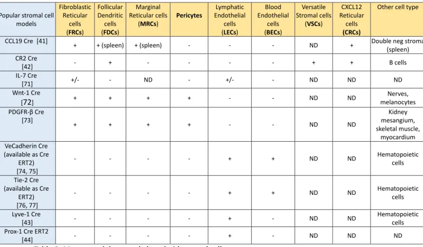
Popular stromal cell models Fibroblastic Reticular cells (FRCs) Follicular Dendritic cells (FDCs) Marginal Reticular cells (MRCs) Pericytes Lymphatic Endothelial cells (LECs) Blood Endothelial cells (BECs) Versatile Stromal cells (VSCs) CXCL12 Reticular cells (CRCs)
Other cell type
CCL19 Cre [41] + + (spleen) + (spleen) - - - ND + Double neg stroma
(spleen) CR2 Cre [42] - + - - - - + + B cells IL-7 Cre [71] +/- - ND - +/- - ND ND ND Wnt-1 Cre [72] + + + + - - ND ND melanocytesNerves, PDGFR-β Cre [73] + + + + - - ND ND mesangium, Kidney skeletal muscle, myocardium VeCadherin Cre (available as Cre ERT2) [74, 75] - - - - + + ND ND Hematopoietic cells Tie-2 Cre (available as Cre ERT2) [76, 77] - - - - + + ND ND Hematopoietic cells Lyve-1 Cre [43] - - - - + - ND ND Hematopoietic cells
Prox-1 Cre ERT2
[44] - - - - + - ND ND ND
Table 2: Mouse models to study lymphoid stromal cells. + : expressed, - : not expressed, ND : not determined
Trends Box
Lymphoid stromal cells are more than architectural cells: they are pivotal regulators of adaptive immunity.
While we begin to understand the functions of lymphoid stromal cells at the population level, we critically lack information on the heterogeneity of individual stromal cells.
Stromal cells are difficult to study: animal models are rare and flow cytometry approaches are limited.
Neuroscientists face similar technical limitations and have developed tools to decipher and manipulate the neuronal network: let them be an inspiration to us.
Outstanding Questions Box
During an immune response, stromal cells divide and remodel to accompany LN growth. Are all stromal cells able to divide equally or do equivalents to embryonic LTo exist as quiescent precursors in the adult?
An increasing number of lymphoid mesenchymal subsets is being identified. Are all these stromal cells truly different populations or do they represent subtle adaptations of a single mesenchymal cell type to distinct LN microenvironments?
Which mechanisms drive re-establishment of LN stromal cell quiescence at the end of an immune response?
Upon return to LN quiescence, superfluous stromal cells need to be deleted. Is this pruning stochastic or selective?