HAL Id: hal-03162042
https://hal-amu.archives-ouvertes.fr/hal-03162042
Submitted on 8 Mar 2021
HAL is a multi-disciplinary open access
archive for the deposit and dissemination of
sci-entific research documents, whether they are
pub-lished or not. The documents may come from
teaching and research institutions in France or
abroad, or from public or private research centers.
L’archive ouverte pluridisciplinaire HAL, est
destinée au dépôt et à la diffusion de documents
scientifiques de niveau recherche, publiés ou non,
émanant des établissements d’enseignement et de
recherche français ou étrangers, des laboratoires
publics ou privés.
Distributed under a Creative Commons Attribution| 4.0 International License
Rare Pathogenic Variants in Mitochondrial and
Inflammation-Associated Genes May Lead to
Inflammatory Cardiomyopathy in Chagas Disease
Maryem Ouarhache, Sandrine Marquet, Amanda Frade, Ariela Ferreira,
Barbara Ianni, Rafael Almeida, Joao Paulo Silva Nunes, Ludmila Rodrigues
Pinto Ferreira, Oliveira-Carvalho Rigaud, Darlan Cândido, et al.
To cite this version:
Maryem Ouarhache, Sandrine Marquet, Amanda Frade, Ariela Ferreira, Barbara Ianni, et al.. Rare
Pathogenic Variants in Mitochondrial and Inflammation-Associated Genes May Lead to
Inflamma-tory Cardiomyopathy in Chagas Disease. Journal of Clinical Immunology, Springer Verlag, 2021,
�10.1007/s10875-021-01000-y�. �hal-03162042�
ORIGINAL ARTICLE
Rare Pathogenic Variants in Mitochondrial
and Inflammation-Associated Genes May Lead to Inflammatory
Cardiomyopathy in Chagas Disease
Maryem Ouarhache1&Sandrine Marquet1,2&Amanda Farage Frade3,4&Ariela Mota Ferreira5&Barbara Ianni6&
Rafael Ribeiro Almeida3,4&Joao Paulo Silva Nunes3,4&Ludmila Rodrigues Pinto Ferreira3,4,7&
Vagner Oliveira-Carvalho Rigaud3&Darlan Cândido3&Charles Mady6&Ricardo Costa Fernandes Zaniratto3&
Paula Buck6&Magali Torres2&Frederic Gallardo2&Pauline Andrieux2&Sergio Bydlowsky3,4&Debora Levy3,4&
Laurent Abel8,9&Clareci Silva Cardoso10&Omar Ribeiro Santos-Junior11&Lea Campos Oliveira12&
Claudia Di Lorenzo Oliveira10&Maria Do Carmo Nunes11&Aurelie Cobat8,9&Jorge Kalil3,4&Antonio Luiz Ribeiro11&
Ester Cerdeira Sabino12&Edecio Cunha-Neto3,4,13 &Christophe Chevillard1,2
Received: 20 November 2020 / Accepted: 15 February 2021 # The Author(s) 2021
Abstract
Cardiomyopathies are an important cause of heart failure and sudden cardiac death. Little is known about the role of rare genetic variants in inflammatory cardiomyopathy. Chronic Chagas disease cardiomyopathy (CCC) is an inflammatory cardiomyopathy
prevalent in Latin America, developing in 30% of the 6 million patients chronically infected by the protozoanTrypanosoma
cruzi, while 60% remain free of heart disease (asymptomatic (ASY)). The cytokine interferon-γ and mitochondrial dysfunction are known to play a major pathogenetic role. Chagas disease provides a unique model to probe for genetic variants involved in inflammatory cardiomyopathy.
Methods We used whole exome sequencing to study nuclear families containing multiple cases of Chagas disease. We searched for rare pathogenic variants shared by all family members with CCC but absent in infected ASY siblings and in unrelated ASY. Results We identified heterozygous, pathogenic variants linked to CCC in all tested families on 22 distinct genes, from which 20
were mitochondrial or inflammation-related– most of the latter involved in proinflammatory cytokine production. Significantly,
Edecio Cunha-Neto and Christophe Chevillard contributed equally to this work. * Edecio Cunha-Neto edecunha@gmail.com * Christophe Chevillard christophe.chevillard@univ-amu.fr 1
INSERM, Aix Marseille university, UMR_906, Marseille, France
2
Theories and Approaches of Genomic Complexity (TAGC), INSERM, Aix Marseille university, UMR_1090, Parc Scientifique de Luminy, case 928, 163, avenue de Luminy,
13288 Marseille, France
3
Laboratory of Immunology, Heart Institute (InCor), Hospital das Clínicas and Department of Medicine, Faculdade de Medicina FMUSP, Universidade de Sao Paulo, São Paulo, Brazil
4 Institute for Investigation in Immunology, iii-INCT, Instituto
Nacional de Ciência e Tecnologia, São Paulo, Brazil
5 State University of Montes Claros (Universidade Estadual de Montes
Claros), Montes Claros, Minas Gerais, Brazil
6
Unidade Clínica de Miocardiopatias, Heart Institute (Incor) Faculdade de Medicina FMUSP, Universidade de Sao Paulo, São Paulo, Brazil
7 Departamento Morfologia, Instituto de Ciências Biológicas,
Universidade Federal de Minas Gerais, Belo Horizonte, Brazil
8
Laboratory of Human Genetics of Infectious Diseases, Necker Branch, Necker Hospital for Sick Children, Paris, France
9
Imagine Institute, Paris Descartes University, Paris, France
10 School of Medicine, Federal University of São João del-Rei,
Divinopolis, Brazil
11
Hospital das Clínicas and School of Medicine, Universidade Federal de Minas Gerais, Belo Horizonte, Brazil
12
Department of Infectious Diseases, and Division of Laboratory Medicine (LIM03) and Institute of Tropical Medicine, Faculdade de Medicina FMUSP, São Paulo, Brazil
13 Heart Institute, Laboratory of Clinical Immunology and
Allergy-LIM60, University of São Paulo School of Medicine, São Paulo, SP, Brazil
incubation with IFN-γ on a human cardiomyocyte line treated with an inhibitor of dihydroorotate dehydrogenase brequinar (enzyme showing a loss-of-function variant in one family) markedly reduced mitochondrial membrane potential (ΔψM), indicating mitochondrial dysfunction.
Conclusion Mitochondrial dysfunction and inflammation may be genetically determined in CCC, driven by rare genetic variants. We hypothesize that CCC-linked genetic variants increase mitochondrial susceptibility to IFN-γ-induced damage in the myo-cardium, leading to the cardiomyopathy phenotype in Chagas disease. This mechanism may also be operative in other inflam-matory cardiomyopathies.
Keywords Variants . chagas . cardiomyopathy . pathogenic . mitochondria . inflammation
Introduction
Cardiomyopathies are an important cause of cardiovascular death by heart failure and arrhythmia. Familial cardiomyopa-thies are a group of Mendelian genetic disorders associated with rare high-impact gene variants altering protein structure and function, mostly involving genes encoding sarcomeric/ structural and calcium handling proteins. Among the acquired causes of cardiomyopathy, an estimated 30% have an
infec-tious etiology, associated with myocarditis [1].
Little is known about the genetic underpinnings of tious cardiomyopathy. Chagas disease (CD), caused by
infec-tion with the protozoanTrypanosoma cruzi, is the most
com-mon cause of nonischemic cardiomyopathy in Latin America, where 6 million people are infected, causing approximately
10,000 deaths/year due to cardiac compromise [2]. It is
trans-mitted by the reduviid insect vector, by blood transfusion, congenitally, and by ingestion. An estimated 400,000 infected persons live in nonendemic countries, mainly the USA and Europe. Chronic CD cardiomyopathy (CCC) is a chronic in-flammatory cardiomyopathy occurring decades after infection
withT. cruzi [3] in up to 30% of CD patients. ECG
abnormal-ities and heart conduction defects are associated with progres-sive inflammatory fibrotic and hypertrophic myocardial
le-sions including the conducting tissue [4] and precede
ventric-ular arrhythmia/sudden cardiac death (SCD) and/or dilated cardiomyopathy with heart failure (HF), the major causes of
death in CCC [5]. From the remaining CD patients, 60%
per-sist asymptomatic form (ASY), and 10% develop
gastrointes-tinal motility disorders [6,7]. CD patients with ECG
abnor-malities typical of CCC show higher total and cardiac
mortal-ity [8]. No adequate treatment is available to prevent the
de-velopment of chronic heart disease, whose prognosis is worse
than dilated cardiomyopathies of other etiologies [3]. New
approaches to the treatment of CCC are thus sorely needed. The pathogenesis of CCC is still incompletely understood, although myocardial inflammation and reduced mitochondrial energy metabolism are thought to play a major role in CD and
cardiac remodeling and heart failure of any etiology [2,3].
After acute infection, parasitism is partially controlled by the immune response, and low-grade parasite persistence fuels the
production of inflammatory cytokines like IFN-γ and TNF-α,
which is more intense in CCC than ASY patients [9]. A T cell
and monocyte-rich chronic myocarditis with fibrosis and
hy-pertrophy is the histopathological hallmark of CCC [10,11].
IFN-γ is the most abundant cytokine expressed in the CCC myocardium, and transcriptomic analyses of the CCC myo-cardium show a significant IFN-γ transcriptional signature
[10,12]. IFN-γ treatment induces reduced contractility [13]
and fatty acid metabolism of cardiomyocytes [14] and
profibrotic changes in fibroblasts and transgenic mice
overex-pressing IFN-γ develop inflammatory cardiomyopathy [15,
16]. Taken together, evidence suggests IFN-γ is the culprit
of CCC. In addition, the myocardium from CCC patients with ventricular dysfunction displays decreased levels of
mito-chondrial metabolism enzymes [17] and ATP production
[18]. IFN-γ has multiple deleterious effects on the
cardiomyo-cyte mitochondria. It induces TNF-alpha and potentiates TNF-alpha-mediated NF-kB signaling, leading to NOS2
pro-duction of NO [19, 20] which in the presence of IFN-
γ-induced reactive oxygen species turns into peroxynitrite [21]
and ensuing mitochondrial fragmentation and reduction of
mitochondrial membrane potential, lipid beta-oxidation [14],
and ATP generation [22].
High-impact rare gene variants altering protein structure and function underlie Mendelian disease and contribute to complex multifactorial disease. Approximately 10% of acute viral myocarditis (AVM) patients carried rare pathogenic ho-mozygous variants in genes implicated in familial
cardiomy-opathy [23], suggesting an overlap between genetic and
ac-quired forms of myocarditis and cardiomyopathy. However, the evidence for a role of such variants in the pathogenesis of AVM is circumstantial, as no genetic study has compared AVM patients with virus-infected patients that failed to devel-op myocarditis. In CD, however, serological tests can readily
ascertain T. cruzi infection in patients who have developed
cardiomyopathy or have remained asymptomatic, decades af-ter the initial infection. This has allowed genetic association studies of common gene polymorphisms between the two T. cruzi-exposed groups with divergent cardiac phenotypes,
CCC and ASY [2], which have a low individual effect on
effect of rare pathogenic genetic variants in the susceptibility towards developing postinfectious cardiomyopathy in humans.
We hypothesize here that rare genetic variants may lead to progression towards CCC by increasing cardiomyocyte sus-ceptibility to inflammatory damage. Whole exome sequenc-ing (WES) studies in families with multiple disease cases are an unbiased approach that has been used to identify rare path-ogenic variants in Mendelian genetic disorders and complex multifactorial diseases. We used WES to search for rare, high-impact gene variants linked to CCC in nuclear families con-taining multiple cases of CD and involved in pathobiological processes involved in inflammatory cardiomyopathy. Our analysis disclosed rare heterozygous pathogenic variants in inflammation-related and mitochondrial genes linked to CCC cases. Functional testing indicated that IFN-γ caused significant mitochondrial dysfunction on a human cardiomyo-cyte line treated with an inhibitor of dihydroorotate dehydro-genase brequinar (enzyme showing a loss-of-function variant in one family).
Patients, Materials, and Methods
Ethical Issues
This protocol was approved by the INSERM Internal Review Board and by the Brazilian National Ethics in Research Commission (CONEP), and written informed consent was obtained from the patients. All patients enrolled in this study were over 21 years old. Investigations were conformed to the principles outlined in the declaration of Helsinki.
Patients
Probands and their nuclear family members were recruited from the Sami-Trop CD cohort, in rural Minas Gerais state
[24], and the CD outpatient clinic at the Heart Institute/
HCFMUSP.
Nuclear families typically center on a married couple
which may have any number of children. Figure1shows the
pedigrees of the 6 selected nuclear families with multiple CD
cases (n = 25), and Table1 depicts the clinical and
demo-graphic parameters of the studied subjects. Families 1 and 6 came from the Heart Institute, while families 2–5 came from the Sami-Trop CD cohort.
Blood DNA Preparation and Whole Exome
Sequencing
On EDTA vacutainer tubes, 10 ml of blood was collected. The genomic DNA of 25 CCC and ASY patients belonging to the 6 families, plus 14 genetically unrelated ASY controls, was
isolated with the QIAamp DNA Blood Midi Kit (Qiagen, Hilden, Germany). Exome sequencing was performed using the Ion Proton platform (Life Technologies, Villebon sur Yvette, France) according to a protocol previously described
[25].
Variant Prioritization
We excluded synonymous, non-exonic polymorphisms, keep-ing polymorphisms with a minor allele frequency (MAF) of <1% in at least one public database using the VARAFT
filter-ing and annotation tool (https://varaft.eu) on vcf files. Variant
calls with variant quality (QUAL) ≤60, depth of coverage
(DP) <20, and mapping quality (MQ)≤40 were filtered out.
Only exonic nonsynonymous damaging variants were kept for downstream genetic analyses. We searched for variants, inde-pendently, in each family, under an autosomal dominant or under autosomal recessive models. In order to identify gene variants associated with CCC in each family, we selected var-iants that were shared by all CCC patients and absent in any and all ASY patients in a given nuclear family, as well as in 14 unrelated ASY controls. We assessed the pathogenic potential of missense variants using 4 algorithms embedded in VarAft. Only rare variants tagged as pathogenic (or damaging) were retained for downstream genetic analyses.
Polymerase Chain Reaction and Sanger Sequencing
For validation, we designed specific primers for each mutation of interest with Primer3 software (V4.0.0) to amplify genomic
DNAs (online Table3). PCR amplifications were carried out
with GoTaq polymerase (Promega, Charbonnières-les-Bains, France) and 1uM of each primer. On Eppendorf thermocycler,
50 μl reactions were carried out. The PCR products were
visualized on agarose gel (1.5% agarose TBE0.5X) and puri-fied with QIAEXII gel extraction kit (Qiagen) before Sanger sequencing.
Mitochondrial Membrane Potential Assay
Total mitochondria were stained with MitoTracker green, and mitochondrial membrane potential (MMP) was evaluated using the TMRE dye that accumulates in fully polarized, but
not in depolarized mitochondria [26]. We measured TMRE
fluorescence on MitoTracker green-labeled mitochondria. Cell viability was assessed using LIVE/DEAD™ Fixable Aqua Dead Cell Stain Kit. All fluorescent dyes were from Thermo Fisher Scientific, and assays were performed using live microscopy with the ImageXpress Micro instrument (Molecular Devices).
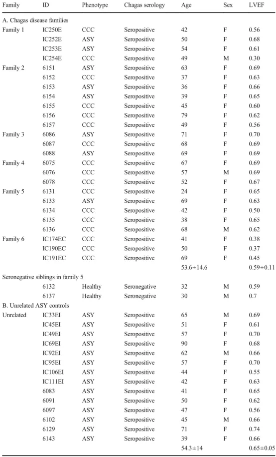
Table 1 Description of the study
phenotypes Family ID Phenotype Chagas serology Age Sex LVEF
A. Chagas disease families
Family 1 IC250E CCC Seropositive 42 F 0.56
IC252E ASY Seropositive 50 F 0.68
IC253E ASY Seropositive 54 F 0.61
IC254E CCC Seropositive 49 M 0.30
Family 2 6151 ASY Seropositive 63 F 0.69
6152 CCC Seropositive 37 F 0.63 6153 ASY Seropositive 36 F 0.66 6154 ASY Seropositive 39 F 0.65 6155 CCC Seropositive 45 F 0.60 6156 CCC Seropositive 79 F 0.62 6157 CCC Seropositive 49 F 0.56
Family 3 6086 ASY Seropositive 71 F 0.70
6087 CCC Seropositive 68 F 0.69 6088 ASY Seropositive 69 F 0.69 Family 4 6075 CCC Seropositive 67 F 0.69 6076 CCC Seropositive 57 M 0.69 6078 CCC Seropositive 52 F 0.67 Family 5 6131 CCC Seropositive 24 F 0.65 6133 ASY Seropositive 69 F 0.63 6134 CCC Seropositive 42 F 0.50 6135 CCC Seropositive 38 F 0.65 6136 CCC Seropositive 68 M 0.62
Family 6 IC174EC CCC Seropositive 41 F 0.38
IC190EC CCC Seropositive 50 F 0.37
IC191EC CCC Seropositive 69 F 0.45
53.6±14.6 0.59±0.11
Seronegative siblings in family 5
6132 Healthy Seronegative 32 M 0.59
6137 Healthy Seronegative 30 M 0.7
B. Unrelated ASY controls
Unrelated IC33EI ASY Seropositive 65 M 0.69
IC45EI ASY Seropositive 51 F 0.61
IC49EI ASY Seropositive 57 F 0.70
IC69EI ASY Seropositive 90 F 0.68
IC92EI ASY Seropositive 62 M 0.66
IC95EI ASY Seropositive 57 F 0.70
IC106EI ASY Seropositive 44 F 0.55
IC111EI ASY Seropositive 42 F 0.63
6083 ASY Seropositive 41 F 0.65 6091 ASY Seropositive 50 F 0.62 6097 ASY Seropositive 47 F 0.56 6102 ASY Seropositive 45 M 0.66 6129 ASY Seropositive 71 F 0.74 6143 ASY Seropositive 39 F 0.66 54.3±14 0.65±0.05
Results
We performed whole exome sequencing and assessed rare pathogenic gene variants associated with CCC in six nuclear families containing multiple cases of CD families (25 patients) and in a group of unrelated ASY patients (n = 14) who came from CD-endemic rural areas in Brazil. The average age in the CCC and ASY patients in the family groups and the ASY unrelated control were similar, around 54 ± 14 years
(Table1). Pedigrees are shown in Fig.1.
Whole exome sequencing disclosed that on average, each patient sample contained 41,780 gene variants. Among them, on average, 11,651 variants were located in coding (exonic or splicing) regions and nonsynonymous. We focused on vari-ants characterized by a minor allele frequency < 1% in the databases (ESP6500, 1000G, and ExAC). Under a hypothesis of complete penetrance, for each given family, we selected nonsynonymous exonic or splicing variants shared by all fam-ily members with CCC but absent from ASY famfam-ily members as well as by the unrelated ASY controls. For families 1 to 6, we found 39, 8, 108, 81, 9, and 76 variants fulfilling the above
criteria, respectively, comprising a total of 321 CCC-specific nonsynonymous exonic rare variants. We performed a
Reactome (reactome.org) pathways analysis of the 321
CCC-specific variants, prior to any prioritization. We found 8 enriched pathways with a significant false discovery ratio (FDR < 0.05). Pathways were based on a very limited number of genes (6/8 with one or two genes) and thus of limited relevance. At any event, the 8 pathways were related either to interferon signaling or to endosomal/antigen processing/ presentation pathways (HLA molecules, BTK) (online
Table 4). After the application of our pathogenicity filter
with multiple algorithms, we found 102 CCC-specific rare pathogenic variants. After filtering for evolutionary conserva-tion, we found 87 variants. At this point, we prioritized the 87 gene variants in 9 pathobiological processes associated to CD. It highlighted 23 of our candidate genes (corresponding to 25 variants). From these variants, 88% (22/25, contained in 20
genes) were confirmed by Sanger sequencing (Table2, online
Fig.1). All these 22 variants showed CADD scores above 15,
consistent with pathogenicity. Indeed, for information, the
CADD score of each variant is also shown in Table2b. Two
Fig. 1 Pedigrees of the six nuclear families included in this study. All
patients underwent detailed clinical interview andT. cruzi serological
tests.T. cruzi-seropositive individuals were considered as CCC patients
when presenting major ECG abnormalities according to the Minnesota
Code classification, modified by Ribeiro et al. [8]. Major ECG
abnormalities are cited in online Table1. The presence of any one of
these major ECG changes was associated with a twofold increase in
mortality [8].T. cruzi-seropositive individuals presenting major ECG
abnormalities, according to the Minnesota Code classification, modified
b y R i b e i r o e t a l . [8] w e r e c o n s i d e r e d a s C C C p a t i e n t s .
T. cruzi-seropositive patients without such ECG findings, with normal echocardiography, and no clinical signs were considered indeterminate forms of CD (ASY) cases
variants have a CADD score above 15, while the other 20 variants have a CADD score above 20. Markers with a CADD score over 20 are usually included in the top 1% del-eterious variants in the genome. For the remaining 62 genes, we performed a second gene set enrichment analysis using Ingenuity Pathway Analysis and Reactome, but no significant pathways were identified. Each family had CCC-associated 1 to 7 pathogenic variants in 1 to 6 genes. Each of the 22 CCC-specific variants only appeared in a single nuclear family. However, for the APOB gene, two variants were detected in
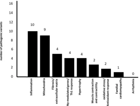
two different families. Figure2 shows the number of genes
containing associated variants in each pathophysiological pro-cess. Some genes may be common to several pathways.
Table2shows the detailed information on the 22 gene variants
(2A), pathogenicity and conservation scores (2B), participa-tion in pathobiological processes relevant for inflammatory cardiomyopathy (2C), and their frequency in the databases (2D). The frequencies of our variants of interest in Latino
reference subpopulations is described in online Table6. We
found a striking accumulation of CCC-associated variants in inflammation-related and mitochondrial genes (17 out of the 20 genes). All families carried at least one variant in mito-chondrial or inflammation-associated genes; five families car-ried variants in mitochondrial genes and 5 in inflammation-related genes. A total of 10 pathogenic variants were found in 9 mitochondrial genes (ADCY10, DHODH, GIT1, MRPS18B, RPUSD3, LEPR, UMPS, MOCS1, and OBSCN). A total of 11 pathogenic variants were located in 10 inflammation-associated genes (ADGRG6, AKAP13, L E P R , L I L R A 2 , M A M L 1 , M A P 4 K 4 , S L C 1 1 A 1 ,
TNFRSF4, APOB, and DHODH) (Figs.2and 3, Table 2).
This accumulation of genes was not an artifact of selection, since the number of prioritized variants in each pathway was not proportional to the total number of genes in the pathway/ process. For instance, while we found 10 variants in 1532 mitochondrial genes, there were 4 variants in the 951 genes belonging to the fibrosis/extracellular matrix process.
Conversely, 3 variants were found in the 178 genes
belong-ing to the contraction/contractility process. Figure3shows the
participation of the variant genes in different mitochondrial and inflammation-related pathways. Eight out of 9 mitochon-drial genes with CCC-specific variants are involved directly or indirectly with energy generation, in processes including mi-tochondrial biogenesis, mimi-tochondrial DNA-encoded gene t r a n s l a t i o n , f a t t y a c i d o x i d a t i o n , a n d o x i d a t i v e phosphorylation/electron transfer chain; interestingly, two genes are involved in pyrimidine biosynthesis. Of note, 8 out of 10 inflammation-related genes are involved in proin-flammatory cytokine production via activation of NF-kB and MAP kinase pathways. Two mitochondrial genes (DHODH
and LEPR) are also involved in inflammation (Table2, Fig.3
in bold). On the whole, 5 of the inflammation-related genes and 5 of the mitochondrial genes also played roles in other
cardiomyopathy-related pathobiological processes. The RPUSD3 gene, involved in the assembly of the mitochondrial ribosome, displayed a stopgain variant at exon 8 in family 4, creating a truncated version lacking 24% of its C-terminal sequence.
The GIT1 gene showed two variants in family 3 that seg-regated together, suggesting compound heterozygosity. Only 3 variants occurred in genes that were not mitochondrial and/ or inflammation-related: PKHD1 and SERPINE2, which belonged to the ECM/fibrosis process, and RNLS, belonging to the hypertrophy and contraction processes. Only one vari-ant gene, OBSCN, had previously been described in genetic/ familial cardiomyopathy. No variants were detected in arrhythmia/ion channel–related genes. Patients carrying het-erozygous gene variants had a normal childhood and reported no debilitating disease before developing CCC as adults, and we inferred that the variants by themselves alone were not able to induce childhood-onset mitochondriopathy. Detailed
infor-mation on the variants is described in Table2.
We have performed Sanger sequencing in two healthy, eutrophic, seronegative siblings from family 5 searching for the CCC-associated variants in genes DHODH and MAML1. One of them (subject 6137) carried the heterozygote variant DHODH C/T (R135C) shared by the CCC family members,
while the other (subject 6132) carried the“wild-type”
homo-zygous DHODH n.403 C/C (R135). Regarding MAML1, the second variant gene in Family 5, both 6132 and 6137 carried
the“wild-type” homozygous MAML1 n. 407 G/G (G136).
Since both IFN-γ and loss-of-function mitochondrial mu-tations cause mitochondrial dysfunction, we studied the effect
of IFN-γ on cardiomyocytes made deficient in mitochondrial
enzyme dihydroorotate dehydrogenase (DHODH) activity with the inhibitor Brequinar. This enzyme is important for the electron transport chain and showed a loss-of-function mutation (DHODH R135C) linked with CCC in one of the studied families. We found that both IFN-γ and brequinar treatment significantly reduced mitochondrial membrane po-tential (ΔψM) as expressed by mitochondrial TMRE fluores-cence on the AC16 human cardiomyocyte cell line and that cells treated simultaneously with IFN-γ and brequinar showed
an even larger decrease onΔψM (Fig.4).
Discussion
In this study of whole exome sequencing of six nuclear fam-ilies with multiple cases of CD, we found 22 CCC-associated rare heterozygous nonsynonymous high-impact pathogenic variants in 20 genes belonging to pathways relevant to inflam-matory cardiomyopathy. Only individuals that were both se-ropositive and carriers of the heterozygous pathogenic vari-ants developed CCC, but not seropositive patients carrying the wild-type sequences, nor seronegative siblings carrying the
Table 2 Descrip tion o f p athogenic v arian ts ide ntified o n the 6 nuclear families . A . G enetic data. B . p athogenicity and cons ervatio n. C. Participation in se lect path obiological processes. D. F requ ency o f var iant s in di ff er ent d at aba ses A Fa mi ly Ge ne acr o -ny m Ch r S ta rt En d R ef er en ce /m u tated all ele Lo ca liz ati o n T yp e o f m ut ati o n N uc le ic ac id cha nge Am ino ac id ch an ge av sn p14 7 1 LEPR 1 66, 081 ,79 1 66, 081 ,79 1 C/T E x onic n o n syn exon 14 2 096 C > T (NM_ 00 119 868 7) T6 99M rs 34 499 590 1 ADCY10 1 167 ,83 0 ,2 54 167 ,83 0 ,2 54 T/ C E x onic n o n syn exon 12 1 205 A > G (NM _ 00 116 774 9) Y4 02C rs 14 066 302 9 1 M OCS1 6 39, 877 ,66 6 39, 877 ,66 6 G/ A E x onic n o n syn exon 8 1 0 15C > T (N M_0 059 43) R33 9 W rs14 857 988 6 1 ADGRG6 6 142,724,940 142 ,724,940 G/ A E xonic; sp licing no n syn exon 13 1 873 G > A (NM _ 00 103 239 4) A6 25T rs 18 423 521 3 1 AKAP13 15 86, 124,694 86, 124,694 T/ C E xonic n on sy n exon7 3 395 T > C (NM_00673 8) L1132S rs7 45783128 2 O BSCN 1 228 ,46 4 ,2 67 228 ,46 4 ,2 67 G/ T E x onic n o n syn exon 22 6 337 G > T (NM _ 00 109 862 3) G2 113 C rs74 623 201 3 A POB 2 21, 247 ,99 6 21, 247 ,99 6 C/A E x onic ; sp licing no n syn exon 16 2 245 G > T (NM_ 000 384 ) D 7 49Y . 3 M RPS18B 6 30, 590 ,61 2 30, 590 ,61 2 G/ A E x onic ; sp licing no n syn exon 5 3 5 8 G > A (NM_0 140 46) V1 20M rs 11 652 493 6 3 P KHD1 6 51, 947,999 51, 947,999 G/ A E xonic n on sy n exon3 1 07C > T (NM_13869 4) T36M rs137852944 3 R NLS 1 0 90, 122 ,34 4 90, 122 ,34 4 C/T E x onic n o n syn exon 5 6 6 5 G > A (NM_ 00 103 170 9) R22 2 H rs19 173 313 3 3 G IT 1 1 7 27, 901 ,77 3 27, 901 ,77 3 C/T E x onic n o n syn exon 20 2 233 G > A (NM_ 014 030 ) A 7 45T . 3 G IT 1 1 7 27, 910 ,55 9 27, 910 ,55 9 C/T E x onic n o n syn exon 2 1 2 8 G > A (NM_ 00 108 545 4) R43H . 3 L IL RA2 1 9 55, 098 ,71 5 55, 098 ,71 5 C/T E x onic n o n syn exon 6 1 2 67C > T (N M_ 00 129 027 0) R42 3 C rs14 958 079 7 4 M AP4 K 4 2 102 ,44 0 ,4 80 102 ,44 0 ,4 80 A/ G E x onic n o n syn exon 4 2 7 1 A > G (NM_ 00 124 255 9) K91E . 4 S LC11A1 2 219 ,25 7 ,7 28 219 ,25 7 ,7 28 C/T E x onic n o n syn exon 12 1 189 C > T (NM_ 000 578 ) R 39 7C rs 74 906 275 4 R PUSD3 3 9,8 80, 802 9,8 80, 802 C/T E x onic sto pga in exon 8 8 0 6 G > A (NM_ 00 135 173 8) W26 9 X rs14 298 451 5 4 U M P S 3 124 ,44 9 ,4 06 124 ,44 9 ,4 06 A/ G E x onic n o n syn exon 1 8 8 A > G (NM _00 037 3) S30 G rs 17 843 776 5 M AML1 5 179 ,19 2 ,4 18 179 ,19 2 ,4 18 G/ A E x onic n o n syn exon 2 4 0 7 G > A (NM_0 147 57) G1 36E rs 14 638 219 8 5 DHODH 16 72, 048,540 72, 048,540 C/T E xonic n on sy n exon3 4 03C > T (NM_00136 1) R135C rs201230446 6 T NFRSF4 1 1 ,1 47, 467 1,1 47, 467 G/ C E x onic n o n syn exon 5 4 8 9 C > G (NM _00 332 7) D1 63E . 6 A POB 2 21, 230 ,41 9 21, 230 ,41 9 G/ C E x onic n o n syn exon 26 9 321 C > G (NM_ 000 384 ) N 3 107 K rs72 653 101 6 S ERPI NE2 2 224 ,86 6 ,4 27 224 ,86 6 ,4 27 A/ G E x onic n o n syn exon 2 1 9 1 T > C (NM_ 00 113 652 8) M 64T rs 34 078 713 B Fami ly Gene Ami n o ac id ch an -ge av snp 147 Po lyp h en 2 HDIV pred ict ion Pol yphen2 HVAR prediction SIFT pr ed ict ion UMD p re dic tio n C ADD pr edi cti on sc or e C on sur f co nser v a-ti o n sc or e 1 LEPR T 69 9M rs3 449 959 0 D am agi n g D am ag in g D am ag in g P rob P at ho 26 .5 9 1 ADCY10 Y 40 2C rs1 406 630 29 Dam agi ng Da ma gin g Da ma gin g Poly 22 .2 6 1 M OCS1 R33 9 W rs1 485 798 86 Dam agi ng Da ma gin g Da ma gin g Path o 3 3 .0 8 1 ADGRG6 A625T rs184235213 Damagi ng Damaging Tolerat e Patho 2 6.0 9 1 AKAP13 L1132S rs7457831 28 Damagi ng Prob Dam D amaging P rob P atho 15.6 7 2 O BSCN G21 13C rs7 462 320 1 D am agi n g D am ag in g D am ag in g P rob P o ly 2 5 .0 9
Tab le 2 (continued) APOB D749Y . D amaging D amaging D amaging P atho 28.5 7 3 MRP S 18-B V120M r- s116 524936 Damaging Prob Dam D am aging P rob P atho 29.4 3 3 P KH D1 T36M r- s137 852944 Damaging Damaging Da maging Patho 30.0 9 3 R NLS R 222H r- s191 733133 Damaging Damaging Damaging Prob Patho 33.0 7 3 G IT1 A 745T . D amaging D amaging D amaging P atho 33.0 9 3 G IT 1 R 43H . D amaging D amagi n g D am ag ing P ol y 35. 0 9 3 L ILRA2 R 423C r- s149 580797 Damaging Damaging Da maging Prob Poly 24.5 8 4 M AP4K4 K 91E . D amaging D amaging D amaging P atho 24.4 3 4 S LC 11A1 R 397C rs74906275 Damaging Da maging Damaging Poly 33.0 6 4 R PUSD3 W 269X r- s142 984515 Damaging Damaging Da maging Prob Poly 27.9 4 4 U MPS S 30G rs17843776 P rob Dam P rob D am Damaging Poly 23.4 9 5 M AML1 G136E r- s146 382198 Damaging Prob Dam T olerate P atho 15.3 3 5 D HODH R135C r- s201 230446 Damaging Damaging Da maging Patho 34.0 9 6 T NFRSF 4 D163E . D amaging D amaging D amaging P oly 22.3 9 6A P O B N3107- K rs72653101 Damaging Damaging Damaging Prob Poly 22.9 7 6 SER P IN -E2 M64T rs34078713 P rob Dam P rob D am Damaging Prob Patho 26.7 8 C Fami l-y Gen e Am ino ac id change avsnp147 Inflamma tion M itochondrial ge n es IF N γ modulated genes/Th1 re-sponse Hy pertrophy Mu scle contraction and cont ra cti lity Fibrosis extr ac ell u lar ma tri x O x ida tive st re ss / antioxidant re-sponse Famil ial CMP genes 1 L EPR T 699 M rs34499590 X X X X
Tab le 2 (continued) 1 A DC Y10 Y 402C r- s140 663029 X 1 M OCS1 R339W r- s148 579886 X 1 A DGRG6 A 625T r- s184 235213 X 1 A KAP13 L 113 2S r- s745 783128 XX X 2 O BS CN G2113- C rs74623201 X X 3 A POB D 749Y . X X 3 MRP S 18-B V120M r- s116 524936 X 3 P KH D1 T36M r- s137 852944 X 3 R NLS R 222H r- s191 733133 XX 3 G IT1 A 745T . X X X 3 G IT1 R 43H . X X X 3 L ILRA2 R 423C r- s149 580797 X 4 M AP4K4 K 91E . X X 4 S LC 11A1 R 397C rs74906275 X X X 4 R PUSD3 W 269X r- s142 984515 X 4 U MPS S 30G rs17843776 X X 5 M AML1 G136E X
Tab le 2 (continued) r- s146 382198 5 D HODH R135C r- s201 230446 XX X 6 T NFRSF 4 D163E . X 6A P O B N3107- K rs72653101 X X 6 SER P IN -E2 M64T rs34078713 X D Fami l-y Gen e Am ino ac id change avsnp147 P o lymorphism frequency in the ESP6500 database Polymorph ism frequency in the 1 000G databas e Polymorphism frequency in theE xAC database Polymorphism freq u en cyi nt h e Br azi lia n g enomic variant cohort (A Br aOM ) 1 L EPR T 699 M rs34499590 1.28% 0.98% 0.32% 0.82% 1 A DC Y10 Y 402C r- s140 663029 0.04% 0.080% 0.02% 0.16% 1 M OCS1 R339W r- s148 579886 0.02% 0.040% 0.04% NA 1 A DGRG6 A 625T r- s184 235213 0.24% 0.26% 0.07% 0.08% 1 A KAP13 L 113 2S r- s745 783128 NA NA 0.0008% NA 2 O BS CN G2113- C rs74623201 0.59% 0.50% 0.16% 0.08% 3 A POB D 749Y . N A N A N A N A 3 MRP S 18-B V120M r- s116 524936 NA 0.02% 0.002% NA 3 P KHD1 T36M 0.03% 0.02% 0.05% NA
Tab le 2 (continued) r- s137 852944 3 R NLS R 222H r- s191 733133 NA 0.02% 0.002% NA 3 G IT1 A 745T . N A N A N A N A 3 G IT1 R 43H . N A N A N A N A 3 L ILRA2 R 423C r- s149 580797 0.21% 0.30% 0.09% 0.33% 4 M AP4K4 K 91E . N A N A N A N A 4 S LC 11A1 R 397C rs74906275 0.04% 0.04% 0.03% NA 4 R PUSD3 W 269X r- s142 984515 0.008% NA 0.007% NA 4 U MPS S 30G rs17843776 0.02% 0.36% 0.38% NA 5 M AML1 G136E r- s146 382198 0.008% 0.08% 0.07% 0.49% 5 D HODH R135C r- s201 230446 0.04% 0.04% 0.04% 0.08% 6 T NFRSF 4 D163E . N A N A N A N A 6A P O B N3107- K rs72653101 NA NA 0.0008% NA 6 SER P IN -E2 M64T rs34078713 0.78% 0.30% 0.75% 1.15% Prob , p robable o r p robably; Pat ho , p athogen ic; Po ly = poly = polymorphism; Dam , d amaging CMP , cardiomyopathy We excluded synon ymo u s, n on-exonic pol ymorphisms, keeping p o lymo rph isms with a m inor allele frequency (MAF) of <1% in at least o ne p ublic databases (ESP65 00; NHLBI G O E xome S equencing P roject (E VS, E SP6500SI-V2 rele ase o n http://evs.gs.wa shington.edu/EVS/ ); 100 0 G enomes (April 2 014 data release o n http://browser.10 00genomes.org ); and E xome A ggregation C onsortium (ExAC, January 2015 Ver sion 0 .3 data relea se o nhttp:// ex ac .broadins titut e.org )) using the VA R A FT filte ring and annota tion tool ( ht tps://var aft.e u ) o n v cf file s. Varia n t calls with V ariant quality (Q UAL) ≤ 60, depth o f coverag e (DP) <20, and m apping quality (M Q) ≤ 40 were filtered out. W e also assessed an available B razili an exome d ata b as e o f 609 elderly individuals fr om the city of Sao P aulo [ 18 ]. Only exo n ic nonsy nonymous damaging varia n ts w ere kept for downstrea m g enetic analyses. W e searc h ed fo r v aria nts, independently, in each family, u nd er an autos o mal d ominant o r u nder au tosomal rece ssive models. In o rder to identify g ene v ari ant s associate d wi th CCC in each fa mily, w e sel ected variants that wer e shared by all CCC pa tients and ab sent in any and all IF p atients in a given nuclear fa mi ly, as w ell as in 1 4 unrelated IF controls. W e ass ess ed the pathogenic potential o f m issense v ariants u sin g 4 alg orith ms embedded in V arAft: SIFT ( https://sift.bii.a -star.e du.sg/ ), Polyphen 2 HumDiv and P olyphen 2 HumVar ( http://genetic s.bwh.ha rvard.edu/pph2/ index.shtml ), and U MD-P redictor ( http://umd-pre di ctor.eu/ ). Only rare variants tagged as p ath ogenic (or damag ing) or pos sibly p athog en ic (o r p robably d amaging) in at least three datab as es and patho g enic/ dama g ing in at lea st one algorithm w ere re tained fo r downstream g enet ic an aly ses. We v alid at ed the abovementioned p athogenicity filter w it h the Co m bin ed A nnotation-Dependent Depletion (C A DD) too l ( http://ca d d.gs.wa shington.edu/score ). Evolutionary conser vation in the position w as deter m ined with the C onsur f server ( https://consurf.tau.a c.il )
pathogenic variant. A striking accumulation of CCC-specific variants (86%) occurred in mitochondrial or inflammation-related genes, and all studied families displayed at least one CCC-associated variant gene belonging to these pathways. The finding that only a single family carried a variant in a gene previously associated with familial cardiomyopathy in-dicates the genetic landscape, and pathogenesis of CCC is distinct from that of familial cardiomyopathy. Importantly, we also found that IFN-γ potentiated mitochondrial dysfunc-tion caused by an inhibitor of DHODH, a mitochondrial en-zyme bearing a loss-of-function mutation (DHODH R135C)
linked with CCC in one of our families, indicating synergy in the induction of mitochondrial dysfunction. To our knowl-edge, this is the first report that rare heterozygous pathogenic variants in mitochondrial and inflammation-related genes are linked to the development of complex multifactorial inflam-matory cardiomyopathy.
Among the 9 mitochondrial genes showing CCC-specific pathogenic variants, 8 are involved in processes leading to mitochondrial ATP production (biogenesis, translation, fatty acid oxidation-FAOx, and the electron transfer chain/ oxidative phosphorylation (OXPHOS)). The R135C variant Fig. 2 Number of genes containing pathogenic variants for each
biological process related to cardiomyopathy. Pathway analyses were performed with the Ingenuity Pathways Analysis (IPA®, Qiagen,
Redwood City, USA) or Reactome (reactome.org); we also interrogated
whether genes participated in 9 biological processes related to the
pathophysiology of CCC: inflammation, IFNγ-modulated genes/Th1
re-sponse, fibrosis/extracellular matrix, contractility of heart, hypertrophy, arrhythmia, oxidative stress/antioxidant response, familial
cardiomyopathy, and mitochondria-related genes. For these nine addi-tional biological processes, we merged IPA Knowledge Base (IKB) gene
lists and published gene lists including the IFNγ induced/repressed gene
lists, and Nrf2-modulated genes where appropriate. Mitochondrial gene list was a combination of genes contained in the Mitochondrion Gene Ontology term and Mitocarta 2.0. The gene list for these pathways/
processes in online Table2
Fig. 3 Features of the main mitochondrial and inflammation-related genes containing deleteri-ous pathogenic variants associat-ed to CCC
observed in the DHODH gene was the only variant known to lead to complete loss-of-function and mitochondrial
dysfunc-tion [27]. Genetic truncation or expression of
dominant-negative variants of DHODH, GIT1, RPUSD3, ADCY10, and MOCS1 in cells was shown to cause mitochondrial dys-function, including reduced mitochondrial membrane poten-tial, respiratory chain/oxidative phosphorylation (OXPHOS)
activity, and ATP production [28–30]. Interestingly, patients
carrying mutations or animals genetically deficient in 6 genes (DHODH, UMPS, MRPS18B, GIT1, OBSCN, and LEPR)
developed cardiac phenotypes [27,30–34]. Parents of patients
with Miller syndrome (homozygous DHODH loss-of-function R135C or other variants; 70% with congenital cardi-ac abnormalities) carry one copy of the defective gene as
members of family 5 but are nevertheless healthy [27].
Animals genetically deficient in MRPS18B and GIT1 showed evidence of mitochondrial dysfunction, altered mitochondrial
morphology, and reduced myocardial contractility [35].
Details about the function of affected genes are shown in
online Table5.
Genetic disorders involving mitochondrial genes are the m o s t c o m m o n c o n g e n i t a l g e n e t i c s y n d r o m e s . Mitochondriopathies are caused by homozygous pathogenic rare variants in nuclear-encoded mitochondrial genes or in mitochondrial DNA. Homozygous pathogenic variants of more than 250 of the ~1500 nuclear-encoded mitochondrial genes were identified as causally related to mitochondrial
dis-orders [35]. Each leads to drastic mitochondrial dysfunction
and energetic and functional impairment in tissues of high energy demand, causing syndromes affecting single organs
or organ combinations that usually present in early childhood; each clinical syndrome is associated with variants in specific
mitochondrial genes [35], and clinical penetrance is variable.
The diverse clinical presentation of mitochodriopathies is re-lated to genetic heterogeneity and the central role of mitochon-dria in multiple processes, including energy generation, anti-oxidant defenses, Ca++ homeostasis, intracellular signaling, macromolecule and nucleotide biosynthesis, reprogramming nuclear gene expression, and cell death. Interestingly, up to 30% of mitochondriopathy patients develop cardiomyopathy, heart conduction defects, ventricular arrhythmia or sudden cardiac death, and autonomic nervous system imbalance
[36], while up to 15% develop gastrointestinal motility
disor-ders including achalasia/megaesophagus and megacolon [37].
The striking similarity between the clinical presentation and p r o p o r t i o n o f c a r d i a c a n d d i g e s t i v e d i s o r d e r s i n
mitochondriopathies and the clinical spectrum of CD [3]
sug-gested the pathogenesis of CCC may be dependent on mito-chondrial dysfunction. Indeed, CCC myocardium displays signs of reduced mitochondrial activity and energy
produc-tion. Decreased mitochondrial rRNA [10], rDNA [38], and
in vivo ATP production [18] were observed in the CCC
myo-cardium. Myocardial levels and activity of mitochondrial en-ergy metabolism enzymes ATP synthase and creatine kinase
are even lower than in CCC than other cardiomyopathies [17],
which might contribute to the worse prognosis of CCC. This is
in line with Nisha Garg’s group findings implicating
myocar-dial mitochondrial dysfunction and oxidative stress in the
pathogenesis of murine models of CCC (reviewed in [39]).
IFN-γ and brequinar treatment significantly reduced
Fig. 4 IFN-γ and brequinar treatment significantly reduced
mitochondrial membrane potential. AC16 human cardiomyocyte cell
line was stimulated with or without IFN-γ and with or without brequinar
for 48 h. Total mitochondria were stained with MitoTracker green, and mitochondrial membrane potential (MMP) was evaluated using the TMRE dye that accumulates in fully polarized, but not in depolarized
mitochondria. We measured TMRE fluorescence on MitoTracker green-labeled mitochondria. Cell viability was calculated as the ratio of the amount of live cells (propidium iodide-negative) and total cells (propidium iodide-negative plus propidium iodide-positive cells) × 100.
mitochondrial membrane potential (ΔψM) on the AC16 hu-man cardiomyocyte cell line. Significantly, cells treated
simul-taneously with IFN-γ, and brequinar showed an even larger
decrease onΔψM. This result is consistent with an increased
susceptibility of cells with reduced mitochondrial enzyme
ac-tivity to IFN-γ. Even though the identified genes play
impor-tant roles in mitochondrial energy generation and physiology, the heterozygous mitochondrial variants did not cause child-h o o d s i g n s o f a l t e r e d d e v e l o p m e n t t y p i c a l o f mitochondriopathies and were only associated with
progres-sion to cardiac disease inT. cruzi-seropositive patients, whose
age group was typical from CCC patients in general. Taken
together, our results suggest thatT. cruzi-induced IFN-γ is
necessary to cause clinically significant disease in patients with heterozygous variants. We propose that long-term expo-sure to high levels IFN-γ associated with CD – particularly
high in CCC myocardium [40]– is the additional
mitochon-drial insult suffered by carrier of heterozygous mitochonmitochon-drial
variants that leads them to mitochondrial dysfunction. IFN-γ
has multiple deleterious effects on cardiomyocyte mitochon-dria. It induces TNF-alpha and potentiates TNF-alpha-mediated NF-kB signaling, leading to NOS2 production of
NO [41], which in the presence of IFN-γ-induced reactive
oxygen species turns into peroxynitrite [21], and ensuing
mi-tochondrial fragmentation and reduction of mimi-tochondrial
membrane potential, lipid beta-oxidation [14], and ATP
gen-eration [22], leading to cardiomyocyte dysfunction and
apo-ptosis [15]. Variants in the inflammation-related genes may
also lead to a mitochondrial dysfunction phenotype. Gain-of-function variants of genes linked to NF-kB or MAP kinase signaling could lead to increased proinflammatory cytokine production, which might lead to further mitochondrial
dys-function in cardiomyocytes [22], and this mitochondrial
dam-age can be further fueled by IFN-γ [42]. It is thus possible that
the common final pathogenic mechanism for mitochondrial and inflammation-associated gene variants might involve en-hancement of IFN-γ-induced cardiac mitochondrial damage. As mitochondrial damage and release of damage-associated molecular patterns can itself induce the production of proin-flammatory cytokines, a self-perpetuating positive feedback
loop between inflammation and mitochondrial damage [43]
may contribute to the chronicity of inflammation in CCC heart tissue.
Our study has limitations. CCC patients included in the pedigrees mostly have major diagnostic ECG changes without left ventricular dysfunction. However, follow-up studies show that asymptomatic chagasic individuals with ECG abnormal-ities typical of CCC show higher total and cardiac mortality in relation to chagasic individuals with normal ECG; up to 50%
of deaths in this groups are due to SCD [6, 8, 44–46].
Similarly, conduction defects and arrhythmias can be the
pre-senting symptoms of genetic mitochondriopathies [47]. Even
though CCC has a progressive character and inflammatory
mediators and mitochondrial dysfunction are key participants
of arrhythmia [48] and heart failure [43], we can only
hypoth-esize that the genetic variants identified in our study may play
a direct role in the inflammation [10,11] and mitochondrial
dysfunction [17,38] observed in end-stage CCC.
Conclusion
Results indicate that the genetic contribution to CCC is poly-genic and driven by several rare variants in genes that differ between families but are related to mitochondria and inflam-mation. Results imply that mitochondrial dysfunction and in-flammation, key processes in the pathophysiology of CCC, are at least in part genetically determined. To our knowledge, this is the first report that rare variants in mitochondrial and inflammation-related genes are linked to complex multifacto-rial cardiomyopathy. Our results also support the notion of a two-hit mechanism where IFN-γ and proinflammatory cyto-kines induced by chronic infection trigger mitochondrial dys-function and clinical disease in carriers of heterozygous mito-chondrial gene variants. Indeed, modulation of mitomito-chondrial damage induced by IFN-γ and other cytokines could perhaps be a suitable therapeutic target in CCC. Treatment with mitochondria-protective agents such as antioxidants or ago-nists of sirtuin-1 and AMP-activated protein kinase (AMPK) was found to attenuate or even reverse cardiac damage in mouse models of CCC, by reducing NF-kB activation and
the intensity of chronic myocarditis (reviewed in [38]). To
conclude, it is possible that a similar two-hit mechanism, whereby genetic variants may increase mitochondrial suscep-tibility to inflammatory cytokine-induced dysfunction, may be relevant for the pathogenesis of other inflammatory cardiomy-opathies and degenerative diseases associated with mitochon-drial dysfunction.
Abbreviations T. cruzi, Trypanosoma cruzi; CCC, Chronic Chagas
car-diomyopathy; CD, Chagas disease; ASY, Asymptomatic form; DCM, Dilated cardiomyopathy; ECG, Electrocardiography; IFN, Interferon; TNF, Tumor necrosis factor; NO, Nitrite oxide; ATP, Adenosine triphos-phate; AVM, Acute viral myocarditis; WES, Whole exome sequencing; SCD, Sudden cardiac death; HF, Heart failure; LEPR, Leptin receptor; ADCY10, Adenylate cyclase 10; MOCS1, Molybdenum cofactor syn-thesis 1; ADGRG6, Adhesion G protein-coupled receptor G6; AKAP13, AKinase-anchoring protein 13; OBSCN, Obscurin; APOB , Apolipoprotein B; MRPS18B , Mitochondrial ribosomal protein S18B; PKHD1 , Polycystic kidney and hepatic disease 1; RNLS , Renalase, FAD-dependent amine oxidase; GIT1, G protein-coupled receptor kinase
interactor 1; LILRA2, Leukocyte immunoglobulin–like receptor A2;
MAP 4 K4, Mitogen-activated protein kinase kinase kinase kinase 4; SLC11A1, Solute carrier family 11 member 1; RPUSD3, RNA pseudouridine synthase D3; UMPS , Uridine monophosphate synthetase; MAML1, Mastermind like transcriptional coactivator 1; DHODH , Dihydroorotate dehydrogenase; TNFRSF4, TNF receptor superfamily member 1; SERPINE2, Serpin family E member 2
Supplementary Information The online version contains supplementary
material available athttps://doi.org/10.1007/s10875-021-01000-y.
Acknowledgments We thank all the patients and their relatives.
Availability of Data and Material Raw data will be available on request.
Authors’ Contribution Designed the study: JK, LAR, ECS, ECN, and
CC.
Supervised the study as a whole: ECN and CC.
Participated in the recruitment of patients and sample collection: AFF, BI, AMF, CM, PB,
ClC, ORSJ, LCO, CDLO, MDCN, and LAR.
Performed experiments: MO, SM, RRA, JPN, LRPF, SB, DL, VOCR, DC, RCFZ, MT, FG, and PA.
Performed and supervised computational studies: LA, AC, ECN, and CC.
Wrote the manuscript: ECN and CC.
All authors reviewed the results and approved the final version of the manuscript.
Funding This work was supported by the Institut National de la Santé et de la Recherche Médicale (INSERM); the Aix-Marseille University
(grant number: AMIDEX“International_2018” MITOMUTCHAGAS);
the French Agency for Research (Agence Nationale de la
Recherche-ANR (grant numbers:“Br-Fr-Chagas,” “landscardio”); the CNPq
(Brazilian Council for Scientific and Technological Development); the FAPESP (São Paulo State Research Funding Agency Brazil (grant num-bers: 2013/50302–3, 2014/50890–5); and the National Institutes of Health/USA (grant numbers: 2 P50 AI098461–02 and 2U19AI098461– 06). This project has received funding from the Excellence Initiative of
Aix-Marseille University (A*Midex) a French“Investissements d’Avenir
programme”- Institute MarMaRa AMX-19-IET-007. ECN, JK, ALR,
and ECS are recipients of productivity awards by CNPq. The funders do not play any role in the study design, data collection and analysis, decision to publish, or preparation of the manuscript.
Declarations
Conflict of Interest The authors declare that they have no conflict of
interest.
Open Access This article is licensed under a Creative Commons Attribution 4.0 International License, which permits use, sharing, adap-tation, distribution and reproduction in any medium or format, as long as you give appropriate credit to the original author(s) and the source, pro-vide a link to the Creative Commons licence, and indicate if changes were made. The images or other third party material in this article are included in the article's Creative Commons licence, unless indicated otherwise in a credit line to the material. If material is not included in the article's Creative Commons licence and your intended use is not permitted by statutory regulation or exceeds the permitted use, you will need to obtain permission directly from the copyright holder. To view a copy of this
licence, visithttp://creativecommons.org/licenses/by/4.0/.
References
1. Schultheiss HP, Fairweather D, Caforio ALP, Escher F,
Hershberger RE, Lipshultz SE, et al. Dilated cardiomyopathy. Nat Rev Dis Primers. 2019;5(1):32.
2. Chevillard C, Nunes JPS, Frade AF, Almeida RR, Pandey RP,
Nascimento MS, et al. Disease tolerance and pathogen resistance genes may underlie trypanosoma cruzi persistence and differential progression to Chagas disease cardiomyopathy. Front Immunol. 2018;9:2791.
3. Bocchi EA, Bestetti RB, Scanavacca MI, Cunha Neto E, Issa VS.
Chronic Chagas heart disease management: from etiology to
car-diomyopathy treatment. J Am Coll Cardiol. 2017;70(12):1510–24.
4. Casado J, Davila DF, Donis JH, Torres A, Payares A, Colmenares
R, et al. Electrocardiographic abnormalities and left ventricular sys-tolic function in Chagas' heart disease. Int J Cardiol. 1990;27(1):
55–62.
5. Barbosa MP, da Costa Rocha MO, de Oliveira AB, Lombardi F,
Ribeiro AL. Efficacy and safety of implantable cardioverter-defibrillators in patients with Chagas disease. Europace.
2013;15(7):957–62.
6. Baroldi G, Oliveira SJ, Silver MD. Sudden and unexpected death in
clinically‘silent’ Chagas’ disease. A hypothesis. Int J Cardiol.
1997;58(3):263–8.
7. Higuchi ML, De Morais CF, Pereira Barreto AC, Lopes EA, Stolf
N, Bellotti G, et al. The role of active myocarditis in the
develop-ment of heart failure in chronic Chagas’ disease: a study based on
endomyocardial biopsies. Clin Cardiol. 1987;10(11):665–70.
8. Ribeiro AL, Marcolino MS, Prineas RJ, Lima-Costa MF.
Electrocardiographic abnormalities in elderly Chagas disease pa-tients: 10-year follow-up of the Bambui cohort study of aging. J Am Heart Assoc. 2014;3(1):e000632.
9. Abel LC, Rizzo LV, Ianni B, Albuquerque F, Bacal F, Carrara D,
et al. Chronic Chagas' disease cardiomyopathy patients display an increased IFN-gamma response to trypanosoma cruzi infection. J
Autoimmun. 2001;17(1):99–107.
10. Cunha-Neto E, Dzau VJ, Allen PD, Stamatiou D, Benvenutti L,
Higuchi ML, et al. Cardiac gene expression profiling provides ev-idence for cytokinopathy as a molecular mechanism in Chagas'
disease cardiomyopathy. Am J Pathol. 2005;167(2):305–13.
11. Nogueira LG, Santos RH, Fiorelli AI, Mairena EC, Benvenuti LA,
Bocchi EA, et al. Myocardial gene expression of T-bet, GATA-3, Ror-gammat, FoxP3, and hallmark cytokines in chronic Chagas disease cardiomyopathy: an essentially unopposed TH1-type re-sponse. Mediat Inflamm. 2014;2014:914326.
12. Laugier L, Rodrigues Pinto Ferreira L, Moraes Ferreira F,
Cabantous S, Farage Frade A, Ribeiro Almeida R, et al. miRNAs may play a major role in the control of gene expression in key pathobiological processes in Chagas disease cardiomyopathy. Plos Negl Trop Dis. 2020; In press.
13. Patten M, Kramer E, Bunemann J, Wenck C, Thoenes M, Wieland
T, et al. Endotoxin and cytokines alter contractile protein expression
in cardiac myocytes in vivo. Pflugers Arch. 2001;442(6):920–7.
14. Ni C, Ma P, Wang R, Lou X, Liu X, Qin Y, et al.
Doxorubicin-induced cardiotoxicity involves IFNgamma-mediated metabolic
reprogramming in cardiomyocytes. J Pathol. 2019;247(3):320–32.
15. Levick SP, Goldspink PH. Could interferon-gamma be a
therapeu-tic target for treating heart failure? Heart Fail Rev. 2014;19(2):227–
36.
16. Soares MP, Gozzelino R, Weis S. Tissue damage control in disease
tolerance. Trends Immunol. 2014;35(10):483–94.
17. Teixeira PC, Santos RH, Fiorelli AI, Bilate AM, Benvenuti LA,
Stolf NA, et al. Selective decrease of components of the creatine kinase system and ATP synthase complex in chronic Chagas dis-ease cardiomyopathy. PLoS Negl Trop Dis. 2011;5(6):e1205.
18. Leme AM, Salemi VM, Parga JR, Ianni BM, Mady C, Weiss RG,
et al. Evaluation of the metabolism of high energy phosphates in
patients with Chagas' disease. Arq Bras Cardiol. 2010;95(2):264–
70.
19. Silva JS, Vespa GN, Cardoso MA, Aliberti JC, Cunha FQ. Tumor
infection in mice by inducing nitric oxide production in infected gamma interferon-activated macrophages. Infect Immun.
1995;63(12):4862–7.
20. Vila-del Sol V, Punzon C, Fresno M. IFN-gamma-induced
TNF-alpha expression is regulated by interferon regulatory factors 1 and
8 in mouse macrophages. J Immunol. 2008;181(7):4461–70.
21. Rakshit S, Chandrasekar BS, Saha B, Victor ES, Majumdar S,
Nandi D. Interferon-gamma induced cell death: regulation and con-tributions of nitric oxide, cJun N-terminal kinase, reactive oxygen species and peroxynitrite. Biochim Biophys Acta. 2014;1843(11):
2645–61.
22. Buoncervello M, Maccari S, Ascione B, Gambardella L, Marconi
M, Spada M, et al. Inflammatory cytokines associated with cancer growth induce mitochondria and cytoskeleton alterations in
cardiomyocytes. J Cell Physiol. 2019;234(11):20453–68.
23. Belkaya S, Kontorovich AR, Byun M, Mulero-Navarro S, Bajolle
F, Cobat A, et al. Autosomal recessive cardiomyopathy presenting
as acute myocarditis. J Am Coll Cardiol. 2017;69(13):1653–65.
24. Cardoso CS, Sabino EC, Oliveira CD, de Oliveira LC, Ferreira AM,
Cunha-Neto E, et al. Longitudinal study of patients with chronic Chagas cardiomyopathy in Brazil (SaMi-trop project): a cohort pro-file. BMJ Open. 2016;6(5):e011181.
25. Marquet S, Bucheton B, Reymond C, Argiro L, El-Safi SH, Kheir
MM, et al. Exome sequencing identifies two variants of the alkylglycerol monooxygenase gene as a cause of relapses in viscer-al leishmaniasis in children, in Sudan. J Infect Dis. 2017;216(1):22– 8.
26. Khan R, Lee JE, Yang YM, Liang FX, Sehgal PB. Live-cell
imag-ing of the association of STAT6-GFP with mitochondria. PLoS One. 2013;8(1):e55426.
27. Fang J, Uchiumi T, Yagi M, Matsumoto S, Amamoto R, Takazaki
S, et al. Dihydro-orotate dehydrogenase is physically associated with the respiratory complex and its loss leads to mitochondrial dysfunction. Biosci Rep. 2013;33(2):e00021.
28. Arroyo JD, Jourdain AA, Calvo SE, Ballarano CA, Doench JG,
Root DE, et al. A genome-wide CRISPR death screen identifies genes essential for oxidative phosphorylation. Cell Metab. 2016;24(6):875–85.
29. Grings M, Seminotti B, Karunanidhi A, Ghaloul-Gonzalez L,
Mohsen AW, Wipf P, et al. ETHE1 and MOCS1 deficiencies: disruption of mitochondrial bioenergetics, dynamics, redox homeo-stasis and endoplasmic reticulum-mitochondria crosstalk in patient fibroblasts. Sci Rep. 2019;9(1):12651.
30. Pang J, Xu X, Getman MR, Shi X, Belmonte SL, Michaloski H,
et al. G protein coupled receptor kinase 2 interacting protein 1 (GIT1) is a novel regulator of mitochondrial biogenesis in heart. J
Mol Cell Cardiol. 2011;51(5):769–76.
31. Abella V, Scotece M, Conde J, Pino J, Gonzalez-Gay MA,
Gomez-Reino JJ, et al. Leptin in the interplay of inflammation, metabolism and immune system disorders. Nat Rev Rheumatol. 2017;13(2):
100–9.
32. Ding Y, Liu W, Deng Y, Jomok B, Yang J, Huang W, et al.
Trapping cardiac recessive mutants via expression-based
insertion-al mutagenesis screening. Circ Res. 2013;112(4):606–17.
33. Marston S. Obscurin variants and inherited cardiomyopathies.
Biophys Rev. 2017;9(3):239–43.
34. Miyazawa S, Furuta S, Hashimoto T. Reduction of beta-oxidation
capacity of rat liver mitochondria by feeding orotic acid. Biochim
Biophys Acta. 1982;711(3):494–502.
35. Kohda M, Tokuzawa Y, Kishita Y, Nyuzuki H, Moriyama Y,
Mizuno Y, et al. A comprehensive genomic analysis reveals the genetic landscape of mitochondrial respiratory chain complex defi-ciencies. PLoS Genet. 2016;12(1):e1005679.
36. Finsterer J, Kothari S. Cardiac manifestations of primary
mitochon-drial disorders. Int J Cardiol. 2014;177(3):754–63.
37. Finsterer J, Frank M. Gastrointestinal manifestations of
mitochon-drial disorders: a systematic review. Ther Adv Gastroenterol.
2017;10(1):142–54.
38. Wan X, Gupta S, Zago MP, Davidson MM, Dousset P, Amoroso A,
et al. Defects of mtDNA replication impaired mitochondrial bio-genesis during trypanosoma cruzi infection in human cardiomyocytes and chagasic patients: the role of Nrf1/2 and anti-oxidant response. J Am Heart Assoc. 2012;1(6):e003855.
39. Lopez M, Tanowitz HB, Garg NJ. Pathogenesis of chronic Chagas
disease: macrophages, mitochondria, and oxidative stress. Curr Clin Microbiol Rep. 2018;5(1):45–54.
40. Reis MM, Higuchi Mde L, Benvenuti LA, Aiello VD, Gutierrez PS,
Bellotti G, et al. An in situ quantitative immunohistochemical study of cytokines and IL-2R+ in chronic human chagasic myocarditis: correlation with the presence of myocardial trypanosoma cruzi
an-tigens. Clin Immunol Immunopathol. 1997;83(2):165–72.
41. Kauppinen A, Suuronen T, Ojala J, Kaarniranta K, Salminen A.
Antagonistic crosstalk between NF-kappaB and SIRT1 in the reg-ulation of inflammation and metabolic disorders. Cell Signal.
2013;25(10):1939–48.
42. Lee HJ, Oh YK, Rhee M, Lim JY, Hwang JY, Park YS, et al. The
role of STAT1/IRF-1 on synergistic ROS production and loss of mitochondrial transmembrane potential during hepatic cell death induced by LPS/d-GalN. J Mol Biol. 2007;369(4):967–84.
43. Zhou B, Tian R. Mitochondrial dysfunction in pathophysiology of
heart failure. J Clin Invest. 2018;128(9):3716–26.
44. Manzullo EC, Chuit R. Risk of death due to chronic chagasic
car-diopathy. Mem Inst Oswaldo Cruz. 1999;94(Suppl 1):317–20.
45. Pimenta J, Valente N, Miranda M. Long-term follow up of
asymp-tomatic chagasic individuals with intraventricular conduction dis-turbances, correlating with non-chagasic patients. Rev Soc Bras
Med Trop. 1999;32(6):621–31.
46. Rodriguez-Salas LA, Klein E, Acquatella H, Catalioti F, Davalos
VV, Gomez-Mancebo JR, et al. Echocardiographic and clinical p r e d i c t o r s o f m o r t a l i t y i n c h r o n i c C h a g a s ' d i s e a s e . Echocardiography. 1998;15(3):271–8.
47. Bonnet D, Martin D, Pascale De L, Villain E, Jouvet P, Rabier D,
et al. Arrhythmias and conduction defects as presenting symptoms of fatty acid oxidation disorders in children. Circulation.
1999;100(22):2248–53.
48. Montaigne D, Marechal X, Lacroix D, Staels B. From cardiac
mi-tochondrial dysfunction to clinical arrhythmias. Int J Cardiol.
2015;184:597–9.
Publisher’s Note Springer Nature remains neutral with regard to