1428
|
wileyonlinelibrary.com/journal/ejn Eur J Neurosci. 2020;51:1428–1440. R E S E A R C H R E P O R TTraining‐, muscle‐ and task‐specific up‐ and downregulation of
cortical inhibitory processes
Wolfgang Taube
1|
Albert Gollhofer
2|
Benedikt Lauber
1,2This is an open access article under the terms of the Creative Commons Attribution License, which permits use, distribution and reproduction in any medium, provided the original work is properly cited.
© 2019 The Authors. European Journal of Neuroscience published by Federation of European Neuroscience Societies and John Wiley & Sons Ltd. Edited by Christoph M. Michel.
Abbreviations: AMT, Active Motor Threshold; BT, Balance Training; CSE, Corticospinal Excitability; CSP, Cortical Silent Period; EMG,
Electromyography; GABA, Gamma‐aminobutyric Acid; H‐reflex, Hoffmann Reflex; LG, Gastrocnemius Lateralis; MEP, Motor Evoked Potential; MG, Gastrocnemius Medialis; MT, Motor Threshold; MVC, Maximal Voluntary Force; RMT, Resting Motor Threshold; RTD, Rate of Torque Development; SICI, Short‐interval Intracortical Inhibition; SOL, Soleus; ST, Strength Training; TA, Tibialis Anterior; TMS, Transcranial Magnetic Stimulation. 1Department of Neurosciences and
Movement Science, University of Fribourg, Fribourg, Switzerland
2Department of Sport and Sport Science, University of Freiburg, Freiburg, Germany
Correspondence
Benedikt Lauber, Department of Neurosciences and Movement Sciences, University of Fribourg, Fribourg, Switzerland.
Email: benedikt.lauber@unifr.ch
Abstract
Motor cortical contribution was shown to be important for balance control and for ballistic types of movements. However, little is known about the role of cortical inhibitory mechanisms and even less about long(er)‐term adaptations of these inhibi-tory processes. Therefore, the aim of the present study was to investigate the role of intracortical inhibition before and after four weeks of explosive or balance training. Two groups of subjects participated for four weeks either in an explosive training programme of the plantar flexor muscles or in a balance training programme on unstable devices. Adaptations in short‐interval intracortical inhibition (SICI) were assessed by applying paired‐pulse TMS to the soleus muscle during dynamic plantar flexions, balance perturbations and at rest. Furthermore, SICI was assessed for the untrained tibialis anterior muscle. The results show task‐, muscle‐ and group‐specific adaptations in SICI after the training (p = .021) with significantly increased SICI after balance training in the balance task and decreased SICI after explosive training in the ballistic task. The training also caused task‐ and group‐specific behavioural adaptations indicated by improved balance performance after balance training and increased ballistic performance after explosive training. There were no changes in SICI when measured at rest or in the untrained tibialis anterior muscle. This study shows that long(er)‐term training improves the ability to modulate cortical inhibitory processes in a task‐ and muscle‐specific manner.
K E Y W O R D S
cortical inhibition, long(er)‐term adaptations, task specificity The peer review history for this article is
available at https ://publo ns.com/ publon/10.1111/EJN.14538
1
|
INTRODUCTION
Motor learning is the result of plastic changes within the neural system, and it is well known that the primary motor cortex plays a crucial role (Classen, Liepert, Wise, Hallett, & Cohen, 1998; Lotze, Braun, Birbaumer, Anders, & Cohen, 2003; Muellbacher, Ziemann, Boroojerdi, Cohen, & Hallett, 2001; Muellbacher et al., 2002). Recent observations fur-ther indicate that adaptations of the intracortical inhibitory system are closely linked to progresses of motor learning as several studies have shown that short‐interval intracortical in-hibition (SICI) is significantly reduced following short‐term motor learning (Camus, Ragert, Vandermeeren, & Cohen, 2009; Cirillo, Todd, & Semmler, 2011; Leung, Rantalainen, Teo, & Kidgell, 2015; Perez, Lungholt, Nyborg, & Nielsen, 2004). For example, Leung et al. (2015) demonstrated that a single session of metronome‐paced strength training caused a reduction in SICI while self‐paced strength training did not. Furthermore, Perez et al. (2004) showed that SICI was signifi-cantly reduced after 32 min of motor skill training, but unaf-fected by nonskill or passive training. This suggests that SICI is generally decreased during the initial phase of motor learn-ing. However, apart from a study of Leung, Rantalainen, Teo, and Kidgell (2017) that measured SICI during a low‐level con-traction, all other previously mentioned studies measured SICI at rest despite evidence of a task‐specific modulation of in-hibitory processes (Opie & Semmler, 2016; Papegaaij, Taube, Hogenhout, Baudry, & Hortobagyi, 2014; Sidhu, Cresswell, & Carroll, 2013; Soto, Valls‐Sole, Shanahan, & Rothwell, 2006). With regard to long‐term effects of motor learning on SICI, there are only inconsistent reports. Rosenkranz, Kacar, and Rothwell (2007) investigated the effects of five consecu-tive days of motor learning of a rapid thumb abduction task, and they showed that SICI was reduced on day five compared to the first day of practice. Similarly, longer‐term strength training seems to reduce the amount of intracortical inhibition as the study by Weier, Pearce, and Kidgell (2012) showed that four weeks of heavy load squat strength training resulted in a 32% reduction in SICI when tested during weak isometric contractions. In contrast, a cross‐sectional study comparing elite badminton players with novices demonstrated higher lev-els of SICI in the athletes (Dai et al., 2016). The authors spec-ulated that coordinative training such as playing badminton strengthens inhibitory processes. In line with this, a recently published longitudinal study demonstrates higher levels of SICI after balance training (Mouthon & Taube, 2019). In this study, participants trained exclusively on one specific balance device where they had to keep a freely rotating platform in a horizontal position as long as possible. Interestingly, the sub-jects with the greatest improvements in the balance task also showed the largest increases in SICI when measured after the 2‐week training period.
Thus, it may be assumed that specific markers for the ac-tivity of the cortical inhibitory system (i.e. SICI and the cor-tical silent period) as well as corticospinal excitability adapt task specifically. More precise, intracortical inhibition was assumed to be increased during coordinative tasks (i.e. bal-ancing) and decreased in tasks where high levels of muscle activity are required (i.e. explosive tasks). Therefore, the primary aim of the present study was to test this hypothe-sis of a task‐specific up‐ and downregulation of inhibitory mechanisms (i.e. increase and decrease). For this purpose, the influence of explosive and balance training on SICI was compared before and after four weeks of balance or explo-sive training as it is known that training of these tasks results in different(ial) neural adaptations. For instance, corticospi-nal excitability was shown to increase after ballistic strength training (Hinder, Schmidt, Garry, & Summers, 2010; Kidgell & Pearce, 2010; Lee, Hinder, Gandevia, & Carroll, 2010) whereas a decreased excitability was reported after balance training (for review see Taube, Gruber, & Gollhofer, 2008). Whether these adaptations are caused by plastic changes within parts of the cortical inhibitory system is unknown and is the subject of the present study. Therefore, one group of subjects participated in a ballistic explosive training pro-gramme (ET) of the plantar flexor muscles for four weeks while the other group participated in a balance training pro-gramme (BT) with the same duration. Inhibitory activity of the motor cortex was tested using the SICI paradigm during activity (plantar flexions, balance perturbations) and during control conditions (sitting/rest, standing) and in a muscle that was not trained. This allowed us to evaluate task‐ and muscle‐specific adaptations in SICI in both training groups. It was hypothesized that SICI will increase its activity after balance training (in line with Mouthon & Taube, 2019), a task with high coordinative demands and that SICI would decrease after explosive training where high levels of corti-cal drive are beneficial to enhance task performance (Leung et al., 2017). In addition, these adaptations were expected to be task‐ and muscle‐specific.
2
|
MATERIALS AND METHODS
2.1
|
Study participants
A total of 26 subjects agreed to participate in this study, but only 24 subjects (23.3 ± 2.4 years, 10 female) completed the study and were included in the analyses. Sample size was based on an a priori power analyses using G*Power (Faul, Erdfelder, Lang, & Buchner, 2007) with the following as-sumptions: effect size 0.20, alpha 0.05, power 80%, number of groups 2, number of measurements 2, correlation among repeated measures 0.8, nonsphericity correction ε 1, with a repeated measures ANOVA study design. Before the pre‐ test, subjects gave written informed consent and the study
was approved by the ethics committee of the University of Freiburg (418/16) and was in accordance with the latest ver-sion of the Declaration of Helsinki.
Depending on the rate of torque development (RTD) and balance performance of the pre‐test, subjects were randomly assigned into two groups from which one group performed four weeks of explosive training (ET) while the second group participated in four weeks of balance training (BT). The post‐ test was performed three days after the last training session to avoid influences of fatigue.
2.2
|
Electromyography (EMG)
The skin of the subject's right leg was shaved and cleaned with disinfectant before surface EMG electrodes (Blue sensor P, Ambu, Bad Nauheim, Germany) were attached to the mus-cle bellies of the m. soleus (SOL), m. gastrocnemius medialis (MG), m. gastrocnemius lateralis (LG) and the m. tibialis ante-rior according to SENIAM guidelines. The interelectrode dis-tance was 2 cm, and the reference electrode was placed on the tibial plateau. The EMG recordings were amplified (×500), band‐pass‐filtered (10–1000 Hz) and sampled at 4 kHz.
2.3
|
Transcranial magnetic stimulation
(TMS)
Transcranial magnetic stimuli were applied over the left hem-isphere motor cortex via a MagPro X100 with MagOption magnetic stimulator connected with a 95‐mm focal ‘butter-fly‐shaped’ coil (D‐B80, both MagVenture A/S). The coil was mounted to a helmet (Petzl) which could be adjusted to the individual head size. The handle of the coil was pointing backwards, and the stimulator was programmed to induce a posterior–anterior current flow in the motor cortex. The coil was fixed to a custom‐made pin‐jointed system made of light but rigid plastic allowing movements of the coil relative to the head. The coil as well as the helmet were additionally secured with straps to the chin and the back of the head of the subjects (Taube, Gruber, et al., 2008), minimizing movements of the coil relative to the head. The cable of the coil was fixed to a pulley system which was attached to the ceiling to minimize forces acting onto the helmet. The initial stimulation point was set 0.5 cm anterior to the vertex and over the midline for each subject individually. Subsequently, the final coil posi-tion was established by moving the coil anterior and left from the vertex during which the size of the motor evoked potential in the SOL was permanently checked. The final coil position was then marked directly on the subject's head using a felt pen, and the position was permanently controlled throughout the entire experiment. The participants were asked to keep the mark for the duration of the study to ensure that the coil was placed over the same area of M1 in the pre‐ and post‐test as no neuronavigation system was available.
2.4
|
TMS during the plantar flexions and
balance perturbations
For the plantar flexions, a red target line representing 70% of the maximal rate of RTD was presented on a computer screen in front of the subject which had to be reached by a black line representing the actually exerted torque during the plantar flexion. Subjects were instructed to contract as fast as pos-sible (Maffiuletti et al., 2016) and hit the target as precisely as possible, and subjects relaxed again once they hit the target line. The TMS stimulation was triggered on the rising EMG of the SOL corresponding to 50% of the maximal rectified EMG obtained during the submaximal contractions. This level for triggering the EMG has been successfully used pre-viously to study the influence of SICI during cycling (Sidhu et al., 2013). This trigger level was kept constant throughout the entire experiment (i.e. plantar flexions and balance per-turbations) to ensure the same level of muscle activation at the time of stimulation. In the post‐test, the trigger threshold was set at the same EMG level as in the pre‐test.
2.5
|
Active motor threshold (AMT)
During the plantar flexions, balance perturbations and quiet standing, AMT was defined as the stimulation intensity to evoke MEPs <100 μV in three out of five consecutive tri-als (Papegaaij et al., 2014). The AMT was individually es-tablished for each condition. For the plantar flexions, AMT was established during isometric plantar flexions while for the balance perturbations, the AMT was established during anterior–posterior balance perturbations that were applied to the subjects (see TMS during the plantar flexions and balance perturbations). The AMT was individually established for each condition, and stimulation intensity was set at 80% AMT for the conditioning pulse. Establishing individual AMTs for each condition is very important as they slightly differ between experimental conditions (Table 1), and because it was shown that when the intensity of the conditioning pulse is very close to the AMT (approximately 95%), SICI can be contaminated by short‐interval intracortical facilitation (Peurala, Muller‐Dahlhaus, Arai, & Ziemann, 2008).
2.6
|
Intracortical inhibition
Intracortical inhibition was tested using a paired‐pulse TMS protocol where the suprathreshold single TMS pulse at 1.2MT was preceded by the subthreshold (0.8MT) TMS pulse by 2.5 ms (Kuhn, Keller, Ruffieux, & Taube, 2016; Lauber, Gollhofer, & Taube, 2018; Papegaaij, Baudry, Negyesi, Taube, & Hortobagyi, 2016). While the first pulse serves as a conditioning pulse which is applied at intensities below the threshold to evoke a MEP (subthreshold TMS), the second pulse evokes a clearly visible MEP (suprathreshold
TMS). It is assumed that the first pulse activates intracorti-cal inhibitory interneurons which then reduce the MEP am-plitude of the second pulse and this phenomenon is known as short‐interval intracortical inhibition (SICI). The peak‐to‐ peak amplitude of the conditioned MEP is then compared to the unconditioned MEP. There is indeed evidence that SICI is a cortical phenomenon from direct recordings of descend-ing corticospinal activity from the spinal cord of patients (Di Lazzaro, Oliviero, Ferrara, Mazzone, & Rothwell, 1998; Di Lazzaro & Rothwell, 2014; Weise et al., 2013). These stud-ies show a suppression of later I‐waves (synaptically evoked corticospinal volleys) by the conditioning pulse when the primary motor cortex is stimulated at subthreshold intensi-ties. An increase in SICI would therefore represent an in-crease in intracortical inhibition, while dein-crease in SICI indicates a reduced amount of intracortical inhibition.
In the present study, SICI was tested during rest (control experiments) as well as during activity (trained and untrained muscles). Because of the changes in the level of muscle ac-tivation from rest to activity, the stimulation intensity was adjusted for each condition and muscle. This ensured that there were no visible MEPs following the subthreshold TMS neither during the rest (control) nor during the active condi-tions. In contrast, the suprathreshold TMS resulted in clearly visible MEPs in the SOL.
Furthermore, cortical silent period (CSP), a measure for cortical GABAB in contrast to SICI most likely representing GABAA, was also analysed to test whether the two receptor
types were differently affected by the training programmes.
2.7
|
SICI during plantar flexions
Once the AMT for the SOL was established, subjects per-formed 30 submaximal isometric ballistic contractions
during which 15 paired‐pulse (conditioned MEPs) and 15 single‐pulse MEPs (unconditioned MEPs) were applied in a randomized order with the subthreshold TMS pulse at 0.8 AMT followed by the suprathreshold stimulus at 1.2 AMT. Subjects received visual feedback about their torque (see TMS during the plantar flexions and balance perturbations), and contractions were performed every 20s according to the beat of a metronome. After 15 contractions, subjects rested for 3 min.
For the TMS measurements during the isometric plan-tar flexions, subjects were seated in a rigid chair with the hip at 90 degrees while the knee was almost fully ex-tended. The ankle angle was held 100 degrees, and the foot was attached to the footplate of a custom‐made ankle ergometer measuring plantar flexor torque. The foot was strapped to the footplate to exclude movements of the ankle. The hip and the trunk of the subjects were strapped to the backrest of the chair to avoid trunk movements. The seating of the chair was big enough to provide support of the thigh, while the shank was unsupported. To familiar-ize with the task, subjects performed a set of 5 to 7 sub-maximal contractions. Then, the subjects performed five contractions and were instructed to contract as fast and as hard as possible for approximately 1 s and were allowed to rest for 30 s between the contractions (Maffiuletti et al., 2016). The actual force and the target torque level were visually displayed on a computer screen 2 m in front of the subjects. The subjects were instructed to start the contrac-tions according to the beat of a metronome indicating the start of a new contraction every 20s. Data were sampled at 4 kHz. SICI was also tested during rest (sitting without movements of the ankle).
2.8
|
SICI during balance perturbations
The balance perturbations were applied, while subjects stood on a motor‐driven perturbation platform (Mornieux, Gehring, Tokuno, Gollhofer, & Taube, 2014; Weltin, Mornieux, & Gollhofer, 2015) that accelerated with 1.6 m/ s2 over a distance of 21 cm in posterior direction with a rise
time of 60 ms. The platform perturbation caused a rapid increase in the SOL EMG, and the TMS was triggered on the same level of SOL muscle activation (50% of maximum EMG obtained during submaximal ballistic contractions) as during the dynamic plantar flexions and was kept constant in the pre‐ and post‐test. Intracortical inhibition was tested during a total of 30 perturbations comprising 15 paired‐ pulse stimulations to test for SICI and 15 unconditioned single‐pulse MEPs. It is important to note that the stimula-tion intensity (0.8 and 1.2AMT) was individually adjusted for each active condition (i.e. plantar flexion and balance perturbation).
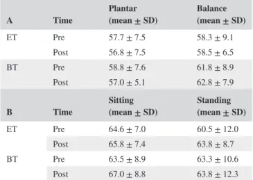
TABLE 1 gives an overview of the AMT (A) for the plantar flexion and balance perturbation, and the RMT (B) in the control conditions in the ET and BT during at the pre‐ and post‐test
A Time Plantar (mean ± SD) Balance (mean ± SD)
ET Pre 57.7 ± 7.5 58.3 ± 9.1 Post 56.8 ± 7.5 58.5 ± 6.5 BT Pre 58.8 ± 7.6 61.8 ± 8.9 Post 57.0 ± 5.1 62.8 ± 7.9
B Time Sitting (mean ± SD) Standing (mean ± SD)
ET Pre 64.6 ± 7.0 60.5 ± 12.0 Post 65.8 ± 7.4 63.8 ± 8.7 BT Pre 63.5 ± 8.9 63.3 ± 10.6
2.9
|
TMS during control conditions
2.9.1
|
Resting motor threshold (RMT)
The RMT was defined as the lowest possible stimulation in-tensity to evoke MEPs <50 μV in three out of five consecu-tive trials (Rossini et al., 1994) during the control conditions sitting and standing.
2.9.2
|
SICI in control conditions
In addition to the plantar flexions and balance perturbations, TMS was also applied during two control conditions. In the first control condition (rest‐sitting), subjects were in the same seated position as during the plantar flexions but were in-structed to rest. Then, SICI was tested by applying 15 single‐ and 15 paired‐pulse TMS stimulations that were applied at 0.8 and 1.2RMT.
The second control condition was during quiet upright standing where subjects were instructed to stand as quietly as possible while 15 single‐ and 15 paired‐pulse TMS stim-ulations were applied at 0.8 and 1.2AMT as there was back-ground activation of the SOL. It is again important to note that the RMT for the sitting and the AMT for the standing condition were individually adjusted so that the stimulation intensity was specific for each condition tested in this study.
The control conditions were included in the experiment as it provided the baseline measure of cortical inhibition and al-lowed us to calculate the range of cortical inhibition between the control and the active conditions.
2.9.3
|
SICI in untrained muscle (TA)
SICI in the TA was measured according to the exact same procedures as for the SOL, but coil position, AMT, RMT and thus the stimulation intensity for the SICI measurements dur-ing rest as well as durdur-ing activity (dorsiflexions and standdur-ing) were individually adjusted for the TA.
2.10
|
Behavioural measures
2.10.1
|
Explosive training (ET)
Before the first training session, the 1 repetition maximum (RM) was defined for each exercise. During the ET, four sets with six repetitions at 40% of the individual 1 RM had to be performed (Van Cutsem, Duchateau, & Hainaut, 1998) for each exercise with a rest of 2.5 min between sets and types of exercise. The explosive training took place three times per week over a period of four weeks. After a 15‐min warm‐up, the training consisted of four exercises (half‐squats with a barbell on the shoulders, box jumps from a squatting posi-tion, lateral steps with kettlebells and plantar flexions while
sitting with barbell on the thigh) and it was always taken care of that the concentric phase was ballistic and ended in a plan-tar‐flexed position.
2.10.2
|
Balance training (BT)
The BT was performed three times per week over a four‐week period. After a 15‐min warm‐up, subjects had to stand on a two‐dimensional swinging platform, wobbling boards, spin-ning tops and soft mats. Each traispin-ning consisted of 3–6 sets with 30‐s balancing on each of the devices. This resulted in 12 trials per leg at the beginning of the training and was then increased to 24 trails in the last week of the training (Taube, Gruber, et al., 2007). Furthermore, task difficulty was pro-gressively increased (standing with eyes closed, catching a ball while balancing, etc.). There was a 30‐s rest between tri-als and 5‐min break between sets. Subjects stood barefoot on one leg on the devices, while the arms were held akimbo. The training ended with a 15‐min cool‐down, and the same balance training has been applied in a number of previous experiments (Gruber et al., 2007; Taube et al., 2006; Taube, Gruber, et al., 2007; Taube, Kullmann, et al., 2007).
2.10.3
|
Rate of torque development (RTD)
The maximal rate of torque development without TMS stim-ulation was measured during isometric plantar flexion using an isokinetic device (Isomed 2000®, D&R GmbH, Hemau, Germany). Therefore, subjects were lying on their back with the hip at 90 degrees and the knee at 180 degrees (fully ex-tended). The foot of the subjects was strapped to the footplate of the device at an ankle angle of 100 degrees, and the foot was tightly secured to exclude movements of the ankle. The hip and shoulders were strapped to the backrest of the isoki-netic device to avoid trunk movements. Initially, subjects were allowed to perform a set (5–7) of submaximal contrac-tions to get accustomed to the task. Then, subjects performed 5 contractions and were instructed to contract as fast and as hard as possible for approximately 1 s with 30‐s rest between the contractions (Maffiuletti et al., 2016). Data were sampled at 2 kHz.
2.10.4
|
Maximal voluntary force (MVC)
The subjects performed three isometric maximum voluntary contractions (MVC) using the same device as for the RTD measurements. The plantar flexion MVCs consisted of a gradual increase in force from zero to maximum over a 3‐s time span. The maximal force was held for 2 s, and subjects were verbally encouraged to achieve maximal force. After each trial, subjects were allowed to rest for 90 s. The position of the body was the same as for the RTD measurements, and data were sampled at 2 kHz.
2.10.5
|
Strength in untrained muscle (TA)
The set‐up and the procedures for the RTD and MVC meas-ures in the TA were identical to the measmeas-ures in the SOL, but for the TA, subjects performed dorsiflexions instead of plantar flexions.
2.10.6
|
Balance performance
The balance performance was tested using a balance device (Posturomed) with a good test–retest reliability (Boeer, Mueller, Krauss, Haupt, & Horstmann, 2010) allowing platform sway in the transversal plane (Muller, Gunther, Krauss, & Horstmann, 2004). To minimize any short‐term learning effects, subjects were given 2 min on the de-vice to familiarize with the task (Keller, Pfusterschmied, Buchecker, Muller, & Taube, 2012). Performance was tested under two conditions, while the participants stood with the right leg on the free‐swinging device and were in-structed to sway as little as possible. In the first condition, subjects stood quietly for a period of 30s while the platform sway was measured. In the second condition, anterior–pos-terior balance perturbations were applied and subjects had to counteract the perturbations for 20 s. Anterior–posterior and medio‐lateral sway paths were recorded by joystick potentiometers connected to the moveable platform. The cumulative sway paths of three trials were averaged for each condition, and values obtained before and after train-ing were compared.
2.11
|
Data analyses and statistics
2.11.1
|
Motor cortical inhibition
The size of the motor evoked potential was quantified by peak‐to‐peak analysis of the conditioned MEP (paired‐pulse stimulation) compared to the unconditioned MEP during the active (plantar flexion and balance perturbations) and the control conditions (sitting and quite standing). SICI was ex-pressed as percentage inhibition of the conditioned in relation to the unconditioned MEP using the formula: 100 – (con-ditioned MEP/ uncon(con-ditioned MEP x 100), which has been used previously (Kuhn et al., 2016; Lauber et al., 2018).
Cortical silent period was measured in the trials with sin-gle‐pulse TMS. The duration of the CSP was measured from the onset of the MEP, and the endpoint coincided with the reoccurrence of EMG activity in individual trials via visual inspection (Kimiskidis et al., 2005).
2.11.2
|
Corticospinal excitability
Corticospinal excitability (CSE) was quantified by the peak‐to‐peak amplitude of the MEP resulting from the
unconditioned (single‐pulse) TMS stimulation measured dur-ing the control and active conditions.
2.11.3
|
Background EMG activity (bEMG)
Muscle activation at the time of the TMS stimulation was calculated in a time window of 50 ms prior to each stimula-tion. EMG signals were rectified, and root mean square val-ues were calculated and averaged.
2.11.4
|
Rate of torque development
The RTD of the ballistic plantar flexion was defined as the maximal slope of the force–time curve in each trial (Gruber et al., 2007; Lauber et al., 2013). The best three out of the five trials were used for comparisons (Maffiuletti et al., 2016).
2.11.5
|
Balance performance
The cumulative sway paths of three trials without perturba-tion and the three trials with perturbaperturba-tion were individually averaged for each condition.
All data were analysed using custom‐written MATLAB scripts (MathWorks Inc.).
2.11.6
|
Statistical comparison
The Kolmogorov–Smirnov test was used to test for the normal distribution of the data. To test for group‐ and task‐specific adaptations in SICI (paired‐pulse TMS) and corticospinal ex-citability (single‐pulse TMS) after the training, a three‐way repeated measures ANOVA with factors time (pre, post), condition (balance perturbation, plantar flexion) and group (ET, BT) was calculated. To evaluate changes in motor be-haviour, the Z‐transformations (Kleinbaum, Kupper, Azhar, & Muller, 1978; Taube, Leukel, et al., 2008) were used before comparing results of the balance and strength tests by cal-culating a three‐way ANOVA with factors time (pre, post), condition (balance performance, RTD) and group (ET, BT).
To test for differences in the control experiment, separate two‐way repeated measures ANOVAs with factors time (pre, post) and group (ET, BT) were calculated for the sitting and standing condition.
Changes in RMT, AMT, SICI and RTD in the TA were calculated with separate repeated measures ANOVAs with factors time (Pre, Post), group (ET, BT) and condition (RTD, balance perturbations).
Changes in bEMG, AMT and RMT were investigated by separate three‐way repeated measures ANOVAs with fac-tors time (pre, post), condition (balance perturbation, plantar flexion) and group (ET, BT).
In the case of significant F‐values (p ≤ .05), Bonferroni‐ corrected Student's t tests were calculated to assess
differences between conditions. All data are reported as means ± standard deviation, effect sizes are shown as partial eta square, and the level of significance was set at p ≤ .05. SPSS 24 software was used for all statistical comparisons.
3
|
RESULTS
3.1
|
Neural adaptations
3.1.1
|
SICI during activity
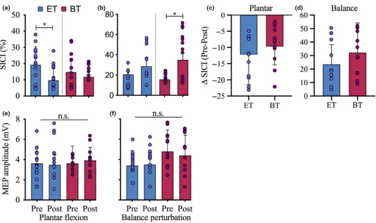
The ANOVA showed a significant time*condition*group interaction (F2,22 = 6.2, p = .021, η2 = 0.21)
demonstrat-ing a task‐specific adaptation in SICI as a consequence of the different training programmes (Figure 1). Post hoc tests indicated that this interaction was due to a significant reduc-tion in SICI during plantar flexions after ET (19.3 ± 11.1 vs. 9.6 ± 8.1%; p = .032), while BT did not show a sig-nificant change (BT: 14.8 ± 6.5% vs. 11.8 ± 8.4%, p = .37; Figure 2a,c).
For the balance perturbations, the results also demon-strated a task‐ and group‐specific adaptation in SICI as post hoc tests showed that there was a significant increase in SICI during the balance perturbations after BT (15.6 ± 5.5 vs. 35.0 ± 6.6%, p = .01) while ET did not show a signifi-cant change in SICI (20.5 ± 5.7% vs. 28.7 ± 6.9%, p = .32; Figure 2b,d).
There was no significant change in CSP in any of the groups between the pre‐ and the post‐test as we did not ob-serve a time effect (F1,22 = 0.3, p = .87, η2 = 0.001).
3.1.2
|
Corticospinal excitability (CSE)
The ANOVA showed that there was no significant change in CSE between the pre‐ and the post‐test as there was no
time*condition*group interaction (F2,22 =.001, p = .99,
η2 = 0.001; Figure 1e+f) as well as no significant main
effect of time (F1,22 = .07, p = .41, η2 = 0.03). When
the MEPs were normalized to the maximal M‐wave of the soleus obtained during the standing condition, there
were no significant changes (F2,22 = 1.2, p = .290,
η2 = 0.053).
3.1.3
|
Background EMG
The ANOVA showed that there was no significant change in bEMG before and after the training in any of the condi-tions showing that the EMG was similar prior to stimulation (time*condition*group interaction: F2,22 = 0.001, p = .99,
η2 = 0.001). There was also no condition (F
1,22 = 1.01, p = .33, η2 = 0.05) or time effect (F
1,22 = 1.96, p = .18,
η2 = 0.09)
3.1.4
|
Active motor threshold
An overview about the AMT can be found in Table 1 (A). The ANOVA revealed no significant time*condition*group interaction (F2,22 = 0.23, p = .64, η2 = 0.01).
3.1.5
|
Resting motor threshold
The ANOVA revealed no significant condition (F1,22 = 1.6, p = .22, η2 = 0.068) and no time*group effect (F
1,22 = 0.13, p = .73, η2 = 0.006). There was a trend towards a FIGURE 1 Representative EMG traces of one exemplary subject from the ST (a+b) and the BT (c+d). a+c display the MEPs measured during the plantar flexions after single‐pulse (black) and paired‐pulse stimulation (red) from a subject from the ST (a) and BT (c). b+d show MEP trances obtained during the balance perturbations after single‐pulse (black) and paired‐pulse stimulation (blue) from a subject from the ST (b) and BT (d). The coloured lines represent the mean of all stimulations, while the grey lines represent individual responses to TMS. [Color figure can be viewed at wileyonlinelibrary. com]
FIGURE 2 SICI during the active conditions in the pre‐ and the post‐test. (a) After the training, there was a significant decrease in SICI during the plantar flexions in the ST group (blue) as well as in the BT group (red). There was no significant difference between the groups. (b) Changes in SICI were measured during the balance perturbations. There was a significant increase in SICI in the BT as well as in the ST. There was no significant difference between the groups. (c) The change in SICI (decline) during the plantar flexions was more pronounced in the ST than the BT, while d shows a greater increase in SICI in the BT than in the ST when comparing the pre‐ with the post‐values. (d) There was no significant change in the CSE during the plantar flexions (e) as well as during the balance perturbations (f) in any of the groups (*p < .05). [Color figure can be viewed at wileyonlinelibrary.com]
FIGURE 3 SICI measures obtained during the control experiments in the ET (top panel) and BT (lower panel). There was no significant difference between the pre‐ and the post‐test in the sitting (a) and standing condition (b). There was no significant difference in SICI in the TA before and after the training (c) in the sitting (blue), standing (purple) nor during the dorsiflexion (orange). [Color figure can be viewed at wileyonlinelibrary.com]
time*group*condition effect (F1,22 = 3.79, p = .064,
η2 = 0.14). An overview about the RMT can be found in
Table 1 (B).
3.2
|
Results from Control Experiments
3.2.1
|
Sitting
There was no significant change in SICI after the train-ing between the groups (time*group: F1,22 = 0.01, p = .92,
η2 = 0.001; Figure 3a).
3.2.2
|
Standing
There was no significant change in SICI after the train-ing between the groups (time*group: F1,22 = 0.49, p = .53,
η2 = 0.01; Figure 3b).
3.2.3
|
Untrained muscle TA
To test whether the observed changes were caused by the training or a general effect, SICI was also measured in the TA during sitting, dorsiflexions and standing. The results showed no time*group*condition interaction (F2,22 = 0.09, p = .89,
η2 = 0.005) supporting task‐ and muscle‐specific adaptations
in SICI (Figure 3c).
3.3
|
Behavioural adaptations
Both groups improved in balance as well as explosive strength (time effect (F2,22 = 181.1, p = .0001, η2 = 0.89). However, a
corrected post hoc test only reached statistical significance for the increase in RTD in the ET group (ET: 667.2 ± 62.4 Nm/s vs.
827.5 ± 65.6 Nm/s; p = .03) and not the BT (696.3 ± 65.1 Nm/s vs. 792.9 ± 82.7 Nm/s; p = .18; Figure 4a). On the contrary, corrected post hoc test for the increase in dynamic balance con-trol was only significant in the BT group (BT: 43.9 ± 6.6 cm vs. 33.6 ± 4.5 cm, p = .05) but not the ET (37.5 ± 6.3 cm vs. 22.8 ± 4.3 cm; p = .12; Figure 4b).
4
|
DISCUSSION
The main aim of the present study was to identify task‐, muscle‐ and long(er)‐term training‐specific adaptations in inhibitory motor cortical control. We speculated that tasks that require maximal motor drive such as a ballistic force task would result in reduced levels of SICI whereas tasks that require complex coordination such as balancing would lead to enhanced levels of SICI. So far, most of the previ-ous research investigating changes in the cortical inhibi-tory system as a result of motor learning used short‐term learning paradigms rather than several weeks of learning (Camus et al., 2009; Cirillo et al., 2011; Leung et al., 2015; Perez et al., 2004). These short‐term studies have consist-ently shown reduced levels of intracortical inhibition, sug-gesting a general decrease in SICI during the initial phase of motor learning. However, with ongoing motor prac-tice, some studies reported decreases (Weier et al., 2012) whereas others demonstrated increases (Dai et al., 2016; Mouthon & Taube, 2019) in SICI. In the current study, we therefore tested the hypothesis that the inhibitory system adapts depending on the task being learned. The current results confirm this assumption showing significantly re-duced SICI levels during the execution of ballistic con-tractions after explosive training. In contrast, SICI was
FIGURE 4 (a) Rate of torque development of the ST (blue) and the BT (red) measured during the pre‐ and the post‐test. Both groups significantly increased their RTD (*p < .05). The bars indicate group mean values, while the small dots represent individual results. (b) Balance performance during the dynamic balance task of the ST (blue) and the BT (red) measured during the pre‐ and the post‐test showing that both groups significantly reduced their postural sway during the balance perturbations (*p < .05) while there was no difference between the groups. [Color figure can be viewed at wileyonlinelibrary.com]
significantly enhanced during the execution of balance exercises after balance training. These neural adaptations were also reflected in the behavioural outcomes as the explosive training group showed a much greater increase in their explosive performance while the balance train-ing group showed greater improvements in their balance performance. These task‐specific adaptations resemble the ones reported previously in the study of Gruber et al. (2007) who showed that four weeks of explosive training caused an increase of 48 ± 16% in RFD while balance training over the same duration caused only an increase of 14 ± 5%. In the present study, the mean increase in RTD was also higher (+31.1 %) in the ET compared to the BT (+14.6%).
At first sight, it may be surprising that the present study found a reduced level of SICI after the explosive training as a recent review by Berghuis, Semmler, Opie, Post, and Hortobagyi (2017) failed to identify changes in SICI as a consequence of ballistic motor learning in young as well as old subjects. From a functional perspective, however, it makes sense that cortical inhibition is reduced after explosive training in order to ensure high levels of cortical excitatory drive (Kidgell, Bonanno, Frazer, Howatson, & Pearce, 2017). Furthermore, the difference to the results of Berghuis et al. (2017) might simply be explained by the observation that in the majority of studies included in this review, SICI had been tested at rest rather than during activity even though inhibition is well known to be modulated in a task‐dependent manner (Opie & Semmler, 2016; Papegaaij et al., 2016; Sidhu et al., 2013; Soto et al., 2006). In the present study, however, there was no change in SICI when assessed during rest but only when tested during the previously trained ballistic plantar flexions. This a) highlights the task specificity of how SICI is modu-lated and b) emphasizes the importance of the appropriate test condition in order to draw sound conclusions about changes in inhibitory motor control. Supporting this, the balance training group demonstrated increases in SICI exclusively during the execution of balance tasks while no changes occurred when SICI was tested at rest. This supports the findings of a previ-ous balance training study demonstrating increased SICI only during balancing but not at rest (Mouthon & Taube, 2019). The functional relevance of an increased inhibition during balancing may be to avoid unnecessary co‐activations and/or co‐movements (Mouthon & Taube, 2019). Furthermore, the ability to modulate cortical inhibition may be important to allow shifting motor control from cortical to more subcorti-cal centres (for review, see Taube, Gruber, et al., 2008). This scenario would allow—for example with increases in postural task complexity—that cortical control could be reinforced by decreasing the level of intracortical inhibition.
The high task specificity of the training‐induced changes in the present study resembles the one reported in a study by Schubert et al. (2008) who also compared ballistic training with balance training. This study showed that changes in
corticospinal transmission were only seen when tested in the trained task but not during an untrained motor task and also not at rest. More specifically, conditioned H‐reflexes were increased after balance training while they were decreased after ballistic strength training indicating a pathway‐specific adaptation with respect to the type of training (Schubert et al., 2008). Even though the present study tested for adaptations in SICI and, thus, a different neurophysiological mechanism, the data also indicate very task‐specific adaptations as a conse-quence of explosive training and balance training which seem to be mainly taking place in the GABAA and not GABAB
net-work as we only found changes in SICI but not in the CSP. Furthermore, the present results indicate that motor learning does not result in a strict up‐ (balance training) or downreg-ulation (explosive training) of inhibitory processes but may rather help to enlarge the modulatory capacity of intracortical inhibition. This means that the baseline level of SICI mea-sured at rest remains unaltered, but training seems to enlarge the range of inhibition and the type of exercise seems to de-termine the direction of this adaptation. In this sense, balance trained subjects are better able to upregulate SICI during coor-dinative balance tasks whereas explosive trained subjects can better downregulate SICI during explosive tasks. It therefore seems that the range of modulation (modulatory bandwidth) of inhibitory control may be essential for an adequate motor control rather than a strict up‐ or downregulation of SICI.
This might explain why deficits in the ability to mod-ulate SICI have been related to behavioural declines in motor functions such as prolonged reaction times (Levin, Fujiyama, Boisgontier, Swinnen, & Summers, 2014) as well as impairments in postural control (Papegaaij et al., 2016). The modulatory capacity might be important to in-crease motor cortical contribution in order to fulfil complex motor tasks. It might be speculated that the capacity for inhibition may allow subjects to discharge cortical motor centres and shift motor control to more subcortical areas. The modulatory capacity might therefore be important to increase motor cortical contribution in order to fulfil com-plex motor tasks. Thus, a high baseline level of inhibition allows for a broad modulatory range which might be im-portant for both learning of new motor skills and execution of complex motor skills.
5
|
CONCLUSION
The present study is the first to compare changes in the GABAAergic cortical inhibitory system after the learning of
two different motor tasks for several weeks. Depending on the trained task (balance vs. explosive contractions), intracortical inhibition was either up‐ or downregulated when measured during the acquired activity but not at rest. This is an important finding as it indicates that training mainly improves the ability
to modulate inhibitory processes task specifically hereby in-creasing the modulatory bandwidth of cortical inhibition. Our results also explain some of the discrepancies in the literature as they emphasize the need to assess intracortical inhibition during activity and not solely at rest. Measurements during activity are more difficult to perform and to control, which might explain why most previous studies assessed intracorti-cal inhibition exclusively at rest. However, it seems rational that the functional benefit of changing the ‘baseline level’ of intracortical inhibition (i.e. inhibition at rest) is very limited compared to a task (and muscle)‐specific adaptation of inhibi-tory processes that meet the movement‐related requirements.
5.1
|
Limitations
In the present study, SICI as well as cortical silent period was measured as markers of cortical inhibition. While it is
assumed that SICI is an indicator for the GABAA‐mediated
inhibitory activity, silent period is thought to represent corti-cal inhibition due to the activity of the GABAB system. Thus,
the two measures provide substantial information about the activity of the cortical inhibitory system. Nevertheless, it needs to be noted that there are additional mechanisms such as long‐interval intracortical inhibition (LICI) which might have influenced the results but was not included in the proto-col to reduce the total amount of stimulations. Additionally, SICI and single‐pulse TMS were recorded during rest as well as during activity. Thus, the different test conditions resulted in different levels of muscle activation which af-fects the activity of the corticospinal system. Nevertheless, we considered it to be important to test cortical inhibition not only at rest but also during activity because it was shown that the activity of inhibitory system changes in a task‐de-pendent manner (Opie & Semmler, 2016; Papegaaij et al., 2016; Sidhu et al., 2013; Soto et al., 2006).
ACKNOWLEDGEMENTS
There are no conflicts of interest between authors, compa-nies or manufacturers. The results of the study are presented clearly, honestly and without fabrication, falsification or in-appropriate data manipulation.
CONFLICT OF INTEREST
The authors declare that there are no conflicts of interest.
AUTHOR CONTRIBUTIONS
WT, AG and BL designed the experiment; BL executed the experiment and analysed the data; WT and BL drafted the manuscript; WT, AG and BL revised and finalized the manuscript.
DATA ACCESSIBILITY
Data are made available upon direct request to the author.
ORCID
Benedikt Lauber https://orcid.org/0000-0001-9684-3675
REFERENCES
Berghuis, K. M. M., Semmler, J. G., Opie, G. M., Post, A. K., & Hortobagyi, T. (2017). Age‐related changes in corticospinal excit-ability and intracortical inhibition after upper extremity motor learn-ing: a systematic review and meta‐analysis. Neurobiology of Aging,
55, 61–71.
Boeer, J., Mueller, O., Krauss, I., Haupt, G., & Horstmann, T. (2010). Reliability of a measurement technique to characterise standing properties and to quantify balance capabilities of healthy subjects on an unstable oscillatory platform (Posturomed). Sportverletzung
Sportschaden, 24, 40–45.
Camus, M., Ragert, P., Vandermeeren, Y., & Cohen, L. G. (2009). Mechanisms controlling motor output to a transfer hand after learn-ing a sequential pinch force skill with the opposite hand. Clinical
Neurophysiology, 120, 1859–1865.
Cirillo, J., Todd, G., & Semmler, J. G. (2011). Corticomotor excitability and plasticity following complex visuomotor training in young and old adults. European Journal of Neuroscience, 34, 1847–1856. Classen, J., Liepert, J., Wise, S. P., Hallett, M., & Cohen, L. (1998).
Rapid plasticity of human cortical movement representation induced by practice. Journal of Neurophysiology, 79, 1117–1123.
Dai, W., Pi, Y. L., Ni, Z., Tan, X. Y., Zhang, J., & Wu, Y. (2016). Maintenance of balance between motor cortical excitation and inhi-bition after long‐term training. Neuroscience, 336, 114–122. Di Lazzaro, V., Oliviero, D. R. A., Ferrara, P. P. L., Mazzone, A. I. P.,
& Rothwell, P. T. J. C. (1998). Magnetic transcranial stimulation at intensities below active motor threshold activates intracortical in-hibitory circuits. Experimental Brain Research, 265–268.
Di Lazzaro, V., & Rothwell, J. C. (2014). Corticospinal activity evoked and modulated by non‐invasive stimulation of the intact human motor cortex. Journal of Physiology, 592, 4115–4128.
Faul, F., Erdfelder, E., Lang, A.‐G., & Buchner, A. (2007). G*Power 3: a flexible statistical power analysis program for the social, behavioral, and biomedical sciences. Behavior Research Methods, 39, 175–191. Gruber, M., Gruber, S. B. H., Taube, W., Schubert, M., Beck, S. C.,
& Gollhofer, A. (2007). Differential effects of ballistic versus sen-sorimotor training on rate of force development and neural activa-tion in humans. Journal of Strength and Condiactiva-tioning Research /
National Strength & Conditioning Association, 21, 274–282.
Hinder, M. R., Schmidt, M. W., Garry, M. I., & Summers, J. J. (2010). The effect of ballistic thumb contractions on the excitability of the ip-silateral motor cortex. Experimental Brain Research. Experimentelle
Hirnforschung. Expérimentation Cérébrale, 201, 229–238.
Keller, M., Pfusterschmied, J., Buchecker, M., Muller, E., & Taube, W. (2012). Improved postural control after slackline training is accom-panied by reduced H‐reflexes. Scandinavian Journal of Medicine
and Science in Sports, 22, 471–477.
Kidgell, D. J., Bonanno, D. R., Frazer, A. K., Howatson, G., & Pearce, A. J. (2017). Corticospinal responses following strength
training: a systematic review and meta‐analysis. European Journal
of Neuroscience, 46, 2648–2661.
Kidgell, D. J., & Pearce, A. J. (2010). Corticospinal properties follow-ing short‐term strength trainfollow-ing of an intrinsic hand muscle. Human
Movement Science, 1–11.
Kimiskidis, V. K., Papagiannopoulos, S., Sotirakoglou, K., Kazis, D. A., Kazis, A., & Mills, K. R. (2005). Silent period to transcranial magnetic stimulation: construction and properties of stimulus‐re-sponse curves in healthy volunteers. Experimental Brain Research,
163, 21–31.
Kleinbaum, D., Kupper, L., Azhar, N., & Muller, K. (1978) Applied
regression analysis and other multivariable methods. Boston, MA:
Duxbury Applied.
Kuhn, Y. A., Keller, M., Ruffieux, J., & Taube, W. (2016). Adopting an external focus of attention alters intracortical inhibition within the primary motor cortex. Acta Psychologica.
Lauber, B., Gollhofer, A., & Taube, W. (2018). Differences in motor cortical control of the Soleus and Tibialis. Journal of Experimental
Biology, 221, https ://doi.org/10.1242/jeb.174680
Lauber, B., Lundbye‐Jensen, J., Keller, M., Gollhofer, A., Taube, W., & Leukel, C. (2013). Cross‐limb interference during motor learning.
PLoS ONE, 8(12), e81038.
Lee, M., Hinder, M. R., Gandevia, S. C., & Carroll, T. J. (2010). The ipsilateral motor cortex contributes to cross‐limb transfer of perfor-mance gains after ballistic motor practice. Journal of Physiology,
588, 201–212.
Leung, M., Rantalainen, T., Teo, W. P., & Kidgell, D. (2015). Motor cortex excitability is not differentially modulated following skill and strength training. Neuroscience, 305, 99–108.
Leung, M., Rantalainen, T., Teo, W.‐P., & Kidgell, D. (2017). The corti-cospinal responses of metronome‐paced, but not self‐paced strength training are similar to motor skill training. European Journal of
Applied Physiology, 117, 2479–2492.
Levin, O., Fujiyama, H., Boisgontier, M. P., Swinnen, S. P., & Summers, J. J. (2014). Aging and motor inhibition: a converging perspective provided by brain stimulation and imaging approaches. Neuroscience
and Biobehavioral Reviews, 43, 100–117.
Lotze, M., Braun, C., Birbaumer, N., Anders, S., & Cohen, L. G. (2003). Motor learning elicited by voluntary drive. Brain, 126, 866–872.
Maffiuletti, N. A., Aagaard, P., Blazevich, A. J., Folland, J., Tillin, N., & Duchateau, J. (2016). Rate of force development: physiological and methodological considerations. European Journal of Applied
Physiology, 116, 1091–1116.
Mornieux, G., Gehring, D., Tokuno, C., Gollhofer, A., & Taube, W. (2014). Changes in leg kinematics in response to unpredictability in lateral jump execution. European Journal of Sport Science, 14, 678–685.
Mouthon, A., & Taube, W. (2019). Intracortical inhibition increases during postural task execution in response to balance training.
Neuroscience, 401, 35–42.
Muellbacher, W., Ziemann, U., Boroojerdi, B., Cohen, L., & Hallett, M. (2001). Role of the human motor cortex in rapid motor learning.
Experimental Brain Research, 136, 431–438.
Muellbacher, W., Ziemann, U., Wissel, J., Dang, N., Kofler, M., Facchini, S., … Hallett, M. (2002). Early consolidation in human primary motor cortex. Nature, 415, 640–644.
Muller, O., Gunther, M., Krauss, I., & Horstmann, T. (2004). Physical characterization of the therapeutic device Posturomed as a measuring
device–presentation of a procedure to characterize balancing ability.
Biomed Tech (Berl), 49, 56–60.
Opie, G. M., & Semmler, J. G. (2016). Intracortical inhibition assessed with paired‐pulse transcranial magnetic stimulation is modulated during shortening and lengthening contractions in young and old adults. Brain Stimul, 9, 258–267.
Papegaaij, S., Baudry, S., Negyesi, J., Taube, W., & Hortobagyi, T. (2016). Intracortical inhibition in the soleus muscle is reduced during the control of upright standing in both young and old adults.
European Journal of Applied Physiology, 116, 959–967.
Papegaaij, S., Taube, W., Hogenhout, M., Baudry, S., & Hortobagyi, T. (2014). Age‐related decrease in motor cortical inhibition during standing under different sensory conditions. Frontiers in Aging
Neuroscience, 6, 126.
Perez, M. A., Lungholt, B. K., Nyborg, K., & Nielsen, J. B. (2004). Motor skill training induces changes in the excitability of the leg cortical area in healthy humans. Experimental Brain Research, 159, 197–205.
Peurala, S. H., Muller‐Dahlhaus, J. F., Arai, N., & Ziemann, U. (2008). Interference of short‐interval intracortical inhibition (SICI) and short‐interval intracortical facilitation (SICF). Clinical
Neurophysiology, 119, 2291–2297.
Rosenkranz, K., Kacar, A., & Rothwell, J. C. (2007). Differential mod-ulation of motor cortical plasticity and excitability in early and late phases of human motor learning. Journal of Neuroscience, 27, 12058–12066.
Rossini, P. M., Barker, A. T., Berardelli, A., Caramia, M. D., Caruso, G., Cracco, R. Q., … Tomberg, C. (1994). Non‐invasive electrical and magnetic stimulation of the brain, spinal cord and roots: basic principles and procedures for routine clinical application. Report of an IFCN committee. Electroencephalography and Clinical
Neurophysiology, 91, 79–92.
Schubert, M., Beck, S., Taube, W., Amtage, F., Faist, M., & Gruber, M. (2008). Balance training and ballistic strength training are associ-ated with task‐specific corticospinal adaptations. European Journal
of Neuroscience, 27, 2007–2018.
Sidhu, S. K., Cresswell, A. G., & Carroll, T. J. (2013). Short‐interval intracortical inhibition in knee extensors during locomotor cycling.
Acta Psychologica, 207, 194–201.
Soto, O., Valls‐Sole, J., Shanahan, P., & Rothwell, J. (2006). Reduction of intracortical inhibition in soleus muscle during postural activity.
Journal of Neurophysiology, 96, 1711–1717.
Taube, W., Gruber, M., Beck, S., Faist, M., Gollhofer, A., & Schubert, M. (2007). Cortical and spinal adaptations induced by balance train-ing: correlation between stance stability and corticospinal activa-tion. Acta Psychologica, 189, 347–358.
Taube, W., Gruber, M., & Gollhofer, A. (2008). Spinal and supraspinal adaptations associated with balance training and their functional rel-evance. Acta Physiologica, 1–16.
Taube, W., Kullmann, N., Leukel, C., Kurz, O., Amtage, F., & Gollhofer, A. (2007). Differential reflex adaptations following sensorimotor and strength training in young elite athletes. International Journal
of Sports Medicine, 28, 999–1005.
Taube, W., Leukel, C., Schubert, M., Gruber, M., Rantalainen, T., & Gollhofer, A. (2008). Differential modulation of spinal and cortico-spinal excitability during drop jumps. Journal of Neurophysiology,
99, 1243–1252.
Taube, W., Schubert, M., Gruber, M., Beck, S., Faist, M., & Gollhofer, A. (2006). Direct corticospinal pathways contribute to the
neuromuscular control of perturbed stance. Journal of Applied
Physiology, 420–429.
Van Cutsem, M., Duchateau, J., & Hainaut, K. (1998). Changes in single motor unit behaviour contribute to the increase in contraction speed after dynamic training in humans. Journal of Physiology, 513 (Pt
1, 295–305.
Weier, A. T., Pearce, A. J., & Kidgell, D. J. (2012). Strength training reduces intracortical inhibition. Acta Psychologica, 206, 109–119. Weise, D., Mann, J., Ridding, M., Eskandar, K., Huss, M., Rumpf, J. J.,
… Classen, J. (2013). Microcircuit mechanisms involved in paired associative stimulation‐induced depression of corticospinal excit-ability. Journal of Physiology, 591, 4903–4920.
Weltin, E., Mornieux, G., & Gollhofer, A. (2015). Influence of gender on trunk and lower limb biomechanics during lateral movements.
Research in Sports Medicine (Print), 23, 265–277.
How to cite this article: Taube W, Gollhofer A, Lauber
B. Training‐, muscle‐ and task‐specific up‐ and downregulation of cortical inhibitory processes. Eur J
Neurosci. 2020;51:1428–1440. https ://doi.org/10.1111/ ejn.14538