REVIEW ARTICLE
SPECT/CT for imaging of the spine and pelvis in clinical
routine: a physician
’s perspective of the adoption of SPECT/
CT in a clinical setting with a focus on trauma surgery
Max J. Scheyerer&Carsten Pietsch&Stefan M. Zimmermann&
Georg Osterhoff&Hans-Peter Simmen&Clement M. L. Werner
Received: 23 May 2013 / Accepted: 19 August 2013 / Published online: 21 September 2013 # Springer-Verlag Berlin Heidelberg 2013
Abstract Injuries of the axial skeleton are an important field of work within orthopaedic surgery and traumatology. Most lesions following trauma may be diagnosed by means of conventional plain radiography, computed tomography or magnetic resonance imaging. However, for some aspects SPECT/ CT can be helpful even in a trauma setting. In particular, the combination of highly sensitive but nonspecific scintigraphy with nonsensitive but highly specific computed tomography makes it particularly useful in anatomically com-plex regions such as the pelvis and spine. From a trauma surgeon’s point of view, the four main indications for nuclear medicine imaging are the detection of (occult) fractures, and the imaging of inflammatory bone and joint diseases, chronic diseases and postoperative complications such as instability of instrumentation or implants. The aim of the present review was to give an overview of the adoption of SPECT/CT in a clinical setting.
Keywords SPECT/CT . Spine . Pelvis . Fracture . Trauma
Introduction
Injuries of the axial skeleton and associated late sequelae are an important field of work within orthopaedic surgery and traumatology. Despite the relatively small proportion of spinal
fractures compared to all fractures, they are of great impor-tance due to potentially severe consequences. The estimated incidence of spinal fractures reported in the literature is 64 per 100,000 persons, with young men and elderly women being predominantly affected. In young patients, spinal fractures usually occur in the presence of a substantial external force. Depending on the direction and position of the main rotation axis, the result is a compression fracture, a flexion/distraction injury, a rotational injury or a combined injury. In the elderly, spinal fractures are the most common first manifestation of osteoporosis and may result not only from simple falls from a standing height, but may also occur during activities of daily living such as lifting objects or merely bending over [1]. The result is usually a compression fracture, which according to the AO/ASIF (Association for the Study of Internal Fixation) classification may be classified as a type A fracture. The consequences of such a fracture may frequently be underestimated, but these injuries are of substantial relevance in the elderly and mortality rates are comparable to those recently demonstrated in femoral neck fractures [2].
Other frequent locations for osteoporotic fractures include lesions of the pelvis, especially the pubic rami and sacrum. They are associated with chronic pain and increased morbidity and mortality [3–5]. This is believed to be due to occult fractures of the posterior pelvic ring, which is far more im-portant for stability than the anterior structures [6–8]. Timely therapeutic intervention following an accident may substan-tially lower morbidity and mortality rates. For this reason, as well as a thorough patient history and physical examination, a sufficient diagnostic work-up is also essential. This usually involves a step-by-step process. In addition to digital conven-tional X-ray imaging, the use of computed tomography (CT) has been established especially in the acute phase following trauma. Current guidelines strongly recommend a whole-body CT scan following primary survey and basic life support
M. J. Scheyerer (*)
:
S. M. Zimmermann:
G. Osterhoff:
H.<P. Simmen
:
C. M. L. WernerDepartment of Surgery, Division of Trauma Surgery, University Hospital Zurich, Raemistrasse 100, 8091 Zürich, Switzerland e-mail: [email protected]
C. Pietsch
Department of Medical Radiology, Division of Nuclear Medicine, University Hospital Zurich, Zurich, Switzerland
measures during management of heavily injured patients in the resuscitation room [9]. This approach has led to a signif-icant decrease in mortality [10].
Conventional X-ray imaging has lost importance in the context of spinal and pelvic injuries as well as in polytrauma due to the higher sensitivity and specificity of CT [11,12]. Lesions of the posterior pelvic ring, for example, are frequent-ly missed on conventional radiographs [8,12]. As well as CT, magnetic resonance imaging (MRI) is also an important ele-ment in detecting lesions of the axial skeleton. When neuro-logical deficits are present, the use of MRI may be indicated even in the acute phase following trauma in order to rule out relevant spinal cord injuries or a spinal cord compression. During the course of treatment, MRI is used for differentiating between stable and unstable spinal fractures as well as explo-ration of various differential diagnoses of back pain. With the help of fluid-sensitive sequences, even subtle injuries such as bone bruises may be visualized. Due to these developments, scintigraphy has become increasingly less relevant in posttraumatic musculoskeletal imaging. Hybrid imaging tech-niques, however, have gained ground in recent years. In this context, the highly sensitive but nonspecific planar scintigra-phy or single photon emission CT (SPECT) is combined with very specific but less sensitive CT. This has led to new possible application areas far from the field of oncological indications [13–19].
From a trauma surgeon’s point of view, there are four main types of indication for nuclear medicine imaging as listed in Table 1: for detecting fractures, for imaging inflammatory bone and joint diseases and posttraumatic or postoperative complications, and for evaluating chronic diseases [20].
Trauma
Most osseous lesions following trauma may be diagnosed by means of conventional plain radiography or CT. However, several studies have demonstrated that planar scintigraphy is able to reveal additional initially missed fractures in more than
half of all examined polytraumatized patients [21, 22]. However, nuclear medicine imaging technologies still play a minor role in the early diagnostic process. On the other hand, they are of great value when searching for occult fractures and subtle lesions of the ribs, sternum or pelvic region. In these regions, bone metabolism is locally increased and perifocal haematoma and necrosis, as well as calcification, may occur. The resulting phosphate complex then bonds with the99m Tc-methylenediphosphonate which is utilized in both scintigraphy and SPECT [23,24].
The moment when a fracture can first be detected with SPECT/CT is dependent on the fracture location. Juxta-articular fractures, for example, may be visualized earlier than shaft fractures. Using bone scanning, a fresh fracture can be diagnosed within 48 h with a sensitivity of 95 % and after 72 h with a sensitivity of 100 % [25,26] in patients with normal bone metabolism rates. The time to initial fracture detection may be increased up to 1 week in elderly patients. Within 6– 9 months, the visualized bone uptake continuously decreases as bone healing progresses [27], but may be visible up to 3 years following a fracture.
Pelvic ring
In elderly patients, fractures of the pelvic ring are frequently missed and certainly underestimated considering their sub-stantial morbidity and mortality [3–5] (Fig. 1). Usually a type-A fracture (according to the AO classification) is found, which is considered stable. However, the rehabilitation of such patients is commonly difficult and takes a long time. This is believed to be due to occult lesions of the posterior pelvic ring [3,5,28]. With the use of scintigraphy, Gertzbein and Chenoweth were able to demonstrate that apparently isolated fractures of the pubic rami were in fact associated with additional osseous lesions to the posterior pelvic ring in all of six patients examined [6]. Other studies subsequently confirmed this finding and showed that given the adequacy of the imaging modality, an additional lesion in seemingly iso-lated fractures of the pubic rami can be found in almost every
Table 1 Indications of SPECT/CT with focus on trauma surgery
Acute trauma Posttraumatic/postoperative Infections Chronic diseases
Occult fractures Loosening of implants Osteomyelitis Primary malignant bone diseases
Stress fractures Instability of instrumentation Osteitis Secondary malignant bone diseases
Insufficiency fractures Nonunion Joint infection Osteoarthrosis
Pathological fractures Low-grade infectiona Spondylodiscitis Osteochondrosis
Evaluation of fracture age Osteonecrosis Soft-tissue infections Rheumatoid arthritis
Sudeck’s atrophy Postoperative infections Degenerative changes
Prosthetic infections
a
case [7,8]. In addition to CT, other suitable imaging modal-ities include SPECT/CT as it is not only possible to detect a fracture, but also estimate the age of the injury. Furthermore, reactive changes within the surrounding soft tissue may addi-tionally be causing pain and can be visualized with SPECT/ CT (Fig.2).
Spine
Traumatic spondylolysis, which results from a fractured pars interarticularis of the vertebral body, is frequently not visible on conventional radiographs [29]. Further, CT of the lumbar spine is not sensitive for detecting early acute stress reactions
in the pars interarticularis [30,31]. This lesion is believed to be caused by repetitive axial loading in combination with hyperextension movements of the spine. The resulting in-creased metabolic turnover in the affected region of the bone can be made visible by SPECT. The sensitivity and specificity are therefore superior even to those of MRI [29]. The lesion can readily be anatomically localized when combined with CT: SPECT/CT [29,32]. Additionally, acute injuries can be differentiated from chronic injuries [32] and inflammatory changes from degenerative changes.
This distinction is of additional value in osteoporotic com-pression fractures of the spine (Fig.3). Conventional radio-graphs as well as CT scans of the spine often reveal multiple
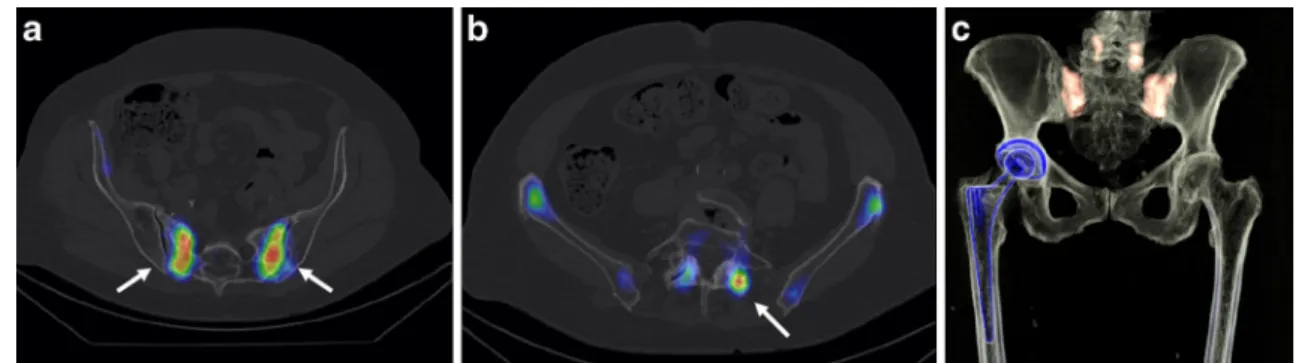
Fig. 1 SPECT/CT imaging in a 77-year-old patient with suspicion of iliosacral joint arthritis 3 years after right total hip joint arthroplasty (a, b axial and c volume-rendered images). Tracer uptake is seen alongside
bilateral longitudinal sacral fractures and focal uptake due to bilateral hypertrophic spondylarthrosis of L4/5. The hip implant does not show increased signal uptake
Fig. 2 SPECT/CT imaging in a 61-year-old patient with bilateral
iliosacral joint arthropathy. a–c Follow-up imaging 7 months after
per-cutaneous minimally invasive iliosacral S1 and S2 joint fusion with the iFuse Implant System (a paracoronal, b sagittal and c volume-rendered images). The iFuse System is fully incorporated without any residual signal uptake. There was residual pain due to activated erosive
osteochondrosis and bilateral facet L5/S1 joint arthritis (arrow). d, e
Follow-up imaging 4 months after decompressive L5/S1 laminectomy, L5/S1 discectomy, interbody fusion with capstone and dorsal
instrumen-tation and L4–S1 spondylodesis (d paracoronal and e volume-rendered
images). There is minimal postoperative reactive tracer uptake alongside the incorporated material
spinal deformities such as compression fractures in elderly patients. Since the tracer accumulates in the region of a fresh fracture, SPECT/CT can aid in distinguishing between fresh and old lesions. To assess fracture age it is necessary to acquire three scintigraphic phases normally as spot images (flow , blood-pool and late phases). Further, SPECT/CT of the suspected fracture region is performed to evaluate morpho-logical as well as metabolic changes. During the first 4 weeks after trauma the fracture shows diffuse tracer uptake during the flow and blood-pool phases. During the following 8 weeks the activity is more focused on the late phase images, while activity in flow and blood-pool phase images decreases. Usually, normalization in the flow and blood-pool phases can be observed after 6 months. A decrease in the late phase is visible after 3–18 months, and sometimes uptake persists for years.
As well as diagnostic benefits, this information is of value when choosing a suitable treatment. For example, many pa-tients with osteoporotic compression fractures of the spine may experience pain reduction following a stabilization pro-cedure such as percutaneous kyphoplasty of a fresh fracture [33–36]. In this context, it is crucial that the fracture is not older than 6 weeks, since a reduced therapeutic benefit has been demonstrated when operatively treating such a fracture beyond this time [37,38]. Information concerning the approx-imate age of the fracture may be obtained either using the short tau inversion recovery sequence (STIR) in MRI or by assessing tracer uptake on SPECT/CT. This makes estimation of the benefit of a planned surgical intervention possible [39,
40]. A clear advantage for SPECT/CT imaging compared to
conventional SPECT has been demonstrated as SPECT/CT has shown additional information in 62.5 % of patients [40]. Furthermore, SPECT/CT allows a precise differentiation be-tween malignant and benign compression fractures, which is of key importance in therapy and prognosis [41]. These two entities are usually distinguished using MRI [42].
Compression fractures result in lesions within the bone mar-row which lead to minor signal intensity changes on the T1-weighted sequences and a high signal intensity on the STIR sequence. This however may cause difficulties in distinguishing between malignant and benign processes [43]. Furthermore, MRI is not an option in patients suffering from claustrophobia or patients with a cardiac pacemaker, which is why other equivalent diagnostic methods are necessary. SPECT/CT may be a valuable alternative with proven equal sensitivity and specificity to MRI [41].
Femoral neck fractures
Early detection and treatment of femoral neck fractures is crucial because of their associated high morbidity and mortal-ity. A twofold increase in mortality has been reported when operative treatment of a femoral neck fracture is postponed by only 2 days [44]. Nondisplaced fractures are partly not seen on plain radiography and CT, as well as when there is an infrac-tion. Consecutively, the partial lack of weight-bearing may lead to a secondary dislocation mandating an operation at a later time with increased perioperative risk. Due to their high sensitivity and specificity, nuclear medicine technologies are a
Fig. 3 Imaging in a 79-year-old patient with osteoporotic compression
fractures of the spine. a–c CT (a), SPECT/CT volume-rendered (b) and
SPECT/CT (c ) images obtained 14 days after percutaneous balloon
kyphoplasty of Th5–7. Kyphoplasties of Th11, L3 and L4 are older than
1 year and do not show signal enhancement. The Th8 endplate is clearly
visible with corresponding tracer enhancement as a sign of increased
loading (arrow). Due to pain and a high risk of fracture of adjacent
segments kyphoplasty was carried out. d Follow-up plain radiograph 4 weeks after kyphoplasty of Th8 and vertebroplasty of Th9 and Th10
true alternative to MRI, especially in patients in whom MRI is not an option [15,25].
Stress fractures
As well as obvious acute traumatic fractures, repetitive stress in certain regions may lead to an imbalance between contin-uous bone resorption and reformation. These disrupted remodelling processes may result in a stress fracture. In addi-tion to the tibia and tarsal bones, further locaaddi-tions for such a fracture are the vertebral bodies and the pelvic region. The incidence of a spinal stress fracture is estimated at 6 % and most commonly occurs in the fourth or fifth lumbar vertebra [15]. Young athletic patients are frequently affected [45] and present with acute onset of pain. These fractures are missed even with CT in up to 15 % of patients [46], whereas such lesions are readily visible on SPECT/CT due to increased local bone uptake due to increased osteoblast activity [47]. SPECT/CT also reveals additional information concerning metabolic activity which in turn allows a prognosis regarding the healing process [48].
Insufficiency fractures
Insufficiency fractures can be considered a subgroup within stress fractures and are defined as osseous lesions resulting from minor trauma. The most common underlying reason is osteoporosis. Further risk factors include rheumatoid arthritis, fibrous dysplasia, osteomalacia, hyperparathyroidism and long-term corticosteroid administration. The axial skeleton is most commonly affected. Within the pelvic region, the pubic rami and the sacrum are predominantly involved [1]. Other locations include the supraacetabular region and fractures of the superomedial portion of the ilium [49]. H-formed fractures of the sacrum are quite common and are missed particularly often on conventional plain radiography. Apart from MRI, nuclear medicine techniques may be a helpful alternative in these cases [50–52]. Additionally, SPECT/CT may be helpful when ruling out malignancies. Furthermore, it has been esti-mated that more than half of all compression fractures of the spine are in fact insufficiency fractures [1]. As well as its ability to assess fracture location and age, SPECT/CT may also be helpful in diagnosing other painful degenerative or neoplastic diseases [53]. SPECT may also be useful when planning a specific treatment. A targeted injection into a single activated facet joint, for instance, may relieve pain.
Posttraumatic and postoperative indications
From a trauma or orthopaedic surgeon’s perspective, the assessment of potential implant loosening is of great value. This question is usually addressed by conventional plain
radiographs or CT scans. Despite recent developments regard-ing new sequence modalities, MRI—due to signal artefacts— often leads to inconclusive results [54]. Nuclear medicine tech-niques, especially SPECT, provide a solid alternative since the visualized metabolic activity is not altered by metal artefacts [55–57] (Table1).
Spine
Some patients report persistent pain following treatment of degenerative or traumatic afflictions of the spine and pelvis with consecutive iliosacral arthrodesis or dorsal spine instru-mentation and fusion. Studies have shown that up to 14 % of patients with spinal implants may require an additional oper-ation within 4 years due to persistent pain [58]. Because the results of such a reoperation are inferior to those of a primary operation, the indication must be well-founded [59]. Important questions to be answered by the chosen imaging technique include loosening of pedicle screws, instability of the instrumentation, non-union of spondylodesis, low-grade infection and finally evidence of epifusional and subfusional degeneration. With this knowledge, a well-founded judge-ment can be made as to whether a patient might profit from an additional operation or whether conservative treatment is the better choice. Furthermore, the type of operation can be better planned.
As mentioned above, in the postoperative setting following spinal instrumentation, MRI has a much lower diagnostic value in the presence of metal artefacts. When evaluating osseous consolidation or active degenerative changes, SPECT/CT again shows better sensitivity and specificity than CT (Fig.4) [60,61].
Radiological documentation of spinal fusion is usually possible after 6–9 months on conventional radiographs or CT scans. However, complete healing may be expected only after 2 years. With the help of nuclear medicine, suspected pseudoarthrosis can be visualized through increased tracer uptake as early as 1 year postoperatively [56,60]. This in turn may explain persistent pain and warrant additional surgical interventions. Usually a consistent decrease in tracer uptake is recorded beginning within the third postoperative month [61], with the total tracer accumulation being dependent on general bone metabolism.
As well as increased tracer uptake within the field of operation in pseudoarthrosis, implant loosening and infection may be detected (Fig.5). Adjacent segments may show in-creased uptake as well, as in the presence of epi- or subfusional degeneration with (erosive) osteochondrosis [61]. Whether there is also a signal increase in adjacent segments following percutaneous procedures such as kyphoplasty still remains undetermined (Fig.3). This would be of great value when estimating the risk of adjacent segment fractures
which can consistently be found in a substantial number of patients [62,63].
Osteonecrosis
Posttraumatic osteonecrosis of the femoral head may occur in up to 14 % of patients with impacted, nondisplaced femoral neck fractures and 50 % of patients following a displaced fracture [64]. SPECT/CT may be helpful in such cases as sensitivity is superior even to that of MRI [65].
Infection
Visible changes on CT or MRI due to infection are usually evident only during a late stage and are rather nonspecific in
nature. Furthermore, the presence of implant-associated arte-facts may lead to a substantial deterioration in image quality. Compared to other nuclear medicine techniques, SPECT/CT not only allows the detection of infection but also its exact location, and is thus of great value in implant-associated infections [66]. The sensitivity and specificity are dependent on the radiopharmaceutical agent used. For this purpose, dicarboxypropandiphosphonate (DPD) scintigraphy com-bined with radioactively marked leucocytes is considered the gold standard [66]. Antigranulocyte scintigraphy is more pop-ular in Europe and has been proven to be of equal value [67]. Depending on the body region, sensitivity and specificity may vary. This is why FDG PET/CT is currently still considered the standard for diagnosing infections of the spine, but SPECT/CT is superior to PET/CT or MRI for imaging an infection of an extremity [68,69]. It is also highly sensitive for detection of osteomyelitis and has a higher specificity than
Fig. 5 Imaging in a 46-year-old patient with persistent lower lumbar
pack pain 2 years after removal of dorsal Th11–L5 instrumentation
following a flexion-distraction type L1/2 injury and compression
frac-tures of L2–L4 (a SPECT/CT volume-rendered, b SPECT/CT coronal
and c axial CT images). Strong tracer accumulation can be seen around
the site of the L5 pedicle screws, on the left more than the right, with subtle signs of loosening around these pedicle screws. The other segments show signs of consolidation and allograft integration with nonpathological mild tracer accumulation
Fig. 4 Imaging in a 76-year-old patient 11 months after L4–S1 implant
removal and L1–L5 reinstrumentation and currently suffering from lower
lumbar pack pain (a coronal SPECTCT/CT, b axial L4/5 SPECT/CT and c L4/5 CT images). Nonpathological perioperative tracer uptake is seen
around the intervertebral spacers as well as within segment L1/2 with osteochondrosis. Strong focal tracer uptake is seen within the right facet joint L4/5, which shows no signs of consolidation
planar nuclear medicine techniques [69,70]. Since a detailed anatomical correlation is possible, it is also helpful in differ-entiating between osteomyelitis and soft tissue infections [71].
Conclusion
The combination of highly sensitive but nonspecific scintigra-phy and nonsensitive but highly specific CT can be very helpful in a trauma setting. Compared to planar scintigraphy, additional insight is possible as anatomical and pathomorphological find-ings are coupled with metabolic information. This is especially important in anatomically complex regions such as the pelvis and spine [23,24,26].
Conflicts of interest None.
References
1. Cooper C, Atkinson EJ, O’Fallon WM, Melton 3rd LJ. Incidence of
clinically diagnosed vertebral fractures: a population-based study in
Rochester, Minnesota, 1985-1989. J Bone Miner Res. 1992;7:221–7.
2. Cauley JA, Thompson DE, Ensrud KC, Scott JC, Black D. Risk of mortality following clinical fractures. Osteoporos Int. 2000;11:
556–61.
3. Hill RM, Robinson CM, Keating JF. Fractures of the pubic rami. Epidemiology and five-year survival. J Bone Joint Surg Br. 2001;83:
1141–4.
4. Koval KJ, Aharonoff GB, Schwartz MC, Alpert S, Cohen G, McShinawy A, et al. Pubic rami fracture: a benign pelvic injury? J
Orthop Trauma. 1997;11:7–9.
5. van Dijk WA, Poeze M, van Helden SH, Brink PR, Verbruggen JP. Ten-year mortality among hospitalised patients with fractures of the
pubic rami. Injury. 2009;41:411–4.
6. Gertzbein SD, Chenoweth DR. Occult injuries of the pelvic ring. Clin
Orthop Relat Res. 1977;(128):202–7.
7. Isler B, Ganz R. Classification of pelvic ring injuries. Injury. 1996;27
Suppl 1:S-A3–12.
8. Scheyerer MJ, Osterhoff G, Wehrle S, Wanner GA, Simmen HP, Werner CM. Detection of posterior pelvic injuries in fractures of the
pubic rami. Injury. 2012;43:1326–9.
9. Woltmann A, Buhren V. Shock trauma room management of spinal injuries in the framework of multiple trauma. A systematic review of
the literature. Unfallchirurg. 2004;107:911–8.
10. Huber-Wagner S, Lefering R, Qvick LM, Korner M, Kay MV, Pfeifer KJ, et al. Effect of whole-body CT during trauma resuscitation on survival: a retrospective, multicentre study. Lancet. 2009;373:
1455–61.
11. Hauser CJ, Visvikis G, Hinrichs C, Eber CD, Cho K, Lavery RF, et al. Prospective validation of computed tomographic screening of the
thoracolumbar spine in trauma. J Trauma. 2003;55:228–34,
discus-sion 34–5.
12. Rommens PM, Vanderschot PM, Broos PL. Conventional radiogra-phy and CT examination of pelvic ring fractures. A comparative
study of 90 patients. Unfallchirurg. 1992;95:387–92.
13. Groves AM, Bird N, Tabor I, Cheow HK, Balan KK. 16-Detector multislice CT-skeletal scintigraphy image co-registration. Nucl Med
Commun. 2004;25:1151–5.
14. Gregory PL, Batt ME, Kerslake RW, Scammell BE, Webb JF. The value of combining single photon emission computerised tomogra-phy and computerised tomogratomogra-phy in the investigation of
spondylolysis. Eur Spine J. 2004;13:503–9.
15. Van der Wall H, Fogelman I. Scintigraphy of benign bone disease.
Semin Musculoskelet Radiol. 2007;11:281–300.
16. Hirschmann MT, Iranpour F, Davda K, Rasch H, Hügli R, Friederich NF. Combined single-photon emission computerized tomography and conventional computerized tomography (SPECT/CT): clinical value for the knee surgeons? Knee Surg Sports Traumatol Arthrosc.
2010;18:341–5.
17. Hirschmann MT, Iranpour F, Konala P, Kerner A, Rasch H, Cobb JP, et al. A novel standardized algorithm for evaluating patients with painful total knee arthroplasty using combined single photon emis-sion tomography and conventional computerized tomography. Knee
Surg Sports Traumatol Arthrosc. 2010;18:939–44.
18. Hirschmann MT, Davda K, Rasch H, Arnold MP, Friederich NF. Clinical value of combined single photon emission computerized tomography and conventional computer tomography (SPECT/CT) in sports medicine. Sports Med Arthrosc. 2011;19:174–81. 19. Konala P, Iranpour F, Kerner A, Rasch H, Friederich NF, Hirschmann
MT. Clinical benefit of SPECT/CT for follow-up of surgical treat-ment of osteochondritis dissecans. Ann Nucl Med. 2010;24:621–4. 20. Welsch M, Welsch F, Grünwald F. Nuclear medicine techniques in
the diagnosis of orthopaedic diseases. Orthopade. 2006;35:632–40, 642–3.
21. Spitz J, Becker C, Tittel K, Weigand H. Clinical relevance of whole body skeletal scintigraphy in multiple injury and polytrauma patients. Unfallchirurgie. 1992;18:133–47.
22. Heinrich SD, Gallagher D, Harris M, Nadell JM. Undiagnosed frac-tures in severely injured children and young adults. Identification with technetium imaging. J Bone Joint Surg Am. 1994;76:561–72. 23. Love C, Din AS, Tomas MB, Kalapparambath TP, Palestro CJ.
Radionuclide bone imaging: an illustrative review. Radiographics. 2003;23:341–58.
24. Palestro CJ, Love C, Schneider R. The evolution of nuclear medicine and the musculoskeletal system. Radiol Clin N Am. 2009;47:505–32. 25. Holder LE, Schwarz C, Wernicke PG, Michael RH. Radionuclide bone imaging in the early detection of fractures of the proximal femur (hip): multifactorial analysis. Radiology. 1990;174:509–15. 26. Lee E, Worsley DF. Role of radionuclide imaging in the orthopedic
patient. Orthop Clin N Am. 2006;37:485–501.
27. Matin P. Bone scintigraphy in the diagnosis and management of
traumatic injury. Semin Nucl Med. 1983;13:104–22.
28. Breuil V, Roux CH, Testa J, Albert C, Chassang M, Brocq O, et al. Outcome of osteoporotic pelvic fractures: an underestimated severity.
Survey of 60 cases. Joint Bone Spine. 2008;75:585–8.
29. Zukotynski K, Curtis C, Grant FD, Micheli L, Treves ST. The value of SPECT in the detection of stress injury to the pars interarticularis in patients with low back pain. J Orthop Surg Res. 2010;5:13. 30. Sairyo K, Katoh S, Takata Y, Terai T, Yasui N, Goel VK, et al. MRI
signal changes of the pedicle as an indicator for early diagnosis of spondylolysis in children and adolescents: a clinical and
biomechan-ical study. Spine J. 2006;31:206–11.
31. Hession PR, Butt WP. Imaging of spondylolysis and spondylolisthesis.
Eur Radiol. 1996;6:284–90.
32. Standaert CJ, Herring SA. Expert opinion and controversies in sports and musculoskeletal medicine: the diagnosis and treatment of spondylolysis in adolescent athletes. Arch Phys Med Rehabil.
2007;88:537–40.
33. Cortet B, Cotten A, Boutry N, Flipo RM, Duquesnoy B, Chastanet P, et al. Percutaneous vertebroplasty in the treatment of osteoporotic vertebral compression fractures: an open prospective study. J
Rheumatol. 1999;26:2222–8.
34. Amar AP, Larsen DW, Esnaashari N, Albuquerque FC, Lavine SD, Teitelbaum GP. Percutaneous transpedicular polymethylmethacrylate
vertebroplasty for the treatment of spinal compression fractures.
Neurosurgery. 2001;49:1105–14, discussion 14–5.
35. Barr JD, Barr MS, Lemley TJ, McCann RM. Percutaneous vertebroplasty for pain relief and spinal stabilization. Spine J.
2000;25:923–8.
36. Werner CM, Osterhoff G, Schlickeiser J, Jenni R, Wanner GA, Ossendorf C, et al. Vertebral body stenting versus kyphoplasty for the treatment of osteoporotic vertebral compression fractures: a
ran-domized trial. J Bone Joint Surg Am. 2013;95:577–84.
37. Buchbinder R, Osborne RH, Ebeling PR, Wark JD, Mitchell P, Wriedt C, et al. A randomized trial of vertebroplasty for painful
osteoporotic vertebral fractures. N Engl J Med. 2009;361:557–68.
38. Kallmes DF, Comstock BA, Heagerty PJ, Turner JA, Wilson DJ, Diamond TH, et al. A randomized trial of vertebroplasty for
osteo-porotic spinal fractures. N Engl J Med. 2009;361:569–79.
39. Maynard AS, Jensen ME, Schweickert PA, Marx WF, Short JG, Kallmes DF. Value of bone scan imaging in predicting pain relief from percutaneous vertebroplasty in osteoporotic vertebral fractures.
AJNR Am J Neuroradiol. 2000;21:1807–12.
40. Sola M, Perez R, Cuadras P, Diaz R, Holgado S, Puyalto P, et al. Value of bone SPECT-CT to predict chronic pain relief after percutaneous
vertebroplasty in vertebral fractures. Spine J. 2011;11:1102–7.
41. Tokuda O, Harada Y, Ueda T, Ohishi Y, Matsunaga N. Malignant versus benign vertebral compression fractures: can we use bone SPECT as a substitute for MR imaging? Nucl Med Commun. 2011;32:192–8.
42. Chan JH, Peh WC, Tsui EY, Chau LF, Cheung KK, Chan KB, et al. Acute vertebral body compression fractures: discrimination between benign and malignant causes using apparent diffusion coefficients. Br J Radiol. 2002;75:207–14.
43. Uetani M, Hashmi R, Hayashi K. Malignant and benign compression fractures: differentiation and diagnostic pitfalls on MRI. Clin Radiol. 2004;59:124–31.
44. Zuckerman JD, Skovron ML, Koval KJ, Aharonoff G, Frankel VH. Postoperative complications and mortality associated with operative delay in older patients who have a fracture of the hip. J Bone Joint Surg Am. 1995;77:1551–6.
45. Kainberger F, Weidekamm C, Matzner M, Trieb K. Sports injury of the spine: imaging diagnosis. Rontgenpraxis. 2006;56:47–57. 46. Congeni J, McCulloch J, Swanson K. Lumbar spondylolysis. A study
of natural progression in athletes. Am J Sports Med. 1997;25:248–53.
47. Roub LW, Gumerman LW, Hanley Jr EN, Clark MW, Goodman M, Herbert DL. Bone stress: a radionuclide imaging perspective.
Radiology. 1979;132:431–8.
48. Ryan PJ, Fogelman I. The role of nuclear medicine in orthopaedics.
Nucl Med Commun. 1994;15:341–60.
49. Peh WC, Khong PL, Yin Y, Ho WY, Evans NS, Gilula LA, et al. Imaging of pelvic insufficiency fractures. Radiographics. 1996;16:
335–48.
50. Ciullo JV, Jackson DW. Pars interarticularis stress reaction, spondylolysis, and spondylolisthesis in gymnasts. Clin Sports Med.
1985;4:95–110.
51. Anderson K, Sarwark JF, Conway JJ, Logue ES, Schafer MF. Quantitative assessment with SPECT imaging of stress injuries of the pars interarticularis and response to bracing. J Pediatr Orthop.
2000;20:28–33.
52. Traughber PD, Havlina Jr JM. Bilateral pedicle stress fractures:
SPECT and CT features. J Comput Assist Tomogr. 1991;15:338–40.
53. Cook GJ, Hannaford E, See M, Clarke SE, Fogelman I. The value of bone scintigraphy in the evaluation of osteoporotic patients with back
pain. Scand J Rheumatol. 2002;31:245–8.
54. Rutherford EE, Tarplett LJ, Davies EM, Harley JM, King LJ. Lumbar spine fusion and stabilization: hardware, techniques, and imaging
appearances. Radiographics. 2007;27:1737–49.
55. Lusins JO, Danielski EF, Goldsmith SJ. Bone SPECT in patients with persistent back pain after lumbar spine surgery. J Nucl Med. 1989;30:
490–6.
56. Gates GF, McDonald RJ. Bone SPECT of the back after lumbar
surgery. Clin Nucl Med. 1999;24:395–403.
57. Larsen JM, Rimoldi RL, Capen DA, Nelson RW, Nagelberg S, Thomas Jr JC. Assessment of pseudarthrosis in pedicle screw fusion: a prospective study comparing plain radiographs, flexion/extension radiographs, CT scanning, and bone scintigraphy with operative
findings. J Spinal Disord. 1996;9:117–20.
58. Martin BI, Mirza SK, Comstock BA, Gray DT, Kreuter W, Deyo RA. Are lumbar spine reoperation rates falling with greater use of fusion
surgery and new surgical technology? Spine J. 2007;32:2119–26.
59. Waddell G, Kummel EG, Lotto WN, Graham JD, Hall H, McCulloch JA. Failed lumbar disc surgery and repeat surgery following
indus-trial injuries. J Bone Joint Surg Am. 1979;61:201–7.
60. Damgaard M, Nimb L, Madsen JL. The role of bone SPECT/CT in the evaluation of lumbar spinal fusion with metallic fixation devices.
Clin Nucl Med. 2010;35:234–6.
61. Rager O, Schaller K, Payer M, Tchernin D, Ratib O, Tessitore E. SPECT/CT in differentiation of pseudarthrosis from other causes of back pain in lumbar spinal fusion: report on 10 consecutive cases. Clin Nucl Med. 2012;37:339–43.
62. Uppin AA, Hirsch JA, Centenera LV, Pfiefer BA, Pazianos AG, Choi IS. Occurrence of new vertebral body fracture after percutaneous vertebroplasty in patients with osteoporosis. Radiology. 2003;226: 119–24.
63. Kim SH, Kang HS, Choi JA, Ahn JM. Risk factors of new compres-sion fractures in adjacent vertebrae after percutaneous vertebroplasty. Acta Radiol. 2004;45:440–5.
64. Calandruccio RA, Anderson WE 3rd. Post-fracture avascular necro-sis of the femoral head: correlation of experimental and clinical studies. Clin Orthop Relat Res. 1980;(152):49–84.
65. Ryu JS, Kim JS, Moon DH, Kim SM, Shin MJ, Chang JS, et al. Bone SPECT is more sensitive than MRI in the detection of early osteonecrosis of the femoral head after renal transplantation. J Nucl Med. 2002;43:1006–11.
66. Navalkissoor S, Nowosinska E, Gnanasegaran G, Buscombe JR. Single-photon emission computed tomography-computed
tomogra-phy in imaging infection. Nucl Med Commun. 2013;34:283–90.
67. Richter WS, Ivancevic V, Meller J, Lang O, Le Guludec D, Szilvazi I, et al. 99mTc-besilesomab (Scintimun) in peripheral osteomyelitis: comparison with 99mTc-labelled white blood cells. Eur J Nucl Med
Mol Imaging. 2011;38:899–910.
68. Stumpe KD, Zanetti M, Weishaupt D, Hodler J, Boos N, Von Schulthess GK. FDG positron emission tomography for differentia-tion of degenerative and infectious endplate abnormalities in the lumbar spine detected on MR imaging. Am J Roentgenol. 2002;179:
1151–7.
69. Linke R, Kuwert T, Uder M, Forst R, Wuest W. Skeletal SPECT/CT
of the peripheral extremities. Am J Roentgenol. 2009;194:W329–35.
70. Horger M, Eschmann SM, Pfannenberg C, Storek D, Vonthein R, Claussen CD, et al. Added value of SPECT/CT in patients suspected of having bone infection: preliminary results. Arch Orthop Trauma
Surg. 2007;127:211–21.
71. Bar-Shalom R, Yefremov N, Guralnik L, Keidar Z, Engel A, Nitecki S, et al. SPECT/CT using 67Ga and 111In-labeled leukocyte