HAL Id: hal-01785438
https://hal.archives-ouvertes.fr/hal-01785438
Submitted on 15 May 2018HAL is a multi-disciplinary open access archive for the deposit and dissemination of sci-entific research documents, whether they are pub-lished or not. The documents may come from teaching and research institutions in France or abroad, or from public or private research centers.
L’archive ouverte pluridisciplinaire HAL, est destinée au dépôt et à la diffusion de documents scientifiques de niveau recherche, publiés ou non, émanant des établissements d’enseignement et de recherche français ou étrangers, des laboratoires publics ou privés.
Linking E-cadherin mechanotransduction to cell
metabolism through force-mediated activation
of AMPK
Jennifer Bays, Hannah Campbell, Christy Heidema, Michael Sebbagh, Kris
Demali
To cite this version:
Jennifer Bays, Hannah Campbell, Christy Heidema, Michael Sebbagh, Kris Demali. Linking E-cadherin mechanotransduction to cell metabolism through force-mediated activation of AMPK. Na-ture Cell Biology, NaNa-ture Publishing Group, 2017, 19 (6), pp.724 - 731. �10.1038/ncb3537�. �hal-01785438�
1 1
Linking E-cadherin mechanotransduction to cell metabolism through
2force mediated activation of AMPK
3Jennifer L. Bays1, Hannah K. Campbell1#, Christy Heidema2#, Michael Sebbagh3, and
4
Kris A. DeMali1,2*
5
1Department of Biochemistry and the 2Interdisciplinary Graduate Program in Molecular and Cellular
6
Biology, Roy J. and Lucille A. Carver College of Medicine, University of Iowa, Iowa City, IA 52242. 7
3Centre de Recherche en Cancérologie de Marseille, Aix Marseille Univ UM105, Inst Paoli Calmettes,
8
UMR7258 CNRS, U1068 INSERM, Cell Polarity, Cell signalling and Cancer - Equipe labellisée Ligue 9
Contre le Cancer, Marseille, France. 10
#These authors contributed equally to this work. 11
*Corresponding Author: Kris DeMali, Department of Biochemistry, University of Iowa, Iowa City, IA 52242, 12
Tel:319-335-7882, Email: [email protected] 13
14
The response of cells to mechanical force is a major determinant of cell
15
behavior and is an energetically costly event. How cells derive energy to resist
16
mechanical force is unknown. Here, we show that application of force to
E-17
cadherin stimulates Liver Kinase B1 (LKB1) to activate AMP-activated protein
18
kinase (AMPK), a master regulator of energy homeostasis. LKB1 recruits AMPK
19
to the E-cadherin mechanotransduction complex, thereby stimulating actomyosin
20
contractility, glucose uptake, and ATP production. The increase in ATP provides
21
energy to reinforce the adhesion complex and actin cytoskeleton so the cell can
22
resist physiological forces. Together, these findings reveal a paradigm for how
23
mechanotransduction and metabolism are linked and provide a framework for
24
understanding how diseases involving contractile and metabolic disturbances
25
arise.
26 27
In response to externally applied forces, cell surface adhesion receptors trigger
28
robust actin cytoskeletal rearrangements and growth of the associated adhesion
29
complex1-3. These changes are energetically costly, requiring approximately 50% of the
30
total ATP in a cell4, 5. Energy homeostasis is controlled by AMP-activated protein kinase
31
(AMPK). Based on this rationale, we tested whether application of force on E-cadherin
2
increased AMPK activity. For this, a well-established approach to directly apply force to
33
cadherins was employed6-12. Magnetic beads were coated with E-cadherin extracellular 34
domains (or IgG as a control) and permitted to adhere to MCF10A epithelial cells. A
35
constant force was then applied for 5 minutes using a permanent ceramic magnet.
36
Following application of force, AMPK was immunoprecipitated and subjected to an in
37
vitro kinase assay with a fusion protein of GST and a SAMS peptide (an AMPK-specific
38
substrate)13. Application of force increased phosphorylation of the SAMS peptide by
4.9-39
fold; a control peptide (SAMA) lacking the second serine phosphorylation site was not
40
phosphorylated (Fig. 1a). Importantly, the peptide phosphorylation was blocked by
41
application of Compound C (a cell permeable AMPK specific inhibitor)14.
42 43
As additional measures of AMPK activation, we examined phosphorylation of
44
AMPK in its activation loop and phosphorylation of the AMPK substrate, acetyl CoA
45
carboxylase. Force increased phosphorylation of AMPK in its activation loop in MCF10A
46
(pAMPK, Fig. 1b) and MDCK (Fig. S1a) cells. The increases in activation loop
47
phosphorylation were blocked when AMPK was inhibited using shRNAs (Fig. 1b) or
48
Compound C (Fig. S1a-c). Phosphorylation of acetyl CoA carboxylase was also
49
elevated (Fig. S1c). Hence by three independent measures, force stimulated AMPK
50
activation.
51 52
To ensure AMPK activation was independent of the method of force application
53
shear stress was applied to MDCK cells using a parallel plate chamber. Alternatively,
54
junctional assembly was triggered using a calcium switch assay—a process that relies
55
on elevations in actin polymerization and myosin II activity15, 16. Both shear stress and 56
junctional assembly stimulated AMPK activation loop phosphorylation (Fig. 1c,S1d).
57 58
To interrogate the contribution of E-cadherin to force-induced AMPK activation,
59
we examined the effects of inhibiting E-cadherin function using a function blocking
60
antibody (HECD-1) or by silencing E-cadherin expression (Fig. 1d, S1e). E-cadherin
61
was required to trigger AMPK activation (Fig. 1d, S1e). Additionally, application of force
62
to another transmembrane adhesion receptor, syndecan-1, failed to enhance AMPK
3
phosphorylation (Fig. S1f). Taken together, these data demonstrate that force on
E-64
cadherin stimulates AMPK activation.
65 66
To investigate the contribution of force to AMPK activation, we examined the
67
effect of promoting and interfering with known mechanically controlled elements. To
68
promote force transmission increases in contractility were stimulated by applying
69
Calyculin A, a phosphatase inhibitor that augments myosin II phosphorylation.
70
Stimulating myosin light chain phosphorylation increased AMPK activation (Fig. 1e). To
71
interfere with force transmission, cells were treated with blebbistatin, a myosin II
72
inhibitor. In the presence of blebbistatin, myosin light chain phosphorylation and
force-73
induced AMPK activation were decreased (Fig. 1d).
74 75
Since activate AMPK localizes to the plasma membrane17 and E-cadherin is
76
membrane-bound18, 19, we examined whether force stimulated AMPK recruitment to the
77
cadherin adhesion complex. To address this possibility, we applied tensile forces to
E-78
cadherin and the level of co-precipitating AMPK and activated AMPK were assessed.
79
AMPK (Fig 1f) and active AMPK (Fig 1g) were recovered with E-cadherin complexes.
80
The recruitment of AMPK to E-cadherin was blocked by preincubation of the cells with
81
blebbistatin (Fig 1f and g), with E-cadherin function blocking antibodies (Fig 1f and g), or
82
by silencing AMPK expression (Fig S1g). Additionally, the recruitment of AMPK to
E-83
cadherin was not dependent on the method of force application as stimulating junctional
84
assembly using a calcium switch assay triggered AMPK co-immunoprecipitation with
E-85
cadherin (Fig. S1h). Taken together, these studies demonstrate AMPK is recruited to
E-86
cadherin in response to force.
87 88
How is AMPK activated and recruited to the cadherin complex? Previous studies
89
from the Schwartz laboratory indicated that LKB1, an AMPK activator, localizes to
90
cadherin-containing complexes in maturing junctions17. Additionally work from Cantley
91
laboratory found that calcium-induced tight junction assembly depends on LKB120.
92
Based on this rationale, we determined if LKB1 associates with the cadherin adhesion
93
complex in response to force. LKB1 was recovered with E-cadherin-coated magnetic
4
beads in a force- and E-cadherin-dependent manner (Fig 2a). Since the buffer
95
conditions used to examine protein recruitment beneath the magnetic beads were less
96
strigent than convention immunoprecipitation studies, we examined if LKB1
co-97
immunoprecipitated with E-cadherin. Robust co-immunoprecipitation of E-cadherin was
98
observed LKB1 immunoprecipitates recovered from cells lysed in a 1%-triton
x100-99
containing buffer, thereby confirming the interaction (Fig 2b). In further support of LKB1,
100
we determined if LKB1 co-localized with E-cadherin in cells. Since the magnetic beads
101
we use in these studies autofluorescence, we examined co-localization in response to
102
application of shear stress to MDCK cells. We observed strong co-localization of LKB1
103
and E-cadherin (Fig 2c).
104 105
Having identified a mechanically-active signaling pathway from E-cadherin to
106
LKB1, we next determined if LKB1 is required for AMPK activation and recruitment to
107
the cadherin adhesion complex. Tensile forces were applied to E-cadherin on MCF10A
108
(Fig 2d) or on MDCKII (Fig 2e) cells. We found the force-induced activation of AMPK
109
was prevented by LKB1 silencing in both cell lines (Fig 2d-e). Similarly, shear
stress-110
induced activation of AMPK was blocked by inhibition of LKB1 (Fig 2f). We next
111
determined whether LKB1 was required for the recruitment of AMPK to E-cadherin.
112
LKB1 inhibition prevented co-precipitation of AMPK and active AMPK with E-cadherin
113
coated magnetic beads (Fig 2g). Taken together, this data demonstrates that LKB1 is
114
needed for AMPK to be recruited to and activated at cadherin-containing sites.
115 116
The observation that LKB1 and AMPK are recruited to the cadherin adhesion
117
complex suggests they may lie in a known contractility pathway. This pathway initiates
118
when E-cadherin activates Abelson tyrosine kinase (Abl) thereby triggering
119
phosphorylation of Y822 vinculin (Fig. 3a) and culminating in elevated RhoA-mediated
120
contractility11. To determine whether LKB1 and/or AMPK are components of this
121
pathway, we examined the effect of their inhibition. As an indicator of Abl activation, we
122
followed phosphorylation the Abl substrate, CrkL, using a phosphospecific antibody
123
against the Abl-specific sites21. Application of tensile forces using the magnetic bead
124
approach stimulated CrkL phosphorylation in MCF10A (Fig 3b and c) and MDCKII cells
5
(Fig. S2a). Inhibition of LKB1 (Fig. 3b, S2a) or AMPK (Fig. 3c, S2b) prevented this
126
increase. Stimulation of vinculin Y822 phosphorylation, a downstream target of Abl, also
127
required LKB1 (Fig. 3d) and AMPK (Fig 3e, S2c). These data indicate that AMPK lies
128
upstream of Abl in the known contractility pathway. Since Abl is activated and AMPK
129
and vinculin are recruited to cadherin-containing junctions in response to force22, we
130
examined whether AMPK is in a complex with these components. For this, we
131
monitored co-immunoprecipitation of vinculin and Abl with AMPK from cells lysed in
132
RIPA buffer. These studies revealed that AMPK forms a complex with Abl and vinculin
133
in a force-dependent manner (Fig. 3f).
134 135
Further downstream in the E-cadherin contractility pathway (Fig. 3a),
RhoA-136
mediates activation of Rho kinase, which promotes phosphorylation of myosin light
137
chain (MLC), thereby stimulating actomyosin contractility23. We measured force-induced
138
RhoA activity in cells in the presence or absence of LKB1 or AMPK. Application of
139
tensile force to E-cadherin stimulated RhoA activation in an LKB1- and
AMPK-140
dependent manner (Fig. 3g). To determine if increases in RhoA were propagated to
141
changes in contractility, we analyzed MLC phosphorylation at a regulatory Ser19 site24.
142
MLC phosphorylation increased 2.7-fold in response to force (Fig 3h). Inhibition of
143
AMPK or LKB1 abrogated these effects (Fig. 3g, 3h, S2d, and S2e). Taken together,
144
these findings demonstrate that AMPK is required to increase RhoA-mediated
145
contractility when E-cadherin experiences force.
146 147
These observations raise the question why cells activate a master regulator of
148
metabolism, such as AMPK, to modulate contractility. When cells experience force,
149
elevations in enzymatic activity, actin polymerization, and actomyosin contractility
150
facilitate the cytoskeletal rearrangements and the growth of adhesions necessary to
151
withstand the force2, 25-28. All of these processes require energy. The preferred energy
152
source for epithelial cells is ATP derived from glucose oxidation29. In other systems,
153
AMPK activation stimulates glucose uptake and oxidation30. Based on this rationale, we
154
hypothesized a consequence of force-induced AMPK is the stimulation of glucose
155
uptake. To test this possibility, tensile forces were applied to E-cadherin and the uptake
6
of a fluorescently labelled, non-hydrolyzable 2-deoxyglucose was monitored. Force
157
stimulated a 2.2-fold increase in glucose uptake in the MCF10A cells (Fig. 4a, Fig. S3a)
158
and a 2.6-fold increase in the MDCK II cells (Fig. 4b, Fig. S3b). Moreover, inhibition of
159
E-cadherin, LKB1, or AMPK prevented the force-induced glucose uptake (Fig. 4a-b, Fig.
160
S3a-b). To ensure that these results were not the consequence of the approach, the
161
effects of shear stress on MDCKII cells or the effects of stimulating junctional assembly
162
(using a calcium switch assay) were monitored. Both methods stimulated an elevation in
163
glucose uptake. The fold activation was similar to the increase observed when tensile
164
forces applied (Fig. 4c, S3c, S3d) and required E-cadherin, LKB1 and AMPK.
165
Collectively, these data demonstrate that force on E-cadherin stimulates glucose uptake
166
in a LKB1- and AMPK-dependent manner.
167 168
In response to many stimuli, glucose is oxidized to ATP. Hence, we tested
169
whether increases in glucose uptake translate to elevations in ATP. Application of force
170
to E-cadherin increased cellular ATP levels by 1.5-fold (Fig. 4d and S3e). The increase
171
in ATP, while slight, was statistically significant and reproducible (Fig. S3e). This
172
modest change is not surprising as ATP levels remain relatively constant, even in the
173
most metabolically active tissues31. Importantly, we found force-induced ATP could be
174
blocked by the shRNAs against LKB1 or AMPK (Fig 4d), the AMPK inhibitor Compound
175
C (Fig. 4e, Fig S3f), or the ATP synthase inhibitor Oligomycin A (Fig. 4e, Fig S3f). To
176
ensure that the ATP produced was derived from glucose, the effect of a
non-177
hydrolyzable, 2-deoxyglucose analog was studied. The 2-deoxyglucose analog blocked
178
force-induced elevations in ATP (Fig. 4e). Taken together, these data indicate that the
179
glucose taken up when E-cadherin experiences force is converted to ATP.
180
181
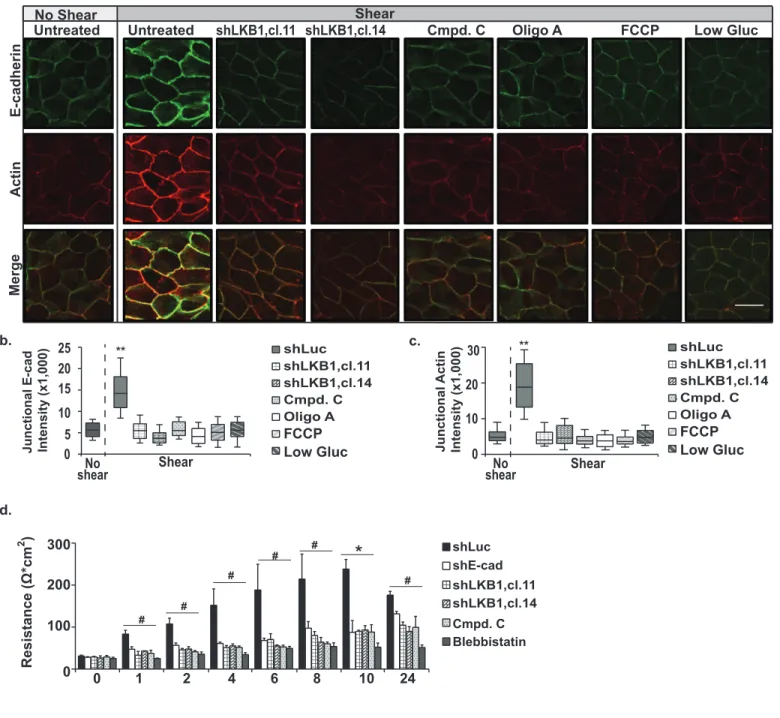
We next tested the possibility that ATP provides the energy necessary to
182
reinforce the actin cytoskeleton and cadherin adhesion complex in response to force. In
183
support of a role for AMPK, we found that A-769662, an AMPK activator, increased
E-184
cadherin and F-actin enrichment in cell-cell junctions (Fig. S4a). To directly test the role
185
for LKB1 and AMPK, we applied shear stress to MCDK cells and monitored F-actin and
186
E-cadherin enrichment in cell-cell junctions using immunofluorescence. A 2.0-fold
7
increase E-cadherin deposition in cell-cell junctions (Fig. 5a and b) and a 3.8-fold
188
increase in junctional actin were observed in cells exposed to shear (Fig 5a and c).
189
These increases were blocked by shRNAs against LKB1 or inhibitors of AMPK (Fig
5a-190
c). Similarly, inhibiting glucose metabolism (by incubating cells in low glucose containing
191
media) or blocking ATP synthesis (using Oligomycin A or Carbonyl
cyanide-4-192
(trifluoromethoxy)phenylhydrazone) dramatically impaired junctional enrichment of
F-193
actin and E-cadherin in shear stress treated cells (Fig. 5a-c). Only modest changes
194
were observed in control cells (Fig. S4b-d).
195 196
To ensure that shear stress applied force to cell-cell junctions, we investigated
197
whether myosin light chain was phosphorylated in response to shear and whether
198
vinculin (an actin binding protein that bears force) was enriched in cell-cell junctions.
199
Both myosin light chain phosphorylation and vinculin localization to cell-cell contacts
200
were increased in response to shear stress (Fig. S4e-h). In further support of a role for
201
force, we found that the enrichment of vinculin in cell-cell junctions was blocked by
202
preincubation of cells with blebbistatin (Fig. S4e-g). This observation is in agreement
203
with previous findings showing vinculin is recruited to endothelial cell-cell junctions in a
204
tension-dependent manner32 and force-dependent vinculin recruitment can be blocked
205
by blebbistatin22. Collectively, these data demonstrate that AMPK-dependent increases
206
in ATP enrich F-actin and E-cadherin in response to force.
207 208
Previous studies show that tension is required for the formation of an epithelial
209
barrier33. To interrogate the physiological significance of the pathway uncovered in this 210
study, the formation of an epithelial barrier was monitored in MDCKII cells after a
211
calcium switch. After readdition of calcium to the medium, control cells gradually formed
212
an epithelial barrier (Fig. 5d). Inhibition of E-cadherin, LKB1, or AMPK or Blebbistatin
213
compromised formation of the barrier function. By 24 h after junctional assembly was
214
initiated, the cells with E-cadherin, LKB1 or AMPK inhibited had only modest (but
215
statistically significant) alterations in their barriers (Fig. 5d). Interestingly, a slight
216
alteration in barrier function at 24h after calcium readdition was observed in the MCDKII
217
cells lacking E-cadherin. The requirement for AMPK is in good agreement with previous
8
studies showing that treatment of epithelial cells with AMPK activators promoted barrier
219
function34. Taken together, these data indicate force-induced activation of LKB1/AMPK 220
is required for efficient formation of an epithelial barrier.
221 222
Cell differentiation, proliferation, gene expression and disease development are
223
all impacted by the forces experienced by the cell35-38. A vast literature shows that cells
224
withstand forces by reinforcing their actin cytoskeletons and growing their adhesion
225
complexes1-3, 26. These events increase enzymatic activity, actin polymerization, and
226
actomyosin contractility26-28, yet it is unknown how the cell derives the vast amount of
227
energy it needs to support these events. Here, we demonstrate that LKB1-mediated
228
activation of AMPK is a key player in a junctional contractility pathway that increases
229
glucose uptake and ATP synthesis. This is a mechanism for how cells signal to increase
230
energy and use the energy to reinforce their cytoskeletal networks in order to resist
231
applied forces.
232 233
This work establishes AMPK as a critical link between mechanotransduction and
234
metabolism. This information may serve as the impetus for future studies aimed at
235
establishing further links between mechanotransduction and the metabolic machinery
236
and defining mechanisms of regulation. This work also has the potential to have far
237
reaching medical implications as AMPK is a negative regulator of diseased states with
238
metabolic disturbances. Our observation that E-cadherin force transmission activates
239
AMPK demonstrates that mechanical forces on E-cadherin may protect against the
240
metabolic disturbances associated with diseases such as cardiovascular disease39, 241
diabetes40, and cancer38. 242
243
Affiliations: Department of Biochemistry, Roy J. and Lucille A. Carver College of
244
Medicine, University of Iowa, Iowa City, IA, 52242.
245
Contributions: J.B. designed and performed experiments, analyzed all the data, and
246
helped write the manuscript. C.H. and H.C. helped with experimental design and
247
procedures. K.D. helped with the experimental design, wrote the manuscript, and
248
directed the project. All authors provided detailed comments.
9
Acknowledgements: We thank Thomas Moninger and Todd Washington at the
250
University of Iowa for their assistance in performing experiments. Research reported in 251
this publication was supported by The National Institutes of General Medicine (Award Number 252
R01GM112805 to K.A.D) and the National Cancer Institute of the National Institutes of Health 253
(Award Number P30CA086862). Predoctoral fellowships from the American Heart Association 254
(Award Number AHA 16PRE26701111) and National Institutes of Health (Award T32 255
GM067795) support J.B. and C.H., respectively. M.S. supported by “Fondation ARC pour la 256
Recherche sur le Cancer” (ARC SFI20111203781) and CNRS-AMI Mecanobio 257 (Mecanopol_2016-2017). 258 259 Figure Legends. 260
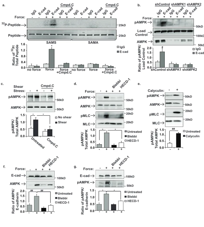
Figure 1. AMPK is activated in response to force applied to E-cadherin. a and b, MCF10A 261
cells were incubated with magnetic beads coated with IgG or E-cadherin extracellular domains 262
(E-cad). The cells were left resting(-) or a magnet was used to generate tensional forces (+). a, 263
AMPK immunoprecipitates were subjected to in vitro kinase assay with its substrate, SAMS 264
peptide. SAMA=control peptide. Cmpd. C indicates cells pretreated with the AMPK inhibitor, 265
Compound C. b, total cell lysates were immunoblotted with antibodies that recognize AMPK or 266
AMPK phosphorylated in its activation loop (pAMPK). shControl indicates cells treated with 267
scrambled shRNAs. shAMPK1and shAMPK2 indicate cells infected with two separate shRNAs 268
targeting AMPK. c, shear stress was applied to MDCK cells, and AMPK and pAMPK were 269
monitored by immunoblotting. d, tensional forces (+) were applied to MCF10A cells pretreated 270
with blebbistatin (Blebbi) or E-cadherin function blocking antibodies (HECD-1). Total cell lysates 271
were probed with antibodies against pAMPK, AMPK, phospho-myosin light chain (pMLC), or 272
MLC. e, MCF10A cells were left resting (-) or treated (+) with Calyculin A (to stimulate myosin II-273
dependent increased contractility). Total cell lysates were immunoblotted as described in d. f 274
and g, Tensional forces were applied to MCF10A cells as described in a. The beads were 275
recovered and co-precipitation of AMPK (f) and pAMPK (g) with E-cadherin were examined by 276
immunoblotting. The graphs beneath the image show the average ± SEM for 3 independent 277
experiments. *, #, and ## indicate p-values of <0.01, 0.05 and 0.005, respectively. Unprocessed 278
scans of blots are shown in Supplementary Figure 5. 279
280
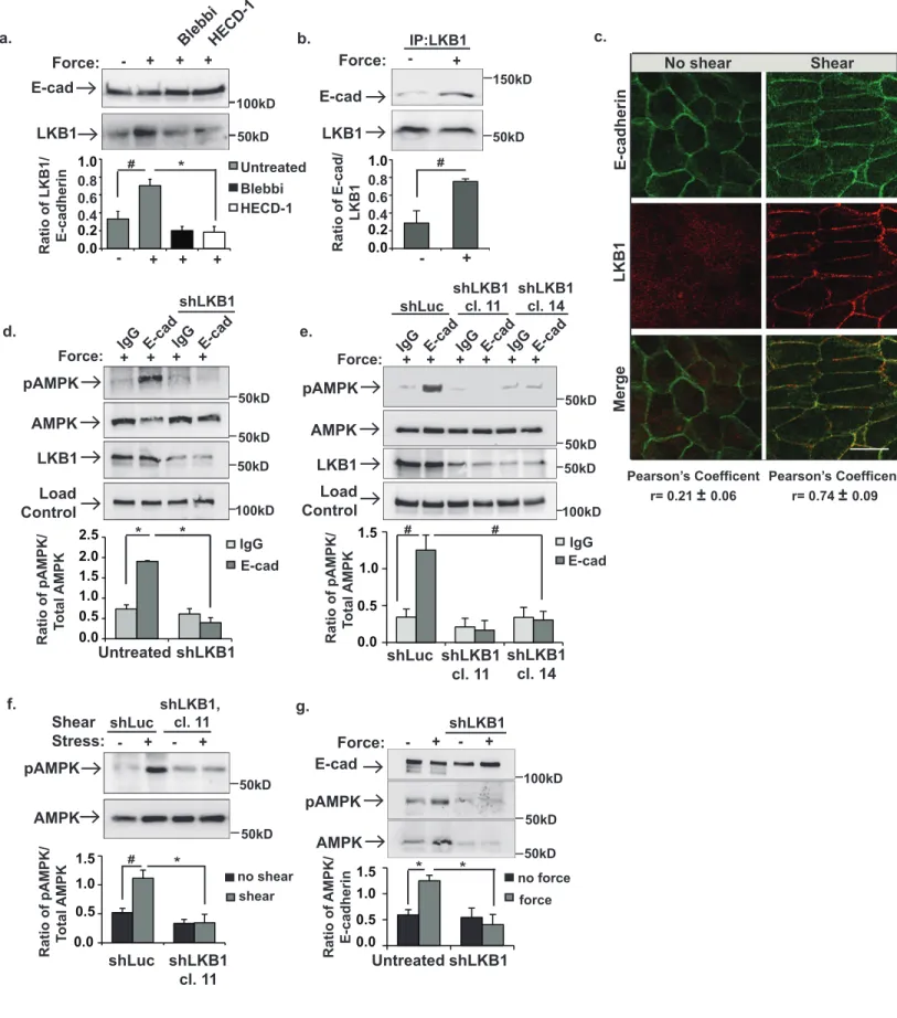
Figure 2. LKB1 is recruited to the cadherin adhesion complex in response to force and 281
activates and recruits AMPK. MCF10A cells (a,b,d,g) or MDCK cells (e) were incubated with 282
beads coated with IgG or E-cadherin extracellular domains (E-cad) and left resting (-) or 283
stimulated (+) with tensional force using a permanent magnet. In other experiments, MDCK 284
cells (c and f) were left resting (-) or exposed to shear stress (+). a, the cells were lysed and co-285
precipitation of LKB1 with the E-cadherin-coated magnetic beads was examined. b, Co-286
immunoprecipitation of E-cadherin (E-cad) with LKB1 was monitored using immunoblotting. c, 287
The cells were fixed, permeabilized and stained with antibodies against E-cadherin or LKB1. 288
The co-localization of LKB1 with E-cadherin was examined using confocal microscopy. Scale 289
10
bar = 20µm. d-f, The cells were lysed, and whole cell lysates were immunoblotted with the 290
indicated antibodies. shLKB1 denotes cells with LKB1 silenced. shLuc indicates cells 291
expressing a vector control cDNA, and cl.11 and cl.14 indicate two clonal cell lines lacking 292
LKB1. g, the cells were lysed, and pAMPK and AMPK co-purification with the E-cadherin-coated 293
magnetic beads was examined by immunoblotting. The graphs beneath each image show the 294
average ± SEM for 3 independent experiments.* and # indicate p-values of <0.01, and <0.05, 295
respectively. Unprocessed scans of blots are shown in Supplementary Figure 5. 296
297
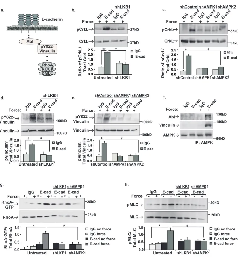
Figure 3. LKB1 and AMPK are upstream of Abl-mediated phosphorylation of Y822 298
vinculin and Rho-mediated contractility. a, schematic of the signal transduction cascade 299
from E-cadherin to Rho-mediated contractility. b-h, MCF10a cells were incubated with beads 300
coated with IgG or E-cadherin extracellular domains (E-cad) and left resting (-) or stimulated (+) 301
with tensional force using a permanent magnet. b,c,d,e, whole cell lysates were probed by 302
immunoblotting with antibodies that recognize phosphorylation of CrkL at the Abl-specific site (b 303
and c, pCrkL) or phosphorylation of vinculin Y822 (d and e, pY822). f, AMPK was 304
immunoprecipitated and vinculin and Abl recruitment were examined by immunoblotting. g, 305
Active Rho (Rho–GTP) was isolated with GST–RBD and analyzed by western blotting. h, total 306
cell lysates were immunoblotted with antibodies against myosin light chain (MLC) or MLC 307
phosphorylated at Serine 19 (pMLC). shLKB1 denotes cells expressing shRNAs against LKB1. 308
shControl indicates cells treated with scrambled shRNAs. shAMPK1and shAMPK2 indicate cells 309
infected with two separate shRNAs targeting AMPK. The graphs beneath each image show the 310
average ± SEM for 3 independent experiments. **, * and # indicate p-values of <0.001, <0.01, 311
and 0.05, respectively. Unprocessed scans of blots are shown in Supplementary Figure 5. 312
313
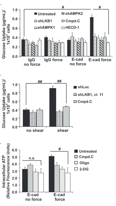
Figure 4. Force-induced AMPK stimulates glucose uptake and increases intracellular ATP 314
levels. a and b, MCF10A (a) or MDCK (b) cells were incubated paramagnetic beads coated 315
with IgG or E-cadherin extracellular domains (E-cad). Tensile forces were applied to the beads 316
using a magnet, the cells were lysed, and the amount of a fluorescently-labelled 2-deoxyglucose 317
analog taken up into the cells was monitored using a fluorimeter. c, MDCK cells were left resting 318
(no shear) or exposed to shear stress (shear), and the amount of glucose taken up into the cells 319
was monitored as described in a. d and e, Total ATP levels in cells treated as described in a 320
and b were monitored as described in the experimental procedures. Cmpd C indicates cells 321
treated with the AMPK inhibitor Compound C, Oligo indicates cells treated with the ATP 322
synthase inhibitor, Oligomycin A, and DG indicates cells incubated in the presence of 2-323
deoxyglucose. The graphs represent average glucose uptake or intracellular ATP for at least 324
three representative experiments ± SEM. # and ## indicate p-values of <0.05 and < 0.005, 325
respectively. n.s. indicates that there is no statistical differences between groups. 326
327
Figure 5. Force-induced increases in ATP reinforce the actin cytoskeleton and the E-328
cadherin adhesion complex to modulate barrier formation. a-c, MDCKII cells (n=80) or two 329
11
clonal MDCKII cells lines (cl.11 and cl.14, n=63 and 52 respectively) lacking LKB1 were left 330
untreated, treated with inhibitors of AMPK (Compound C=Cmpd. C, n=62) or ATP synthesis 331
(Oligo A,n=44 or Carbonyl cyanide-4-(trifluoromethoxy)phenylhydrazone=FCCP, n=26, or 332
incubated in low glucose containing media (Low Gluc, n=25). The cells were then left resting (no 333
shear) or exposed to physiological shear stress. The cells were fixed, stained with antibodies 334
against E-cadherin or Texas-Red phalloidin, and examined by confocal microscopy. The graphs 335
in b and c represent the average corrected fluorescence intensity of E-cadherin (b, E-cad) or F-336
actin (c) in junctions.The data are represented as a box and whisker plot with median, 10th, 25th, 337
75th, and 90th percentiles shown. Scale bars=20 μm. d, MDCKII cells were grown to confluence 338
and then incubated overnight in low calcium containing media. The formation of cell-cell 339
junctions was then stimulated by adding growth media to the cells. The trans-epithelial 340
resistance across the epithelial monolayer was monitored using a voltmeter at the indicated 341
times (hours). **,*, and # indicate p-values of <0.001, <0.01 and <0.05, respectively. 342
343 344
12 References
345 346
1. Borghi, N. et al. E-cadherin is under constitutive actomyosin-generated tension that is increased 347
at cell-cell contacts upon externally applied stretch. Proceedings of the National Academy of 348
Sciences of the United States of America 109, 12568-12573 (2012).
349
2. Liu, Z. et al. Mechanical tugging force regulates the size of cell-cell junctions. Proceedings of the 350
National Academy of Sciences of the United States of America 107, 9944-9949 (2010).
351
3. Chen, C.S., Tan, J. & Tien, J. Mechanotransduction at cell-matrix and cell-cell contacts. Annual 352
review of biomedical engineering 6, 275-302 (2004).
353
4. Bernstein, B.W. & Bamburg, J.R. Actin-ATP hydrolysis is a major energy drain for neurons. The 354
Journal of neuroscience : the official journal of the Society for Neuroscience 23, 1-6 (2003).
355
5. Daniel, J.L., Molish, I.R., Robkin, L. & Holmsen, H. Nucleotide exchange between cytosolic ATP 356
and F-actin-bound ADP may be a major energy-utilizing process in unstimulated platelets. 357
European journal of biochemistry / FEBS 156, 677-684 (1986).
358
6. Guilluy, C. et al. The Rho GEFs LARG and GEF-H1 regulate the mechanical response to force on 359
integrins. Nature cell biology 13, 722-727 (2011). 360
7. Marjoram, R.J., Guilluy, C. & Burridge, K. Using magnets and magnetic beads to dissect signaling 361
pathways activated by mechanical tension applied to cells. Methods 94, 19-26 (2016). 362
8. Barry, A.K. et al. alpha-catenin cytomechanics--role in cadherin-dependent adhesion and 363
mechanotransduction. J Cell Sci 127, 1779-1791 (2014). 364
9. Collins, C. et al. Localized tensional forces on PECAM-1 elicit a global mechanotransduction 365
response via the integrin-RhoA pathway. Current biology : CB 22, 2087-2094 (2012). 366
10. Kim, T.J. et al. Dynamic visualization of alpha-catenin reveals rapid, reversible conformation 367
switching between tension states. Current biology : CB 25, 218-224 (2015). 368
11. Bays, J.L. et al. Vinculin phosphorylation differentially regulates mechanotransduction at cell-cell 369
and cell-matrix adhesions. The Journal of cell biology 205, 251-263 (2014). 370
12. Tzima, E. et al. A mechanosensory complex that mediates the endothelial cell response to fluid 371
shear stress. Nature 437, 426-431 (2005). 372
13. Kishimoto, A., Ogura, T. & Esumi, H. A pull-down assay for 5' AMP-activated protein kinase 373
activity using the GST-fused protein. Molecular biotechnology 32, 17-21 (2006). 374
14. Zhou, G. et al. Role of AMP-activated protein kinase in mechanism of metformin action. The 375
Journal of clinical investigation 108, 1167-1174 (2001).
376
15. Walsh, S.V. et al. Rho kinase regulates tight junction function and is necessary for tight junction 377
assembly in polarized intestinal epithelia. Gastroenterology 121, 566-579 (2001). 378
16. Ivanov, A.I., Hunt, D., Utech, M., Nusrat, A. & Parkos, C.A. Differential roles for actin 379
polymerization and a myosin II motor in assembly of the epithelial apical junctional complex. 380
Molecular biology of the cell 16, 2636-2650 (2005).
381
17. Sebbagh, M., Santoni, M.J., Hall, B., Borg, J.P. & Schwartz, M.A. Regulation of LKB1/STRAD 382
localization and function by E-cadherin. Current biology : CB 19, 37-42 (2009). 383
18. Yoshida, C. & Takeichi, M. Teratocarcinoma cell adhesion: identification of a cell-surface protein 384
involved in calcium-dependent cell aggregation. Cell 28, 217-224 (1982). 385
19. Nagafuchi, A., Shirayoshi, Y., Okazaki, K., Yasuda, K. & Takeichi, M. Transformation of cell 386
adhesion properties by exogenously introduced E-cadherin cDNA. Nature 329, 341-343 (1987). 387
20. Zheng, B. & Cantley, L.C. Regulation of epithelial tight junction assembly and disassembly by 388
AMP-activated protein kinase. Proceedings of the National Academy of Sciences of the United 389
States of America 104, 819-822 (2007).
13
21. Zipfel, P.A., Zhang, W., Quiroz, M. & Pendergast, A.M. Requirement for Abl kinases in T cell 391
receptor signaling. Current biology : CB 14, 1222-1231 (2004). 392
22. le Duc, Q. et al. Vinculin potentiates E-cadherin mechanosensing and is recruited to actin-393
anchored sites within adherens junctions in a myosin II-dependent manner. The Journal of cell 394
biology 189, 1107-1115 (2010).
395
23. Chrzanowska-Wodnicka, M. & Burridge, K. Rho-stimulated contractility drives the formation of 396
stress fibers and focal adhesions. The Journal of cell biology 133, 1403-1415 (1996). 397
24. Amano, M. et al. Phosphorylation and activation of myosin by Rho-associated kinase (Rho-398
kinase). The Journal of biological chemistry 271, 20246-20249 (1996). 399
25. Puszkin, S. & Rubin, E. Adenosine diphosphate effect on contractility of human muscle 400
actomyosin: inhibition by ethanol and acetaldehyde. Science 188, 1319-1320 (1975). 401
26. Shewan, A.M. et al. Myosin 2 is a key Rho kinase target necessary for the local concentration of 402
E-cadherin at cell-cell contacts. Molecular biology of the cell 16, 4531-4542 (2005). 403
27. Mehta, D. & Gunst, S.J. Actin polymerization stimulated by contractile activation regulates force 404
development in canine tracheal smooth muscle. The Journal of physiology 519 Pt 3, 829-840 405
(1999). 406
28. Cipolla, M.J., Gokina, N.I. & Osol, G. Pressure-induced actin polymerization in vascular smooth 407
muscle as a mechanism underlying myogenic behavior. FASEB J 16, 72-76 (2002). 408
29. Nash, R.W., McKay, B.S. & Burke, J.M. The response of cultured human retinal pigment 409
epithelium to hypoxia: a comparison to other cell types. Investigative ophthalmology & visual 410
science 35, 2850-2856 (1994).
411
30. Kahn, B.B., Alquier, T., Carling, D. & Hardie, D.G. AMP-activated protein kinase: ancient energy 412
gauge provides clues to modern understanding of metabolism. Cell Metab 1, 15-25 (2005). 413
31. Balaban, R.S., Kantor, H.L., Katz, L.A. & Briggs, R.W. Relation between work and phosphate 414
metabolite in the in vivo paced mammalian heart. Science 232, 1121-1123 (1986). 415
32. Grashoff, C. et al. Measuring mechanical tension across vinculin reveals regulation of focal 416
adhesion dynamics. Nature 466, 263-266 (2010). 417
33. Kannan, N. & Tang, V.W. Synaptopodin couples epithelial contractility to alpha-actinin-4-418
dependent junction maturation. The Journal of cell biology 211, 407-434 (2015). 419
34. Zhang, L., Li, J., Young, L.H. & Caplan, M.J. AMP-activated protein kinase regulates the assembly 420
of epithelial tight junctions. Proceedings of the National Academy of Sciences of the United 421
States of America 103, 17272-17277 (2006).
422
35. Mammoto, T., Mammoto, A. & Ingber, D.E. Mechanobiology and developmental control. Annual 423
review of cell and developmental biology 29, 27-61 (2013).
424
36. Janmey, P.A., Wells, R.G., Assoian, R.K. & McCulloch, C.A. From tissue mechanics to transcription 425
factors. Differentiation; research in biological diversity 86, 112-120 (2013). 426
37. Klein, E.A. et al. Cell-cycle control by physiological matrix elasticity and in vivo tissue stiffening. 427
Current biology : CB 19, 1511-1518 (2009).
428
38. Levental, K.R. et al. Matrix crosslinking forces tumor progression by enhancing integrin signaling. 429
Cell 139, 891-906 (2009).
430
39. Gemayel, C. & Waters, D. Mechanical or metabolic treatment for coronary disease: synergistic, 431
not antagonistic, approaches. Cardiology in review 10, 182-187 (2002). 432
40. Rice, K.M. et al. Diabetes alters vascular mechanotransduction: pressure-induced regulation of 433
mitogen activated protein kinases in the rat inferior vena cava. Cardiovascular diabetology 5, 18 434
(2006). 435
13 1
Materials and Methods.
2 3
Cell lines. No cell lines used in this study were found in the database of commonly
4
misidentified cell lines maintained by the ICLAD and NCBI. MCF10A human breast
5
epithelial cells and MDCK II canine kidney epithelial cells were purchased from ATCC
6
and were maintained as previously described11,41. Cell lines were used for no more
7
than twelve passages and were tested periodically for mycoplasma contamination
8
(Lonzo MycoAlert). The cell lines were not authenticated. MDCKII lines expressing
9
control shLuc, shLKB1 clones 11 and 14, and shE-cadherin were generous gifts from
10
Dr. Michael Sebbagh17. MDCKII cells were maintained in DMEM (4g/L D-glucose with L-11
Glutamine) with 10% FBS (Atlanta Biologicals) and 1x Penicillin/ Streptomycin (Sigma).
12
These lines were chosen for they are both non-tumorigenic epithelial lines that form
13
strong cell-cell adhesions which have been characterized by our laboratory and
14
others11,41-43. 293GPG cells are a virus-producing cell line that are a derivative of 293T
15
cells and were maintained as described previously11.
16 17
Constructs. shRNA lentiviral particles targeting LKB1 and AMPK were purchased from
18
Santa Cruz (270074-V labeled shLKB1, 29673-V denoted shAMPK1, and 45312-V
19
termed shAMPK2). Additional control shRNA lentiviral particles containing scrambled
20
AMPK targeting regions were purchased from Santa Cruz (108080, referred to as
21
shControl). pLEGFP-vinculin Y822F was generated using site-specific mutagenesis of
22
pLEGFP-WTvinculin11,42.pGEX4T1-SAMS (aattccacatgaggtccgccatgtccggcttgcacctagtaaaac 23
gacgac) and SAMA (aattccacatgaggtccgccatggccggcttgcacctagtaaaacgacgac) were
24
generated by annealing oligonucleotides and ligating oligonucleotides into pGEX4T1
25
vector (GE Healthcare) cut at Xho1 and EcoR1 restriction sites. pGEX-RBD was a
26
generous gift from Dr. Keith Burridge (University of North Carolina).
27 28
Magnetic Bead Force Assays. The application of tensile force to E-cadherin using
29
magnetic beads was performed as previously described11. In brief, paramagnetic beads
14
were coated with Fc-Ecadherin, IgG or syndecan-1 antibodies. For the E-cadherin and
31
IgG coated beads, 1.5 mg Dynabeads Protein A (Invitrogen) were coated with 10 µg
32
purified Fc-E-cadherin44 or IgG. For the syndecan experiments, 0.75 µg protein G
33
Dynabeads (Invitrogen) were coated with 10µg syndecan-1 antibody (281.2; BD
34
Biosciences). The beads were incubated with cells for 40 min at 37°C in the presence or
35
absence of Compound C (10µM, Sigma), Blebbistatin (50µM, Sigma), or HECD-1
36
(200µg/mL, Invitrogen). Tensile forces were applied to beads 5-10 minutes using a
37
permanent ceramic magnet. For all experiments, the magnet was placed parallel to and
38
at a distance of 0.6 cm from the cell surface, so that the force on a single bead was
39
approximately 10 pN6,11. After application of force, the cells were transferred to ice and
40
immediately lysed.
41 42
Shear stress. To examine the cellular response to shear stress, cells were grown to
90-43
95% confluence on 35mm coverslips coated with 10µg/ml fibronectin. Cells were placed
44
in a parallel plate flow chamber (Glycotech) and a Buchler polystatic pump was
45
employed to apply force at 10dyn/cm2 by administering media onto the cells at a rate of
46
3mL/minute. To determine force, the equation τ =6µQ/a2b 45 was used. τ = shear stress, 47
dynes/cm2, µ= apparent viscosity of the media (DMEM-F12= 0.009598 Poise or
48
dynes*sec/ cm2), Q= volumetric flow rate (3 mL/min), a= channel height (0.12 cm), and 49
b= channel width (2 cm). For signaling and cytoskeletal reinforcement studies, fluid was
50
passed along the monolayer of cells for 6 hours in the presence or absence of
51
Compound C (Sigma, 10µM), Oligomycin A (Tocris, 10µM), or Carbonyl
cyanide-4-52
(trifluoromethoxy)phenylhydrazone (i.e. FCCP, Sigma, 1µM) or in low glucose media
53
(0.5 g/L D-Glucose in DMEM). Cells were then immediately lysed in 2X Laemmli sample
54
buffer or fixed in 4% paraformaldehyde. For glucose uptake assays, cells were exposed
55
to shear stress for 2 hours and then allowed to recover for 1 hour with glucose
56
derivative (2-NBDG).
57 58
Calyculin A Treatment. Cells were grown to near confluence and treated with 5nM of
59
Calyculin A (Cell Signaling) for 40 minutes and then lysed.
60 61
15
Calcium-switch assays. The calcium-switch assays were performed by incubating
62
cells in calcium-free media for 12 hours and then restoring calcium-containing media for
63
the times indicated.
64 65
AMPK in vitro kinase assay. Cells with and without force applied were lysed into an in
66
vitro kinase assay buffer (50 mM Tris, pH 7.4, 50 mM NaF, 5 mM Na pyrophosphate, 1
67
mM EDTA, 1 mM EGTA, 250 mM mannitol, 1% (v/v) Triton X-100, 1 mM DTT). AMPK
68
was immunoprecipitated from whole cell lysates a 1:100 dilution of a polyclonal antibody
69
against AMPK (2532), and the immunoprecipitates were washed with 50 mM Tris, pH
70
7.4, 150 mM NaCl, 50 mM NaF, 5 mM Na pyrophosphate, 1 mM EDTA, 1 mM EGTA.
71
GST-SAMS and GST-SAMA fusion proteins were purified according to the
72
manufacturer’s instructions. After elution, proteins were concentrated using the Amicon
73
Ultra 3,000 MWCO system (Millipore). 1 µg of purified SAMS and SAMA proteins were
74
added to a kinase reaction mixture (1X Hepes- Brif buffer, 250 mM Na Hepes, pH 7.4, 5
75
mM DTT, 0.1%% Brij-35, 100 µM cold ATP, 300 µM AMP, 25 mM MgCl2, and 10 µCi
76
32P-ATP). 20µL of the reaction mixture was next added to 5µL of washed protein A
77
beads with bound AMPK. The reactions were incubated for 30 minutes at room
78
temperature and stopped by adding 5X Laemmli sample buffer. The samples were
79
boiled, analyzed by SDS-PAGE, and detected by autoradiography.
80 81
AMPK activator. Cells without force applied were treated with 100 µM of A-769662
82
(Selleck Chemical) which is a potent, reversible allosteric activator of AMPK. Cells were
83
treated with the activator for 2 hours and then fixed and stained for
84
immunofluorescence.
85 86
Immunoprecipitation. To immunoprecipitate E-cadherin or LKB1, cells were
87
solubilized in Extraction Buffer (10 mM Tris-HCl, pH 7.6, 50 mM NaCl, 1% triton X-100,
88
5mM EDTA, 50 mM NaF, 20 µg/ml aprotinin, 2 mM Na3VO4, and 1 mM PMSF). Clarified 89
cell lysates were incubated with 6 μg of E-cadherin (HECD-1, Invitrogen) or a 1:100
90
dilution of a polyclonal LKB1 (Cell Signaling 27D10) antibody, and the resulting antibody
16
complexes were recovered with Protein G or Protein A agarose (Sigma). To
92
immunoprecipitate AMPK, cells were lysed in ice-cold RIPA buffer (50 mM Tris-HCl, pH
93
7.4, 1% NP-40, 0.5% Na-deoxycholate, 0.1% SDS, 150 mM NaCI, 2mM EDTA, 50mM
94
NaF, 20 µg/ml aprotinin, 2 mM Na3VO4, and 1 mM PMSF). Clarified lysates were
95
incubated with a 1:100 dilution of a polyclonal antibody against AMPK (Cell Signaling
96
2532). The complexes were recovered with Protein A agarose (Sigma).
97 98
Pulldown assays. Force was applied to cells using the magnetic bead approach
99
described above with the exception that cells were pretreated for 2 hours with 50µM
100
blebbistatin (Sigma) or 200µg/mL HECD-1 (Invitrogen). Cells were lysed in ice-cold lysis
101
buffer (20 mM Tris at pH 7.6, 150 mM NaCl, 0.1% NP-40, 2 mM MgCl2,
102
20 μg/ ml aprotinin). Cadherin-coated beads were isolated from the lysate using a
103
magnet and washed three times with lysis buffer. The bound proteins were denatured
104
and reduced in 2X Laemmli sample buffer and separated using SDS-PAGE.
105 106
RhoA assays. Active RhoA (RhoA-GTP) was isolated using a GST fusion protein with
107
Rhotekin binding domain (GST-RBD) as detailed in Arthur and Burridge46. The
GST-108
RBD domain binds specifically to GTP-bound, but not GDP-bound, RhoA proteins47.
109
Cells were lysed in 50 mM Tris (pH 7.6), 500mM NaCl, 0.1% SDS, 0.5% DOC, 1% triton
110
X-100, MgCl2 and rotated for 30 minutes with 30 μg of purified GST–RBD bound to
111
glutathione-Sepharose beads. The beads were washed in 50 mM Tris (pH 7.6), 150 mM
112
NaCl, 1%Triton X-100, 10 mM MgCl2 and the bound proteins were separated using
113
SDS–PAGE.
114
Immunoblotting. Cell lysates were fractionated by SDS-PAGE and transferred to
115
PVDF (Immobilon). The membranes were blocked in 5% milk (vinculin, E-cadherin), 5%
116
BSA (AMPK, pAMPK, ACC, pACC) or 1% BSA (pVinculin, pCrkL, CrkL, MLC, pMLC,
117
Abl) and subjected to Western blot analysis. AMPK was recognized using a polyclonal
118
antibody from Cell Signaling (2532) that detects both the endogenous α-1 and α-2
119
isoforms of the catalytic subunit, but not the regulatory γ and β subunits.
Phospho-120
AMPK was detected with an antibody that recognizes AMPK phosphorylated at Thr172
121
(Cell Signaling Technology, 40H9 2535 @ 1:1000 dilution). LKB1 was recognized with a
17
polyclonal antibody from Cell Signaling (27D10 @ 1:1000 dilution). ACC was
123
recognized with a polyclonal antibody from Cell Signaling (C83B10 @ 1:1000 dilution);
124
phospho-ACC was detected with an antibody that recognizes ACC phosphorylated at
125
S79 (Cell Signaling Technology, D7D11@ 1:1000 dilution). Abl kinase was recognized
126
using a polyclonal antibody raised against a peptide mapping the kinase domain from
127
Santa Cruz (clone K-12, @ 1:250 dilution). E-Cadherin was immunoblotted with an
128
HECD-1 mouse monoclonal antibody (Invitrogen 13-1700 @ 1:1000 dilution) or
129
monoclonal antibody from (BD Transduction Labs @ 1:1000 dilution). Vinculin was
130
detected with a monoclonal vinculin antibody (hVIN-1, Sigma @ 1:1000 dilution), and
131
phosphorylated vinculin at Y822 was recognized with a rabbit polyclonal antibody
132
(AB61071, Abcam @ 1:1000 dilution). CrkL was recognized with a polyclonal antibody
133
raised against the C-terminus of human CrkL (C-20, Santa Cruz Biotechnology @ 1:250
134
dilution) and phospho-CrkL was immunoblotted with a polyclonal antibody that
135
recognizes CrkL phosphorylated at Y207 (3181S, Cell Signaling Technology @ 1:1000
136
dilution). Phosphorylated myosin light chain (MLC) was detected with antibodies against
137
phosphorylated serine 19 (3671, Cell Signaling @ 1:1000 dilution). MLC was also
138
recognized with an antibody from Cell Signaling Technology (3672 @ 1:1000 dilution).
139
The blots were visualized using chemoluminescence detection reagents (Pierce), and
140
the signal was detected on x-ray film (Kodak) or a GE Image Quant LAS 400 Imager.
141
Immunoblots were quantified using the ImageJ program, which measures the integrated
142
density of bands corrected for background. Shown is the average ratio density from at
143
least 3 experiments ± standard error of mean. A series of two-tailed student t-tests,
144
heteroscedastic variance, normal distribution, were performed to determine statistical
145
significance.
146 147
Immunofluorescence. Cells were fixed in 4% paraformaldehyde in phosphate buffered
148
saline (PBS), permeabilized in 0.5% Triton X-100 in Universal buffer (UB) (150 mM
149
NaCl, 50 mM Tris pH 7.6, 0.01% NaN3) for 3 minutes, and washed in UB or PBS. Cells 150
were blocked with 5% goat serum in UB for an hour at 37°C, incubated with a primary
151
antibody for 1 hour at 37°C, washed with UB, and then incubated with secondary
152
antibody for 1 hour at 37°C. F-actin was stained using phalloidin conjugated with Texas
18
Red at a 1:200 dilution (Life Technologies). E-cadherin was visualized by staining with
154
HECD-1 (Invitrogen) at a 1:500 dilution, followed by FITC-conjugated goat anti-mouse
155
IgG (H+L) (Jackson ImmunoResearch Laboratories, Inc) at a 1:500 dilution. To examine
156
LKB1, cells were blocked in 1% BSA in UB and stained with LKB1 at 1:400 (Cell
157
Signaling, 27D10). To examine vinculin, cells were blocked with 10% BSA in UB and
158
stained with hVin-1(Sigma) and F79 (Millipore) at a 1:100 dilution and β-catenin (Sigma)
159
at 1:750, was and incubated with Texas Red–conjugated donkey anti-rabbit IgG (H + L)
160
at a 1:500 dilution (Jackson ImmunoResearch Laboratories, Inc.) and FITC–conjugated
161
donkey anti-mouse IgG (H + L) at a 1:300 dilution (Jackson ImmunoResearch
162
Laboratories, Inc.). Fluorescence images were captured at room temperature with a
163
confocal microscope (model LSM 510; Carl Zeiss Micro Imaging, Inc.). We used a 63X
164
objective (Carl Zeiss Micro Imaging, Inc.) with an NA of 1.2. Images were obtained
165
using the LSM Image Browser (Carl Zeiss Micro Imaging, Inc.). To examine vinculin the
166
Leica SP8 confocal microscope was used with a 40X objective. Quantifications of
167
images were made using ImageJ. Fifty junctions were chosen at random measured at
168
random over at least five fields of view. Data analyzer was blinded to image identity. A
169
Dixon Q-test95% was used to determine if data should be excluded. Graphs report the 170
corrected fluorescence intensity of the regions of interest of interest. The corrected
171
fluorescence intensity= integrated density- background (area of measurement times the
172
mean intensity). Data represented as a box and whisker box with 90-10 percentile
173
shown. Fold increase in intensity was calculated from the average corrected
174
fluorescence intensity divided by the corrected fluorescence intensity from the untreated
175
samples and is depicted as the average of 3 independent experiments.
176 177
Glucose uptake assays. Glucose uptake was measured using a kit from Cayman
178
Chemical (600470). To determine uptake in response to tensile forces, 1.0x10⁵
179
cells/well were plated in 24-well plates and grown for two days. One hour prior to assay,
180
cells were transferred to PBS with and without Compound C (10 µM, Sigma) or
181
200µg/mL HECD-1 (Invitrogen). 50 µg of Dynabeads Protein A (Invitrogen) coated in
182
0.4 µg Fc-E-cadherin or IgG were incubated with cells for 45 minutes at 37°C. Just prior
183
to applying force on cells, 33 µg of glucose derivative (2-NBDG) was added to each
19
well. Force was applied to beads for 10 minutes, and then the cells were permitted to
185
recover for 10 minutes at 37°C and lysed in 250µL of 10 mM Tris (pH 7.4), 50mM NaCl,
186
5mM EDTA, 50mM NaF, 1% triton X-100 and protease inhibitors. The lysate were
187
centrifuged at 12,000 rpm for 5 minutes at 4 °C, and the resulting supernatant (200µL)
188
was collected. An equal volume of Cell- Based Assay Buffer (Cayman Chemical,
189
10009322) was added to the collected supernatant. 100 µL of resulting solution were
190
loaded into a 96-well plate in triplicate, and a fluorescence reading at 485/535 nm was
191
taken (Biotek Synergy Neo model NEOALHPA B, Gen 5 software). To evaluate uptake
192
in response to junction formation, 1.0x10⁵ cells/well were plated in 24-well plates and
193
grown for 48 hours in calcium-containing media. After 48 hours, the cells were
194
incubated in calcium-free media for 12 hours and calcium was restored for the times
195
indicated. The cells were lysed and the amount of glucose uptake was measured as
196
described above. The glucose uptake concentration was determined using a standard
197
curve. Results are reported as µg/mL/1x105 cells.
198 199
ATP assays. Cells were plated at a density of 0.75 x10⁵ for 2 days in 35mm dishes. An
200
hour before applying force, cells were treated with Compound C (Sigma 10µM),
201
Oligomycin A (Tocris, 10µM), 2-NBDG (fluorescently-labeled 2-deoxyglucose from
202
Cayman Chemical, 150µg/mL). Dynabeads (0.15mg) coated with 1µg of Fc-E-cadherin
203
or IgG were incubated with the cells for 45 minutes. Force was applied using a ceramic
204
magnet for 10 minutes. Intracellular ATP levels were examined using a Fluorometric
205
ATP assay kit from Abcam (ab83355). Cells were lysed in 200µL of ATP Assay Buffer,
206
centrifuged at 12,000 rpm for 5 minutes at 4°C, and protein was removed from the
207
supernatant using a 10 Kd spin column (Thermo Scientific). 5µL of the de-proteinated
208
sample were added to ATP reaction mix in 96-well plates and a fluorescence reading at
209
535/587nm was made (Biotek Synergy Neo model NEOALHPA B, Gen 5 software).
20
Transepithelial electrical resistance. Cells were plated on Costar® 0.4 µm
211
Polycarbonate membrane Transwell® 24-well plates and grown to confluence. The cells
212
were then incubated in calcium free DMEM overnight. Growth media was added back
213
to the cultures for the indicated times and transepithelial electrical resistance was
214
measured in triplicate using a Millipore Voltmeter (MERS 000 01). Results are in Ω*cm2.
215 216
Statistics and Reproducibility. Statistical differences between groups of data were
217
analyzed using a series of two-tailed unpaired Student t-tests. All experiments were
218
completed at least three independent times. Key findings were repeated by at least two
219
of the authors.
220
Data availability. All data supporting the findings of this study are available from the
221
corresponding author on reasonable request.
222 223
References for Methods
224
41. Maiers, J.L., Peng, X., Fanning, A.S. & DeMali, K.A. ZO-1 recruitment to alpha-catenin--a novel 225
mechanism for coupling the assembly of tight junctions to adherens junctions. J Cell Sci 126, 226
3904-3915 (2013). 227
42. Peng, X., Cuff, L.E., Lawton, C.D. & DeMali, K.A. Vinculin regulates cell-surface E-cadherin 228
expression by binding to beta-catenin. J Cell Sci 123, 567-577 (2010). 229
43. Rodgers, L.S., Beam, M.T., Anderson, J.M. & Fanning, A.S. Epithelial barrier assembly requires 230
coordinated activity of multiple domains of the tight junction protein ZO-1. J Cell Sci 126, 1565-231
1575 (2013). 232
44. Chappuis-Flament, S., Wong, E., Hicks, L.D., Kay, C.M. & Gumbiner, B.M. Multiple cadherin 233
extracellular repeats mediate homophilic binding and adhesion. The Journal of cell biology 154, 234
231-243 (2001). 235
45. Bacabac, R.G. et al. Dynamic shear stress in parallel-plate flow chambers. Journal of 236
biomechanics 38, 159-167 (2005).
237
46. Arthur, W.T. & Burridge, K. RhoA inactivation by p190RhoGAP regulates cell spreading and 238
migration by promoting membrane protrusion and polarity. Molecular biology of the cell 12, 239
2711-2720 (2001). 240
47. Ren, X.D., Kiosses, W.B. & Schwartz, M.A. Regulation of the small GTP-binding protein Rho by 241
cell adhesion and the cytoskeleton. The EMBO journal 18, 578-585 (1999). 242
243 244
Figure 1. Bays et al 2017
b.
f.
IgG E-cadIgG E-cad IgG E-cad
shAMPK1 shAMPK2 AMPK Load Control c. a. Force: - - + + + + - - + + + + 32P-Peptide Peptide Cmpd.C Cmpd.C pAMPK
IgG E-cadIgG E-cadIgG E-cad IgG E-cadIgG E-cadIgG E-cad
shControl shAMPK1 shAMPK2
# 0.0 0.5 1.0 1.5 2.0
Ratio of pAMPK/ Load Control
IgG E-cad shControl Cmpd.C AMPK pAMPK SAMA SAMS Shear Stress: - + - + 25kD 25kD 50kD 100kD 50kD Force: + + + + + + E-cad AMPK Force: - + E-cad pAMPK Force: - + Ratio of AMPK/ E-cadherin
Ratio of pAMPK/ E-cadherin 0.0 0.5 1.0 2.0 0.0 1.5 +BlebbiHECD-1+ +Blebbi+ + - + + * * 0.5 1.0 + - + + 1.5 ## ## g. 50kD d. HECD-1 50kD 50kD 50kD 0.0 0.2 0.4 0.6 0.8 1.0 no force force force
+Cmpd.Cno force force +Cmpd.Cforce
Ratio of 32 P/ Total Peptide 1.6 0.0 0.4 1.2 0.8 * IgG E-cad pAMPK AMPK Force: - + +BlebbiHECD-1 MLC + 50kD 50kD 20kD 20kD pMLC e. 100kD 100kD pAMPK/ Total AMPK 1.5 1.0 0.5 0.0 - + + + Untreated Blebbi HECD-1 Untreated Blebbi HECD-1 Untreated Blebbi HECD-1 * * pAMPK/ Total AMPK 1.5 1.0 0.5 0.0 * * Untreated Cmpd.C No shear Shear pAMPK/ Total AMPK ## AMPK pAMPK Calyculin: - + MLC pMLC 50kD 50kD 20kD 20kD 1.5 + 1.0 0.5 0.0 -Untreated Calyculin
Figure 2. Bays et al 2017. g. E-cad pAMPK Force: - + shLKB1- + Force: -IP:LKB1+ E-cad LKB1 b. Merge a. 0.0 0.2 0.4 1.0 + -0.6 Ratio of E-cad/ LKB1 0.8 IgG E-cad c. pAMPK AMPK LKB1
IgG E-cadIgG E-cad shLKB1 f. AMPK pAMPK shLKB1 Untreated shLKB1, cl. 11 shLuc Shear Stress: - + - + Load Control 0.0 1.5 2.5 2.0 1.0 E-cad Force: - + + +BlebbiHECD-1 LKB1 d.
Ratio of pAMPK/ Total
AMPK E-cadherin LKB1 No shear Shear Ratio of AMPK/ E-cadherin Untreated 1.5 1.0 0.5 0.0 shLKB1 AMPK 50kD 50kD 50kD 100kD 50kD 50kD 50kD 50kD Force: + + + + IgG E-cad
Ratio of pAMPK/ Total
AMPK 0.0 0.5 1.0 1.5 pAMPK AMPK LKB1 Load Control
IgG E-cadIgG E-cad shLKB1 cl. 11 IgG E-cad shLKB1 cl. 14 shLuc # # shLuc shLKB1 cl. 11 shLKB1cl. 14 50kD 50kD 50kD 100kD Force: + + + + + + Pearson’s Coefficent r= 0.21 ± 0.06 Pearson’s Coefficent r= 0.74 ± 0.09 100kD
Ratio of pAMPK/ Total
AMPK shLuc shLKB1 cl. 11 no force force * * 0.5 150kD 50kD 50kD e. Ratio of LKB1/ E-cadherin 0.00.2 - + + + 0.4 1.0 0.6 0.8 UntreatedBlebbi HECD-1 # # * 100kD 1.5 1.0 0.5 0.0 no shear shear * # * *
Figure 3. Bays et al 2017
IgG E-cadIgG E-cad shLKB1 pY822-Vinculin Vinculin pCrkL CrkL pMLC MLC c. d. f. pY822-Vinculin RhoA ROCK pMLC a. E-cadherin Abl AMPK Force: - - + + h. E-cad E-cad IgG IgG
IgG E-cadIgG E-cadIgG
shAMPK1shAMPK2 E-cad Ratio of pCrkL/ Total CrkL IgG E-cad 0.0 0.5 1.0 1.5 2.0 2.5 shAMPK1shAMPK2 * # shControl shControl Abl Force: - + - + RhoA-GTP RhoA - + - + g.
IgG E-cad E-cad E-cadshLKB1shAMPK1
pV inculin/ Total Vinculin 0.0 2.0 1.5 1.0 0.5 # # Untreated shLKB1 37kD 37kD 100kD 100kD
IgG E-cadIgG E-cadIgG E-cad
pY822-Vinculin Vinculin e. b.
IgG E-cadIgG E-cad shLKB1 Ratio of pCrkL/ Total CrkL pCrkL CrkL * IgG E-cad pV inculin/ Total Vinculin 2.0 1.5 1.0 0.5 0.0 shAMPK1shAMPK2 IgG shControl E-cad # # shAMPK1 shAMPK2 shControl 0.0 0.5 1.0 1.5 2.0 2.5 Untreated shLKB1 ** 37kD 37kD 100kD Vinculin 100kD Force: + + + + Force: + + + + + Force: + + + + + +
Force: - + - +IgG E-cad E-cad E-cad- + - + shAMPK1 shLKB1
Force: + + + + +
RhoA-GTP/ Total RhoA
IgG no force E-cad no force IgG force E-cad force 0.0 0.5 1.0 1.5 Untreated # * IgG no force E-cad no force IgG force E-cad force Untreated shAMPK1 shLKB1 shLKB1 shAMPK1 pMLC/ Total MLC 0.0 0.5 1.0 1.5 * # 25kD 25kD 20kD 20kD 150kD 50kD IgG E-cad IP: AMPK 150kD
Figure 4. Bays et al 2017
Cmpd.C shAMPK1
shAMPK2
IgG
no force IgG force no forceE-cad E-cad force
Glucose Uptake (µg/mL)/ 1x10 5 cells 0.0 0.2 0.4 0.6 0.8 1.0 # a. b. 0.0 0.2 0.4 0.6 0.8 1.0 # # Cmpd.C Untreated Untreated shLKB1 HECD-1 # shLuc shLKB1, cl. 11 shLKB1, cl.14 shE-cadherin IgG
no force IgG force no forceE-cad E-cad force
c. d.
e.
0.0
1.0 #
IgG
no force forceIgG no forceE-cad E-cad force
0.0 E-cad
no force E-cad force
1.0 # n.s. 2.0 6.0 3.0 4.0 5.0 Intracellular A TP
(Relative Flourescence Units)
Intracellular
A
TP
(Relative Flourescence Units)
Glucose Uptake (µg/mL)/ 1x10 5 cells 0.0 0.2 0.4 0.6 0.8 6.0 no shear shear shLuc shLKB1, cl. 11 Cmpd.C ## ## Glucose Uptake (µg/mL)/ 1x10 5 cells # 2-DG Oligo Cmpd.C Untreated 1.0 2.0 3.0 4.0 5.0 UntreatedshLKB1 shAMPK2
Figure 5. Bays et al 2017 a. b. Cmpd. C Oligo A Shear No Shear Merge Actin E-cadherin Untreated Untreated
Junctional E-cad Intensity (x1,000) 05
10 15 20 Junctional Actin Intensity (x1,000) c. shLKB1,cl.11 d. shLKB1,cl.14 10 20 30 0 100 200 300 Blebbistatin Cmpd. C shLKB1,cl.11 shLuc shLKB1,cl.14 shE-cad FCCP Low Gluc Resistance (Ω*cm 2 ) 0 1 2 4 6 8 10 24 * # # # # # # ** Shear No shear Oligo A Cmpd. C shLKB1,cl.11 shLuc shLKB1,cl.14 FCCP Low Gluc Oligo A Cmpd. C shLKB1,cl.11 shLuc shLKB1,cl.14 FCCP Low Gluc 0 25 No shear Shear **