HAL Id: tel-03088382
https://tel.archives-ouvertes.fr/tel-03088382
Submitted on 26 Dec 2020
HAL is a multi-disciplinary open access
archive for the deposit and dissemination of sci-entific research documents, whether they are pub-lished or not. The documents may come from teaching and research institutions in France or abroad, or from public or private research centers.
L’archive ouverte pluridisciplinaire HAL, est destinée au dépôt et à la diffusion de documents scientifiques de niveau recherche, publiés ou non, émanant des établissements d’enseignement et de recherche français ou étrangers, des laboratoires publics ou privés.
Characterization of novel proteins regulating and
remodeling lipid fluxes during the intracellular
development of Apicomplexan parasites
Sheena Dass
To cite this version:
Sheena Dass. Characterization of novel proteins regulating and remodeling lipid fluxes during the intracellular development of Apicomplexan parasites. Cellular Biology. Université Grenoble Alpes [2020-..], 2020. English. �NNT : 2020GRALV003�. �tel-03088382�
THÈSE
Pour obtenir le grade de
DOCTEUR DE L’UNIVERSITÉ GRENOBLE ALPES
Spécialité : Biologie cellulaire Arrêté ministériel : 25 mai 2016
Présentée par
Sheena DASS
Thèse dirigée par Cyrille BOTTE
et co-encadrée par Yoshiki YAMARYO-BOTTE
préparée au sein du Laboratoire Apicolipid - Institute for Advanced Biosciences
dans l'École Doctorale Chimie et Sciences du Vivant
Caractérisation de nouvelles protéines
régulant et remodelant les flux lipidiques au
cours du développement intracellulaire des
parasites Apicomplexan
Characterization of novel proteins regulating
and remodeling lipid fluxes during the
intracellular development of Apicomplexan
parasites
Thèse soutenue publiquement le 6 février 2020, devant le jury composé de :
Monsieur CYRILLE BOTTE
DIRECTEUR DE RECHERCHE, CNRS DELEGATION ALPES, Directeur de thèse
Monsieur MALCOLM MCCONVILLE
PROFESSEUR, UNIVERSITE DE MELBOURNE - AUSTRALIE, Rapporteur
Madame DOMINIQUE SOLDATI-FAVRE
PROFESSEUR, UNIVERSITE DE GENEVE - SUISSE, Rapporteur Monsieur JAMES MACRAE
DOCTEUR-CHERCHEUR, INSTITUT FRANCIS CRICK - ROYAUME-UNI, Examinateur
Monsieur PIERRE HAINAUT
PROFESSEUR DES UNIVERSITES, UNIVERSITE GRENOBLE ALPES, Président
1
ACKNOWLEDGEMENT
Foremost, I would like to express my deepest gratitude to my thesis supervisors Dr. Cyrille Botté and Dr. Yoshiki Yamaryo Botté for their full support, expert guidance, understanding and encouragement throughout the course of this study. I am extremely grateful to both for mentoring me to my thesis completion. I thank Yoshiki for her timely counsel and an eye for detail that trained me rigorously during my PhD and Cyrille for allowing me to work with new ideas and for always having his office door open in case of doubts and discussions.
I would like to thank team Apicolipid- Cyrine, Christophe, Serena, Samuel and Nick for making the work environment cheerful. A special thanks to Annie, my first tutor of Plasmodium molecular biology, for the fun times and constant support; and Nick, ParaFrap partner, for being ever ready to help and for answering my unending scientific queries.
I acknowledge ParaFrap for their financial support and coursework during my thesis. I would also like to thank my thesis committee members Dr. Ali Hakimi and Dr. Jose Juan Lopez Rubio and ParaFrap scientific advisory board. Their thoughtful questions and comments are valued greatly.
I also would like to express my gratitude to my thesis jury members Prof. Malcolm McConville, Prof. Dominique Soldati, Dr. James Macrae and Prof. Pierre Hainaut.
I would like to thank my friends and my constant supporters Pratima and Vrushali. It is their unconditional love and patience that helped me ride through this adventurous journey. A special thanks to Keerthi, Dayana and Georgious for all the food binging and fun times. Thank you for your support during both happy and sad times.
This acknowledgement would be incomplete without mentioning my grandfather who showed faith in me and supported me to achieve my goals in life. Finally, I would like to thank my family, my loving parents and brother. Despite being continents apart, their unfailing support, encouragement and love helped me sail through this incredible journey of 3 years.
2
ABSTRACT
3 Apicomplexan parasites are responsible for major human infectious diseases such as malaria and toxoplasmosis against which there are no efficient vaccines and rapid emergence of drug resistance. The propagation and survival of these parasites depends on complex metabolic interactions with their human host cells. Lipid synthesis is one such key pathway crucial for parasite survival, which relies on an essential combination or ‘patchwork’ of fatty acids synthesized de novo and scavenged from the host. Additionally, these parasites can modulate their metabolic capacities depending on the different hosts and their nutritional environments. Availability of the appropriate amount of fatty acids is an essential determinant for successful adaptation of the parasite to various host cells. Since, fatty acid uptake is as essential as de novo fatty acid synthesis for the parasite growth and pathogenesis, therefore these fatty acid homeostatic pathways hold promise for specific drug targets. The molecular mechanism by which parasites combine and regulate the fatty acid flux through effective remodelling of their lipid metabolism, remains unknown. Therefore, to determine the same my PhD project investigated pivotal role of two families of enzymes in parasite fatty acid metabolism: acyl CoA synthetases (ACS) putatively allowing the activation of fatty acids and phosphatidic acid phosphatases (LIPIN) the gatekeepers for the synthesis of central lipid precursors.
Phosphatidic acid (PA), the simplest glycerophospholipid, acts as the rate limiting central lipid molecule in this process of fatty acid flux-based homeostasis. During my PhD, I investigated the role of a Toxoplasma phosphatidic acid phosphatase called TgLIPIN, in metabolising the critical levels of PA through controlled channelling of parasite vs host derived fatty acids.
Toxoplasma tachyzoites are unable to survive a knockdown of LIPIN due to severe replication
defect amongst several other membrane anomalies. This cytosolic enzyme catalyses the synthesis of diacylglycerol from phosphatidic acid, a central lipid precursor and signal transducer, as confirmed by heterologous complementation and lipidomics approaches. The disruption of
TgLIPIN induces loss of parasite lipid storage concomitantly with an increase of the total lipid
content (fatty acids), leading to rapid parasite death by ‘lipotoxicity’. With the help of novel 13C glucose-based fluxometrics approaches using GC-MS, we identified that the enzyme mainly use and controls the use host fatty acids (esp. C18:1), which become a lipotoxic source for the parasite when disrupting TgLIPIN. One of the physiological consequences is a toxic increase of lipids and free fatty acids killing the parasite. On the other hand, the downregulation of TgLIPIN induced a significant reduction of the apicoplast FASII activity indicating its activity regulates the parasite
4 de novo synthetic capacities in response to its scavenging activity. Electron microscopy reveals that TgLIPIN indirectly regulates nuclear membrane morphology and also the inner membrane complex biogenesis, suggesting the importance of lipid balance during the growth of parasite. Overall, we suggest that TgLIPIN protects parasite against FA-induced toxicity allowing a normal replication cycle within its host by regulating the critical levels of phosphatidic acid.
The second project during my PhD focused on the enzymatic pathway involved in the activation of fatty acids within these parasites. In eukaryotes, the activation of fatty acids to their acyl-coA thioesters is a two-step energy requiring biochemical process that involves key enzymes called Acyl-coA synthetases (ACS). Since, fatty acids are virtually involved all aspects of cellular biochemistry, their activation pathway could be an Achilles heel for the parasite survival within its host. In an attempt to the same, we have identified a novel family of 7 enzymes in Toxoplasma
gondii as putative acyl-coA synthases (TgACSs). All the putative TgACSs, tagged endogenously,
localize to non-overlapping sub-cellular compartments of the parasite. Using parasite genetics approaches and GC-MS based lipidomics, TgACS3 was functionally characterised. Genetic ablation of TgACS3 affected parasite replication within its host. TgACS3 depletion reduced the overall fatty acid content within the parasite phospholipids alongside a significant increase in the amount of free fatty acids, further confirming its function as an ACS.
Another major question I addressed in my PhD (including collaborative papers), is the effect of host environmental/nutritional conditions affecting the adaptation of de novo FA synthesis and host FA scavenging capacities of the parasite. Growing these parasites in the presence of growth media supplemented with different amounts of FBS (Fetal bovine serum) mimics different host nutritional environments in vitro. Normally, high nutritional environment (10% FBS) was able to sustain better parasite growth in comparison to low nutritional status, in a concentration dependent manner (10% FBS>1% FBS> 0% FBS). FBS starvation significantly increased the presence of nile red stained lipid droplets within the parasite or the PV in a dose-dependent manner, suggesting adaptive metabolic capacity of the parasite. However, in case of parasites lacking TgLIPIN and
TgACS3, the consequent growth defect was aggravated in the presence of high nutritional
environment i.e. 10% FBS supplemented growth media in comparison to low nutrient conditions (1% and 0% FBS). We determined the reason of this opposite trend (parasite growth rate: 10%FBS<1%FBS<0%FBS) as the increase in the amount of free fatty acids which when
5 supplemented with external rich source resulted in growth defect due to absence of pivotal enzymes involved in FA metabolism.
This thesis presents data that confirm that the intracellular replication and survival of apicomplexan parasites is reliant on effective remodelling of their lipid metabolic capacity by FA flux derived de novo, from the host and its nutritional environment.
6
RESUME
7 Les parasites apicomplexes sont des pathogènes unicellulaires eucaryotes responsables de maladies infectieuses humaines majeures de l’homme telles que le paludisme et la toxoplasmose. Il n’existe à ce jour pas de vaccin efficace contre ces agents pathogènes. De plus, l'émergence des souches parasitaires résistantes aux traitements actuels, tous deux pointent l’urgence actuelle sur l’identification de nouvelles cibles thérapeutiques. La propagation et la survie de ces parasites dépendent des interactions métaboliques complexes des parasites avec leurs cellules hôtes humaines. La synthèse des lipides est une de ces voies clés cruciale pour la survie du parasite, qui repose sur une combinaison essentielle ou «patchwork» d’acides gras synthétisés de novo et récupérés de l’hôte. De plus, ces parasites peuvent moduler leurs capacités métaboliques en fonction des différents hôtes et de leurs environnements nutritionnels. La disponibilité de la quantité appropriée d'acides gras est un déterminant essentiel pour une adaptation réussie du parasite à diverses cellules hôtes. Étant donné que l'absorption des acides gras est aussi essentielle que la synthèse de novo des acides gras pour la croissance et la pathogenèse des parasites, ces voies permettant le transport et l’homéostasie des acides gras représentent donc des cibles thérapeutiques. Le mécanisme moléculaire par lequel les parasites combinent les acides gras issus des deux voies d’acquisition/synthèse et régulent le flux d'acides gras grâce à un remodelage efficace de leur métabolisme lipidique reste inconnu. Mon projet de doctorat a focalisé sur l’étude du rôle supposé pivot et essentiel de deux familles d'enzymes dans le métabolisme des acides gras parasitaires : les acyl synthétases d'acyl (ACS) permettant l’activation des acides gras et les acide phosphatidique phosphatases, (PAP/LIPIN) à l’origine de la synthèse des précurseurs centraux de tous les lipides majeurs du parasite.
L'acide phosphatidique (PA) est le glycérophospholipide le plus simple structurellement. De plus, il est ke précurseur unique pour la synthèse de novo de toutes les classes de glycérophospholipides. Le PA agit comme une classe lipidique centrale dont la synthèse limite la vitesse dans ce processus de flux et d'homéostasie des acides gras. Au cours de ma thèse, j'ai étudié le rôle d'une acide phosphatidique phosphatase (PAP) appelée TgLIPIN. TgLipin régule les niveaux critiques du PA cellulaires une canalisation contrôlée des acides gras issus soit de la synthèse de novo du parasite soit obtenus par vol des ressources de l’hôte. L’arrêt de l’expression de TgLIPIN entraine la mort intracellulaire du parasite par arrêt de la division du parasite et par d’importantes anomalies membranaires. Cette enzyme cytosolique catalyse la synthèse du diacylglycérol à partir de l'acide phosphatidique, un précurseur lipidique central et un transducteur de signal, comme confirmé par
8 les approches de complémentation hétérologue et d’analyses lipidomiques. La perturbation de
TgLIPIN induit une perte des classes des lipides de stockage parasitaires en même temps qu'une
augmentation de la teneur totale en phosphoglycérolipides et des acides gras libres, entraînant une mort rapide des parasites par «lipotoxicité». À l'aide de fluxométrie par marquage au 13C-glucose suivi d’analyse lipidomique en GC-MS, nous avons identifié que la source lipotoxique de ces acides gras toxiques est directement issu l'hôte (en particulier l’acide oléique, C18 :1). D’autre part, l'activité de la voie FASII est fortement réduite lors de la déplétion de TgLIPIN, suggérant que son activité contrôle aussi directement ou indirectement les voies de néosynthèse des acides gras. La microscopie électronique révèle que TgLIPIN régule indirectement la morphologie de la membrane nucléaire et également la biogenèse du complexe membrane interne (IMC), suggérant l'importance de l'équilibre lipidique pendant la croissance du parasite. Nos approches en fluxomique et lipidomique révèlent donc que TgLIPIN régule la balance synthétique du PA et du DAG, controlant ainsi soit la biogénèse membranaire active du parasite soit le stockage des lipides, respectivement. Mes travaux montrent donc que TgLipin est le régulateur métabolique central controllant la synthèse lipidique du parasite et permettant donc sa division intracellulaire controllée et sa survie intracellulaire.
Le second volet de monprojet de ma thèse a porté sur la voie enzymatique impliquée dans l'activation des acides gras au sein de ces parasites. Chez les eucaryotes, l'activation des acides gras en acyl-coA est une réaction active et obligatoire nécessitant des enzymes clés appelées Acyl-coA synthetases (ACS). Étant donné que les acides gras sont pratiquement impliqués dans tous les aspects de la biochimie cellulaire, leur voie d'activation constituerait donc un talon d'Achille pour la survie du parasite au sein de son hôte. J’ai pu identifié une nouvelle famille de 7 acyl-coA putatives (TgACS) chez Toxoplasma gondii. J’ai pu taggé l’ensemble de ces candidats de manie1re endogène et montre que celles-ci localisent dans des compartiments cellulaires indépendants du parasite. En utilisant des approches de d’inactivation moléculaire et d’analyses lipidomiques basées sur la GC-MS, j’ai réalisé la caractérisation moléculaire de candidate ACS majeurs :
TgACS3. L'inactivation génétique de TgACS3 provoque l’arrêt de la division parasitaire au sein
de sa cellule hôte. Le KO de TgACS3 induit une forte réduction de la teneur en phospholipides parasitaires parallèlement à une augmentation significative de la quantité d'acides gras libres, confirmant sa fonction en tant qu'ACS et indiquant son rôle dans l’activation des acides gras nécessaires à la synthèse en masse des phospholipides membranaires du parasite.
9 Une autre question importante que j'ai abordée lors ma thèse (incluant 2 articles collaboratifs), est l'effet des conditions environnementales / nutritionnelles de l'hôte affectant l'adaptation de la synthèse de novo des acides gras et les capacités de piégeage des FA de l’hôte par le parasite. K’ai mis en place des conditions de culture cellulaire mimant les fluctuations nutritionnelles de l’hôte en faisant fluctuer les quantités de sérume de veau Fœtal (FBS), utilisé en tant que source de nutriments dans les mileiux de culture in vitro. En conditions nutritionnelle physiologique, la quantité de nutriments est haute (10%FBS) et permet une croissance soutenue du parasite. Dans le cas de restriction nutritive de l’hôte, les nutriments sont plus faiblement disponibles et impacte directment les taux de division parasitaire au sein de leurs cellules hôtes. Ces mêmes conditions de carence nutritives ont pour conséquence l’augmentation significative de la présence des goutelettes lipidiques au sein du parasite et de sa vacuole parasitophore. Leur présence au sein du parasite est inversement proportionnelle à la quantité du nutriment disponible depuis l’hôte, suggérant une plasticité métabolique du parasite en fonction des conditions nutritives. Lors de l’inactivation de la présence de la protéine TgLIPIN ou de TgACS3, le défaut de croissance qui en résulte fortement aggravé en présence d'un environnement nutritionnel élevé, (10% de FBS). Ceci suggère que la fonction de ces deux protéines est directement liée et impliqué dans l’utilisation des ressources lipidique de l’hôte.
10
T
ABLE OF
C
ONTENTS
ABBREVIATIONS ... 14
CHAPTER I: PHYLUM APICOMPLEXA ... 18
AN INTRODUCTION TO THE PHYLUM ‘APICOMPLEXA’ ... 19
Apicomplexan parasites with importance in human diseases ... 22
a) Plasmodium falciparum ... 22
b) Toxoplasma gondii ... 22
Life cycle of apicomplexan parasites ... 23
Establishment of an infectious niche: active host cell invasion by T. gondii ... 29
Ultrastructural morphology of Toxoplasma tachyzoite ... 30
CHAPTER II: LIPID METABOLISM IN APICOMPLEXAN PARASITES ... 36
LIPIDMETABOLISMINAPICOMPLEXA:BIOSYNTHESIS,UPTAKE AND RECYCLING ... 37
Phosphatidylcholine ... 37
Phosphatidylethanolamine ... 38
Phosphatidylserine ... 39
Phosphatidylthreonine ... 39
Phosphatidylinositol and related phosphoinositides ... 40
Cardiolipin ... 41
Phosphatidylglycerol... 42
Cytidine diphosphate-diacylglycerol ... 42
Sphingomyelin ... 42
Cholesterol and cholesteryl esters ... 43
Phosphatidic acid ... 44
Triacylglycerols ... 52
FATTYACIDMETBOLISMINAPICOMPLEXA ... 55
De novo type II fatty acid biosynthesis pathway ... 56
Fatty acid elongation pathway ... 59
The concept of ‘Patchwork lipids’ in Toxoplasma gondii: FAs derived from host and de novo synthesis ... 60
Acquiring the fats: Potential methods of lipid scavenging by Toxoplasma ... 65
CHAPTER III: METABOLIC REWIRING OF THE HOST TO FACILITATE PARASITE GROWTH ... 69
REFERENCES(CHAPTER I,II AND III) ... 75
HYPOTHESIS AND AIMS OF THE THESIS ... 87
CHAPTER IV: LIPIN, A PIVOTAL NEXUS IN TOXOPLASMA LIPID METABOLISM, CHANNELING HOST FATTY ACID FLUX TO STORAGE AND MEMBRANE BIOGENESIS 89 CHAPTERIVSUMMARY... 90
TITLE: LIPIN, A PIVOTAL NEXUS IN TOXOPLASMA LIPID METABOLISM, CHANNELING HOST FATTY ACID FLUX TO STORAGE AND MEMBRANE BIOGENESIS (IN SUBMISSION) ... 91
11
ABSTRACT ... 91
INTRODUCTION ... 92
RESULTS ... 93
Toxoplasma genome encodes a single lipin, TgLIPIN, which has functional phosphatidate phosphatase activity ... 93
TgLIPIN disruption induces rapid division defects leading to replication arrest and parasite death ... 94
Electron microscopy reveals gross membrane anomalies as an early impact of TgLIPIN downregulation ... 95
TgLIPIN regulates the synthesis of glycerophospholipids by controlling the bulk synthesis of PA and DAG ... 96
TgLIPIN controls critical levels of DAG and free fatty acids towards parasite storage lipids, triacylglycerols ... 97
DISCUSSION ... 100
Metabolic regulation of TgLIPIN ... 100
Implications of lipid changes on phenotype of TgLIPIN mutant: Phospholipid and TAG biosynthesis ... 101
Nutrient sensing and host mediated remodeling of parasite lipids ... 103
METHODSANDMATERIALS ... 104
Sequence analysis and structure generation ... 104
T. gondii strains and cultures ... 105
Generation of HA-tagged and inducible knockdown line for TgLIPIN ... 105
Generation of HA-tagged and inducible knockdown line for PfLIPIN ... 106
Immunofluorescence assay ... 107
Western blot analysis ... 107
Phenotypic analysis ... 108
Electron microscopy ... 108
Nile red staining of lipid droplets ... 109
Heterologous complementation ... 109
Lipidomic analysis ... 109
Stable isotope metabolic labelling experiment ... 111
Statistical analysis for all experiments ... 112
FIGURES... 113
Fig. 1 T. gondii LIPIN (TgLIPIN) is a phosphatidate phosphatase localized to parasite cytoplasm ... 113
Fig. 2 TgLIPIN is indispensable for parasite replication and growth within its host . 114 Fig.3 TgLIPIN depletion results in gross membrane anomalies early on during the process of ATc downregulation ... 115
Fig. 4 TgLIPIN regulates critical levels of PA and other major phospholipids ... 116
Fig. 5 TgLIPIN generated DAG is directed towards neutral lipid storage ... 117
Fig. 6. Monitoring the source of excess fatty acids in TgLIPIN-iKD ... 118
12
REFERENCES(CHAPTER IV) ... 128
CHAPTER V: CHARACTERIZATION OF TOXOPLASMA GONDII ACYL-COA SYNTHETASES REVEAL THE CRITICAL ROLE OF TGACS3 IN PROVIDING ACYL-COA FOR PHOSPHOLIPID SYNTHESIS DURING TACHYZOITE DIVISION ... 132
CHAPTERV:SUMMARY ... 133
TITLE:CHARACTERIZATION OF TOXOPLASMA GONDII ACYL-COA SYNTHETASES REVEAL THE CRITICAL ROLE OF TGACS3 IN PROVIDING ACYL-COA FOR PHOSPHOLIPID SYNTHESIS DURING TACHYZOITE DIVISION (IN PREPARATION) ... 134
ABSTRACT ... 134
INTRODUCTION ... 135
RESULTS ... 137
Identification of seven genes encoding putative acyl-CoA synthetase (ACS) enzymes within the Toxoplasma genome ... 137
Putative TgACSs localize to non-overlapping intracellular compartments of T. gondii tachyzoites ... 138
TgACS3 is critical for tachyzoite intracellular development especially in high host nutrient environments ... 139
Disruption of TgACS3 leads to reduction of parasite phospholipids and concomitant increase of free fatty acid content ... 141
DISCUSSION ... 142
MATERIALSANDMETHODS ... 147
Protein sequence analysis and Phylogeny: Identification of TgACSs ... 147
T. gondii strains and cultures ... 148
Generation of HA-tagged lines for all TgACSs and inducible knockdown line for TgACS2 and TgACS3 ... 148
Immunofluorescence assay ... 151
Confocal Microscopy and 3D reconstruction ... 151
Western blot analysis ... 152
Phenotypic analysis ... 152
Lipidomic analysis ... 153
Statistical analysis for all experiments ... 154
FIGURES... 155
Fig.1. Identification of seven acyl CoA synthetase encoding genes in T. gondii (TgACS). ... 155
Fig.2. Phylogenetic analysis of the seven TgACSs with eukaryotic homologs ... 156
Fig.3. Endogenous localizations of the Toxoplasma ACSs ... 157
Fig.4. TgACS2 localizes to mitochondrial vicinity and is dispensable for growth of T. gondii. ... 158
Fig.5. TgACS3 is required for parasite growth in vitro ... 159
Fig.6. TgACS3 depletion results in specific decrease in the phospholipid fatty acid species within the parasite. ... 160
13 REFERENCES(CHAPTERV) ... 177
CHAPTER VI: GENERAL DISCUSSION AND FUTURE PERSPECTIVES... 182
TGLIPIN: KEY ENZYME REGULATING FA FLUXES VIA PA METABOLISM IN TOXOPLASMA .. 183
IDENTIFYING THE PROTEIN REGULATORS OF THE METABOLIC TAP,TGLIPIN ... 183 DOES THE PRESENCE OF A PREDICTED NUCLEAR LOCALIZING SIGNAL,NLS, IN PLASMODIUM FALCIPARUMLIPIN (PFLIPIN) MEAN THAT PFLIPIN HAS RETAINED A LINK TO
PHOSPHOLIPID GENE REGULATION LIKE ITS HUMAN HOMOLOG LIPIN-1? ... 184 TGACSS:ROLE OF THE FATTY ACID ‘ACTIVATING’ ENZYMES IN TOXOPLASMA LIPID
METABOLISM ... 185 UNDERSTANDING THE BIGGER PERSPECTIVE:METABOLIC CO-EVOLUTION OF THE HOST WITH ITS PARASITE ... 187 REFERENCES(CHAPTER VI) ... 191
14
ABBREVIATIONS
PV Parasitophorous vacuole IMC Inner Membrane Complex MJ Moving Junction
RONs Rhoptry Neck Proteins
ROPs Rhoptry bulbous body proteins IPP Isopentenyl pyrophosphate MEP Methylerythritol phosphate TCA Tricarboxylic acid cycle ETC Electron transport chain
BCKDH Branched chain ketoacid dehydrogenase PC Phosphatidylcholine SDPM Serine-decarboxylase-phosphoethanolamine-methyltransferase PSD Phosphatidylserine decarboxylase PMT Phosphatidylethanolamine methyltransferase PE Phosphatidylethanolamine ER Endoplasmic reticulum
PSD1mt Phosphatidylserine decarboxylase1 (mitochondrial) PSD Phosphatidylserine decarboxylase1 NO Nitric oxide PSS Phosphatidylserine synthase PTS Phosphatidylthreonine synthase PS Phosphatidylserine Pth Phosphatidylthreonine PI Phosphatidylinositol PI3P Phosphatidylinositol-3-monophosphate PI-PLC Phosphatidylinositide-phospholipase C PIP2 Phosphatidylinositol 4,5-bisphosphate
15 DAG sn-1,2-diacylglycerol
GPI Glycosylphosphatidylinositol CDP-DAG Cytidine diphosphate diacylglycerol PA Phosphatidic acid
CL Cardiolipin
PG Phosphatidylglycerol ACBP2 Acyl coA binding protein2
LC-MS Liquid Chromatography Mass spectrometry CDS1 CDP-DAG synthase1
CDS2 CDP-DAG synthase2
EPC Ceramide phosphor ethanolamine SLS Sphingolipid synthase
IPC Inositol phosphorylceramide dhSM dihydrosphingomyelin
ACAT1 acyl CoA: cholesterol acytransferase1 ACAT2 acyl CoA: cholesterol acytransferase2 LDL Low density lipoprotein
PA Phosphatidic acid
GPAT Glycerol-3-phosphate acyltransferase
LPAAT Acyl CoA: lysophosphatidic acid acyltransferase DHAP Dihydroxyacetone phosphate
DGK Diacylglycerol kinase ATP Adenosine triphosphate DAG Diacylglycerol
AGPAT Acylglycerol-3-phosphate acyltransferase G3PDH Glycerol-3-phosphate dehydrogenase GAC Glideosome associated connector GC Guanylate cyclase
TAG Triacylglycerol
16 LD Lipid droplets
ACP Acyl carrier protein
TPT Triose phosphate transporter 3-PGA 3- Phosphoglycerate
PEP Phosphoenol pyruvate PK Pyruvate kinase
BCKDH Branched chain ketoacid dehydrogenase ACC Acetyl coA carboxylase
FabD Malonyl coA:ACP transacylase FabH β-Ketoacyl-ACP synthase III FabG β-ketoacyl:ACP reductase FabZ β-hydroxyacyl-ACP dehydratase FabB/F β-Ketoacyl-ACP synthase I/II FabI Enoyl-ACP reductase
SAM S-adenosylmethionine PDH Pyruvate dehydrogenase FA Fatty acid
DEH Dehydratase ECR Enoyl reductase ACS Acetyl coA synthetase LFCA Long chain fatty acid VLFCA Very long chain fatty acid SCD Stearoyl coA desaturase
GC-MS Gas chromatography coupled mass spectrometry IVN Intravacuolar network
ABCG ATP binding cassette G transporter proteins SCP Sterol carrier protein
FABP Fatty acid binding protein ACBP Acyl coA binding protein MEF Murine embryonic fibroblasts
17 RMVs RBC-derived microvesicles
LPI Lysophosphatidylinositol
BMP Bis(Monoacylglycerol)Phosphate GM3 Monosialodihexosyl-ganglioside EEF Exo-erythrocytic form
RBC Red blood cell
ACT Artemisinin combination therapy PH Pleckstrin homology
18
19
An introduction to the phylum ‘APICOMPLEXA’
The phylum apicomplexa resides within the eukaryotic super-group (superphylum) called the ‘Alveolata’. Alveolata unites three divergent eukaryotic phyla of unicellular organisms, or protists: Dinoflagellata, Ciliophora (Ciliata), and Apicomplexa (Sporozoa) that have successfully colonized most biotopes on earth. A unifying morphological feature linking the evolution of apicomplexans and dinoflagellates with ciliates, thereby forming the infrakingdom alveolate, is the presence of a unique membrane structure formed of flattened vesicles, called ‘cortical alveoli’, tightly apposed and maintained underneath the plasma membrane through a network of underlying sub-pellicular microtubules (Gould et al., 2008). This triple-membrane structure has evolved to provide a unique platform notably allowing their motility. In apicomplexan parasites the cortical alveoli are dubbed as the inner membrane complex (IMC) located underneath the plasma membrane.
Unlike free-living dinoflagellates and ciliates, apicomplexans have adopted an obligate intracellular parasitic lifestyle. Apicomplexa form five dominant groups of morphologically and ecologically diverse protists including several pathogenic organisms (Vot et al., 2017):
a) Gregarinida
These include the understudied Gregarina, which are known to inhabit invertebrate (marine, freshwater and terrestrial intestines, coeloms and reproductive vesicles posing as potential threats to insect farms or in laboratory colonies (Vot et al., 2017).
b) Piroplasmida
This sub-group derives its name after the pear-shaped intra-erythrocytic stages of the parasites. These include Babesia (B. bovis) and Theileria (T. parva and T. annulata) responsible for cattle-inflicting diseases babesiosis and theileriosis, respectively. These intracellular parasites are transmitted by ticks (Ixodidae) (Jalovecka et al., 2018). Piroplasmosis results in low growth, low milk production, death of infected animals and subsequent economic loss (estimated at more than 100 million dollars direct and indirect losses in the USA).
c) Cryptosporidida
Cryptosporidium has been identified as the second most common cause of infant diarrhea
20 responsible for gastrointestinal disease and morbidity within HIV-infected patients. These parasites have a unique epicellular parasitic lifestyle, developing their parasitophorous vacuole (i.e. an intracellular niche which allows its development within the host cell) just on the surface of the invaded host cell.
d) Haemosporidia
Plasmodium, one of the members of Haemosporidia is responsible for enormous human
suffering and economic loss through the disease caused, malaria. e) Coccidia
This group includes parasite Neospora caninum, found worldwide in dogs, cattle, and other mammals, Sarcocytis sp in cattle and Eimeria sp in poultry. One of the most successful coccidian pathogens infecting virtually every mammalian host species, causing Toxoplasmosis, is Toxoplasma gondii. This parasite can cause serious complications including chorioretinitis (eye damage) and encephalitis in fetuses (congenital toxoplasmosis) and in immunocompromised patients (HIV/AIDS).
This large group of protists encompass up to 6000 described and several other undescribed species. As described above, this diverse phylum includes unicellular eukaryotes of prime importance in medicine and agriculture. Evolutionary phylogeny roots apicomplexa to photosynthetic flagellates (Vot et al., 2017). This direct link of apicomplexan evolution to the photosynthetic lineage has recently been confirmed by the discovery of photosynthetic protists, Chromerids. Corallicolids, living as mutualists in corals are also the closely related to apicomplexans parasites (Janouškovec
et al., 2010; Kwong et al., 2019).
Some of the striking characteristic features of the Apicomplexa phylum include:
● Complex parasitic lifestyle alternating between sexual and asexual stages in different host types
● Apical complex central to parasite invasion and for the establishment of infection within the host
● A non-photosynthetic plastid called ‘apicoplast’ responsible for essential metabolic functions
● A highly reduced genome with a respiring mitochondrion or a non-respiring mitosome (e.g.
21 Figure1 Representation of the evolutionary origin and composition of phylum Apicomplexa. a) A scanning electron microscopic image of Gregarine gamonts. (adapted from (Takahashi, Kawaguchi and Toda, 2009). b) Electron microscopic image of Cryptosporidium invading its host (adapted from Elliott and Clark, 2000). c) EM image depicting intra-erythrocyte trophozoite stages of Babesia bovis (adapted from (Todorovic, Wagner and Kopf, 1981). d) Transmission EM image of intracellular Toxoplasma parasite vacuole (adapted from (Amiar et al., 2016). e) TEM of a P. falciparum intra-erythrocytic trophozoite stage (adapted from Francis, Sullivan and Goldberg, 1997).
22
Apicomplexan parasites with importance in human diseases
a) Plasmodium falciparum
This haemosporidian is responsible for malaria, which is one of the most morbidity-causing disease worldwide. According to the WHO malaria report 2019, no significant gain was attained in terms of curbing malaria between the years 2014-2018. Malaria was responsible for 405,000 deaths between 2010-2018, which unfortunately marked children under 5 years of age as the most vulnerable group (approx. 67% malaria deaths) (World Health Organization, 2019). There are at least 5 different species belonging to the genus Plasmodium, including human pathogens P. vivax,
P. ovale, P. malariae, P. knowlesi, P. falciparum and rodent malaria pathogens P. berghei, P. yoelii. Malaria is treatable if diagnosed promptly. The symptoms of malaria can be mild, which
include fever, headache, chills, nausea, vomiting and general fatigue. However, in certain cases the manifestations of the disease can be severe including acute kidney failure, enlargement of spleen, metabolic acidosis, hypoglycemia and severe anemia. Cases where the parasite crosses the blood brain barrier results in cerebral malaria and ultimately death without any known treatment. Existing effective treatment against P. falciparum caused malaria is artemisinin combination therapy (ACT), which includes artemisinin-based compounds in combination with companion drugs like lumefantrine, mefloquine, amodiaquine, sulfadoxine/pyrimethamine, piperaquine and chlorproguanil/dapsone. However, its less notorious siblings like P. vivax and P. ovale are still treated using only chloroquine. The emerging resistance against artemisinin suggests the pressing need of novel drugs (Dondorp et al., 2009).
b) Toxoplasma gondii
T. gondii, responsible for Toxoplasmosis, inhabits approximately one third of the human
population worldwide. The seropositivity of this polyxenous parasite amongst humans ranges from 15-70% (Tenter, Heckeroth and Weiss, 2000). Till date, the drugs available for Toxoplasma treatment include sulfadiazine and pyrimethamine, mainly targeting parasite’s folic acid metabolism. These drugs are active only against the symptomatic acute stage of the parasite known as tachyzoite. The chronic stage bradyzoites of Toxoplasma can persist within their hosts for lifetime, as they remain asymptomatic. Immunocompromised individuals like patients with HIV/AIDS or autoimmune disorders and fetuses can face severe consequences of Toxoplasmosis
23 which results in damage to the brain, eyes, or other organs. Despite being well studied, there are still no vaccines effective against this disease.
Life cycle of apicomplexan parasites
The life cycle of apicomplexans is rather complex and occurs within two different hosts types. This thesis is focused on two apicomplexan parasites responsible for inflicting morbidity in humans, Toxoplasma gondii and Plasmodium falciparum. The life cycle of coccidians and haemosporidians comprises both asexual division (merogony) and sexual division (gamogony) (Vot et al., 2017).
Life cycle of Plasmodium falciparum
This notorious pathogen has a complex life cycle balancing between female Anopheles mosquito and a vertebrate host. The life cycle begins with a blood feed by an infected female Anopheles mosquito where it injects sporozoites life stages into the dermis of the vertebrate host. Some of these sporozoites that escape the host immune response within the dermis are successfully able to invade liver cells (i.e. hepatocytes) via bloodstream. This invasion initiates the liver stage of
Plasmodium life cycle, which is an asymptomatic step, allowing the exponential multiplication of
the parasite population. The active invasion of a single sporozoite into a hepatocyte is preceded by the traversal of these sporozoites through various hepatocytes involving formation of a transient vacuole. Once the sporozoite has invaded a hepatocyte, it converts to an exo-erythrocytic form (EEF) over the subsequent 2–10 days after invasion. These EEFs undergo massive asexual replication during which the karyokinesis precedes cytokinesis, called ‘schizogony’ culminating in the release of up to 40,000 merozoites per hepatocyte into the bloodstream by budding and active egress of parasite-filled vesicles called merosomes (Sturm et al., 2006). The release of merozoites initiates the symptomatic blood stage of the parasite life cycle. These merozoites next encounter and infect red blood cells (RBCs). The RBC invasion by the merozoites is a dynamic multistep process (attachment, apical reorientation and actual invasion) that is complete within 2 min (Cowman et al., 2016). Once erythrocyte infection is established, parasite undergoes another round of schizogony. The invaded parasite then undergoes morphological, cellular and metabolic changes to accomplish the asexual blood stage division cycle, through parasite maturation into stages called ring, trophozoite and schizont over the following 48 h. Such division cycle allows the generation of 16-32 merozoites per erythrocyte. The end of this process is marked by the
24 destructive release of these merozoites into the blood stream initiated by the active egress of parasites out of the RBC to access new host cells. During the several rounds of schizogony in the bloodstream, few parasites commit to sexual differentiation and develop into gametocytes. After commitment, it takes up to 11 days for the gametocytes to mature into infectious forms ready to be transmitted to mosquitos. Upon ingestion through the Anopheles blood feed, these gametocytes develop into female non-motile macrogamete and male exflagellated motile microgametes, within the mosquito mid-gut, likely upon the sensing of environmental signals (like temperature). Furthermore, mating occurs by fusion of micro- and macrogamete resulting in the formation of a zygote, which transforms over next 24 hr into a motile ookinete. The ookinete traverses the mosquito midgut epithelium and encysts to become an oocyst where asexual sporogenic replication occurs (Kuehn and Pradel, 2010). Following oocyst rupture, thousands of motile sporozoites are released into the hemocoel which then pass into salivary glands from where they can be injected into the next human host and restart the deadly life cycle. Illustration of complete life cycle is described in figure.2.
25
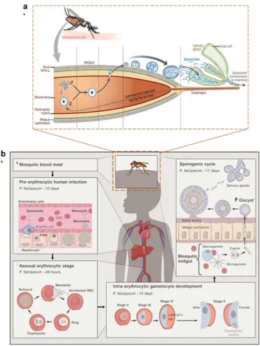
Figure 2 Complete life cycle of Plasmodium falciparum with asexual development in human host cells, hepatocytes and erythrocytes (a), adapted from (Cowman et al. 2016) and sexual development involving life stages female/male gametes, ookinetes, oocysts and sporozoites within female Anopheles mosquito (b), adapted from (Aly, Vaughan, and Kappe 2009). The initiation of asexual life cycle within human host is initiated by the bite of Anopheles injecting sporozoites into the dermis. This is followed by their transport to liver known as the pre-erythrocytic stage where they divide by schizogony (over a period of 10 days) to generate tens of thousands of daughter merozoites are released in packets of merosomes into the bloodstream. Within the bloodstream, these merozoites invade red blood cells carrying out their erythrocytic life stage, a cycle of which lasts for over 48 h. Next, a 15 days period leading to intra-erythrocytic gametocyte development follows which enters the peripheral circulation for ingestion by a mosquito for further transmission. The first part of sexual life cycle occurs in the mosquito midgut where the gametocytes emerge as male and female gametes followed by their fusion into a zygote which transforms into ookinete over a period of 24 h. the ookinete encysts to become oocyst where following sporogony over a period of 10-14 days, thousands of sporozoites are produced. These sporozoites traverse to enter into mosquito salivary gland from where they are transmitted to mammalian host over next blood meal.
26
Life cycle of Toxoplasma gondii
Toxoplasma gondii was first described in 1908 by Nicolle and Manceaux while working in North
Africa, on a semidesert rodent, the common gundi (Ctenodactylus gundi) (Black and Boothroyd, 2000). This intracellular parasite replicates within a nucleated host cell with the goal of producing infectious progeny for further survival and dissemination. The complex life cycle of this apicomplexan parasite is split between two host types: an intermediate and a definitive host (illustration: figure. 3).
1) Intermediate host: Toxoplasma asexual life stage
The intermediate host covers a wide range of warm-blooded animals, including humans, where
Toxoplasma undergoes asexual replication and further differentiation into transmissible forms.
Within the intermediate host, the parasite is able to fancy two different life forms: fast replicating ‘tachyzoites’ and slow dividing ‘bradyzoites’. The tachyzoite is virtually able to infect any nucleated cell at the advent of T. gondii lytic cycle. Although, current data suggest that on top of this large repertoire of host cell type, the parasite can choose and/or use the natural capacities of the host cell to its advantage to improve its dissemination/propagation/dormancy. This asexual life cycle is often called the lytic cycle which proceeds via five cellular steps: attachment, active invasion, vacuole formation notably through the discharge of secretory organelles, replication involving all complex metabolic interactions with the host cell, and active egress (Black and Boothroyd, 2000). After invading the host cell, a tachyzoite surrounds itself with a parasitophorous vacuole (PV) that is the niche and site for interaction between the parasite and its host cell. Replicative tachyzoites undergo several rounds of division, with a generation time of approx. 6-8 h before the final egress out of their inhabitant host cell to further invade its surrounding host cells. The terminology and cellular process used for this replicative process, where daughter progeny is formed within the boundaries of the mother cell that gets consumed at the end of division, is called ‘endodyogeny’ (Francia and Striepen, 2014; White and Suvorova, 2018). Under immune pressure from the host, the acute phase tachyzoites can commit, convert and differentiate into slow growing bradyzoites, finally forming a structure called tissue cyst, which constitutes a resistant and dormant niche filled with bradyzoites. Normally, the tissue cysts appear 7-10 days post infection, which defines the so-called chronic life stage of this parasite. The dormant cyst forms usually occur within the central nervous system and muscle tissue, where they can survive within host for
27 lifetime. Congenital infection occurs in case of vertical transmission of these tachyzoites from maternal blood into fetal tissues (Dubey, 2013).
2) Definitive host: Toxoplasma Sexual life stage
In T. gondii life cycle, the sexual stage is restricted to a feline host, that can acquire Toxoplasma through ingestion of contaminated food. The ingested Toxoplasma invades the feline intestinal epithelium where it differentiates into five morphologically distinct forms of schizont (Pittman and Knoll, 2015). These schizonts then differentiate into merozoites, which is regarded as the first sexual stage of the parasite life cycle. Merozoites then undergo 2-4 rounds of division to further differentiate into microgametes and macrogametes. This process of division where the nucleus undergoes multiple rounds of division prior to cytokinesis is called ‘schizogony’(Ferguson et al., 1974; Francia and Striepen, 2014). These gametes fuse together to form diploid oocyst. These oocysts are shed via cat’s feces into the environment, where they can survive moderate conditions due to their possession of a thick impermeable wall. Cats excrete up to 20 million oocysts per day, after approx. 3-10 days of acquiring Toxoplasma infection. Under favorable environmental conditions, these oocysts undergo mitotic and meiotic divisions to produce haploid sporozoites encysted by the sturdy oocyst wall.
Toxoplasma transmission between various hosts occurs via:
a) Faeco-oral route, by ingestion of food contaminated with Toxoplasma oocysts
b) Congenital acquisition, vertical transmission of parasites from infected mother to fetus
Since, the results of my thesis are majorly focused on T. gondii, therefore further description of parasite morphology and establishment of infection within the host is focused on Toxoplasma.
28 Figure 3. a) Infection of T. gondii within the intermediate host (asexual cycle) occurs through ingestion of contaminated food. The transmissible bradyzoites or sporozoites are released into the intestinal lumen where they invade intestinal enterocytes and differentiate into tachyzoites. Tachyzoites divide rapidly by the process of endodyogeny (b) and then egress from the host, infecting neighboring cells to start another cycle. Tachyzoites convert to dormant stage bradyzoites and then finally to tissue cysts that remain persistent within brain or musculature for almost lifetime of the intermediate host(a). The sexual cycle occurs only in cats, initiated by rupture of ingested tissue cyst that releases bradyzoites into enterocytes. These bradyzoites then undergo a self-limiting number of asexual multiplications, characterized by the development of merozoites within schizont, a process called schizogony (c). This is followed by gamogony and fertilization of the gametes forming an unsporulated oocyst (d). These are releases by with cat feces into the external environment where they undergo sporulation generating at least 4 haploid sporozoites per oocyst (e). (a) adapted from (Pittman and Knoll, 2015); (b,c) adapted from (Francia and Striepen, 2014)
a
b
c
d
e
29
Establishment of an infectious niche: active host cell invasion by T. gondii
Host cell invasion by Toxoplasma is an active process, unlike several other eukaryotic pathogens that embark upon phagocytic engulfment by the host. These parasites employ their actin-myosin machinery for aiding the active entry into their hosts (Carruthers, 2002).
Toxoplasma invasion begins with a reversible attachment of the parasite to the host cell surface, a
process called ‘gliding motility’. Within Toxoplasma, apically located secretory organelles, micronemes and rhoptries act as the main drivers in the process host cell invasion. For instance, the initial binding of the parasite to host cell membrane results in apical release of the micronemal protein MIC2 (Carruthers and Sibley, 1997). The second step is initiated by the discharge of rhoptry proteins in order to form a nascent parasitophorous vacuole. The rhoptries empty their contents apically during the process of host invasion i.e., beginning with the RONs and thereafter ROPs. During the process of invasion, a constriction or tight association, called the moving junction (MJ), is formed between the parasite and host cell membrane (Mordue et al., 1999; Alexander et al., 2005). This MJ associated constriction around an invading parasite migrates from the anterior towards the posterior end. The MJ also serves to exclude all the host cell proteins and parasite secreted micronemes from entering the parasite vacuolar space within its host. The moving junction is formed with a complex of the micronemal protein AMA1 and RONs (RON2,4,5 and 8) within the plasma membrane of the parasite (Besteiro et al., 2009). At the end of the invasion process when the PV formation is complete, the dense granules release their contents called GRA proteins into the PV and beyond to the host cell nucleus (Hakimi and Bougdour, 2015; Mercier and Cesbron-Delauw, 2015). Dense granule proteins (GRA) play important roles in the interaction of the parasite with its host via PVM as well as survival within the host through rewiring of the host cell gene expression.
30
Ultrastructural morphology of Toxoplasma tachyzoite
Toxoplasma derives its name based on its shape, from a Greek work toxon, meaning “bow”. A
typical Toxoplasma tachyzoite is crescent-shaped ranging in the size of 2-6 µm (Dubey, Lindsay and Speer, 1998).
1. The apical complex: machinery guiding the invasion process in T. gondii
The phylum apicomplexa withholds its name due to the presence of an ‘apical complex’ which is central to parasite invasion and subsequent proliferation within its host (Hu et al., 2006). The apical complex is a remarkable structure comprising of a) spirally arranged assembly of fibers called conoid, b)polar ring, located at the parasite apex, which serves as the site for origination of subpellicular microtubules, c) two intraconoidal microtubules and d) the preconoidal rings.
Toxoplasma has several apically located regulated secretory organelles which include micronemes
and rhoptries. Micronemes are small rod-shaped electron dense structures. The micronemes released their contents early on during the process of attachment-invasion, following which there is secretion of rhoptries as the invasion process proceeds. The rhoptries appear as club shaped structures in electron micrographs of T. gondii. This parasite typically has up to 8-12 rhoptries in total which are composed of a bulbous body structure which tapers into a thin duct-like rhoptry neck extending towards the conoid. The rhoptry neck proteins are referred to as RONs whereas the ones residing in bulbous body area are called ROPs. Rhoptries are known to be rich in proteins as well as lipids which they release alongside their secretion during the time of invasion. There are other cytoplasmic secretory organelles called dense granules that are released towards the end after completion of the process of invasion (Black and Boothroyd, 2000).
Figure 5 a) Schematic enlarged view of the apical complex cytoskeleton, showing the conoid (green), preconoidal, and polar rings (brown), and two intraconoid MT (green) (adapted from Hu et al., 2006), b) TEM image of the apical complex of Toxoplasma tachyzoite (adapted from Dubey, Lindsay and Speer, 1998)
31 2) Toxoplasma pellicle: the outer membranes surrounding the parasite
The parasite pellicle is a trilaminar structure comprising of the outer plasma membrane and an inner membrane complex (IMC) comprising two continuous membranes (Dubey, Lindsay and Speer, 1998). IMC is a unique cytoskeletal organelle which comprises of two distinct elements: a sac of flattened vesicles beneath the parasite plasma membrane called alveoli and a supporting rigid network of intermediate filaments (Mann and Beckers, 2001; Chen et al., 2015). The IMC imbibes a highly dynamic organization crucial for parasite development. The IMC of the mother parasite acts as the scaffold used by daughter cells for undergoing the process of endodyogeny (Ouologuem and Roos, 2014). The daughter IMC elongation is based on the recycling of maternal IMC membranes, after emergence of daughters from the mother cell.
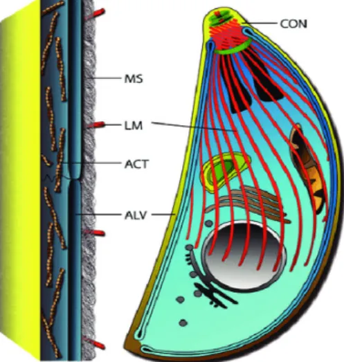
Figure 6 The basic pellicle organization Toxoplasma tachyzoite is characterized by alveolar sacs (ALV) associated with the inner membrane complex beneath the plasma membrane (in yellow) supported by a membrane skeleton (MS) and associated with microtubules structures (red) (adapted from Gould et al., 2011)
3) Metabolically active organelles: Apicoplast and Mitochondria
Similar to their algal ancestors these parasites also possess a single apicoplast and mitochondrion. Apicoplast:
It is a relict chloroplast present in several apicomplexan parasites, acquired in an event involving secondary endosymbiosis. The primary endosymbiotic event that gave rise to chloroplast in plants and algae arose from engulfment of Cyanobacterium by a eukaryotic heterotroph. This was followed by a second endosymbiotic event wherein plastids of chromalveolates were derived from red algae (Lim and McFadden, 2010; van Dooren and Striepen, 2013). Based on its evolutionary
32 origin apicoplast is surrounded by four membranes enriched with phospholipids instead of galactolipids as their algal ancestors (Botté et al., 2013). Apicoplast has time n again proven itself to be parasites Achilles heel due to its role as parasite’s central metabolic hub. This special organelle harbors several metabolic processes crucial for the intracellular development of apicomplexan parasites (van Dooren and Striepen, 2013).
i) Type-II Fatty acid biosynthesis/ FASII pathway: Gene knockout studies of several important enzymes involved in FASII pathway have proven the indispensability of this metabolic pathway for T. gondii. The apicoplast FASII contributes to the total cellular pool of short chain fatty acids (myristate, palmitate) which are further used for membrane biogenesis aiding parasite cell division and hence survival within their host (Mazumdar et al., 2006; Ramakrishnan et al., 2012; Amiar et al., 2016). ii) Isoprenoid precursor assembly pathway: The central precursor required for
isoprenoid biosynthesis is isopentenyl pyrophosphate (IPP). Unlike mammalian cells, the IPP within apicomplexans is synthesized via a non- mevalonate pathway, involving methylerythritol phosphate (MEP), localized within parasite apicoplast. Genetic ablation of the last step of the MEP pathway involving enzyme LytB is lethal to the Toxoplasma tachyzoites (Nair et al., 2011). Isoprenoids act as components of membrane lipids and mitochondrial electron chain coenzyme Q. These compounds also participate in post-translational modifications of several proteins imparting important functions like protein-protein interactions (van Dooren and Striepen, 2013).
iii) Iron-Sulphur cluster biosynthesis: Iron-Sulphur (Fe-S) clusters act as prosthetic groups for proteins involved in various redox reactions (van Dooren and Striepen, 2013). The apicoplast based Fe-S proteins are involved in IPP and fatty acid biosynthetic pathway, thereby suggesting the importance of this metabolic pathway in T. gondii.
iv) Heme biosynthesis pathway: Heme is an essential prosthetic group that functions in many cellular redox reactions including the mitochondrial electron transport chain (Van Dooren, Kennedy and McFadden, 2012). Apicomplexan parasites harbor an unusual pathway of heme biosynthesis spanning three different cellular compartments-cytosol, apicoplast and mitochondria (Kořený, Oborník and Lukeš,
33 2013; Bergmann et al., 2019). Recent study provides the first evidence for presence of a functional heme biosynthesis pathway within Toxoplasma apicoplast (Bergmann et al., 2019).
Mitochondria:
This organelle acts as reserve of essential central carbon metabolic pathways like the tricarboxylic acid cycle (TCA) and the electron transport chain (ETC). Chemical inhibition of mitochondrial TCA cycle with sodium fluoroacetate (NaFAc) is lethal to Toxoplasma tachyzoites (MacRae et
al., 2012). T. gondii is able to utilize both glucose and glutamine as substrates via the TCA cycle.
The catalytic conversion of glycolytic intermediate pyruvate into acetyl coA for further utilization in mitochondrial TCA cycle is facilitated by a branched chain ketoacid dehydrogenase (BCKDH) complex. BCKDH is a functional replacement of mitochondrial PDH in apicomplexan parasites and essential for their intracellular growth (Oppenheim et al., 2014). Another enzyme succinyl coA synthetase, catalyzing the seventh step of TCA cycle succinyl-CoA to succinate, was found to be dispensable for parasite growth (Fleige et al., 2007). A probable reason for this unexpected dispensability could be the leaky expression of the enzyme under control of the anhydrotetracycline regulatable element.
A potential metabolic link between Toxoplasma apicoplast and mitochondria was established by showing that the use of a specific inhibitor of TCA cycle, NaFAc resulted in partial reduction in apicoplast FASII biosynthesis. The reason for the same was justified because of a shunt in which mitochondrial citrate is transported to the apicoplast and converted to a-ketoglutarate with regeneration of NADPH (MacRae et al., 2012)
4)Other organelles covering the endomembrane system include the posteriorly located nucleus,
the tubular network formed by the endoplasmic reticulum extending from the nuclear envelope and the Golgi complex (Nishi et al., 2008). The ER is predominantly located basal end of the parasite while as the Golgi is adjacent to the apical end of the parasite nucleus (Hager et al., 1999).
34 Figure 7 Metabolic pathways present in apicoplast (a) (adapted from Ralph et al., 2004) and mitochondria (b) (adapted from Oppenheim et al., 2014). Apicoplast provides essential metabolites for parasite’s intracellular survival: fatty acids via FASII pathway, isoprenoid biosynthesis (IPP), iron-sulfur cluster biosynthesis and haem synthesis. Mitochondria resident tricarboxylic acid cycle contributes to parasite’s ATP and anabolic needs
35 Figure 8. Electron Micrograph showing Toxoplasma tachyzoite (adapted from Dubey, Lindsay and Speer, 1998), Co, conoid; Dg, electron-dense granule; Go, Golgi complex; Mn, microneme; Nu, nucleus; Pvm, parasitophorous vacuole membrane; Rh, rhoptry; Ap, apicoplast; IMC, inner mitochondrial membrane; Mt, mitochondria. The immunofluorescence images (IFA) using protein markers of various organelles, IMC1, ER marker Der-1, Nucleus-DAPI (adapted from experiments done in this PhD) ; Rhoptry neck protein ROP1 and dense granule protein GRA7 was ( adapted from Wang et al., 2019); conoid (adapted from (Katris et al., 2014) ; Golgi marker GRASP (adapted from (Pfluger et al., 2005); mitochondrial marker TOM40 (adapted from (Van Dooren et al., 2016) ; microneme marker MIC5 (adapted from Huynh, Boulanger and Carruthers, 2014)
36
CHAPTER
II:
LIPID
METABOLISM
IN
APICOMPLEXAN
37
LIPID METABOLISM IN APICOMPLEXA: Biosynthesis, Uptake and Recycling
The survival of these parasites within host is predominantly dependent on lipids which play an essential role by regulating metabolic flux, acting as signaling molecules, storage fuels and structural building blocks of membranes. These parasites meet their high demand of lipids through
de novo synthesis within apicoplast as well as via copious salvage directly from the host and
extracellular environment (Coppens, 2006, 2013). This raises the prospect that lipid homeostatic, trafficking and remodeling pathways in the parasite may abound as potential drug targets. For instance, apicoplast resident fatty acid biosynthetic enzymes have been successfully exploited as antimicrobial targets against Toxoplasma infections. This section summarizes different lipid species present in two important apicomplexan parasites T. gondii and P. falciparum:
Phosphatidylcholine
Phosphatidylcholine (PC) is a neutral glycerophospholipid accounting for the major component of total complex lipids. Quantification of total phospholipid profile within apicomplexa reveals high abundance of phosphatidylcholine in T. gondii (40%-70%) as well as in P. falciparum (40%-50%) (Gupta et al., 2005; Welti et al., 2007; Gulati et al., 2015).
Toxoplasma tachyzoites can synthesize PC via de novo Kennedy pathway involving a cytoplasmic
choline kinase which utilises choline up taken from external environment (Gupta et al., 2005; Sampels et al., 2012). Selective disruption of PC biosynthesis by supplementation of a choline analog, dimethylethanolamine was able to cause dramatic parasite growth arrest. Dimethylethanolamine interferes with choline uptake and subsequent metabolism to phosphatidylcholine, resulting in inhibition of parasite intracellular replication due to inhibition of
PC biosynthesis or massive accumulation of toxic intermediate
phosphatidyldimethylethanolamine (Gupta et al., 2005).
Despite the presence of CDP-choline/Kennedy pathway for major PC biosynthesis, P. falciparum also employs an alternative pathway called serine-decarboxylase-phosphoethanolamine-methyltransferase (SDPM) pathway which uses host serine and ethanolamine as precursors (Elabbadi, Ancelin and Vial, 1997; Pessi and Mamoun, 2006; Witola et al., 2008). This alternative pathway begins with the decarboxylation of host-derived serine to ethanolamine via serine decarboxylase (PfSD). The next step in catalyzed by a phosphoethanolamine methyltransferase
38 (PfPMT) in a three-step methylation reaction starting from substrate phosphoethanolamine to phosphocholine which is further incorporated into phosphatidylcholine (PC) (Pessi, Kociubinski and Ben Mamoun, 2004; Pessi and Mamoun, 2006). The loss of PfPMT by gene knockout studies results in strong growth defects alongside abrogation of PC biosynthesis via SDPM pathway within
P. falciparum (Witola et al., 2008). However, unlike P. falciparum, Toxoplasma cannot use
ethanolamine as a substrate for PC biosynthesis due to the absence of the required methyltransferase enzyme activity (Gupta et al., 2005).
Phosphatidylethanolamine
Phosphatidylethanolamine (PE) is a neutral, second most abundant phospholipid in eukaryotic cells, normally present in the inner bilayer of the plasma membrane. Expectedly, PE is also the second most abundant phospholipid in T. gondii (10%-20%) and P. falciparum (15%-25%) (Welti
et al., 2007; Hartmann et al., 2014; Gulati et al., 2015).
In T. gondii, PE biosynthesis occurs differentially at several organellar sites, in the mitochondrion and in the PV by decarboxylation of phosphatidylserine, and in the ER by fusion of CDP-ethanolamine and diacylglycerol (Gupta et al., 2012; Hartmann et al., 2014). Mitochondrial PE biosynthesis is catalyzed by the enzyme phosphatidylserine decarboxylase (TgPSD1mt). The depletion TgPSD1mt impacts parasite’s optimal growth, which is rescued by the addition of exogenous ethanolamine. However, despite the simultaneous knockdown of TgPSD1 and ethanolamine-depletion the parasites can still egress and form plaques, albeit reduced in terms of area in comparison to wild type parasites (Hartmann et al., 2014). This suggest that the parasite has employed other routes for PE biosynthesis including the high possibility of direct scavenging from its metabolically enriched host. Toxoplasma also secretes a phosphatidylserine decarboxylase (TgPSD1) localizing to the dense granules, into the parasite vacoular space (Gupta et al., 2012). Based on biochemical activity confirmation of TgPSD1, it can be speculated that this enzyme contributes to PE biosynthesis within PV, possibly utilizing host derived precursors.
The PE biosynthesis in P. falciparum is also branched into CDP-ethanolamine/Kennedy pathway and the serine decarboxylation pathway (as described above) (Elabbadi, Ancelin and Vial, 1997; Kilian et al., 2018).
39 Phosphatidylserine
Phosphatidylserine (PS) despite being less than 10% in the total abundance of phospholipids, is still an important anionic lipid. Due to its negative charge, PS is able to impart unique biophysical properties to the membranes and also participates in important biological processes like apoptosis in various eukaryotic cells. In terms of abundance, PS constitutes 5%-10% of the total phospholipid species in both T. gondii and P. falciparum (Welti et al., 2007; Gulati et al., 2015). Intriguingly, phosphatidylserine in T. gondii has been linked to a mechanism of host immune evasion called apoptotic mimicry (dos Santos et al., 2011). Briefly, during this process Toxoplasma expresses PS on the surface thereby decoying itself as an apoptotic cell inside its host macrophages. As a result, the parasite infected macrophages inhibit the synthesis of nitric oxide (NO) via NO synthase degradation, thereby allowing parasite persistence within. Interestingly, Toxoplasma utilizes serine as a substrate for PS biosynthesis through two different enzymes functionally behaving as phosphatidylserine synthases, TgPSS (phosphatidylserine synthase) and TgPTS (phosphatidylthreonine synthase) (Arroyo-Olarte et al., 2015). TgPSS, with strict serine-substrate specificity, localizes towards the parasite ER/mitochondrion intersection.
In P. falciparum PS acts as an important pathogenicity factor by facilitating erythrocyte cytoadherence (Eda and Sherman, 2002). Micro vesicles derived from the blood sample of P.
falciparum infected patients been have shown to have elevated levels of phosphatidylserine (Gulati et al., 2015). The malarial parasite synthesizes PS using host derived serine, most of which is
obtained from parasite mediated host erythrocyte hemoglobin degradation (Wein et al., 2018). As described above PS also serves as a precursor of PE via serine decarboxylation pathway in both T.
gondii and P. falciparum (Elabbadi, Ancelin and Vial, 1997; Hartmann et al., 2014; Kilian et al.,
2018).
Phosphatidylthreonine
Phosphatidylthreonine (PTh) is usually a minor component of the total phospholipid constituent in various eukaryotic cells. Recent study described the existence of Phosphatidylthreonine as a major phospholipid (3-4 times more than phosphatidylserine) in T. gondii facilitating a ‘lipid-mediated virulence’ within its host (Arroyo-Olarte and Gupta, 2016). The parasite employs a novel enzyme phosphatidylthreonine synthase (TgPTS) for generation of PTh within the endoplasmic
40 reticulum. The TgPTS has evolutionarily evolved from TgPSS as both the enzymes are capable for PS synthesis in the presence of serine as a substrate. Unlike, TgPSS, the presence of threonine induces phosphatidylthreonine biosynthesis by the enzymatic action of TgPTS. Genetic disruption of TgPTS (Δtgpts) impairs the parasite lytic cycle and attenuates its growth within murine model. Lipidomic analysis of parasites lacking TgPTS indicate a significant decline in the PTh levels with a concomitant accumulation of phosphatidylserine. The rescue of strong growth defect and PTh synthesis is attained by complementation of Δtgpts parasites with exogenous copy of TgPTS-HA. The main fatty acid components of the T. gondii PTh species included C20:1 and C20:4 (Arroyo-Olarte et al., 2015). The authors of this study further speculate the possible involvement of PTh influencing the calcium flux across the plasma membrane and/or calcium storage organelles (Arroyo-Olarte and Gupta, 2016).
Phosphatidylinositol and related phosphoinositides
Phosphatidylinositol (PI) is a negatively charged important constituent of membrane phospholipids. Phosphate derivatives of phosphatidylinositol, called phosphoinositides have more critical functions within a cell, such as signaling, cell-cell communication, intracellular trafficking. PI comprises approx. 5% of the total phospholipid abundance in both T. gondii and P. falciparum (Welti et al., 2007; Gulati et al., 2015). In Toxoplasma, phosphatidylinositol-3-monophosphate (PI3P) has been shown to be associated with apicoplast protein-shuttling vesicles. Chemical inhibition of PI3P synthesizing kinase, resulted in aberrant apicoplast morphological development suggesting the role of this phospholipid for apicoplast biogenesis (Tawk et al., 2011). A
Toxoplasma phosphatidylinositide-phospholipase C (TgPI-PLC), catalyzes the hydrolysis of
phosphatidylinositol 4,5-bisphosphate (PIP2) to D-myo-inositol 1,4,5-trisphosphate (IP3) and sn-1,2-diacylglycerol (DAG) at the plasma membrane of the parasite (Fang, Marchesini and Moreno, 2006). Recombinant protein of TgPI-PLC showed a preference for phosphatidylinositol (PI) as a substrate rather than PIP2. Another study showed that this protein was localizing to both plasma membrane and cytoplasm within the parasite, suggesting its multiple roles in lipid regulation (Bullen et al., 2016).
In P. falciparum, an enzyme generating 3’ phosphorylated phosphoinositides, phosphoinositide-3-kinase (PI3K) is actively transported across the PVM into its erythrocyte host. Using PI3K