HAL Id: hal-00318871
https://hal.archives-ouvertes.fr/hal-00318871
Submitted on 5 Sep 2008HAL is a multi-disciplinary open access
archive for the deposit and dissemination of sci-entific research documents, whether they are pub-lished or not. The documents may come from teaching and research institutions in France or abroad, or from public or private research centers.
L’archive ouverte pluridisciplinaire HAL, est destinée au dépôt et à la diffusion de documents scientifiques de niveau recherche, publiés ou non, émanant des établissements d’enseignement et de recherche français ou étrangers, des laboratoires publics ou privés.
Activation of a Dimeric Metabotropic Glutamate
Receptor by Inter-Subunit Rearrangement
Carsten Brock, Nadia Oueslati, Stéphan Soler, Laure Boudier, Philippe
Rondard, Jean-Philippe Pin
To cite this version:
Carsten Brock, Nadia Oueslati, Stéphan Soler, Laure Boudier, Philippe Rondard, et al.. Activation of a Dimeric Metabotropic Glutamate Receptor by Inter-Subunit Rearrangement. Journal of Biolog-ical Chemistry, American Society for Biochemistry and Molecular Biology, 2007, 282 (45), pp.33000. �10.1074/jbc.M702542200�. �hal-00318871�
ACTIVATION OF A DIMERIC METABOTROPIC GLUTAMATE
RECEPTOR BY INTER-SUBUNIT REARRANGEMENT
Carsten Brock
1, 2, 3, 4, Nadia Oueslati
1, 2, 3, 4, Stéphan Soler
1, 2, 3, 4, Laure Boudier
1, 2, 3, 4,
Philippe Rondard
1, 2, 3, 4, and Jean-Philippe Pin
1, 2, 3, 4From the
1Institut de Genomique Fonctionnelle, Montpellier, France;
2CNRS
UMR5203, Montpellier, France;
3INSERM, U661, Montpellier, France;
4Université de
Montpellier (IFR3), Montpellier, France.
Running title: mGluR activation by inter-subunit rearrangement
Address correspondence to: Jean-Philippe Pin, Institut de Génomique Fonctionnelle, 141 rue de la Cardonille, 34094 Montpellier, France, Tel. + 33 4 67 14 29 88; Fax. + 33 4 67 54 24 32; E-mail: [email protected]
Although many G protein-coupled receptors (GPCRs) can form dimers, a possible role of this phenomenon in their activation remains elusive. A recent and exciting proposal is that a dynamic inter-subunit interplay may contribute to GPCR activation. Here we examined this possibility using a dimeric metabotropic glutamate receptor (mGluR). We first developed a system to perfectly control their subunit composition, and show that mGluR dimers do not form larger oligomers. We then examined an mGluR dimer containing one subunit in which the extracellular agonist binding domain is uncoupled from the G protein-activating transmembrane domain (TMD). Despite this uncoupling in one protomer, agonist stimulation resulted in symmetric activation of either TMD in the dimer with the same efficiency. This, plus other data, can only be explained by an inter-subunit rearrangement as the activation mechanism. Although well established for other types of receptors such as tyrosine kinase or guanylate cyclase receptors, this is the first clear demonstration that such a mechanism may also apply to GPCRs.
INTRODUCTION
G protein-coupled receptors (GPCRs), the largest family of mammalian genes (~ 1000 members), are involved in a vast variety of physiological and pathological processes, and represent the target of almost 50 % of all modern drugs (1). The common structural feature of all GPCRs is a transmembrane domain (TMD) made of seven transmembrane segments (TM1 - TM7). Despite the vast variety of ligands and the low sequence similarity between GPCRs from various classes, their activation results from similar conformational changes in their TMDs (2-4). In particular, a movement of TM6 likely
opens a crevice allowing interaction with the C-terminus of the G protein α subunit, triggering its activation (5).
Many GPCRs have been shown to form dimers, but the functional role of this remains elusive (6-8). Although a GPCR monomer is sufficient to activate a G protein (8-12), it has been proposed that GPCR dimerization may facilitate their activation, i. e. that allosteric interactions between the protomers, through changes at the dimerization interface or a larger-scale reorientation of the two subunits, may contribute to stabilize the active conformation (13-15). To further elucidate a possible role of an inter-subunit rearrangement in the activation of a dimeric GPCR, we chose a metabotropic glutamate receptor (mGluR) as a model. These GPCRs are clearly established as constitutive dimers, the two subunits being linked by a disulfide bridge (16). Moreover, glutamate and its analogues do not bind to the TMD of an mGluR, but to a distinct extracellular domain called Venus Flytrap (VFT) (17). This separation may help to dissect the relationship between agonist binding and activation within these GPCRs. Agonist binding stabilizes a closed conformation of the VFT (Fig. 1) (18,19), which is both necessary (20) and sufficient (21) for the activation of a class C GPCR. How the VFT closure is in turn transduced into TMD activation, however, is yet unknown. Two mechanisms, not necessarily exclusive, have been proposed.
The first model (Fig. 2A) proposes that the closed VFT directly stabilizes the active conformation of the TMD of the same subunit (22,23). But agonist binding may also induce a relative reorientation of the two VFTs (Fig. 1) (18), and a second model (Fig. 2B) thus proposes that this may in turn yield an activating rearrangement of the two TMDs (24,25). Recent FRET studies are indeed consistent with a
glutamate-induced reorientation of the two TMDs within an mGluR dimer (26). However, this could also simply be a "side effect" not necessarily involved in the activation process. Moreover, in contrast to mGlu1, the relative orientation of the VFTs of mGlu3 appears not to be influenced by agonist binding (Fig. 1) (19), further putting into question this second proposal (Fig. 2B).
Importantly, only in the second (Fig. 2B), but not in the first model (Fig. 2A), agonist binding to one VFT will not only cis-activate the TMD of the same, but also equally well trans-activate the TMD of the other subunit. Using a new system to control the subunit composition, and thereby introduce different mutations specifically into either protomer, we demonstrate here that both cis- and trans-activation within an mGluR dimer occur with the same probability. We show moreover that this trans-activation is not indirect via the other VFT, via the other TMD, or by a domain "swap" between the two subunits (see also Fig. 5). Our data therefore demonstrate that an agonist-induced inter-subunit rearrangement can indeed be responsible for the activation of a dimeric GPCR, a mechanism already well accepted for tyrosine kinase and guanylate cyclase receptors (27-30).
EXPERIMENTAL PROCEDURES
Plasmids for mGlu5 were either previously
described (31-34), or, based on plasmids described in these references, newly constructed by standard molecular biology techniques (PCR, site-directed mutagenesis, subcloning), as described (35). Compared to the previously described -C2 constructs (31), in the -C2KKXX constructs the last 120 amino acid residues are replaced by the sequence KKTN, right after the CC domain. Similarly, in some of the -C1 constructs we have also replaced the last 39 residues by the sequence KKTN, right after the coiled coil (CC) domain. However, we found no significant functional differences between the -C1 and the --C1KKXX constructs (see Supplemental Fig. B). Both types of constructs are therefore collectively referred to as -C1 throughout this paper. The FLAG-V2 plasmid was a generous gift from L. Albizu. CD4 cDNA was a generous gift from B. Schwappach.
Cell culture & transfection of HEK293 cells
was performed as described (32). To avoid any mGluR activation due to ambient glutamate, a plasmid encoding the high-affinity glutamate
transporter EAAC1 was always co-transfected, and the cells were incubated in glutamate-free medium during at least 4 h before the experiments. All experiments were carried out one day after transfection.
Intracellular Ca2+ release was measured as
described (36). In brief, cells were pre-incubated during 1 h with the Ca2+-sensitive Fluo-4 AM (Invitrogen). The fluorescence signals (excitation at 485 nm, emission at 525 nm) were then measured during 60 s (FlexStation, Molecuar Devices). Quis (Tocris) was added after the first 20 s. The Ca2+ response is given as the Quis-stimulated fluorescence increase. Sigmoidal concentration - response curves were fitted using GraphPad Prism.
FRET measurements were conducted as
described (37). In brief, cells were incubated with Eu3+ Cryptate PBP-labeled anti-HA (donor) and AlexaFluor 647-labeled anti-FLAG (acceptor), or with the donor antibody only (negative control). Following excitation at 337 nm, the emission at 665 nm is measured (RubyStar, BMG). The FRET signal is calculated as ∆665, i. e. the difference between the emissions at 665 nm in presence or absence of the acceptor. The fluorophore-labeled antibodies were provided by CIS Bio International (Bagnols sur Cèze, France).
ELISAs anti-HA were performed as described
previously (35). The same protocol was applied for ELISAs FLAG, but using 1 µg/ml anti-FLAG M2 (Sigma) and 0,5 µg/ml anti-mouse-HRP (Amersham Biosciences) instead.
RESULTS
"Heterodimerization" of an mGluR
To study the inter-subunit interactions within a dimeric receptor one first has to control its subunit composition. To that aim, we have previously transferred the "quality control" system of the heterodimeric GABAB receptor
(35,38-40) to the homodimeric metabotropic glutamate receptor 5 (mGlu5) (31). Replacing its C-terminal tail by that of the GABAB1 subunit
(C1) (see also Supplemental Fig. A) generates a chimeric mGlu5-C1 that is retained inside the cell, unless it forms a heterodimer with a chimeric mGlu5-C2 carrying the C-terminal tail of the GABAB2 subunit (C2). In fact, the
intracellular retention signal (the sequence RSR) of the C1 part becomes masked by the specific interaction of the adjacent coiled coil (CC)
domain with the CC domain in the C2 part. The mGlu5-C2 chimera, however, not carrying a retention signal, could still reach the cell surface and function as a homodimer.
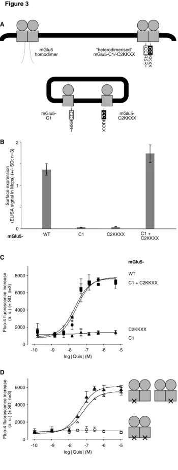
To solve this problem, we have now generated an mGlu5-C2KKXX construct, in which we have replaced the extreme C-terminal tail of the C2 part by an intracellular retention signal (KKXX), right after the coiled-coil (CC) domain (see also Supplemental Fig. A). As a consequence, now both, C1 and mGlu5-C2KKXX, are retained inside the cell when expressed alone (Fig. 3). However, both can reach the cell surface when they are co-expressed in the same cells, demonstrating that the CC interaction results in efficient mutual masking of the two retention signals within the heterodimer. Importantly, this "heterodimerized" mGlu5 is functional, since the Ca2+ response evoked by stimulation with the glutamate analogue Quisqualate (Quis) was similar between a wild-type mGlu5 and an mGlu5-C1/-C2KKXX heterodimer (Fig. 3C). Moreover, the -C1/-C2KKXX parts did not affect the symmetry within the dimer, since the Quis-stimulated Ca2+ response of such "heterodimerized" receptors carrying the G protein-uncoupling mutation F767S (31) in either the -C1 or in the -C2KKXX subunit was similar (Fig. 3D).
mGlu5 is a dimer, not a higher-order oligomer
At least some GPCRs may not only form dimers, but higher-order oligomers (41,42). If this occurred also for mGluRs, this would substantially complicate our analysis of their intra-molecular signal transduction. Our new system to control the subunit composition of cell surface-expressed mGluR dimers (Fig. 3) now permitted us to test this possibility.
We first used a fluorescence resonance energy transfer (FRET) approach using donor- and acceptor fluorophore-labeled HA anti-FLAG antibodies, respectively, to test a possible physical interaction between two distinct mGlu5 dimers at the cell surface (37). We co-expressed mGlu5-C1/-C2KKXX "heterodimers" with only the -C2KKXX subunits carrying an HA or FLAG tag at their extracellular N-termini (Fig. 4A, middle). To reach the cell surface and become accessible to the antibodies, any tagged -C2KKXX subunit must be in a dimer with an untagged -C1 subunit (and not with another tagged -C2KKXX subunit). Only a very low
FRET signal was measured under these conditions (Fig. 4A, middle), similar to that obtained with the V2 vasopressin receptor as a negative control (Fig. 4A, right). A large FRET signal, however, was detected with the positive control with both subunits (-C1 and -C2KKXX) of the same dimer being tagged (Fig. 4A, left). These differences in FRET were not due to different cell surface expression levels, which were controlled in parallel by ELISA. Similar results were obtained with the inverse combination of the -C1 and -C2KKXX subunits (Supplemental Fig. D).
We next tested a possible functional complementation between two non-functional mGlu5 dimers. An mGlu5-C1/-C2KKXX dimer with both TMDs carrying the mutation F767S abolishing G protein activation (31) was coexpressed with an mGlu5-YADA which is not activated by glutamate or its analogues due to a mutation in the VFT (31). To reach the cell surface, the -C1 and -C2KKXX subunits, both carrying the F767S mutation, must necessarily be part of the same dimer. No functional complementation was observed between this dimer and the mGlu5-YADA (Fig. 4B, right), despite the fact that, in line with our previous findings (31), an mGlu5-F767S is principally capable to trans-activate an mGlu5-YADA (Fig. 4B, middle). Again, these differences cannot be accounted for by differences in the cell surface expression levels, which were controlled in parallel by ELISA. Thus, no functional complementation can be observed between two different mGlu5 dimers.
In conclusion, these data revealed that mGlu5 dimers neither physically nor functionally associate into larger higher-order oligomers.
"Direct" trans-activation?
The observed trans-activation between an mGlu5-F767S and an mGlu5-YADA (Fig. 4B, middle) is perfectly in line with the model of an agonist-induced inter-subunit rearrangement as the activating mechanism of an mGluR dimer (Fig. 2B). However, other possibilities also exist. First, this trans-activation could be due to a "swap" between the VFTs and the TMDs of the two subunits (Fig. 5A). Second, this trans-activation could be indirect via the other VFT, i. e. the mutated VFT might nonetheless, through interaction with the agonist-bound VFT, become stabilized in an "active" (closed) conformation, in turn activating its TMD (Fig. 5B). Third, the observed trans-activation might also be indirect
via the other TMD (Fig. 5C). The aim of the
subsequent experiments was to test these possibilities.
Trans-activation not by "swap" between the
VFTs and TMDs within an mGluR dimer
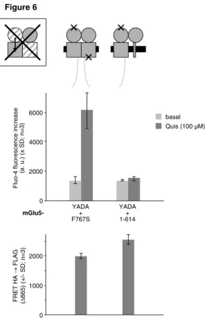
For several glycoprotein hormone GPCRs it has been demonstrated that a receptor with an inactivated exodomain (extracellular hormone binding domain) can be trans-activated by a hormone-bound exodomain devoid of the receptor's endodomain (TMD + cytosolic tail) (43). This suggests that the hormone-bound "isolated" exodomain may "swap" with the inactivated one to activate that subunit's endodomain. Could such a "swap" also occur between the VFTs and TMDs within an mGluR dimer (Fig. 5A)? To answer this question, we now tested whether the VFT-mutated mGlu5-YADA could also be trans-activated by an mGlu5(1-614), devoid of its TMD (34). In this construct, only TM1 was left as a "membrane anchor", while TM2 - TM7 and the cytosolic tail of are lacking. In contrast to the TMD-mutated mGlu5-F767S, this TMD-deficient mGlu5(1-614) did not trans-activate mGlu5-YADA (Fig. 6). This was not due to a lack of heterodimerisation, as controlled by FRET (Fig. 6, bottom panel). Thus, in contrast to what has been proposed for the exo- and endodomains of a glycoprotein hormone receptor (43), there is no "swap" between the two VFTs and TMDs (Fig. 5A), and such a mechanism can therefore not account for the observed trans-activation within an mGluR dimer.
Uncoupling a VFT from its TMD
To further clarify how agonist binding to a VFT leads to activation of a TMD of an mGlu5 dimer, our idea was to introduce another mutation, C240E, that functionally uncouples the VFT from the TMD (33), into one of the two subunits only. The combination with other mutations should then permit to study the functional interactions between the different domains within such a dimer.
We first verified that an mGlu5-C1/-C2KKXX dimer carrying the C240E mutation in only one subunit is still functional (Fig. 7). Indeed, such a receptor was still activated by Quis, although with a reduced maximal response as compared to the receptor not carrying this mutation at similar cell surface expression. Since at the expression levels used in this study the maximal Ca2+ response is directly proportional to the
amount of mGlu5 at the cell surface (see Supplemental Fig. C), this reduced maximal response (at similar cell surface expression) of an mGlu5 with one "uncoupled" VFT therefore indicates a reduced maximal activity. Two different explanations can be proposed. The observed resting activity may simply reflect the activity of the subunit not carrying the C240E mutation, resulting from an intra-subunit transduction (Fig. 2A). Alternatively, the decreased maximal response could also reflect that the disulfide bond involving C240 (33) is important for transmitting the relative movement of the two VFTs to the TMDs (Fig. 2B), which may be less efficient when one disulfide bond is missing. Most importantly, however, the fact that this disulfide bond is not mandatory in both subunits opens the possibility to experimentally refine the route of the intra-molecular signal transduction within an mGluR dimer.
Trans-activation not via the other VFT
Accordingly, we next tested a possible trans-activation between an mGlu5-F767S and an mGlu5-C240E. Indeed, Quis stimulation of an mGlu5 dimer composed of these two subunits still elicits a Ca2+ response (Fig. 8). This demonstrates a trans-activation of one TMD (of the C240E mutant) by the VFT of the other subunit (the F767S mutant), in a way not involving an indirect activation via the other VFT (of the C240E mutant) (Fig. 5B). The maximal response obtained with this combination being lower than that with the heterodimer carrying only the C240E mutation in one subunit is also consistent with the notion that one VFT can both cis-activate the TMD of the same, or trans-activate that of the other subunit, since the cis-activation of the F767S-mutated TMD does not yield any G protein activation.
Trans-activation not via the other TMD
The results presented so far do not exclude the theoretical possibility of an indirect activation
via the other TMD (Fig. 5C). The VFT of the
F767S subunit could first activate the TMD of the same subunit (though incapable of G protein activation), in turn trans-activating the TMD of the other subunit. This pathway, anyway, would also imply an inter-subunit re-arrangement, namely changes at the interface of the two TMDs.
This scenario, however, would be in contrast to recent reports indicating that only one TMD
within a GPCR dimer can reach the active state at a time (12,32,44). We nevertheless verified whether this was also true for mGlu5, in particular with one subunit carrying the C240E mutation, as used in our trans-activation experiment in Fig. 8. To that aim, we now used an mGlu5 subunit with a TMD that, thanks to a triple mutation ("3Ro": P654S, S657C, L743V), can be specifically blocked in its active conformation by the drug Ro01-6128 (45), but without triggering any G protein activation, due to an additional F767S mutation.
Indeed, pretreatment with this drug completely abolished the Quis-stimulated Ca2+ response mediated by an mGlu5 carrying these mutations ("3Ro" + F767S) in one of the two subunits (Fig. 9). This demonstrates that indeed, once the "3Ro"-mutated TMD is in the active state (but without triggering G protein activation due to the F767S mutation) the second TMD cannot reach the active state any more. Thus, also in an mGlu5 dimer, only one TMD can reach the active state at a time. Of note, the effect was independent of the absence or presence of the C240E mutation in the other subunit. Thus, the observed trans-activation of one TMD by the VFT of the other subunit within an mGluR dimer (Fig. 8) cannot be indirect via activation of the second TMD (Fig. 5C).
No preferential intra-subunit transduction
As stated above, the two models of intra-molecular signal transduction from the VFTs to the TMDs of an mGluR dimer (Fig. 2A vs. 2B) are not mutually exclusive. Thus, the above demonstration that the agonist-induced inter-subunit re-arrangement is indeed activating the receptor does not yet exclude that, additionally, the agonist-bound VFT could also activate its associated TMD through an intra-subunit transduction. This can be tested by comparing the cis- and trans-activation within the dimer. The inter-subunit rearrangement induced by agonist binding to one VFT should always cis- and trans-activate equally well both TMDs in the dimer. In contrast, in case of an additional intra-subunit transduction, the cis-activation should be more efficient than the trans-activation. This is not the case, since there is no significant difference between the Quis-stimulated Ca2+ responses mediated by an mGlu5 carrying the C240E and the F767S mutations in the same subunit (only cis-activation can generate a response), or either one of these mutations in each subunit (only
trans-activation) (Fig. 10). This demonstrates that the agonist-induced inter-subunit rearrangement is the only mechanism of intra-molecular signal transduction and activation of a dimeric mGluR, and there is no additional intra-subunit transduction from the VFT to the TMD of the same subunit.
DISCUSSION
In the present study, we examined a possible role of an inter-subunit rearrangement in the activation of a dimeric mGluR. Two models, not mutually exclusive, have been proposed to explain how agonist binding in the VFT leads to activation of the TMD of these receptors. The first model proposes an intra-subunit activation in which the VFT directly activates the TMD of the same subunit (Fig. 2A) (22,23). The second model proposes that the TMD activation may result from an agonist-induced inter-subunit rearrangement (Fig. 2B) (24,25). To experimentally test these models, we first developed a system to perfectly control the composition of an mGluR dimer of two defined protomers each carrying or not different mutations. In particular, we took advantage of a point mutation that functionally disconnects the VFT from its TMD (33). Our data show that in an mGluR dimer carrying this mutation in a single subunit, agonist binding in one subunit activates equally well either of the two TMDs. This is only compatible with the second, but not the first model. Thus, an inter-subunit rearrangement indeed plays a role, and is even crucial for the activation of an mGluR.
Intra-molecular signal transduction within an mGluR dimer
For most GPCRs, agonists bind directly to their TMD, thereby stabilizing its active conformation (23). For class C GPCRs such as the mGluRs where agonists bind to an extracellular domain, it has been proposed that this domain in its agonist-bound conformation could in turn interact with the TMD in a way stabilizing its active conformation (Fig. 2A) (22,46). In line with this proposal, we have recently reported that a disulfide bridge linking the VFT of an mGluR to the rest of the receptor is crucial for the intra-molecular signal transduction (33). However, we now demonstrate that there is no intra-subunit signal transduction within an mGluR dimer, since agonist stimulation results
in the activation of either TMD with the same efficiency, even when only one of the two subunits has this disulfide bond. These data can only be explained by an agonist-induced inter-subunit rearrangement as the mechanism transducing agonist binding to a VFT into activation of a TMD (Fig. 2B).
The role of the crucial disulfide bond linking the VFT to the rest of the receptor (33) is therefore not to ensure an intra-subunit signal transduction, but to allow the transmission of the agonist-induced rearrangement of the VFTs to the TMDs. The reduced maximal activity of an mGluR dimer lacking this disulfide bond in one of the two protomers therefore rather reflects a reduced efficiency of this transmission, i. e. either the relative movement is different, or the active orientation is less well stabilized.
In line with our results, the crystal structure of the mGlu1 VFT dimer revealed that agonist binding indeed induces a relative orientation of the two VFTs (18). When taking into account the positions of the cysteine implicated in the above-mentioned disulfide bond as well as the C-terminus of the VFT that both connect this domain to the rest of the subunit, this agonist-induced reorientation of the two VFTs is indeed expected to induce also a relative movement of the two TMDs within the dimer (18,33). This is also consistent with the observed glutamate-induced changes of the FRET between the CFP- and YFP-labeled TMDs of an mGlu1 dimer, suggesting indeed an agonist-induced rearrangement of the two TMDs (26). However, since the GFP labeling impaired receptor function (G protein activation), these observed FRET changes may not necessarily reflect the "natural" rearrangements within the receptor. Moreover, none of these previous studies has addressed the question whether the agonist-induced inter-subunit rearrangement indeed plays an active role in, or whether it is simply a "side effect" of the mGluR activation. We now demonstrate that the inter-subunit rearrangement does indeed play an "active" role in, and is even crucial for mGluR activation.
However, in contrast to what has previously been reported for mGlu1 (18), it has very recently been reported that the relative orientation of the two VFTs in the crystallized extracellular domains of mGlu3 is not altered by five different agonists (although they do induce the VFT closure) (Fig. 1) (19). This is not consistent with our data. We speculate that the crystallization conditions or the absence of the
TMDs may have prevented the mGlu3 VFT dimer to adopt the correct active orientation.
Absence of higher-order oligomerization
Another intriguing observation with the crystals of the mGlu3 extracellular domains is that the dimers may associate into larger oligomers (19). If this is also the case in the full-length mGluR at the cell surface, this could lead to an alternative, may be more complex, activation mechanism. Indeed, higher-order oligomers have recently been observed for rhodopsin (41) and for α-adrenergic receptors (42). Here, using fluorophore-labeled antibodies, we show that no significant FRET can be measured between mGluR dimers, whereas a high FRET signal is measured between the subunits within a dimer. Because the antibodies used are labeled with 3 to 6 fluorophores, the absence of FRET is very unlikely due to an orthogonal orientation of the donor and acceptor fluorophores. Moreover, absence of FRET due to a too large distance of the fluorophores within the oligomers is also very unlikely. Indeed, the R0 being 65 Å,
absence of FRET would mean a distance larger than 100 Å, incompatible with a direct association between the dimers, especially when considering the size of the antibodies. Thus, mGluR dimers are not in contact with each other at the surface of HEK293 cells. Moreover, we did not observe any trans-activation between two non-functional dimers. Thus, if larger mGluR oligomers can form, these likely require other partners than just the receptor subunits themselves, such as intracellular scaffolding proteins, and these are not required for the correct activation of the receptor. This makes the mGluR dimers the functional unit.
Role of an inter-subunit rearrangement in the activation of other GPCRs?
The activation by inter-subunit rearrangement described here could represent a particularity of class C GPCRs, where this mechanism could primarily represent a means to intra-molecularly transmit the signal from the VFT to the TMD level. Indeed, dimerization appears at least not generally required for GPCR function, since at least some GPCRs can also function as monomers (8-12). Nonetheless, it has been speculated that the dynamic interplay between the two subunits of a GPCR dimer, through changes at the dimerization interface or a larger-scale reorientation of the two protomers relative to each other, could at least contribute to
stabilize the active conformation (13,15). Excitingly, a recent study has nicely demonstrated that activation of the homodimeric D2 dopamine receptor alters its dimerization interface, and, conversely, stabilization of this altered interface by chemical cross-linking results in activation of the receptor even in absence of agonist (14,47). This demonstrates that also a class A GPCR dimer can indeed be activated by an inter-subunit rearrangement. The artificial cross-linking approach used in that study, however, did not permit to conclude whether this may indeed also play a role in the
natural activation of this receptor. Our study now brings the first demonstration that an agonist-induced rearrangement may indeed play an important role also in the natural activation mechanism of a dimeric GPCR.
Of note, activation by agonist-induced inter-subunit rearrangement is a mechanism widely used by other types of transmembrane receptors, namely receptors with associated or intrinsic intracellular tyrosine kinase or guanylate cyclase activity (27-30).
REFERENCES
1. Howard, A. D., McAllister, G., Feighner, S. D., Liu, Q., Nargund, R. P., Van der Ploeg, L. H., and Patchett, A. A. (2001) Trends Pharmacol Sci 22, 132-140
2. Schwartz, T. W., Frimurer, T. M., Holst, B., Rosenkilde, M. M., and Elling, C. E. (2006) Annu
Rev Pharmacol Toxicol 46, 481-519
3. Sheikh, S. P., Vilardarga, J. P., Baranski, T. J., Lichtarge, O., Iiri, T., Meng, E. C., Nissenson, R. A., and Bourne, H. R. (1999) J Biol Chem 274, 17033-17041
4. Binet, V., Duthey, B., Lecaillon, J., Vol, C., Quoyer, J., Labesse, G., Pin, J. P., and Prezeau, L. (2007) J Biol Chem
5. Bourne, H. R. (1997) Curr Opin Cell Biol 9, 134-142
6. Angers, S., Salahpour, A., and Bouvier, M. (2002) Annu Rev Pharmacol Toxicol 42, 409-435 7. Milligan, G. (2004) Semin Cell Dev Biol 15, 261
8. Chabre, M., and le Maire, M. (2005) Biochemistry 44, 9395-9403
9. Bayburt, T. H., Leitz, A. J., Xie, G., Oprian, D. D., and Sligar, S. G. (2007) J Biol Chem 282, 14875-14881
10. Whorton, M. R., Bokoch, M. P., Rasmussen, S. G., Huang, B., Zare, R. N., Kobilka, B., and Sunahara, R. K. (2007) Proc Natl Acad Sci U S A 104, 7682-7687
11. Ernst, O. P., Gramse, V., Kolbe, M., Hofmann, K. P., and Heck, M. (2007) Proc Natl Acad Sci
U S A 104, 10859-10864
12. White, J. F., Grodnitzky, J., Louis, J. M., Trinh, L. B., Shiloach, J., Gutierrez, J., Northup, J. K., and Grisshammer, R. (2007) Proc Natl Acad Sci U S A 104, 12199-12204
13. Breitwieser, G. E. (2004) Circ Res 94, 17-27
14. Guo, W., Shi, L., Filizola, M., Weinstein, H., and Javitch, J. A. (2005) Proc Natl Acad Sci U S
A 102, 17495-17500
15. Ridge, K. D., and Palczewski, K. (2007) J Biol Chem
16. Romano, C., Yang, W. L., and O'Malley, K. L. (1996) J Biol Chem 271, 28612-28616 17. Pin, J. P., Galvez, T., and Prezeau, L. (2003) Pharmacol Ther 98, 325-354
18. Kunishima, N., Shimada, Y., Tsuji, Y., Sato, T., Yamamoto, M., Kumasaka, T., Nakanishi, S., Jingami, H., and Morikawa, K. (2000) Nature 407, 971-977
19. Muto, T., Tsuchiya, D., Morikawa, K., and Jingami, H. (2007) Proc Natl Acad Sci U S A 104, 3759-3764
20. Bessis, A. S., Rondard, P., Gaven, F., Brabet, I., Triballeau, N., Prezeau, L., Acher, F., and Pin, J. P. (2002) Proc Natl Acad Sci U S A 99, 11097-11102
21. Kniazeff, J., Saintot, P. P., Goudet, C., Liu, J., Charnet, A., Guillon, G., and Pin, J. P. (2004) J
Neurosci 24, 370-377
22. Pin, J. P., De Colle, C., Bessis, A. S., and Acher, F. (1999) Eur J Pharmacol 375, 277-294 23. Bockaert, J., and Pin, J. P. (1999) Embo J 18, 1723-1729
24. Pin, J. P., Kniazeff, J., Liu, J., Binet, V., Goudet, C., Rondard, P., and Prezeau, L. (2005) Febs
J 272, 2947-2955
26. Tateyama, M., Abe, H., Nakata, H., Saito, O., and Kubo, Y. (2004) Nat Struct Mol Biol 11, 637-642
27. Livnah, O., Stura, E. A., Middleton, S. A., Johnson, D. L., Jolliffe, L. K., and Wilson, I. A. (1999) Science 283, 987-990
28. Remy, I., Wilson, I. A., and Michnick, S. W. (1999) Science 283, 990-993
29. Moriki, T., Maruyama, H., and Maruyama, I. N. (2001) J Mol Biol 311, 1011-1026 30. van den Akker, F. (2001) J Mol Biol 311, 923-937
31. Kniazeff, J., Bessis, A. S., Maurel, D., Ansanay, H., Prezeau, L., and Pin, J. P. (2004) Nat
Struct Mol Biol 11, 706-713
32. Goudet, C., Kniazeff, J., Hlavackova, V., Malhaire, F., Maurel, D., Acher, F., Blahos, J., Prezeau, L., and Pin, J. P. (2005) J Biol Chem 280, 24380-24385
33. Rondard, P., Liu, J., Huang, S., Malhaire, F., Vol, C., Pinault, A., Labesse, G., and Pin, J. P. (2006) J Biol Chem 281, 24653-24661
34. Liu, J., Maurel, D., Etzol, S., Brabet, I., Ansanay, H., Pin, J. P., and Rondard, P. (2004) J Biol
Chem 279, 15824-15830
35. Brock, C., Boudier, L., Maurel, D., Blahos, J., and Pin, J. P. (2005) Mol Biol Cell 16, 5572-5578
36. Goudet, C., Gaven, F., Kniazeff, J., Vol, C., Liu, J., Cohen-Gonsaud, M., Acher, F., Prezeau, L., and Pin, J. P. (2004) Proc Natl Acad Sci U S A 101, 378-383
37. Maurel, D., Kniazeff, J., Mathis, G., Trinquet, E., Pin, J. P., and Ansanay, H. (2004) Anal
Biochem 329, 253-262
38. Margeta-Mitrovic, M., Jan, Y. N., and Jan, L. Y. (2000) Neuron 27, 97-106
39. Pagano, A., Rovelli, G., Mosbacher, J., Lohmann, T., Duthey, B., Stauffer, D., Ristig, D., Schuler, V., Meigel, I., Lampert, C., Stein, T., Prezeau, L., Blahos, J., Pin, J., Froestl, W., Kuhn, R., Heid, J., Kaupmann, K., and Bettler, B. (2001) J Neurosci 21, 1189-1202
40. Calver, A. R., Robbins, M. J., Cosio, C., Rice, S. Q., Babbs, A. J., Hirst, W. D., Boyfield, I., Wood, M. D., Russell, R. B., Price, G. W., Couve, A., Moss, S. J., and Pangalos, M. N. (2001)
J Neurosci 21, 1203-1210
41. Fotiadis, D., Liang, Y., Filipek, S., Saperstein, D. A., Engel, A., and Palczewski, K. (2003)
Nature 421, 127-128
42. Lopez-Gimenez, J. F., Canals, M., Pediani, J. D., and Milligan, G. (2007) Mol Pharmacol 43. Jeoung, M., Lee, C., Ji, I., and Ji, T. H. (2007) Mol Cell Endocrinol 260-262, 137-143 44. Damian, M., Martin, A., Mesnier, D., Pin, J. P., and Baneres, J. L. (2006) Embo J 25,
5693-5702
45. Knoflach, F., Mutel, V., Jolidon, S., Kew, J. N., Malherbe, P., Vieira, E., Wichmann, J., and Kemp, J. A. (2001) Proc Natl Acad Sci U S A 98, 13402-13407
46. Parnot, C., and Kobilka, B. (2004) Nat Struct Mol Biol 11, 691-692 47. Guo, W., Shi, L., and Javitch, J. A. (2003) J Biol Chem 278, 4385-4388
ACKNOWLEDGEMENTS
This work was supported by CNRS, INSERM, the Ministère de l'Éducation nationale, de l'Enseignement supérieur et de la Recherche, and grants from the program "Actions Concertées Incitatives" (ACI-BCMS328), from the Agence Nationale de la Recherche (ANR-05-PRIB-02502; ANR-BLAN06-3_135092; ANR-05-NEUR-0121-04), from the Fondation de France Comité Parkinson and from the FP6 of the European Community (STREP-GPCR; LSHB-CT-2003-503337). Dr. C. Brock is supported by a grant from the Fondation Recherche Médicale (FRM).
We thank C. Vol and F. Malhaire for expert technical assistance, Dr. D. Maurel for help with FRET experiments, L. Albizu for the generous gift of the FLAG-V2 plasmid, Dr. B. Schwappach for the generous gift of a CD4 plasmid, Dr. A. E. Brady for help with Supplemental Figure A, Dr. J. Perroy and Dr. T. Durroux for critical reading of the mansucript, C. Vol and Dr. L. Prézeau for taking care of the Plate-forme Pharmacology "Criblage-Interactome", and Dr. C. Goudet for his support.
FIGURE LEGENDS
Fig. 1: Different conformations of an mGluR VFT dimer determined by X-ray crystallography.
The conformations observed under various crystallization conditions are represented in the same orientation: lobes I of both protomers on top and lobes II on bottom. Chain A (yellow) is in front and chain B (blue) is in back. Agonist binding stabilizes a closed conformation of the VFT. In case of the mGlu1 VFT dimer, it also induces a relative re-orientation of the two VFTs, bringing their lobes II closer together. No such re-orientation is observed in the crystal structure of the extracellular domain of mGlu3. PDB entries: 1EWT, 1EWK, 1ISR, 2E4U.
Fig. 2: Inter-subunit rearrangement and activation of a dimeric mGluR. A: The VFT in its
agonist-bound conformation could directly stabilize the activate conformation of its associated TMD (intra-subunit transduction). The observed agonist-induced inter-subunit rearrangement could be a simple “side effect” of these conformational changes. B: The agonist-induced inter-subunit rearrangement could represent the mechanism how agonist binding to a VFT is transduced into TMD activation. In this case, agonist binding to one VFT should not only activate the TMD of the same (cis-), but also that of the other subunit (trans-activation).
Fig. 3: Controlling the subunit composition of a dimeric mGluR. A: Schematic representation:
Part of the C-terminal tail of mGlu5 is replaced by parts of the C-terminal tails of either subunit of the heterodimeric GABAB receptor, containing notably their coiled coil (CC) domains, followed each by
an intracellular retention signal (RSR or KKXX). Heterodimerization of the two constructs permits specific interaction of the two CCs, which masks the adjacent retention signals. Thus, monomeric or homodimeric mGlu5-C1 or -C2KKXX are retained inside the cell, and only the heterodimer reaches the cell surface. B: Cell surface expression of the different constructs: ELISA anti-HA on intact (non-permeabilized) cells expressing the different mGlu5 constructs, each carrying an HA tag at their extracelluar N-terminus. Mcps, million counts per second. C: The "heterodimerized" mGlu5 is
functional: Cells from the same transfections as in B were stimulated with various concentrations of
the glutamate analogue quisqualate (Quis), and the resulting Ca2+ response was measured as fluorescence increase of the Ca2+-sensitive dye Fluo-4. D: The "heterodimerized" mGlu5 is
symmetric: Quis-stimulated Ca2+ response of an mGlu5-C1/-C2KKXX heterodimer carrying the G
protein-uncoupling mutation F767S (×) in both subunits (open circles), only in the -C1 subunit (open triangles), or only in the -C2KKXX subunit (filled triangles). All constructs carry an HA tag at their extracellular N-termini, and similar cell surface expression was verified by ELISA on cells from the same transfection. HA ELISA signals were within in a range of ± max. 15 % around 2.2 Mcps.
Fig. 4: mGluRs are dimers, not higher-order oligomers. A: FRET between donor- and acceptor
fluorophore-labeled antibodies directed against HA and FLAG tags, respectively, placed at the extracellular N-termini of different receptor constructs. Left: The two tags are placed on the two subunits within the same mGlu5-C1/-C2KKXX dimer. Right: One tag is placed on one of the mGlu5 dimer subunits, the other one on a V2 vasopressin receptor (dashed). Middle: The two tags are placed on each one subunit of two different mGlu5 dimers. They cannot be within the same dimer, since these two subunits carry the same CC domain, and therefore cannot mutually mask their retention signals. To reach the cell surface (and be recognized by the antibodies), either HA- or FLAG-tagged mGlu5-C2KKXX construct must dimerize with an untagged mGlu5-C1. Similar cell surface expression of the different constructs was verified in parallel by anti-HA and anti-FLAG ELISAs on cells from the same transfections. HA ELISA signals were in a range of ± max. 17 % around 2.7 Mcps; FLAG ELISA signals were in a range of ± max. 29 % around 2.4 Mcps. B: Functional complementation between an mGlu5 mutant not activated by glutamate and its analogues (mGlu5-YADA) and an mGlu5 mutant incapable of G protein activation (mGlu5-F767S). Coexpression of the two mutants restores a Quis-stimulated Ca2+ response (middle), indicating the formation of functional heterodimers. No such functional complementation is observed upon coexpression of mGlu5-YADA with an mGlu5 dimer in which both subunits carry the F767S mutation (right). All mGlu5-F767S constructs carry an HA tag, and mGlu5-YADA carries a FLAG tag at their extracellular N-termini, and similar cell surface expression levels were verified by ELISAs on cells from the same transfections. HA ELISA signals
were within a range of ± max. 8 % around 4.2 Mcps; FLAG ELISA signals were within a range of ± max. 22 % around 1.4 Mcps.
Fig. 5: Possible mechanisms for trans-activation within an mGluR dimer. The hypothetical
mechanisms in A - C are consistent with the model in Fig. 2A. Only when excluding these three possibilities, trans-activation within an mGluR dimer can stand as a proof for the inter-subunit rearrangement as the activation mechanism as presented in Fig. 2B.
Fig. 6: Trans-activation not by domain "swap". Quis-stimulated Ca2+ response (middle panel) upon coexpression of mGlu5-YADA (not activated by glutamate or Quis) either with mGlu5-F767S (incapable of G protein activation) or with mGlu5(1-614) (devoid of its TMD). mGlu5-YADA carries an N-terminal FLAG tag, the two other constructs carry an N-terminal HA tag, and efficient heterodimerisation was verified by FRET on intact cells from the same transfections (bottom panel; negative control: FLAG-mGlu5-YADA + HA-CD4: ∆665 = 674 ± 42). Similar cell surface expression levels were verified by ELISAs on cells from the same transfections. HA ELISA signals were within a range of ± max. 13 % around 4.2 Mcps; FLAG ELISA signals were within a range of ± max. 21 % around 0.9 Mcps.
Fig. 7: Uncoupling of one VFT from its TMD within an mGluR dimer does not prevent its
activation. Activation of an mGlu5-C1/-C2KKXX dimer carrying the mutation C240E (×)
(functionally uncoupling the VFT from the TMD) in both (open circles), in none (filled circles), or in only one of the two subunits (open triangles: mGlu5-C240E-C1 + mGlu5-C2KKXX; filled triangles: mGlu5-C1 + mGlu5-C240E-C2KKXX), as measured by the Quis-stimulated Ca2+ response. All constructs carry an HA tag at their extracellular N-termini, and similar cell surface expression was verified by ELISA on cells from the same transfections. HA ELISA signals were within a range of ± max. 25 % around 1.8 Mcps.
Fig. 8: Trans-activation not via the other VFT. Quis-stimulated Ca2+ response mediated by mGlu5-C1/-C2KKXX dimers composed of one subunit carrying the C240E mutation (functionally uncoupling the VFT from its TMD) (mGlu5-C240E-C1) and one subunit carrying the same mutation (open circles), or the F767S mutation (uncoupling the TMD from the G protein; filled triangles), or none of these two mutations (open triangles). All constructs carry an HA tag at their extracellular N-termini, and similar cell surface expression was verified by ELISA on cells from the same transfections. HA ELISA signals were within a range of ± max. 10 % around 1.8 Mcps.
Fig. 9: Trans-activation not via the other TMD: Only one TMD reaches the active state at a time.
Quis-stimulated Ca2+ response mediated by mGlu5-C1/-C2KKXX dimers composed of one subunit carrying the F767S mutation uncoupling its TMD from the G protein and the "3Ro" triple mutation rendering it sensitive to the drug Ro01-6128 (mGlu5-3Ro-F767S-C2KKXX), and one subunit carrying (triangles) or not (circles) the C240E mutation (uncoupling the VFT from the TMD), in absence (continued lines, filled symbols) or presence (dashed lines, open symbols) of 100 µM Ro01-6128. Ro01-6128 directly stabilizes the active conformation of the TMD of the mGlu5-3Ro-F767S-C2KKXX subunit, but, due to the F767S mutation, without triggering G protein activation. Accordingly, Ro01-6128 alone had no effect (not shown). Preincubation with Ro01-6128 abolishes the Quis-stimulated Ca2+ response mediated by both mGlu5 dimers, demonstrating that the second TMD cannot reach the active state at the same time as the Ro01-6128-bound TMD. All constructs carry an HA tag at their extracellular N-termini, and similar cell surface expression was verified by ELISA on cells from the same transfections. HA ELISA signals were within a range of ± max. 1 % around 2.7 Mcps.
Fig. 10: Equal cis- and trans-activation of a TMD by either VFT within an mGluR dimer.
Quis-stimulated Ca2+ response mediated by mGlu5-C1/-C2KKXX dimers carrying the G protein-uncoupling mutation F767S in one subunit (mGlu5-F767S-C2KKXX), and the mutation C240E (functionally uncoupling the VFT from the TMD) in the same (open triangles), in the other (filled
triangles), or in neither of the two subunits (circles). All constructs carry an HA tag at their extracellular N-termini, and similar cell surface expression was verified by ELISA on cells from the same transfections. HA ELISA signals were within a range of ± max. 10 % around 3.2 Mcps.
Supplemental Fig. A: Complete sequences of the -C1 and -C2KKXX parts. C1 is the cytosolic
C-terminal tail of the GABAB1 subunit of the heterodimeric GABAB receptor, comprising notably a
coiled coil (CC) domain, directly followed by the sequence RSR, an RXR type intracellular retention signal. C2KKXX is derived from the cytosolic C-terminal tail of the GABAB2 subunit of the
heterodimeric GABAB receptor, by replacing its extreme C-terminus by the sequence KKTN, a
KKXX type intracellular retention signal, right after the coiled coil (CC) domain. Specific interaction between the -C1 and -C2KKXX coiled coil domains results in mutual masking of the two adjacent retention signals.
Supplemental Fig. B: No difference between mGlu5-C1 and -C1KKXX constructs. Similar
experiment as in Fig. 3D: Quis-stimulated Ca2+ response of an mGlu5-C1/-C2KKXX heterodimer (circles) or an mGlu5-C1KKXX/-C2KKXX heterodimer (triangles) carrying the G protein-uncoupling mutation F767S (×) only in the -C1(KKXX) subunit (filled symbols) or only in the -C2KKXX subunit (open symbols). All constructs carry an HA tag at their extracellular N-termini, and similar cell surface expression was verified by ELISA on cells from the same transfection. HA ELISA signals were within in a range of ± max. 14 % around 1.6 Mcps.
Supplemental Fig. C: Linear relationship between cell surface expression and Ca2+ response.
Cells were transfected with different quantities of an HA-mGlu5 plasmid. Cell surface expression was quantified by ELISA anti-HA on intact (non-permeabilized) cells, and the Ca2+ response evoked by 10
-5
M Quis (filled circles) (negative control: buffer, open circles) was assayed in parallel on cells from the same transfections.
Supplemental Fig. D: Absence of higher-order oligomerization. Similar experiment as in Fig. 4A,
but with the inverse combination of -C1 and -C2KKXX. Similar cell surface expression of the different constructs was verified in parallel by anti-HA and anti-FLAG ELISAs on cells from the same transfections. HA ELISA signals were in a range of ± max. 16 % around 3.0 Mcps; FLAG ELISA signals were in a range of ± max. 34 % around 2.3 Mcps.
mGlu1 VFT dimer empty or with antagonist
mGlu1 VFT dimer empty or with agonist
mGlu1 VFT dimer with agonist + Gd3+
mGlu3 VFT dimer with agonist
A B trans cis inter-subunit rearrangement activation as a con-sequence VFT TMD agonist binding VFT TMD agonist binding activation by intra-subunit transduction inter-subunit rearrangement as a side effect Figure 2
Figure 3 A CC KKX X “heterodimerised” mGlu5-C1/-C2KKXX mGlu5 homodimer mGlu5-C1 CC KKX X mGlu5-C2KKXX C 8000 6000 4000 0 2000 mGlu5-WT C1 C2KKXX C1 + C2KKXX Fl uo-4 fl uorescence in cr ease (a . u. ) (± S D ; n = 3) -10 -9 -8 -7 -6 -5 log [Quis] (M) Sur face expr essi on (ELISA si gnal in M cps) ( + /-SD ; n= 3) 0 1 2 WT C1 C2KKXX C1 + C2KKXX mGlu5-B CC RS R CC RS R D Fl uo-4 fl uorescence in cr ease (a . u. ) (± S D ; n = 3) 6000 4000 0 2000 -10 -9 -8 -7 -6 -5 log [Quis] (M)
0 1000 2000 3000 4000 5000 basal Quis (100 µM) YADA F767S YADA + F767S F767S-C1 + F767S-C2KKXX YADA + F767S-C1 + F767S-C2KKXX mGlu5-CC KKX X CC KKX X B Fl uo-4 fl uorescence in cr ease (a . u. ) (± S D ; n = 3) CC KKX X FLA G HA CC KKX X HA CC KKX X FLA G CC KKX X HA FLA G A FRET HA → FLA G (∆ 665) ( + /-SD ; n= 3) 0 1000 2000 3000 4000 -mGlu5-C1FLAG -mGlu5-C2KKXX + HA mGlu5-C1 -mGlu5-C2KKXX + HA -mGlu5-C2KKXX + FLAG mGlu5-C1 -mGlu5-C2KKXX + HA -V2 + FLAG CC R S R CC R S R CC R S R CC R S R CC RS R CC RS R Figure 4
Figure 5
trans-activation by “domain swap”
A
trans-activation via the other VFT
B
trans-activation via the other TMD
C
“direct” trans-activation
D
Compatible with the model
in Fig. 2A.
Only compatible with the model
Figure 6 YADA + F767S YADA + 1-614 mGlu5-4000 2000 0 6000 Fl uo-4 fl uorescence in cr ease (a . u. ) (± S D ; n = 3) basal Quis (100 µM) FRET HA → FLA G (∆ 665) ( + /-SD ; n= 3) 0 1000 2000
8000 6000 4000 0 2000 Fl uo-4 fl uorescence in cr ease (a . u. ) (± S D ; n = 3) -10 -9 -8 -7 -6 -5 log [Quis] (M) Figure 7
4000 3000 2000 0 1000 -10 -9 -8 -7 -6 -5 log [Quis] (M) Fl uo-4 fl uorescence in cr ease (a . u. ) (± S D ; n = 3) Figure 8
4000 3000 2000 0 1000 -10 -9 -8 -7 -6 -5 log [Quis] (M) 3Ro 3Ro 5000 Fl uo-4 fl uorescence in cr ease (a . u. ) (± S D ; n = 3) 3Ro 3Ro without Ro01-6128 preincubation with Ro01-6128 Figure 9
Fl uo-4 fl uorescence in cr ease (a . u. ) (± S D ; n = 3) 6000 4000 0 2000 -10 -9 -8 -7 -6 -5 log [Quis] (M) activation trans- cis-Figure 10
Supplemental Figure A K K T N K T Q N Q K E D S K T S T Q F A S E L RS T L H N E SG Q R L R M K I T E L D K D L EE T V T D M Q L Q PE S V T S V N Q Coiled coil (CC) domain KKXX type retention signal Coiled coil (CC) domain NT S N N E R KS E E L RK EN E L E L E K I I A E K E E R V S E L R S H Q Q L Q Q L R S RR H P P T P P D P S G G L P R G P S E P P D R L S C D G S R V H L L Y K K T M G S R RXR type retention signal C1: C2KKXX:
Supplemental Figure B Fl uo-4 fl uorescence in cr ease (a . u. ) (± S D ; n = 3) 4000 3000 2000 0 1000 -10 -9 -8 -7 -6 -5 log [Quis] (M)
Supplemental Figure C
0 1 2
0
anti-HA ELISA signal (Mcps) (+/- SD; n=3) 4000 3000 2000 1000 Fl uo-4 fl uorescence in cr ease (a . u. ) (+ /-S D ; n = 3)
Supplemental Figure D CC KKX X CC R S R CC KKX X CC R S R CC R S R CC KKX X CC R S R FLA G HA HA FLA G HA FLA G FRET HA → FLA G (∆ 665) ( + /-SD ; n= 3) -mGlu5-C2KKXXFLAG -mGlu5-C1 + HA mGlu5-C2KKXX -mGlu5-C1 + HA -mGlu5-C1 + FLAG mGlu5-C2KKXX -mGlu5-C1 + HA -V2 + FLAG 0 1000 2000 3000 CC KKX X