HAL Id: hal-03020123
https://hal.sorbonne-universite.fr/hal-03020123
Submitted on 23 Nov 2020HAL is a multi-disciplinary open access archive for the deposit and dissemination of sci-entific research documents, whether they are pub-lished or not. The documents may come from teaching and research institutions in France or abroad, or from public or private research centers.
L’archive ouverte pluridisciplinaire HAL, est destinée au dépôt et à la diffusion de documents scientifiques de niveau recherche, publiés ou non, émanant des établissements d’enseignement et de recherche français ou étrangers, des laboratoires publics ou privés.
Nonalcoholic Fatty Liver Disease: Modulating Gut
Microbiota to Improve Severity?
Judith Aron-Wisnewsky, Moritz Warmbrunn, Max Nieuwdorp, Karine
Clement
To cite this version:
Judith Aron-Wisnewsky, Moritz Warmbrunn, Max Nieuwdorp, Karine Clement. Nonalcoholic Fatty Liver Disease: Modulating Gut Microbiota to Improve Severity?. Gastroenterology, WB Saunders, 2020. �hal-03020123�
Nonalcoholic fatty liver disease: modulating gut microbiota to improve severity?
Judith Aron-Wisnewsky
1,2,3, Moritz Warmbrunn
3, Max Nieuwdorp
3,4, Karine Clément
1,2Affiliations :
1Sorbonne Université, INSERM, UMRS U1269, Nutriomics research unit, Paris, France
2Assistance Publique Hôpitaux de Paris, Nutrition department, Pitié-Salpêtrière hospital, CRNH Ile de France, Paris
3Amsterdam UMC, location AMC, dept of Vascular Medicine, University of Amsterdam, Amsterdam, the Netherlands
4Amsterdam UMC, location VUMC, dept of Internal Medicine, Free University, Amsterdam, the Netherlands
Corresponding authors:
Dr Judith Aron-wisnewsky: [email protected]
46-83 boulevard de l’hôpital 75003 paris. Tel : +33142177541 Pr Karine Clément: [email protected]
46-83 boulevard de l’hôpital 75003 paris. Tel : +33142177928
Key words: NAFLD, microbiota, probiotics, FMT, exercise, polyphenols
Abstract
Gut microbiota plays a role in the pathophysiology of metabolic diseases which also include nonalcoholic fatty liver diseases (NAFLD), through the gut-liver axis. To date, clinical
guidelines recommend a weight loss goal of 7 to 10% to improve features of NAFLD. Nevertheless, since this target is not easily achieved by all patients, alternative therapeutic options are currently being evaluated. This review focusses on therapeutics that aims to modulate the gut microbiota and the gut-liver axis. We will herein discuss how probiotics, prebiotics, synbiotic, fecal microbiota transfer, polyphenols, specific diets and exercise interventions have been shown to modify gut microbiota signatures, improve NAFLD outcomes and detail, when available, the different mechanisms by which these beneficial outcomes might occur. Apart from probiotics which have already been tested in human RCTs, most of these potential therapeutics have been studied in animals. Their efficacy still warrants confirmation in humans using appropriate design.
Introduction
Non-alcoholic fatty liver disease (NAFLD) prevalence is increasing worldwide1, partly due metabolic disease progression such as insulin-resistance, type 2 diabetes (T2D) and overweight/obesity2.NAFLD also strongly relates to our current lifestyle. Diets rich in saturated-fatty acids, sugar-sweetened beverages, refined carbohydrates, fructose, Western diet and high caloric intake promote both obesity and NAFLD3. However, the pathophysiology of NAFLD is highly complex and involves numerous pathways including insulin-resistance, inflammation, lipotoxicity, increased de novo-lipogenesis and oxidative stress4,5. The contributive part and kinetic of such events in NAFLD development and progression need to be better understood, as also molecular events.
The gut microbiota (GM) is considered a novel organ involved in NAFLD pathophysiology. GM has been studied in obesity and T2D where its role was proposed based on phenotypes’ transmission using fecal microbiota transfer (FMT) experiments from mice or humans into mice recipients6,7. Moreover long-term dietary habits shapes the GM composition8–10, and diet modifications strongly modulate its composition11. Noteworthy, this effect could partly explain the link between adverse diet profiles and NAFLD.
tThe study of the GM within NAFLD now paves the way to (i) demonstrate its causal contribution, (ii) better understand its pathophysiological mechanism s and finally (iii) decipher microbial signatures associated with the disease and its severity stages. Therefore, research evolved towards therapeutic tools to modulate the GM in order to improve NAFLD and/or limit its progression. Several means are now available and have been tested in NAFLD, mostly in animal models. This review aims to detail the different options to modulate the GM, show how these “therapeutic” actions can improve NAFLD and describe the involved mechanisms, mostly related to the gut-liver axis.
Gut dysbiosis in metabolic diseases: impact on NAFLD?
GM has been studied deeply these past ten years, using different high-throughput technics each presenting pros and cons as reviewed12 and described briefly in Table 1. GM changes during obesity and T2D sand reveal severe dysbiosis (i.e. alteration of GM composition and function with negative effects on host metabolism). Obesity is characterized by decreased microbial gene richness (MGR)11,13. Furthermore, the prevalence of patients with low MGR increases with the severity of obesity, from 40% during mild obesity to 75% of individuals with severe obesity14. Noteworthy, low MGR is associated with a proinflammatory status15 as well as with an altered clinical phenotype: worse adiposity11,13 with abdominal distribution and propensity to metabolic alterations (including T2D and hypertension)14. All these factors are involved in NAFLD physio-pathology16. Furthermore, M GM undergoes drastic changes in composition during obesity with for example higher capacity to increase energy storage17, albeit this alteration was not always replicated. Obese individuals with low MGR also display a reduction in bacteria (i) producing short chain fatty acids (SCFA), (ii) involved in hydrogen and methane production and (iii) with the potential to manage oxidative stress13. Concomitantly, bacteria involved in intestinal mucus remodeling are changing as illustrated by the decrease in Akkermansia muciniphila, seen in some overweight, obese18 and prediabetic individuals19. Patients with low MGR also display increased bacteria able to synthetize lipopolysaccharide (LPS), which was further related to insulin-resistance and adverse lipidomic profile20, both of which are also involved in NAFLD pathogenesis16. In T2D, GM similarly undergoes profound changes in terms of reduced MGR as well as composition
and functional changes21,22. Interestingly, this modified GM profile also translates in differential metabolite production between T2D, pre-diabetes and normoglycemic individuals, which is further observed in the systemic 23,24 and even in the portal blood24.
Thus, metabolic alteration-related GM changes could then contribute to NAFLD development. Since, NAFLD now represents the liver component of the metabolic syndrome2,25, one would anticipate that liver disease is also associated with GM dysbiosis and indeed, NAFLD-related GM signatures have recently been reported26. Several studies have analyzed and compared the GM composition of NAFLD or NASH patients to that of control subjects, summarized elsewhere27. In brief, NAFLD is frequently associated with increased Proteobacteria28–32 at the phylum level, while at the family level, Rikenellaceae30,33 and Rumminoccaceae29–32 are decreased and Enterobacteriaceae 29,32 increased. Finally, at the genera level, NAFLD is marked by increased Escherichia28,32 and Dorea30,34 and d e c r e a s ed A n a e r o s p o r o b a c t e r3 3 , 3 5, C o p r o c o c c u s2 8 , 3 2 , 3 3, E u b a c t e r i u m2 8 , 3 2,
Faecalibacterium32,35 and Prevotella28,36. Likewise, microbial signatures of NAFLD-related fibrosis have been published. Compared to either healthy or low to mild fibrosis, patients with advanced fibrosis (F3-F4) display increased Gram-negative microbes, decreased Firmicutes and increased Proteobacteria abundance at the phylum level, while at the species level, E. coli and B. vulgatus were the most abundant and E.rectale 31 was decreased, a signature already observed during metabolic diseases14,37. Finally, species within the Enterobacteriaceae family38 and the Streptococcus genera38,39 are the most enriched in NAFLD-cirrhosis patients, the end-stage of the severity spectrum.
Nevertheless, discrepant results are also seen across studies35,40–42 in terms of NAFLD- and fibrosis-related microbial signatures, being dependent or not on the existence of metabolic disorders. These aspects have been discussed in length in (Aron-wisnewsky et al Nature Review hepatol and gastro: in press) and GM signature variability appears to depend on study designs that involved different types of control, severity of obesity, the presence and severity of other related-metabolic alterations and their specific treatment, ethnicity, the stage of NAFLD and the methods used for NAFLD diagnosis. Nonetheless, models combining several microbial species and a few clinical parameters accurately predict patients with NAFLD-related advanced fibrosis31 or NAFLD-cirrhosis38,39. These models have been
validated in several independent cohorts31,38, suggesting that non-invasive microbiota-related biomarkers could be useful to identify diseased patients, but probably combined with other information. Interestingly, since (i) first-degree relatives of patients diagnosed with NAFLD-cirrhosis have a 12-fold increased risk of advanced fibrosis43 and (ii) shared-housing individuals also share a large similarity in their microbiome38,44, it might, in the future, be interesting to screen patient’s family members after the proband has been diagnosed with NAFLD-cirrhosis using these non-invasive microbiota models. Nevertheless, this should be further validated in independent cohorts and cost effectiveness should be evaluated.
Rationale for a role of the gut microbiota in NAFLD physio-pathogenesis
Although, deciphering a NAFLD-related microbial signature Dhas some interest for biomarker development and future use in routine care, demonstrating the causal role of GM in NAFLD development remains critical to improve understanding of its pathophysiology, identify new pathways and subsequent adequate therapeutic interventions targeting identified novel pathways.
GM role in NAFLD originates fromn mouse studies using FMT or co-housing experiments45. FMT from metabolically-altered mice into germ-free mice reproduced some NAFLD histology features, but not all. Specifically, FMT from conventional mice upon high-fat diet (HFD) that developed metabolic alterations including steatosis, into germ-free mice, translated into a 3-fold increase in liver triglyceride content and increased expression of liver lipogenesis genes in the recipients6,46. Conversely, germ-free mice receiving FMT from obese weight-matched mice without steatosis maintained their healthy liver, thus showing that some NAFLD-related microbiota alterations eare, at least partly, involved in liver injury46. These findings were confirmed in humans47. FMT from NAFLD patients or healthy individuals into gnotobiotic recipient mice induced in the former group a 4.6-fold increase in liver triglyceride contents, authentical liver histological alterations (i.e. steatosis and inflammation), increased expression of liver genes involved in inflammatory pathways resulting in increased systemic inflammation and LPS concentration (endotoxemia). These alterations were even further exacerbated in mice fed a HFD47. Overall, these reports propose a contribution of the GM in NAFLD pathophysiology.
Gut microbiota-related mechanism involved in NAFLD
If NAFLD complex pathogenesis include many factors48, several mechanisms, now termed the gut-liver axis, involve the GM as reviewed herein49–52. In brief, they include50,53,54 the enterohepatic circulation of bile acids55, increased intestinal permeability, systemic inflammation per-se and altered immunity as well as the role of microbial-related
metabolites56,57. Frequently cited metabolites include choline metabolite58, phospholipids54, microbial-associated molecular patterns, SCFA, microbially-produced ethanol59,60 and 3-(4-hydroxyphenyl) lactate61 reviewed in (Aron-wisnewsky et al Nature Review hepatol and gastro: in press).
Since this review focuses on potential therapeutics involving GM modulation to improve NAFLD, we chose to solely detail pathways demonstrated to be ameliorated after GM-targeted interventionsM associated with a proven beneficial effect on NAFLD.
Intestinal permeability / endotoxemia
Human studies have demonstrated increased intestinal permeability in biopsy-proven NAFLD patients compared to healthy controls62. Intestinal permeability can be measured using a lactulose/mannitol test60,63 or the urinary excretion of Cr-ethylene diamine tetraacetate64. Increased intestinal permeability leads to translocation of total or parts of bacteria membrane, subsequently leading to increased concentration of LPS65,66. Endotoxemia rises in humans with increasing liver severity from NAFLD towards NASH as seen with the direct measure of systemic LPS concentration63,67,68 or the indirect measure of LBP69, which is the LPS-binding protein. Interestingly, intestinal permeability increases as early as 1-week after a HFD in mice (as seen with decreased ZO-1 intestinal protein expression and FITC-dextran fluorescent probe associated with increased bacterial translocation to the lamina propria)70. A longer HFD duration translated into similar increased intestinal permeability yet associated with liver alterations (i.e. steatosis, fibrosis and inflammation) and the presence of intra-liver parenchyma bacteria, thus confirming bacterial translocation. These results suggest that gut barrier dysfunction represents an early event in NAFLD pathogenesis that is further followed by bacterial translocation and liver alterations.
To assess the role of GM therein, FMT from mice submitted to 1-week HFD or chow diet were performed in specific-pathogen free (SPF) mice fed a chow diet. Increased intestinal permeability was only observed in SPF mice receiving the FMT of mice fed high-fat diet, suggesting the major impact of the diet on the GM that subsequently transfer in clinical adverse outcomes. Disruption of gut vascular barrier (GVB) is mandatory to induce NASH in the recipient mice (as demonstrated with a cre-lox model of mice on the WNT/β-catenin system, involved in GVB)70. These experiments in mice demonstrate that altered GVB is an early event, necessary to induce NASH and is linked to the HFD-induced GM dysbiosis. These recent results confirm a previous study in mice genetically deficient for intestinal junctional adhesion molecule A (JAM-A), which developed more severe NAFLD and NASH upon HFD than the control mice71.
TLR4/ NLRP3
In-vitro and in-vivo studies have demonstrated that LPS increases intestinal
permeability through TLR4/MyD88 pathways, and can be prevented in TLR4-KO mice72 or using small interfering TLR4 silencing73. LPS activation increases TLR4 and CD14 intestinal expression73,74, as well as liver TLR4 expression67. Importantly, NLRP3 inflammasome, which plays a role in intestinal homeostasis and GM composition75, is involved in this pathway76,77. NLRP3 inflammasome-deficient mice display GM dysbiosis, increased portal TLR4 agonist, increased TNFα liver expression (a downstream inflammatory mediator of TLR pathway), which further leads to exacerbated liver alterations45. This phenotype can be recapitulated using co-housing experiments with NLRP3-deficient and wild-type mice45. Noteworthy, increased TNFα liver expression was confirmed in humans with NASH compared to those with simple steatosis69. Furthermore, NLRP3-deficient mice upon high-fructose diet develop more severe NASH than wild-type mice and up-regulation of genes involved in fatty acid uptake and de novo-lipogenesis. Moreover, the worsened liverd phenotype observed in NLRP3-deficient mice upon high-fructose diet is associated with increased intestinal permeability and bacterial translocation, lower levels of intestinal Angiogenin-4 (an anti-microbial peptide involved in intestine homeostasis) and increased liver TLR4 expression and macrophage infiltration78. The role of NLRP3-dependant GM dysbiosis was further confirmed since antibiotics treatment prevented liver damage. Overall
NAFLD pathogenesis involves GM dysbiosis, bacterial translocation, TLR4 activation leading to increased liver lipid uptake and lipogenesis as well as increased inflammation (Figure 1), suggesting that targeting these pathways through GM modulation could be beneficial to NAFLD development.
Modulations of the gut microbiota to improve NAFLD
European clinical guidelines recommend lifestyle intervention79 as the best therapeutic option for human NAFLD80. A randomized control trial (RCT)81 has indeed demonstrated that ≥7% weight-loss significantly improves steatosis, lobular inflammation and ballooning, thus resulting in a significant decrease in NAFLD activity score (NAS82, a validated biopsy-based semi-quantitative system to evaluate NAFLD severity80). However, only 40% of the patients in the intensive arm achieved this goal and improved NAFLD at 48 months, demonstrating the complexity of reaching this target in everyday life81. Noteworthy, since diet impacts the GM composition10,44, weight-loss, through diet intervention thus associated with both quantitative and qualitative changes in food intake, has indeed been shown to induce GM modulation83. However, this is beyond the topic of this review, where we will focus only on new therapeutic interventions aiming at modulating the GM to improve NAFLD, without targeting weight loss.
Probiotics
International expert consensus define probiotics , as "live microorganisms which when administered in adequate amounts confer host health benefits"84. Murine studies have accumulated large pieces of evidence regarding their beneficial effects on NAFLD and deciphered the underlying mechanistic pathways, reviewed in85. Herein, we have chosen to select only human studies with RCT design (displayed in Table 2), testing probiotics vs. placebo without the adjunction of a diet intervention, including mostly biopsy-proven NAFLD. Noteworthy, no intervention study performed a second liver biopsy after the intervention, thus limiting the final conclusion regarding the potential beneficial effects of probiotics on liver histology. RCT generally assessed steatosis changes using non-invasive tools, yet with
proven efficacy to evaluate liver fat evolution as an end-point in NASH clinical trials86. A small pilot study, using proton magnetic resonance spectroscopy found a major and significant reduction in intra-hepatic triglyceride contentny, 6 months after a probiotic cocktail87. In confirmation, t another RCT randomized individuals with steatosis (defined by MRI-PDFF value≥5.0) to a cocktail of 6 bacteria or placebo for 12 weeks and evaluated the changes in their MRI-assessed intra-hepatic fat88. Probiotics induced a significant yet small reduction in liver fat which differed from the placebo group for whom liver fat displayed a small non-significant increase. Importantly, the response displayed major variability among individuals within the probiotic group, with 40% of good responders, 47% of patients with stable evolution, while the remaining subjects had higher liver fat content88. The probiotic cocktail induced GM composition changes and a beneficial clinical response (i.e. the increase in Agathobaculum, Doreaa, Blautia, Ruminococcus were associated with steatosis reduction). Similarly, a Chinese RCT where NAFLD patients received Bifidobacterium,
Lactobacillus and Enterococcus capsules or placebo for 3 months, enabled a significant reduction in liver fat measured with ultrasound, concomitantly with improved liver enzymes and reduced endotoxemia89 in the treatment arm. Likewise, a RCT performed in children submitted to a 4-months probiotic cocktail (i.e. VSL#3) displayed significant improvement in liver steatosis (measured by ultrasound)90 in the treated groups as compared to a worsening in the placebo arm. In adults, a 3 months intervention with VSL#3 also resulted in improved liver enzymes, albeit this being an indirect NAFLD severity marker91. The mechanistic effects of VSL#3 have been investigated in murine models and involve improvement in insulin signaling, insulin-resistance and gut barrier permeability92 as well as reduced liver collagen accumulation93 or reduced lipid peroxidation in humans91. Other trials observed an improvement of indirect NAFLD-biomarkers after probiotic intervention, specifically
Lactobacillus bulgaricus and Streptococcus thermophilus for 3 months significantly
decreased ALT94 whereas multiprobiotic “Symbiter” decreased FLI yet with major variability in individual responses95.
Overall, these RCTs show heterogeneity in length of treatment or in the type of probiotic cocktail composition or dosage. Despite that, a short duration probiotic cocktail
intervention is safe and appears able to slightly improve liver steatosis or at least prevent further worsening as compared to placebo. Nevertheless, most studies commonly describe einter-individual response variability, yet no predictor of good response has been studied and should probably be the focus of future research. Overall, steatosis improvement involves a series of pathways (Figure 1): modification of GM dysbiosis88,96, reduced endotoxemia89 probably suggesting improved intestinal barrier function, minor yet significant reduction in BMI88,90,97 (yet not with all the tested cocktail) and improvement in insulin-sensitivity parameters97. Importantly, short-term intervention trials do not seem to impact liver fibrosis evolution generally measured by transient elastography88 in humans. Intervention studies are thus needed to evaluate whether longer-term probiotic supplementation could induce more important liver fat reduction and eventually translate in slowing down fibrosis evolution, which represents the critical prognosis parameter in NAFLD98,99. Interestingly, an on-going RCT will evaluate whether 24-weeks of a probiotic mix efficiently reduces liver fibrosis by transient elastography in NAFLD patients.100.
Next-generation probiotics
The GM field rapidly evolves and has moved towards Next-Generation Probiotics (NGPs), defined as “live commensal microorganisms, identified upon comparative microbiota analyses, that when administered in adequate amounts, confer a host health benefit”101. NGPs are seen as disease specific. Their safety and effects need to be demonstrated and their mechanism of action understood102. Several candidates (some more advanced than others) are rising within the metabolic field102 and should be further tested in NAFLD since they have shown beneficial effects in NAFLD-related mechanisms. They include
Akkermansia muciniphila103 with a possible beneficial role in glucose metabolism or insulin-resistance104–106, or Christensenella minuta and Parabacteroides goldsteinii. C.minuta abundance is lower in obese compared to lean individuals in independent cohorts107. FMT from an obese human donor into a recipient mice supplemented with C.minuta (108 C.minuta cells/day during 21 days) induced lower weight and adiposity than non-supplemented recipient, concomitant with modified GM composition, suggesting that C.minuta could induce
in the NAFLD field. Another example is P.goldsteinii which is increased after prebiotic
supplementation in mice (i.e. water extract of Ganoderma lucidum mycelium=WEGL). In mice upon HFD, WEGL prevented weight gain, reduced liver inflammation and systemic endotoxemia and decreased the expression of genes involved in liver lipogenesis108,109. Therefore, P.goldsteinii was further tested as a NGP. Mice supplemented with P. goldsteinii
(4×10⁷ colony-forming units/day for 8 weeks), submitted to HFD display weight-loss, fat mass reduction, improved gut barrier integrity, insulin-resistance and inflammation110, all of which are involved in NAFDL pathophysiology. Therefore, testing whether P.goldsteiniis could be
beneficial in NAFLD improvement is now warranted.
A.muciniphila represents the most studied bacteria within the metabolic field with
advanced translational research. A.muciniphila is a commensal bacteria involved in intestinal
mucus remodeling thus playing a role in gut barrier integrity111. Several recent mice studies have tested the effects of different forms of A.muciniphila (live, pasteurized or through the
infusion of its immunomodulatory outer-membrane protein ‘Amuc_1100’)112. This probiotic enables reduction in body weight and fat mass, improvement in insulin-resistance and liver insulin signaling pathways as well as improved intestinal permeability and reduced endotoxemia112–114. Surprisingly, the pasteurized form, a safer galenic where bacteria is treated 30 min at 70 °C, thus limiting the denaturation of its cellular components, provides the stronger clinical benefit112. Live (1010 bacteria/day112) and pasteurized (1010bacteria/day112)
A.muciniphila were further safely tested during three months, in humans with obesity and T2D (thus individuals at risk of NAFLD, although not assessed at baseline) and recapitulated several beneficial effects observed in mice, but did not show any beneficial effect on insulin-sensitivity in the subjects treated with alive or pasteurized strains115. However, pasteurized A.
muciniphila significantly reduced AST, GGT and endotoxemia as well as ALT (approaching
significance)115. Noteworthy, Liraglutide, a GLP-1 agonist, tested in murine models of NAFLD, reduced hepatic fat content and liver inflammatory cell infiltration and amongst GM modification, associates with an 346%-fold increase in A.muciniphila and a 9% reduction in Proteobacteria116. Liraglutide also improved intestinal epithelium (increased number of goblet cells which has already been linked to A.muciniphila113), thus again suggesting the potential
beneficial role of this bacteria in the NAFLD context. Furthermore, obese individuals with metabolic syndrome who display low feces abundance of A.muciniphila have poorer liver
status, as seen with altered liver enzyme, compared to healthy obese patients with high
A.muciniphila18. Whether the individual abundance of A.muciniphila is important in the response to its therapeutic administration still needs to be explored. Finally, A.muciniphila
abundance decreases with the severity of alcohol steatohepatitis (ASH) in humans and mice and its supplementation in mice both prevents the appearance of ASH as well as therapeutically improve pre-existing ASH and protect gut barrier integrity117. These pioneering results are in favor of testing this future NGP in NAFLD, in mouse models prior to transfer them to human clinical trials.
Polyphenols
Polyphenols are plant-derived components found in fruit and vegetable that constitute a large group of bioactive phytochemicals with proven health benefits in some chronic non-communicable diseases118. While a certain percent is absorbed in the small intestine, a large amount is found in the colon where the GM processM them into metabolites, potentially acting on the host119. Polyphenols also modulates GM thus further impacting host health120. Animal studies mostly, now suggests that different types of polyphenols can reverse or improve features of NAFLD, through GM modifications and modulation of the gut-liver axis. The most studied polyphenols in NAFLD treatment are flavonoids’ compounds. For example, mice upon HFD submitted to high dose of raw bowl tea (containing 7 polyphenol compounds) reduce their body weight, liver steatosis and triglyceride content, liver enzyme and systemic inflammation to levels similar to that of control mice on chow diet121. These mouse data confirm previous reports in HFD-fed Zucker rats122. Furthermore, raw bowl tea reversed the HFD-induced small intestine alterations (namely, reduced immune cell infiltration within the lamina propria and increased tight-junction together with reduced endotoxemia, thus suggesting reduced intestinal permeability)121. Another murine study confirmed the benefice of polyphenols (found in green tea) on HFD-induced NAFLD histologic alterations and liver triglyceride content123. Green tea induced GM modifications with a major increase in Verrucomicrobia phylum among which A.muciniphila123, and a reduction in Proteobacteria123
(often found increased in NAFLD28–31). These observations also corroborate similar results observed with a tea polyphenol extract: epigallocatechin-3-gallate124. Green tea corrected HFD-induced altered bile acid profile, which was associated with some of the modified bacteria, suggesting that polyphenol improves NAFLD also through bile acid modulation. A 16-weeks Quercitin (i.e. a well-known flavonoid), supplementation also partly reverse HFD-induced NAFLD liver alterations (i.e. steatosis and ballooning), liver triglyceride and insulin-resistance125. Quercitin also induces GM modification, specifically, by decreasing HFD-induced increase in Proteobacteria and normalizes HFD-HFD-induced altered intestinal SCFA production, gut barrier and endotoxemia125. As discussed above, whereas HFD increased liver TLR4 gene expression and NLRP3 inflammasome45, Quercitin restored their expression to levels observed in controls125. Interestingly, HFD fed germ-free mice receiving FMT from mice upon HFD+quercitin were protected against liver alterations compared to those receiving FMT from mice solely upon HFD , suggesting that quercitin-induced GM modification is partially responsible for the beneficial liver clinical outcomes obtained after polyphenol supplementation126. Improved steatosis dand gut barrier function, reduced endotoxemia and GM modulation were also obtained in a rat model upon HFD supplemented with 4-weeks of curcumin (i.e. a polyphenolic compound)127. Loquat fruit extract containing several polyphenols, also reduces steatosis, endotoxemia, improves gut barrier function and partially restores GM dysbiosis in rat fed a high-fructose diet128. Another fruit containing polyphenol (Red pitaya betacyanins) also improves HFD-induced NAFLD histologic liver alteration, insulin-resistance, systemic inflammation together with the tincrease in
A.muciniphila129. Overall, these results highlight how several polyphenols can counteract NAFLD alterations via modulating the GM and the gut-liver axis albeit mostly in rodents (Figure 2). Whether these studies have translational significance in humans need serious investigations before their use can be recommended in patients. In humans with NAFLD, resveratrol has been the most investigated polyphenol, yet using indirect biomarkers of NAFLD severity rather than diagnostic criteria (i.e. liver enzymes). By contrast to data obtained in rodents, meta-analysis of human RCT using resveratrol in NAFLD failed to observe a positive effect130,131.
Prebiotics
Prebiotics defined as a “substrate that is selectively utilized by host microorganisms conferring a health benefit”132, indeed modulates the GM both in mice66,105 and humans37,133. In general, prebiotics improve mouse metabolic health by reducing weight, insulin-resistance, endotoxemia and improving gut barrier function134. Human studies are still controversial regarding their effects on metabolic health. While some show beneficial outcomes37, others do not observe any difference between the treated or placebo arm133, despite similar intervention duration, yet with different type of prebiotics possibly explaining the discrepant results. Prebiotics interventions in humans with NAFLD are still scarce, but some promising effects on pathways involved in NAFLD pathogenesis are already available in animal studies.
In-vivo studies demonstrated that oligofructose (OFS)135 (a prebiotic composed of nondigestible/fermentable fructo-oligosaccharides136) as well as inulin137 (a fructan dietary fiber) decreased de novo-lipogenesis in HFD rats, by modulating the expression of lipogenic enzyme gene136. An 8-weeks fructan supplementation in Zucker rats translated in reduced steatosis both measured with NMR spectroscopy and histology concomitantly with reduced levels of portal propionate138. These results were confirmed with only 3-weeks inulin supplementation to HFD-fed rats137. Further in-vitro study demonstrated that reduced liver triglyceride content originated from decreased de novo-lipogenesis due to higher portal propionate levels138. Likewise, OFS supplementation for 3-weeks to rats fed a high-sucrose diet139, or for 4-weeks in rats fed a high-fructose diet140, translated in reduced liver weight and reduced FAS enzyme activity139 thus suggesting reduced steatosis due to decreased lipogenesis139,140. Finally, WEGL, composed of polysaccharides, thus acting as a prebiotic, given to HFD mice enables major metabolic health improvement. Specifically, it reduces liver weight and steatosis as well as liver lipogenic gene expression, hepatic inflammation, endotoxemia and TLR4 signaling in the liver. Furthermore, it restores intestinal permeability and reverses HFD-induced GM dysbiosis. Moreover, FMT from WEGL treated mice into recipient mice upon HFD replicated the reduced liver weight, thus suggesting that the beneficial effects of this prebiotic originates from GM modulation108.
Turning to humans, several small studies have been performed. An 8-weeks RCT with OFS or placebo in 7 biopsy-proven NAFLD translated in a minor yet significant reduction in AST, yet no significant change in ultrasound-measured steatosis141. The absence of significant results could originate either from the small number of individuals orm the heterogeneity of baseline liver lesions severity. By contrast, a RCT including 14 biopsy-proven NAFLD individuals randomized to a 9-months OFS or placebo intervention demonstrated a significant improvement in steatosis and inflammation, confirmed on follow-up biopsies. OFS induced changes in GM composition, specifically an increase in
Bifidobacterium142, which has since been tested in most probiotic studies87–91,95. On-going RCTs including NAFLD patients (diagnosed upon ALT and Ultrasound) will evaluate the effects of a 6-months combined oligofructose+inulin associated with weight loss intervention on liver fibrosis (assessed by Fibroscan® and Fibrotest®) and on steatosis (measured by MRI)143. Prebiotics have proven efficient to improve metabolic diseases in mice (Figure 2), nevertheless few data have yet focused on specific NAFLD outcomes. More research is warranted specifically in humans to gain more insights into the importance of the effects and their underlying mechanisms.
Synbiotic
Synbiotics, defined as a combination of both pro and prebiotics,e recently emerged and some have been evaluated in NAFLD. Although, studies are still scarce and endpoints of published data are often indirect markers of NAFLD (liver enzymes or GGT144), some have quantified the change in steatosis or liver stiffness. Ultrasound-proven NAFLD patients randomized to a 24-weeks synbiotic cocktail of Bifidobacterium animalis and inulin or placebo, observed a significant reduction in ultrasound-assessed steatosisd (with a major effect for individuals with the most severe grade), concomitant with improved liver enzymes145. Another 28-weeks RCT with placebo or a probiotic cocktail and OFS displayed significantly reduced ALT and liver fibrosis measured by transient elastography146. An on-going RCT will evaluate the effect of a 1-year treatment with placebo or synbiotic (OFS+Bifidobacterium animalis lactis) on liver fat content (assessed with MRI and NMR spectroscopy) and fibrosis (assessed by non-invasive algorithm and transient elastography)
in 104 biopsy-proven NAFLD individuals147. Overall, although promising, this fieldt warrants larger studies in human with outcomes focusing on validated markers of NAFLD or using a second biopsy.
FMT
FMT modulates the GM composition and function148 and represents a novel therapeutic tool in NAFLD context. Firstly described for its ability to cure antibiotic-resistant
Clostridium Difficile infection149, it is now widely tested in other diseases150 including metabolic diseases 151. FMT can also be safely performed using oral capsule instead of more invasive routes with similar beneficial effects at least in Clostridium Difficile infection152,153. Within the metabolic field, two studies performed in humans with metabolic syndrome showed that FMT from lean healthy donors significantly improved peripheral insulin-sensitivity, yet with major inter-individual responses154,155. Beneficial response was only observed in individuals with low MGR before the allogenic transfer, which significantly increased MGR six weeks after154. Yet, this beneficial effect is not prolonged in time154 and suggests that repeated FMTs might be necessary in chronic diseases. No human studies have however observed any effect of FMT in weight reduction in overweight154,155 or obese individuals156. In none of these studies, due to logistic reasons, stratification on GM profile was done prior to FMT.
Interestingly, allogenic FMT induced a significant yet minor change in fecal acetate level and fecal bile acid pool154,156,157 and a reduced expression of inflammatory genes within the adipose tissue157. In line, metabolic characteristics of FMT donors (either post bariatric surgery or metabolic syndrome allogenic donors) also majorly affects insulin-sensitivity as well as bile acid metabolism157, both epathways being involved in NAFLD development but to date princeps FMT studies have only been performed in murine models. As such, FMT originating from mice fed a chow diet, into HFD-mice reduces liver triglyceride content and improves liver histological alterations as well as circulating liver enzymes, independently of insulin-resistance status. FMT also improves intestinal permeability as seen with increased gene and protein ZO-1 expression, potentially explaining the observed reduced systemic endotoxemia. FMT also partially corrects HFD-induced GM dysbiosis and increases fecal
butyrate concentration158. These first results observed in mice seem promising and warrant further confirmation in humans with NAFLD/NASH. Two studies, not yet recruiting, are registered on clinical trial (NCT03803540 and NCT02469272) to evaluate the potential benefice of FMT on liver histological alterations (NCT03803540) and the reduction of MRI-assessed steatosis (NCT02469272)e. FMT is now being tested in phase 1 trials in patients with end-stage liver disease and liver-related complications. In patients with cirrhosis and hepatic encephalopathy, enema159 or oral capsulized160 FMTs from healthy donors were well tolerated and accompanied by improvement in EncephalApp score159,160 concomitant with a reduced inflammatory tone (i.e. reduced LBP)160. Although promising, further FMT studies are still needed in earlier stages of NAFLD to formerly evaluate its efficacy on liver histological alterations and then test whether it could slow its progression.
Exercise:
Physical activity is encouraged and prescribed to individuals with overweight/ obesity161 to improve cardiovascular diseases162 and T2D163. An 8-week individualized training program, including combined endurance and strength exercise 3-5 times/week in patients with biopsy-proven NAFLD significantly improves NAFLD-related non-invasive biomarkers. Indeed, exercise lowers markers of systemic inflammation, steatosis (FLI), fibrosis-related parameters (FiB-4, and transient elastography) and liver enzymes displayed a 15%-fold reduction, suggesting the overall benefit of exercise in NAFLD patients164. Thus, both aerobic exercise and resistance training are now recommended in the latest European guidelines and its prescription should be personalized according to patients’ profile80. Literature now shows that physical activity modulates the GM.
Indeed, endurance training drastically modifies GM composition and microbial-related circulating metabolites in healthy individuals running the half-marathon165. Whether such changes also occur in individuals with metabolic alteration still needs to be deciphered, yet this was assessed in several animal models and can therefore improve our understanding as to how physical activity could be beneficial for NAFLD through the microbiome. A recent study166 performed in juvenile rats upon HFD, explored the effects of a 5-weeks combined aerobic and resistance training protocol on NAFLD evolution. Compared to the control group
(i.e. sedentary), the exercise group improved NAFLD (i.e. reduced triglyceride liver content and micro and macro-vesicular steatosis, both of which became similar to rats upon chow diet)166. These histological improvements were associated with decreased expression of genes involved in lipid metabolism (SREBP-1c, FAT/Cd36 and C/EBPα) compared to the control group (HFD+sedentary)166. Going further, whereas HFD induced a well-known GM dysbiosis, the exercise corrected the microbiota imbalanced composition (i.e. the abundance of some bacteria went back to levels observed in sedentary chow-fed rats)166. This later result thus partly confirms results found in mice upon HFD or chow diet with or without exercise, where indeed exercise counteracted the HFD-induced GM dysbiosis for some specific bacteria. Nevertheless, the picture appears more complex, since for some other microbiome features, adding exercise to HFD subsequently shifted the microbiome to yet another state that even differs significantly from the controls fed a chow diet with or without exercise167.However, another mice study evaluating the effects of intensive interval training after a HFD found that exercise reversed some of the HFD-induced microbial alterations168. Specifically, exercise increased MGR in the most distal part of the intestine. This last finding was further confirmed in a human study, where 8-weeks training significantly increased MGR164. The changes in some phyla after the intervention correlated with the importance of clinical response164. More important than microbiota composition, exercise acts on several pathways involved in NAFLD pathophysiology. While HFD impairs GM functions, specifically related to metabolism and TCA cycle, exercise restores these genetic capacities to the level of control mice on chow diet168, possibly contributing to the improved metabolic alterations including NAFLD. Furthermore, exercise restores HFD-induced gut barrier dysfunction and altered mucosa alteration and reduces endotoxemia to levels observed in controls166. Likewise, mouse submitted to moderate swimming session, display maintained intestinal integrity, improved RRS-induced intestinal permeability and reduced bacterial translocation169. Exercise improves HFD-induced activation of the gut-liver axis, by decreasing TLR-4 (within the intestine and liver) and its subsequent inflammatory response in both organs166. Physical activity improves many features involved in NAFLD pathogenesis in mice (Figure 2), nevertheless, its effect on microbiota composition and function warrants further well-controlled studies and confirmation in humans with metabolic disease and
NAFLD. Finally, disentangling potential exercise-induced GM switch from that of dietary action is still needed..
Diet
Specific diet interventions contribute to the treatment of NAFLD2, by modulating the gut-liver axis, with or without targeting weight-loss. Especially, Crete, Italian or Greek170–172 Mediterranean diets (detailed composition170 din Table 3) have demonstrated their ability to improve metabolic parameters and reduce weight. Furthermore, compared to caloric restriction, a 2-weeks low-carbohydrate diet intervention is more efficient in reducing excessive intrahepatic triglycerides, one of the hallmarks of NAFLD173. Therefore, studies have tested the effects of each or the combination of these approaches (Mediterranean diet and/or low-carbohydrate) on NAFLD. A 2-year Mediterranean or a low-carbohydrate diet intervention, in moderately obese subjects resulted in greater weight-loss and decreased ALT compared to a low-fat diet174. The association of Mediterranean diet and low-carbohydrate intake, decreased hepatic fat content and cardiometabolic risk parameters more than a low-fat diet175. Finally, a 6-months intervention with Mediterranean diet or its association with mid-day rest and exercise both resulted in more significant reduction in body weight and liver stiffness than the control group176. Interestingly, solely the combined exercise and Mediterranean diet group significantly decreased ALT176.
Some data are now available suggesting that high-adherence to the Mediterranean diet modulates the GM177–180 towards a healthier state as seen with lower Escherichia coli counts179, higher bifidobacterial/E. coli ratio179, increased fecal SCFA177,179,180 and increased
Prevotella, also associated with a healthier status15. Several compounds of the Mediterranean diet such as whole grains and monounsaturated fatty acids (MUFA), which influence GM composition have been attributed to NAFLD improvement ,wcs . Other compounds are reviewed in detail elsewhere181. The beneficial effects of whole grains are generally related to specific phytochemicals (including fiber and polyphenols). They reduce energy intake and modulate GM (i.e. increase Bifidobacteria and Lactobacilli), both used in
several probiotic studies with proven beneficial effects on NAFLD87–91,95. Whole grains increase Clostridium leptuml182–185, which is involved in butyrate production from fibers186. Therefore, one can hypothesize that whole grains improves NAFLD through GM-related butyrate production, which has been shown to decrease insulin-resistance and have anti-inflammatory effects in animal models187. In accordance, butyrate producing probiotics reduce NAFLD and endotoxemia in rats188. Furthermore, as discussed in the probiotic section, the administration of Bifidobacteria and Lactobacilli improves liver transaminases levels and histological lesions189. They also improve some NAFLD-features (i.e. improved glucose-induced insulin secretion, glucose tolerance and inflammatory status in mice190). Thus, proposing diet that enriches those bacteria in NAFLD patient needs evaluation, specifically ond their effects on liver outcomes.
MUFA enrichment represent another characteristic of the Mediterranean dietA. An enriched-MUFA diet is more efficient in reducing hepatic liver fat than a high-carbohydrate/ high-fiber/low-glycemic index diet in subjects with TD2, thus at risk of NAFLD191. It has been hypothesized that a MUFA-enriched diet decreases hepatic steatosis through increased fat oxidation192, since plasma β-hydroxybutyrate, a surrogate marker for β-oxidation, was
increased in the MUFA groups192. n MUFA supplementation during high-fat feeding also
restored intestinal HFD-induced GM dysbiosis (i.e. significant decrease in Enterobacteriales and increase in Bifidobacterium spp.) in mice193. Nevertheless, available data on the effects
of MUFA on the GM are still scare and somewhat discrepant194 thus warranting further
studies in humans. Overall, the current literature suggests that interventions involving a high-degree adherence to a Mediterranean diet with lifestyle changes contribute to improved NAFLD outcomes80 together with its well-known reduction in metabolic and cardiovascular risk. This might be due to caloric restriction, improvement of GM composition, higher fiber intake or a combination of the aforementioned factors. However, more research is needed to confirm the current findings in humans and optimize the effect of diet interventions targeting NAFLD treatment.
Available NAFLD therapeutic option are scare apart from efficient weight-loss, which is not easily achieved by all patients even in RCT, let alone in real life. Since GM is involved in NAFLD pathogenesis, targeting the GM to improve liver alterations seem an ideal alternative to weight management. We have reviewed several therapeutics or interventions acting on the GM with proven efficacy in improving NAFLD-related parameters. To date, the probiotic field is the most advanced with human RCT showing small yet significant improvement in NAFLD mostly assessed with non-invasive yet effective methods86. Murine studies have already deciphered mechanistic pathways to explain the observed beneficial outcomes. Most importantly, (single strain) probiotics seem at least able to prevent time-related NAFLD exacerbation. Nevertheless, longer-term studies are warranted to evaluate their efficacy on fibrosis, which represent the adverse liver alteration associated with NAFLD-related complication and progression. Regarding prebiotics, synbiotic, polyphenols or FMT, although data obtained in-vitro or in-vivo seem promising, there is a need for human translational research with harmonized and validated NAFLD methods to diagnose patients at inclusion. In the future, combining several of these options, as already tested with synbiotic, with specific lifestyle and diet intervention could prove even more efficient but should be formerly evaluated. Finally, since NAFLD pathophysiology involves multiple pathways with major inter-individual variability among patients, deciphering microbial-related or other biomarkers of response in these interventions is warranted to subsequently propose personalized approaches to modulate the gut microbiome in order to improve NAFLD.
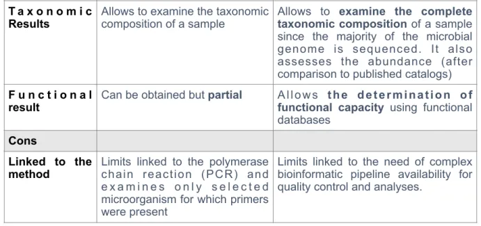
Table 1: Differences in sequencing methodologies: focus on pros and cons
16S-pyrosequencing Shotgun metagenomic sequencing Method 16S pyrosequencing approach
amplifies and further sequences the highly conserved 16S-rDNA gene, present in every bacterium thus enabling the design of universal primer
Untargeted shotgun sequencing breaks the complete DNA material from a sample, into small and defined-size pieces that are subsequently sequenced
Pros
Use widely used Increasingly used
Table 2: RCT using probiotic to improve NAFLD T a x o n o m i c
Results Allows to examine the taxonomic composition of a sample Allows to examine the complete taxonomic composition of a sample
since the majority of the microbial genome is sequenced. It also assesses the abundance (after comparison to published catalogs) F u n c t i o n a l
result Can be obtained but partial A l l o w s t h e d e t e r m i n a t i o n o f functional capacity using functional
databases Cons
Linked to the
method Limits linked to the polymerase chain reaction (PCR) and e x a m i n e s o n l y s e l e c t e d microorganism for which primers were present
Limits linked to the need of complex bioinformatic pipeline availability for quality control and analyses.
Probiotic P a t i e n t ’ s
number
Design RCT
Duration Effects
Lepicol probiotic formula:
Lactobacillusplantarum, Lactobacillus deslbrueckii, Lactobacillus acidophilus, Lactobacillus rhamnosus and
Bifidobacterium bifidum 20 Adults with biopsy-proven NAFLD Vs.
placebo 6 M ↓ intra-hepatic triglyceride content (proton-magnetic resonance spectroscopy)87
L. acidophilus CBT LA1, L. rhamnosus CBT LR5, L. paracasei CBT LPC5, P. pentosaceus CBT SL4, B. lactis CBT BL3 and B. breve CBT BR3 75 Adults with biopsy-proven NAFLD Vs.
placebo 12W ↓ intra-hepatic triglyceride content MRI-PDFF88
Bifidobacterium, Lactobacillus and
Enterococcus 120 Adults with biopsy-proven NAFLD
Vs.
placebo 3M ↓ steatosis (US) ↓ liver enzyme ↓endotoxemia89
VSL#3 = Streptococcus
thermophilus, bifidobacteria [B. breve, B. infantis, B. longum], Lactobacillus acidophilus, L. plantarum, L. paracasei, and L. delbrueckii subsp. Bulgaricus
44 Children with biopsy-proven NAFLD Vs.
VSL#3 = Streptococcus
thermophilus, bifidobacteria [B. breve, B. infantis, B. longum], Lactobacillus acidophilus, L. plantarum, L. paracasei, and L. delbrueckii subsp. Bulgaricus
22 NAFLD Adults with biopsy-proven NAFLD Vs. VSL#3 given to 56 patients with other liver disease 3M ↓ liver enzyme91
Lactobacillus bulgaricus and
Streptococcus thermophilus 30 adults with biopsy-proven NAFLD
Vs.
starch 3M ↓ liver enzyme
94
Multiprobiotic
“Symbiter” (concentrated biomass of 14 probiotic bacteria genera Bifidobacterium, Lactobacillus, Lactococcus, Propionibacterium) 58 adults with T2D with US-proven NAFLD Vs.
placebo 8W ↓ fatty liver index (FLI) ↓ liver enzyme95
On-going RCT trial:
(Lactobacillus acidophillus,
Bifidobacterium lactis, Lactobacillus rhamnosus, Lactobacillus paracasei) 46 adults with/without biopsy-proven NAFLD Vs.
Table 3: Mediterranean diet composition 170
Legend
Figure 1: NAFLD pathophysiology: focus on the liver axis and the effects of probiotics
HFD or western diet induces gut microbiota dysbiosis and is associated with increased intestinal permeability (due to reduced intestinal tight junctions), which translates into translocation of bacteria and LPS leading to increased endotoxemia. LPS triggers TLR4, activates inflammatory pathways in the liver and increased lipogenesis which translates into NAFLD development and its exacerbation into NASH. Probiotics have proven beneficial to improve gut barrier function, reduce endotoxemia and reduce TLR4 activation thus leading to improved NAFLD features.
Figure 2: Effects of other therapeutic options modulating the gut microbiota and the gut liver axis to improve NAFLD
Mostly originating from murine studies, polyphenols, exercise and prebiotics improve some features of NAFLD via their action on the gut-liver axis. Polyphenols, prebiotics and exercise improve NAFLD through improvement of gut microbiota dysbiosis, restore the gut barrier function, decrease endotoxemia. Prebiotics and exercise also reduce liver lipogenesis.
High intake H i g h intake H i g h intake Moderate intake Moderate intake Moderate intake L o w intake L o w intake Vegetables Cereals C o l d pressed olive oils
Meat Red wine D a i r y product
Sweets eggs
Legumes Nuts Fish
Acknowledgments
EU litmus grant (to M. N.and K.C), Also the authors were additionally supported by Le Ducq consortium grant 17CVD01 to M. N. and K.C., JPI-HDHL MICRODIET consortium grant to M.N and K.C. JAW received a grant from Bettencourt shueller foundation. Finally, M. Nieuwdorp is supported by a personal ZONMW-VIDI grant 2013 (016.146.327), M.W is supported by a CVON grant (CVON 2O18.27). M.W. is owner of Nature Plus.
References
1. Estes C, Razavi H, Loomba R, et al. Modeling the epidemic of nonalcoholic fatty liver disease demonstrates an exponential increase in burden of disease. Hepatology 2018;67:123–133.
2. European Association for the Study of the Liver (EASL), European Association for the Study of Diabetes (EASD), European Association for the Study of Obesity (EASO). EASL-EASD-EASO Clinical Practice Guidelines for the management of non-alcoholic fatty liver disease. J Hepatol 2016;64:1388–1402.
3. Barrera F, George J. The role of diet and nutritional intervention for the management of patients with NAFLD. Clin Liver Dis 2014;18:91–112.
4. Day CP, James OF. Steatohepatitis: a tale of two “hits”? Gastroenterology 1998;114:842–845.
5. Neuschwander-Tetri BA. Hepatic lipotoxicity and the pathogenesis of nonalcoholic steatohepatitis: the central role of nontriglyceride fatty acid metabolites. Hepatology 2010;52:774–788.
6. Bäckhed F, Ding H, Wang T, et al. The gut microbiota as an environmental factor that regulates fat storage. Proc Natl Acad Sci USA 2004;101:15718–15723.
7. Ridaura VK, Faith JJ, Rey FE, et al. Gut microbiota from twins discordant for obesity modulate metabolism in mice. Science 2013;341:1241214.
8. De Filippo C, Cavalieri D, Di Paola M, et al. Impact of diet in shaping gut microbiota revealed by a comparative study in children from Europe and rural Africa. Proc Natl Acad Sci USA 2010;107:14691–14696.
9. Wu GD, Chen J, Hoffmann C, et al. Linking long-term dietary patterns with gut microbial enterotypes. Science 2011;334:105–108.
10. Falony G, Joossens M, Vieira-Silva S, et al. Population-level analysis of gut microbiome variation. Science 2016;352:560–564.
11. Cotillard A, Kennedy SP, Kong LC, et al. Dietary intervention impact on gut microbial gene richness. Nature 2013;500:585–588.
12. Thomas V, Clark J, Doré J. Fecal microbiota analysis: an overview of sample collection methods and sequencing strategies. Future Microbiol 2015;10:1485–1504.
13. Le Chatelier E, Nielsen T, Qin J, et al. Richness of human gut microbiome correlates with metabolic markers. Nature 2013;500:541–546.
14. Aron-Wisnewsky J, Prifti E, Belda E, et al. Major microbiota dysbiosis in severe obesity: fate after bariatric surgery. Gut 2018.
15. Vandeputte D, Kathagen G, D’hoe K, et al. Quantitative microbiome profiling links gut community variation to microbial load. Nature 2017;551:507–511.
16. Marjot T, Moolla A, Cobbold JF, et al. Non-alcoholic fatty liver disease in adults: Current concepts in etiology, outcomes and management. Endocr Rev 2019. 17. Zhang H, DiBaise JK, Zuccolo A, et al. Human gut microbiota in obesity and after
gastric bypass. Proc Natl Acad Sci USA 2009;106:2365–2370.
18. Dao MC, Everard A, Aron-Wisnewsky J, et al. Akkermansia muciniphila and improved metabolic health during a dietary intervention in obesity: relationship with gut microbiome richness and ecology. Gut 2015.
19. Allin KH, Tremaroli V, Caesar R, et al. Aberrant intestinal microbiota in individuals with prediabetes. Diabetologia 2018;61:810–820.
20. Kayser BD, Prifti E, Lhomme M, et al. Elevated serum ceramides are linked with obesity-associated gut dysbiosis and impaired glucose metabolism. Metabolomics 2019;15:140.
21. Karlsson FH, Tremaroli V, Nookaew I, et al. Gut metagenome in European women with normal, impaired and diabetic glucose control. Nature 2013;498:99–103.
22. Qin J, Li Y, Cai Z, et al. A metagenome-wide association study of gut microbiota in type 2 diabetes. Nature 2012;490:55–60.
23. Pedersen HK, Gudmundsdottir V, Nielsen HB, et al. Human gut microbes impact host serum metabolome and insulin sensitivity. Nature 2016;535:376–381.
24. Koh A, Molinaro A, Ståhlman M, et al. Microbially Produced Imidazole Propionate Impairs Insulin Signaling through mTORC1. Cell 2018;175:947-961.e17.
25. Yki-Järvinen H. Non-alcoholic fatty liver disease as a cause and a consequence of metabolic syndrome. Lancet Diabetes Endocrinol 2014;2:901–910.
26. Brandl K, Schnabl B. Intestinal microbiota and nonalcoholic steatohepatitis. Curr Opin Gastroenterol 2017;33:128–133.
27. Tilg H, Zmora N, Adolph TE, et al. The intestinal microbiota fuelling metabolic inflammation. Nat Rev Immunol 2019.
28. Hoyles L, Fernández-Real J-M, Federici M, et al. Molecular phenomics and metagenomics of hepatic steatosis in non-diabetic obese women. Nat Med 2018;24:1070–1080.
29. Shen F, Zheng R-D, Sun X-Q, et al. Gut microbiota dysbiosis in patients with non-alcoholic fatty liver disease. HBPD INT 2017;16:375–381.
30. Raman M, Ahmed I, Gillevet PM, et al. Fecal microbiome and volatile organic compound metabolome in obese humans with nonalcoholic fatty liver disease. Clin Gastroenterol Hepatol 2013;11:868-875.e1–3.
31. Loomba R, Seguritan V, Li W, et al. Gut Microbiome-Based Metagenomic Signature for Non-invasive Detection of Advanced Fibrosis in Human Nonalcoholic Fatty Liver Disease. Cell Metab 2017;25:1054-1062.e5.
32. Zhu L, Baker SS, Gill C, et al. Characterization of gut microbiomes in nonalcoholic steatohepatitis (NASH) patients: a connection between endogenous alcohol and NASH. Hepatology 2013;57:601–609.
33. Wang B, Jiang X, Cao M, et al. Altered Fecal Microbiota Correlates with Liver Biochemistry in Nonobese Patients with Non-alcoholic Fatty Liver Disease. Sci Rep 2016;6:32002.
34. Del Chierico F, Nobili V, Vernocchi P, et al. Gut microbiota profiling of pediatric nonalcoholic fatty liver disease and obese patients unveiled by an integrated meta-omics-based approach. Hepatology 2017;65:451–464.
35. Wong VW-S, Tse C-H, Lam TT-Y, et al. Molecular characterization of the fecal microbiota in patients with nonalcoholic steatohepatitis--a longitudinal study. PLoS ONE 2013;8:e62885.
36. Boursier J, Mueller O, Barret M, et al. The severity of nonalcoholic fatty liver disease is associated with gut dysbiosis and shift in the metabolic function of the gut microbiota. Hepatology 2016;63:764–775.
37. Dewulf EM, Cani PD, Claus SP, et al. Insight into the prebiotic concept: lessons from an exploratory, double blind intervention study with inulin-type fructans in obese women. Gut 2013;62:1112–1121.
38. Caussy C, Tripathi A, Humphrey G, et al. A gut microbiome signature for cirrhosis due to nonalcoholic fatty liver disease. Nat Commun 2019;10:1406.
39. Qin N, Yang F, Li A, et al. Alterations of the human gut microbiome in liver cirrhosis. Nature 2014;513:59–64.
40. Mouzaki M, Comelli EM, Arendt BM, et al. Intestinal microbiota in patients with nonalcoholic fatty liver disease. Hepatology 2013;58:120–127.
41. Michail S, Lin M, Frey MR, et al. Altered gut microbial energy and metabolism in children with non-alcoholic fatty liver disease. FEMS Microbiol Ecol 2015;91:1–9.
42. Da Silva HE, Teterina A, Comelli EM, et al. Nonalcoholic fatty liver disease is associated with dysbiosis independent of body mass index and insulin resistance. Sci Rep
2018;8:1466.
43. Caussy C, Soni M, Cui J, et al. Nonalcoholic fatty liver disease with cirrhosis increases familial risk for advanced fibrosis. J Clin Invest 2017;127:2697–2704.
44. Rothschild D, Weissbrod O, Barkan E, et al. Environment dominates over host genetics in shaping human gut microbiota. Nature 2018;555:210–215.
45. Henao-Mejia J, Elinav E, Jin C, et al. Inflammasome-mediated dysbiosis regulates progression of NAFLD and obesity. Nature 2012;482:179–185.
46. Le Roy T, Llopis M, Lepage P, et al. Intestinal microbiota determines development of non-alcoholic fatty liver disease in mice. Gut 2013;62:1787–1794.
47. Chiu C-C, Ching Y-H, Li Y-P, et al. Nonalcoholic Fatty Liver Disease Is Exacerbated in High-Fat Diet-Fed Gnotobiotic Mice by Colonization with the Gut Microbiota from Patients with Nonalcoholic Steatohepatitis. Nutrients 2017;9.
48. Arab JP, Arrese M, Trauner M. Recent Insights into the Pathogenesis of Nonalcoholic Fatty Liver Disease. Annu Rev Pathol 2018;13:321–350.
49. Mouzaki M, Loomba R. Insights into the evolving role of the gut microbiome in
nonalcoholic fatty liver disease: rationale and prospects for therapeutic intervention. Therap Adv Gastroenterol 2019;12:1756284819858470.
50. Tripathi A, Debelius J, Brenner DA, et al. The gut-liver axis and the intersection with the microbiome. Nat Rev Gastroenterol Hepatol 2018;15:397–411.
51. Aron-Wisnewsky J, Gaborit B, Dutour A, et al. Gut microbiota and non-alcoholic fatty liver disease: new insights. Clin Microbiol Infect 2013;19:338–348.
52. Kolodziejczyk AA, Zheng D, Shibolet O, et al. The role of the microbiome in NAFLD and NASH. EMBO Mol Med 2019;11.
53. Leung C, Rivera L, Furness JB, et al. The role of the gut microbiota in NAFLD. Nat Rev Gastroenterol Hepatol 2016;13:412–425.
54. Anjani K, Lhomme M, Sokolovska N, et al. Circulating phospholipid profiling identifies portal contribution to NASH signature in obesity. J Hepatol 2015;62:905–912.
55. Chávez-Talavera O, Tailleux A, Lefebvre P, et al. Bile Acid Control of Metabolism and Inflammation in Obesity, Type 2 Diabetes, Dyslipidemia, and Nonalcoholic Fatty Liver Disease. Gastroenterology 2017;152:1679-1694.e3.
56. Ji Y, Yin Y, Li Z, et al. Gut Microbiota-Derived Components and Metabolites in the Progression of Non-Alcoholic Fatty Liver Disease (NAFLD). Nutrients 2019;11.
57. Canfora EE, Meex RCR, Venema K, et al. Gut microbial metabolites in obesity, NAFLD and T2DM. Nat Rev Endocrinol 2019;15:261–273.
58. Dumas M-E, Barton RH, Toye A, et al. Metabolic profiling reveals a contribution of gut microbiota to fatty liver phenotype in insulin-resistant mice. Proc Natl Acad Sci USA 2006;103:12511–12516.
59. Yuan J, Chen C, Cui J, et al. Fatty Liver Disease Caused by High-Alcohol-Producing Klebsiella pneumoniae. Cell Metab 2019;30:675-688.e7.
60. Volynets V, Küper MA, Strahl S, et al. Nutrition, intestinal permeability, and blood ethanol levels are altered in patients with nonalcoholic fatty liver disease (NAFLD). Dig Dis Sci 2012;57:1932–1941.
61. Caussy C, Hsu C, Lo M-T, et al. Link between gut-microbiome derived metabolite and shared gene-effects with hepatic steatosis and fibrosis in NAFLD. Hepatology 2018. 62. Luther J, Garber JJ, Khalili H, et al. Hepatic Injury in Nonalcoholic Steatohepatitis
Contributes to Altered Intestinal Permeability. Cell Mol Gastroenterol Hepatol 2015;1:222–232.
63. Giorgio V, Miele L, Principessa L, et al. Intestinal permeability is increased in children with non-alcoholic fatty liver disease, and correlates with liver disease severity. Dig Liver Dis 2014;46:556–560.
64. Miele L, Valenza V, La Torre G, et al. Increased intestinal permeability and tight
junction alterations in nonalcoholic fatty liver disease. Hepatology 2009;49:1877–1887. 65. Cani PD, Amar J, Iglesias MA, et al. Metabolic endotoxemia initiates obesity and insulin