HAL Id: inserm-00586840
https://www.hal.inserm.fr/inserm-00586840
Submitted on 18 Apr 2011
HAL is a multi-disciplinary open access
archive for the deposit and dissemination of
sci-entific research documents, whether they are
pub-lished or not. The documents may come from
teaching and research institutions in France or
abroad, or from public or private research centers.
L’archive ouverte pluridisciplinaire HAL, est
destinée au dépôt et à la diffusion de documents
scientifiques de niveau recherche, publiés ou non,
émanant des établissements d’enseignement et de
recherche français ou étrangers, des laboratoires
publics ou privés.
with Dioxin-Mediated Up-Regulation of CYP1A1
Activity.
David Gilot, Nolwenn Le Meur, Fanny Giudicelli, Marc Le Vée, Dominique
Lagadic-Gossmann, Nathalie Théret, Olivier Fardel
To cite this version:
David Gilot, Nolwenn Le Meur, Fanny Giudicelli, Marc Le Vée, Dominique Lagadic-Gossmann, et
al.. RNAi-Based Screening Identifies Kinases Interfering with Dioxin-Mediated Up-Regulation of
CYP1A1 Activity.. PLoS ONE, Public Library of Science, 2011, 6 (3), pp.e18261.
�10.1371/jour-nal.pone.0018261�. �inserm-00586840�
RNAi-Based Screening Identifies Kinases Interfering with
Dioxin-Mediated Up-Regulation of CYP1A1 Activity
David Gilot1*¤, Nolwenn Le Meur1, Fanny Giudicelli1, Marc Le Ve´e1, Dominique Lagadic-Gossmann1, Nathalie The´ret1, Olivier Fardel1,2
1 EA 4427 Signalisation et Re´ponse aux Agents Infectieux et Chimiques, Universite´ de Rennes 1, Institut de Recherche Sante´, Environnement et Travail; Institut Fe´de´ratif de Recherche 140, Rennes, France,2 De´partement He´matologie, Immunologie et The´rapie Cellulaire, Hopital Pontchaillou, CHU Rennes, Rennes, France
Abstract
Background:The aryl hydrocarbon receptor (AhR) is a transcription factor activated by several environmental pollutants, such as 2,3,7,8-tetrachlorodibenzo-p-dioxin (TCDD), and involved in carcinogenesis and various physiological processes, including immune response and endocrine functions. Characterization of kinases-related AhR transduction pathway remains an important purpose.
Results:We performed a kinome-wide siRNA screen in human mammary MCF-7 cells to identify non redundant protein kinases implicated in the up-regulation of cytochrome P-450 (CYP) 1A1 activity, an AhR referent target, in response to TCDD exposure. To this aim, we monitored CYP1A1-related ethoxyresorufin-O-deethylase (EROD) activity and quantified cell density. This normalization was crucial since it allowed us to focus only on siRNA affecting EROD activity and discard siRNA affecting cell density. Analyses of the cell density data allowed us to identify several hits already well-characterized as effectors of the cell cycle and original hits. Collectively, these data fully validated the protocol and the siRNA library. Next, 22 novel candidates were identified as kinases potentially implicated in the up-regulation of CYP1A1 in response to TCDD, without alteration of cell survival or cell proliferation. The siRNA library screen gave a limited number of hits (approximately 3%). Interestingly, four of them are able to bind calmodulin among which the IP3 kinase A (ITPKA) and pregnancy up-regulated non-ubiquitously expressed CaM kinase (PNCK, also named CaMKIb). Remarkably, for both proteins, their kinase activity depends on the calmodulin binding. Involvement of ITPKA and PNCK in TCDD-mediated CYP1A1 up-regulation was further validated by screening-independent expression knock-down. PNCK was finally shown to regulate activation of CaMKIa, a CaMKI isoform previously reported to interplay with the AhR pathway.
Conclusions:These data fully support a role for both IP3-related kinase and CaMK isoforms in the AhR signaling cascade. More generally, this study also highlights the interest of large scale loss-of-function screens for characterizing the molecular mechanism of action of environmental contaminants.
Citation: Gilot D, Le Meur N, Giudicelli F, Le Ve´e M, Lagadic-Gossmann D, et al. (2011) RNAi-Based Screening Identifies Kinases Interfering with Dioxin-Mediated Up-Regulation of CYP1A1 Activity. PLoS ONE 6(3): e18261. doi:10.1371/journal.pone.0018261
Editor: Eliana Abdelhay, Instituto Nacional de Caˆncer, Brazil
Received January 10, 2011; Accepted February 23, 2011; Published March 29, 2011
Copyright: ß 2011 Gilot et al. This is an open-access article distributed under the terms of the Creative Commons Attribution License, which permits unrestricted use, distribution, and reproduction in any medium, provided the original author and source are credited.
Funding: This work was supported by the Programme National de Recherche sur les Perturbateurs Endocriniens (PNRPE) from the Ministe`re de l’Ecologie, de l’Energie, du De´veloppement Durable et de la Mer (http://www.developpement-durable.gouv.fr). The funders had no role in study design, data collection and analysis, decision to publish, or preparation of the manuscript.
Competing Interests: The authors have declared that no competing interests exist. * E-mail: [email protected]
¤ Current address: CNRS UMR 6061 Institut de Ge´ne´tique et De´veloppement de Rennes, Equipe Re´gulation Transcriptionnelle et Oncogene`se, Rennes, France
Introduction
Halogenated aromatic hydrocarbons (AH) such as TCDD (2,3,7,8-tetrachlorodibenzo-p-dioxin) represent a major class of environmentally relevant toxicants. In mice and rats, TCDD exposure leads to a broad spectrum of adverse effects including the development of cancers and the impairment of endocrine functions and immunity. TCDD similarly exerts various major deleterious effects towards human health [1,2,3]. Using aryl hydrocarbon receptor (AhR)2/2mice, the pleiotropic toxicity of TCDD has been mainly linked to AhR, a basic-helix-loop-helix (bHLH) transcription factor activated after binding to cognate ligands exemplified by TCDD [4]. AhR also has important physiological roles, although the endogenous ligand(s) mediating
these functions is/are as yet unidentified. For example, in AhR2/2 mice, vascular differentiation is severely disrupted, reproductive ability is impaired, and liver and immune abnormalities are observed [5].
After TCDD binding, AhR moves to the nucleus, dissociates from the chaperone complex, and forms a heterodimer with the AhR nuclear translocator [1]. This heterodimer binds to specific xenobiotic responsive elements found in the promoter of target genes and subsequently regulates their transcription [6]. In this way, TCDD and other AhR agonists markedly induce expression of the drug-metabolizing enzyme cytochrome P-450 (CYP) CYP1A1, which is commonly considered as a paradigm of AhR gene targets and known to detoxify and to bioactivate carcinogens [1]. AH toxicity appears to be fundamentally a consequence of dysregulation
of gene expression mediated by AhR [4,7]. Therefore the complete elucidation of AhR signaling remains an important issue.
In response to xenobiotic exposure, including TCDD and other AhR agonists, a large number of signaling pathways seems to be modulated, especially second messengers [8,9] and protein kinases [10,11]. However, the involvement of their modulation in regulation of target genes by xenobiotics, including that of CYP1A1 in response to TCDD, remains relatively poorly documented. These studies are recurrently based on chemical inhibition of these signaling mediators [10], although chemical inhibitors are rarely specific of only one family of kinases [12,13]. Moreover, we and others have demonstrated that caution may be required when using chemical inhibitors to study AhR signaling since many of them are agonist/ antagonist of AhR and/or inhibitor of CYP1A1 activity [14,15,16]. Ideally, a combination of chemical and genetic (knock-down or knock-out) inhibitions should be used to demonstrate the role of a candidate. To our knowledge, rare candidates have been identified via this double approach. Among them, members of the calmodulin-dependent protein kinase (CaMK) family are particularly interesting [17,18,19]. CaMKs correspond to structurally-related serine/ threonine protein kinases that play important roles in proliferation [20] and differentiation [21]. We previously reported that activity of CaMKIa, one of the CaMK isoforms, is most likely required for TCDD-triggered nuclear translocation of AhR and subsequent up-regulation of AhR target genes, especially of CYP1A1 [19]. Such data therefore highlight the Ca2+/CaM/CaMKIa pathway as an important contributing factor to AhR function. It has also been hypothesized that other kinases, including c-src [22], CDK4 [23] and MAPKs [24], may be involved in the AhR signaling pathway. In order to better and more extensively characterize the implication of kinases in the AhR cascade, we have analyzed the consequences of kinase expression knock-down on TCDD-mediated up-regulation of CYP1A1 activity, using a siRNA library covering the human kinome. This led us to identify 22 novel candidates as kinases potentially implicated in the up-regulation of CYP1A1 in response to TCDD, without alteration of cell survival or cell proliferation.
Results
A kinome RNAi-based screen allows excluding protein kinases implicated in MCF-7 cell survival and proliferation
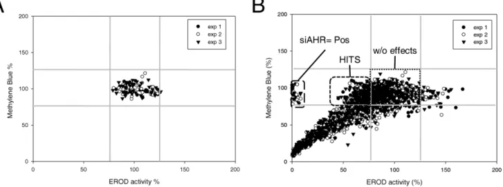
To identify nonredundant determinants of the up-regulation of CYP1A1-related ethoxyresorufin O-deethylase (EROD) activity in response to TCDD exposure, we designed a robust siRNA screen targeting 712 kinases of the human kinome. SiRNA were rehydrated and reverse transfected into the ERa-positive breast cancer cell line MCF-7 using Dharmafect I (Fig. 1). After 3 days, cells were exposed to 5 nM TCDD for 6 h, which usually allowed a strong induction of CYP1A1 transcription and consecutive EROD activity [19]. EROD activity was measured in kinetic in the presence of the CYP1A1 substrate ethoxyresorufin. To identify siRNA affecting cell proliferation and/or cell survival, cell density was evaluated using Methylene blue dye after the analysis of EROD activity. This normalization was crucial in our screen since it allowed us to focus only on siRNA affecting EROD activity determination and discard siRNA affecting cell density. The two sets of data (EROD activity and Methylene blue assays) were collected and analyzed using the R package cellHTS2 especially developed for cell-based high-throughput RNAi screens [25]. The methylene blue values and EROD activity values have been represented in the same plot (in x and y axis, respectively), for the negative controls (Fig. 2A) and all siRNA (Fig. 2B). These plots allowed us to visualize the global distribution of all siRNA in a
same plot (Fig. 2B) and to quickly distinguish the proportion of hits. Negative controls are constituted of non-targeting siRNA (NT1)-transfected cells and Dharmafect1-exposed cells (without transfec-tion of siRNA, (Fig. 1)). Positive controls correspond to MCF-7 cells transfected with siRNA targeting AhR, which abolished EROD activity without affecting cell density, demonstrating the efficiency of our protocol (Fig. 2B and Fig. S1). A majority of siRNA did not display any effect (Fig. 2B), as found in numerous RNAi screens [26]. A large part of siRNA affected cell survival and/or cell proliferation leading to a logical reduction of EROD activity. For the three experiments, results were quite similar (correlation factors: experiment 1 versus experiment 2: 0.79, experiment 2 versus experiment 3: 0.81 and experiment 1 versus experiment 3: 0.78, Fig. S1). It is noteworthy that each siRNA was looked at as independent of the two other siRNA targeting the same kinase, knowing that three independent screens were performed.
To further evaluate the quality of the screen and the siRNA library, a cellHTS2 analysis has been performed based on Methylene blue data (Fig. S2, Tables S1 and S2). These data have been next annotated (http://kinase.com/human/kinome/) [27], allowing the identification of 3 overrepresented kinase families (Tec, Aur and BRD) (Fig. S2C). Among these candidates, several are already known to be implicated in cell proliferation and carcinogenesis, such as Aurora kinase B and ALK [28,29]. Interestingly, various kinases identified as regulator of cell proliferation and/or viability of MCF-7 cells can be targeted by chemical inhibitors (Fig. S2D). For example, BTK and ITK, that belong to the same Tec family, may be targeted by the LFM-A13 inhibitor [30], whereas the multikinase inhibitor AT9283 [31], a small-molecule inhibitor of kinases with potential antineoplastic activity and currently in phase II clinical trial, is able to block kinase activity of Aurora kinase B and JAK2. Altogether, these data highlighted the robustness of our RNAi-based screen approach based on loss-of-cell functions.
RNAi-based screen identifies putative protein kinases regulating TCDD-induced EROD (CYP1A) activity in mammary MCF-7 cells
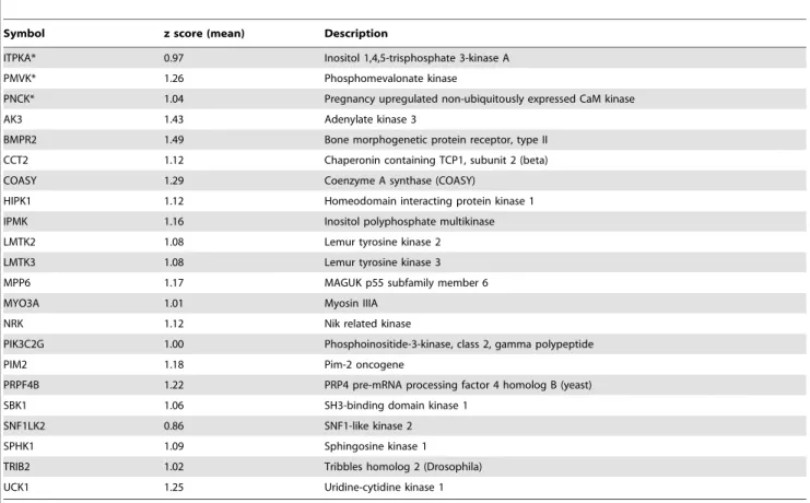
To identify kinases regulating TCDD-induced EROD activity, we performed a second cellHTS2 analysis using the preprocessing work-flow for two-channel screens (EROD and methylene blue data) (Fig. S3). At the end of this analysis, the module cellHTS2 displayed two complementary plots to observe hit distribution on quantile-quantile plot (Fig. 3A), and boxplots of z-scores (Fig. 3B) for the different types of probes (sample, Pos or Neg). The latest plot is a screen-wide image plot (Fig. 3C) to visualize the position of hits in plates (here, the picture corresponds to z-scores calculated from 3 experiments for individual siRNA). As illustrated in Fig. 3A, we selected the 150 siRNA causing the most significant reduction of EROD activity without affecting cell density. Next, we considered a kinase as a potential hit when at least 2 out of 3 siRNA were found into these 150 z-scores, since it is generally considered that observation of a phenotype caused by two distinct siRNA sequences indicates that it is unlikely to be the result of an off-target effect [32]. This led us to identify 22 kinases (Table 1) as candidates potentially implicated in up-regulation of EROD activity in response to TCDD, without affecting cell survival and proliferation. By contrast, with our criteria (at least 2 out of 3 siRNA), we did not identified kinases which negatively regulate the AhR pathway (that is, siRNA gene knock-down that would increase EROD activity). The siRNA library screen gave therefore a limited number of hits (approximately 3%) interfering with up-regulation of CYP1 activity.
Gene ontology analysis was performed to characterize these hits (Table 2). Ten are classified as protein serine/threonine kinases
and two as protein tyrosine kinases. Interestingly, four of them are able to bind calmodulin (PNCK, ITPKA, MYO3A and SPHK1). For ITPKA (IP3 kinase A) and PNCK (pregnancy up-regulated non-ubiquitously expressed CaM kinase), the kinase activity is dependent on the calmodulin binding. In addition, two hits are able to convert IP3 to IP4 (ITPKA and IPMK). The other hits (Table 1) correspond to poorly characterized kinases (i.e. Lemur tyrosine kinase 2 and 3) or non-redundant kinases AK3, PMVK, COASY, SPHK1, MYO3A. A meta-analysis was performed to
integrate this list with gene expression data and protein-protein interaction data but no convincing associations were found (data not shown).
Silencing of ITPKA and PNCK decreases TCDD-induced EROD activity
Among the 22 kinases, we focused our attention on ITPKA and PNCK since they exhibited a significant impact on EROD activity whatever the siRNA sequences tested (i.e., 3 out of 3).
Figure 1. Kinome-scale siRNA screen. Flow diagram of the strategy used to identify kinases interfering with MCF-7 cell density and up-regulation of CYP1A1 activity in human TCDD-exposed cells. Negative controls (siNeg) are non-targeting siRNA (NT1)-transfected cells and Dharmafect1-exposed cells (without siRNA) (wells A12, B12 and G12, H12). Two positive controls are constituted of cells transfected with siRNA targeting AhR (siPos, G1, H1). doi:10.1371/journal.pone.0018261.g001
Figure 2. Validation of screen protocol. A, Scatter plot showing the global distribution of control siRNA in function of EROD activity and methylene blue data. Three experiments were shown in the same plot. For each plate, individual values of negative controls were expressed in percentage in function of the mean value of the four controls, which was set at 100%.B, Visualization of global distribution of all siRNA of the screen as explained for A. Negative controls: wells A12, B12 and G12, H12.
doi:10.1371/journal.pone.0018261.g002
Figure 3. Putative kinases affecting TCDD-induced EROD (CYP1A) activity. A, Hit distribution on quantile-quantile plot and B, boxplots of z-scores for the different types of probes (sample, Pos or Neg).C, a screen-wide image plot to visualize the position in plates of hits. Here, the picture corresponds to z-scores calculated from 3 experiments for individual siRNA. One experiment corresponds to 30 plates. Pos for siRNA targeting AhR, Neg for negative controls constituted of non-targeting siRNA (NT1)-transfected cells and Dharmafect1-exposed cells (without siRNA) and Sample for 36712 siRNA targeting mRNA of human kinases. Hits corresponded to the top 150 z-scores.
Interestingly, we and others have already shown that calcium, calmodulin, IP3 [33,34], and members of the CaMKs family [9,17–19] actively participate to the AhR/CYP1A1 cascade.
Using two siRNA out of 3 available in our library, we confirmed that knock-down of ITPKA and PNCK expression reduced CYP1A activity in TCDD-treated MCF-7 cells. In addition, to compare the effects of ITPKA and PNCK knock-down on EROD activity to our previous published data with CaMKIa [13,16,19], we also used 2 other well-characterized
siRNA sequence targeting this kinase [16,19] (siCaMKIa-1 and a-2, respectively, Fig. 4A). Knock-down consequences were compared to the same controls (siNT1 and siAhR) as those used in the screen.
The knock-down of PNCK or ITPKA significantly reduced TCDD-mediated EROD activity, thus fully validating the results of the screen (Fig. 4A). SiCaMKIa-transfected cells exposed to TCDD also displayed lower EROD activity than their TCDD-treated counterparts transfected with control siNT1, in agreement Table 1. List of 22 kinases potentially implicated in the up-regulation of EROD activity in response to TCDD.
Symbol z score (mean) Description
ITPKA* 0.97 Inositol 1,4,5-trisphosphate 3-kinase A PMVK* 1.26 Phosphomevalonate kinase
PNCK* 1.04 Pregnancy upregulated non-ubiquitously expressed CaM kinase AK3 1.43 Adenylate kinase 3
BMPR2 1.49 Bone morphogenetic protein receptor, type II CCT2 1.12 Chaperonin containing TCP1, subunit 2 (beta) COASY 1.29 Coenzyme A synthase (COASY)
HIPK1 1.12 Homeodomain interacting protein kinase 1 IPMK 1.16 Inositol polyphosphate multikinase LMTK2 1.08 Lemur tyrosine kinase 2 LMTK3 1.08 Lemur tyrosine kinase 3 MPP6 1.17 MAGUK p55 subfamily member 6 MYO3A 1.01 Myosin IIIA
NRK 1.12 Nik related kinase
PIK3C2G 1.00 Phosphoinositide-3-kinase, class 2, gamma polypeptide PIM2 1.18 Pim-2 oncogene
PRPF4B 1.22 PRP4 pre-mRNA processing factor 4 homolog B (yeast) SBK1 1.06 SH3-binding domain kinase 1
SNF1LK2 0.86 SNF1-like kinase 2 SPHK1 1.09 Sphingosine kinase 1 TRIB2 1.02 Tribbles homolog 2 (Drosophila) UCK1 1.25 Uridine-cytidine kinase 1
The mean z-scores were calculated from three individual z-scores (3 siRNA). A kinase was considered as a potential hit when 2/3 or 3/3(*) siRNA were found into the 150 top z-scores using cellHTS2 package.
doi:10.1371/journal.pone.0018261.t001
Table 2. Gene ontology of the 22 kinases potentially implicated in the up-regulation of EROD activity in response to TCDD.
GO GO Coverage Kinase P-value
calmodulin binding 4/140 = 2% SPHK1;PNCK;ITPKA;MYO3A 5.14E205 calmodulin-dependent protein kinase activity 2/14 = 14% PNCK;ITPKA 4.16E204 inositol trisphosphate 3-kinase activity 2/7 = 28% IPMK;ITPKA 1.08E204 uridine kinase activity 1/4 = 25% UCK1 9.30E203 nucleoside triphosphate adenylate kinase activity 1/1 = 100% AK3 3.26E203 phosphomevalonate kinase activity 1/1 = 100% PMVK 3.26E203 pantetheine-phosphate adenylyltransferase activity 1/1 = 100% COASY 3.26E203 D-erythro-sphingosine kinase activity 1/1 = 100% SPHK1 3.26E203 plus-end directed microfilament motor activity 1/1 = 100% MYO3A 3.26E203 Annotation has been performed from http://brcabase.icr.ac.uk. P-value: right-tailed Fischer’s exact test p-value adjusted for false discovery rate - the expected proportion of false discoveries amongst the rejected hypotheses. GO coverage: percentage of hits/GO genes.
doi:10.1371/journal.pone.0018261.t002
with our previous data [16,19]. Next, knock-down efficiencies and specificity of these siRNA were evaluated using RT-qPCR assays (Fig. 4B–E). Transfection of siRNA targeting ITPKA or PNCK resulted in a significant decreased expression of their targets (Fig. 4B and C). For siRNA targeting ITPKA, the mRNA expression level of the 2 other isoforms (ITPKB and ITPKC) of ITPK family was tested. ITPKA siRNAs were unable to decrease mRNA expression of these ITPK isoforms (Fig. 4D). In a similar
manner, PNCK siRNAs failed to decrease mRNA expression of CaMKIa (Fig. 4E).
PNCK governs phosphorylation state of CaMKIa on thr177
Considering the consequence of the knock-down of PNCK in the AhR/CYP1A1 pathway, we hypothesized that PNCK may be able to phosphorylate CaMKIa on Threonine 177. Such
Figure 4. Hit confirmation and selectivity of siRNA. A, Knock-down of ITPKA, PNCK, CaMKIa and AhR in MCF-7 cells. In this confirmation step, 2 siRNA among 3 of the library were selected for ITPKA and PNCK and effects were compared to those of 2 siRNA targeting CaMKIa and AhR (Positive controls) and to negative control (siNT1). 72 h after transfection, cells were exposed to 5 nM TCDD for 6 h and EROD activity was measured as explained in Materials and Methods. Knock-down efficiencies were evaluated by RT-qPCR assays for ITPKA (B) and PNCK (C). Evaluation of off-target effects of siRNA targeting ITPKA or PNCK through mRNA quantification of ITPKB, ITPKC (D) and CaMKIa (E). mRNA level data are expressed in function of the values of mRNA levels found in siNT1-transfected cells exposed to TCDD, arbitrarily set to 1; they correspond to the means 6 S.E.M. of three independent experiments. * p-value,0.05 compared to NT1-transfected cells.
phosphorylation allows a full activation of CaMKIa as already demonstrated in rat cells [35]. MCF-7 cells were transfected with siRNA targeting PNCK or their control (NT1) and 72 h later, cells were exposed to the calcium ionophore ionomycin, a strong activator of the Ca2+/CaM/CaMKs cascade [36]. Next, Western-blots were performed to analyze the phosphorylation state of CaMKIa on Thr177 (Fig. 5). siRNA targeting PNCK strongly affected the phosphorylation state of CaMKIa on Thr177, thus demonstrating that PNCK is able to govern the phosphorylation state of CaMKIa on Thr177 in human cells.
Discussion
To discover cellular factors implicated in up-regulation of CYP1A activity in response to TCDD exposure, we performed a kinome-wide siRNA screen, revealing 22 potential cellular targets that were not previously linked to this environmental pollutant. In addition, we provided the identification of a relevant set of kinases, which interfere with cell proliferation and should be consequently potential targets for breast cancer therapy.
In accordance with numerous screens using loss of cell functions [26], the number of hits down-modulating the up-regulation of CYP1A1 activity was rather limited (approximately 3%), mostly due to false negatives resulting either from the insufficient activity of some siRNA, from the high stability of some proteins, or from the redundancy of some gene functions. This RNAi-based screen failed to identify CaMKIa as interfering with TCDD-triggered EROD up-regulation. The reason for this discrepancy is rather unclear. It is not related to an alteration of cell proliferation (Table S1) and may reflect an inefficient knock-down of CaMKIa expression by the siRNA used in the screen.
In the present study we developed a more selective approach to identify truly kinase involved in EROD regulation. We first considered a kinase as a potential hit when at least 2 out of 3 siRNA were found into the 150 top z-scores (Table 1), since it is generally considered that observation of a phenotype caused by two distinct siRNA sequences indicates that it is unlikely to be the result of an off-target effect [32]. Second, we focused only on siRNA influencing the up-regulation of CYP1A1 activity in response to TCDD exposure, without concomitantly affecting cell survival and proliferation. We admitted the fact that human kinases may be necessary for these two processes. As example, the knock-down of MEK1(MAP2K1), which has been linked to AhR signaling pathway [37], strongly affected MCF-7 cell density, evaluated by methylene blue assay (Table S1). In addition, it
should be kept in mind that inactivation of kinases may affect the TCDD-mediated CYP1A1 activity through an AhR-unrelated manner, such as, for example, impairment of CYP1A1 protein synthesis or catalytic activity [17,38,39]. Interestingly, we have not identified kinase, which negatively regulates the AhR pathway according to our criteria (at least 2 efficient siRNA out of 3) among siRNA gene knock-down that increased EROD activity (Fig. 2B).
Among the 22 kinases identified as potential hits interfering with the up-regulation of CYP1A1 activity by our RNAi screen, we confirmed the implication of PNCK and ITPKA through screen-independent transfection of efficient siRNA.
PNCK/CaMKIb is a member of the calcium/calmodulin-dependent protein kinase family of protein serine/threonine kinases [40], known to efficiently activate CaMKIa in rat cells via Thr177 phosphorylation [35]. Interestingly, we and others previously demonstrated that Ca2+/CaM/CaMKIa pathway is likely an important contributing factor to AhR-mediated genomic response in MCF-7 cells, mouse hepatoma Hepa-1c1c7 cells and cortical neurons [16,17,18,19], without involvement of CaMK kinases (CaMKKs), the classical upstream kinases of CaMKs [16,19]. Intriguingly, the knock-down of only one CaMKI isoform, PNCK/CaMKIb or CaMKIa allowed a reduction of TCDD-induced CYP1A1 activity, but not a nearly complete inhibition as obtained with siRNA targeting AhR (Fig. 4A). These effects have likely to be compared to knock-down efficiencies (siRNA targeting PNCK, ,40–50%, CaMKIa, ,75%, AhR, ,85% at mRNA levels (Fig. 4C and data not shown)), and need further investigation to control the knock-down efficiencies at protein level. However, the knock-down of PNCK strongly attenuated the detection of phospho-CaMKIa on Thr177, suggesting that siRNA-mediated knock-downs are efficient for impairing functional kinase activities. This is in agreement with our previous study, in which we demonstrated that knock-down of CaMKIa abolished CaMKIa activity [19]. Finally, it is tempting to postulate that both PNCK and CaMKIa are required for Ca2+/ CaMKI/AhR signaling pathway (see hypothetical scheme, Fig. S4). Here, PNCK was demonstrated to be able to govern the phosphorylation state of CaMKIa on Thr177 in human cells. This suggests that PNCK, unlike CaMKKs [16,19], may activate CaMKIa cells in TCDD-treated cells.
ITPKA regulates inositol phosphate metabolism through phosphorylation of the second messenger IP3 to IP4. Interestingly, both calcium/calmodulin and protein phosphorylation mecha-nisms control its activity [41]. Here, transfection of siRNA targeting ITPKA decreased EROD activity in TCDD-treated cells. In addition, inositol polyphosphate multikinase (IPMK) [41], which is also able to produce IP4, has been identified as a hit. Therefore, at least two kinases converging to the same pathway were revealed as interfering with the up-regulation of CYP1A activity, suggesting a role for IP3/IP4 in the AhR/CYP1A1 pathway. Further investigations are required to elucidate the exact role of ITPKA, IPMK and of inositol polyphosphates into the AhR signaling cascade (see hypothetical scheme, Fig. S4).
In summary, our RNAi-based screen of the human kinome identifies 22 potential cellular kinases interfering with the up-regulation of CYP1A activity in response to TCDD, especially PNCK and ITPKA. These kinases were not linked to this environmental pollutant so far and could be new targets. Finally, in accordance with literature, our results showed evidence for the role of calcium, IP3, calmodulin and CaMK in the AhR pathway. Overall, our RNAi-based screen methodology likely represents a novel opportunity to characterize signaling pathways activated by environmental pollutants.
Figure 5. Involvement of PCNK in CaMKIa phosphorylation. MCF-7 cells were transfected with siRNA targeting PNCK or control siRNA (NT1) and 72 h later, cells were exposed to the ionophore ionomycin, a strong activator of the Ca2+/CaM/CaMKs pathway. Next, Western-blots were performed to analyze the phosphorylation state of CaMKIa on Thr177. Hsc70 was used as loading control. The data shown are representative of two independent experiments.
doi:10.1371/journal.pone.0018261.g005
Materials and Methods Chemicals and reagents
Ethoxyresorufin and methylene blue were purchased from Sigma-Aldrich (St Louis, MO). TCDD was obtained from Cambridge Isotope Laboratories (Cambridge, MA). Ionomycin was obtained from Calbiochem (La Jolla, CA). TRIzol reagent was obtained from Life Technologies (Cergy Pontoise, France). Monoclonal mouse anti-Hsc70 and rabbit anti-phospho-CaMKIa (Thr177) Abs were purchased from Santa Cruz Biotechnology (La Perray en Yvelines, France). Chemicals were commonly used as stock solution in dimethyl sulfoxide (DMSO). Final concentration of solvent did not exceed 0.2% (v/v); control cultures received the same volume of solvent as for treated counterparts.
Cell culture
The estrogen receptor positive cell line MCF-7 was purchased from European Collection of Cell Cultures (ECACC, Wiltshire, United Kingdom) and cultured in D-MEM medium with 4500 mg/ L D-glucose, 110 mg/L sodium pyruvate and non-essential amino acids, supplemented with 100 U/ml penicillin, 100 U/ml strepto-mycin and 10% fetal calf serum, as previously described [19].
Ethoxyresorufin O-deethylase (EROD) activity assay
EROD activity, corresponding to the O-deethylation of ethoxyresorufin, and mainly supported by CYP1A1 enzyme in living MCF-7 cells, was measured as previously described [19]. Briefly, MCF-7 cells were incubated in phosphate-buffered saline pH 7.4, containing 5mM ethoxyresorufin, and kinetic reading was performed at 37uC with a SpectraMax Gemini SX spectrofluorometer over a 15 min-period.
Methylene Blue Assay
Cell viability was assessed by a methylene blue colorimetric assay [42] immediately after EROD activity assay. Briefly, cells were fixed for at least 30 min in 95% ethanol. Following removal of ethanol, fixed cells were dried and stained for 30 min with methylene blue dye (1% in borate buffer). After two washes with tap water, 100mL of 0.1 N HCl per well were added. Plates were next analyzed with a spectrophotometer at 620 nm.
Reverse transfection of siRNA
The protein kinase siRNA library targeting 712 kinases of the human kinome (MISSION siRNA Human Kinase Panel, 3 siRNA per target in 3 individual wells, Table S3) has been produced and aliquoted in 96-well plates by Sigma-Genesys (St. Quentin, France). 21mer siRNA duplexes with dTdT overhangs have been produced using Rosetta Inpharmatics design algorithm. The following siRNA were purchased from Sigma-Genesys: The positive control siRNA targeting AhR (iAHR), 59-AAGUCGGU-CUCUAUGCCGCtt-39, control siRNA (iNT1) corresponding to non-targeting siRNA, 59-UGGUUUACAUGUCGACUAAtt-39, siRNAs directed against CaMKIa (CaMKIa-1, 59-GCGGUU-ACCCUCCCUUCUAtt-39, and CaMKIa-2, SASI_Hs01_ 00226307), siRNAs directed against PNCK (PNCK-2, 59-CCC-UUUGAGGACUCGAAGAtt-39, and PNCK-3, 59-GCGUCUA-CGAGAUCCGCGAtt-39) and siRNAs directed against ITPKA (ITPKA-2, 59-CCUUGUGUGCUCGACUGCAtt-39, and ITP-KA-3:59-GGCAGAAGAUCCGGACCAUtt-39 ).
HTS Method
Per well of 96 multiwell plate, 10 pmol of siRNAs were mixed with 0.3ml of Dharmafect I reagent (PERBIO SCIENCES
FRANCE, Brebie`res) diluted in 45ml of transfection medium (Opti-MEM, Invitrogen, Paisley, UK) for 40 min at room temperature. Next, 30.000 MCF-7 cells, diluted in complete culture medium (100mL), were added per well. 72 h later, transfected cells were exposed to 5 nM TCDD for 6 h. MCF-7 cells were next used for CYP1A-related EROD activity assay and methylene blue assay.
CellHTS2 analysis
The dual-channel cell-based high-throughput screens (HTS) were analyzed using the R package cellHTS2 [25]. This package was especially developed to pre-process cell-based assays and is freely available on the Bioconductor project website (http:// bioconductor.org/packages/2.5/bioc/html/cellHTS2.html). Briefly, data were normalized, and ratio were next calculated (ERODActiv-ity/Methylene blue, Fig. S3). Then, data were summarized and finally z-scores were estimated, according to the preprocessing work-flow for two-channel screens, http://bioconductor.org/packages/ 2.5/bioc/vignettes/cellHTS2/inst/doc/twoChannels.pdf).
RNA isolation and analysis
Total RNAs, extracted using the TRIzol method, were subjected to reverse transcription-real time quantitative polymer-ase chain reaction (RT-qPCR) analyses as previously described [19]. Relative quantification of mRNA levels was performed after normalization of the total amount of cDNA tested to an 18 S RNA endogenous reference. The sequences of the primers used for RT-qPCR analysis were the following: 18S sense 59-CGCCGCTA-GAGGTGAAATTC-39, 18S antisense 59-TTGGCAAATGCTT-TCGCT-39,
ITPKA sense 59-GCATCGAGGGCATCAAGAAAG-39 ITPKA antisense GATACCGCCTCAGCACTTCC39, ITPKB sense 59-TGGCAGGACACGCAGGGAGT-39, ITPKB antisense 59-GGC-GAGTCGAAGTCGGCCAG-39, ITPKC sense 59-TGCCAGC-GATCCCGAGGACA-39, ITPKC antisense 59-GGCTGCGCTG-CTCACACTGA-39, CaMKIa sense 59-CAAAGATTTCATCC-GGCACT-39, CaMKIa antisense 59-TTGAAGGCTTGCTTCA-CTT-39, PNCK sense 59-ATGACATCTCAGAATCAGCCAA-AG-39, PNCK antisense 59-TTCCAGTGTGTCCGAGCAAAG-39. Gene expression was calculated relative to expression of 18S endogenous control, and adjusted relative to expression in siRNA control (iNT1) transfected cells.
Cellular protein extracts and immunoblotting analysis
Cellular protein extracts were prepared using a cell lysis buffer containing 50 mM HEPES, 150 mM NaCl, 1 mM EGTA, 0.1% Tween 20, 10% glycerol and 100mM phenylmethylsulfonyl fluoride and supplemented with an EDTA-free cocktail protease inhibitor (Roche Diagnostic, Meylan, France). Protein samples (50mg) were next subjected to electrophoresis in a 10% acrylamide gel and electrophoretically transferred to a nitrocellulose mem-brane (Bio-Rad, Marne la Coquette, France). After blocking with Tris-buffered saline containing 4% bovine serum albumin and 0.1% Tween 20 for 1 h at room temperature, membranes were incubated overnight at 4uC with primary Ab. After incubation with appropriate horseradish peroxidase-conjugated secondary Ab for 1 h, immuno-labelled proteins were visualized by autoradiog-raphy using chemiluminescence.
Statistical analysis
Quantitative data were usually given as means 6 SD of values from, at least, three independent experiments. Significant
differences were routinely evaluated with the paired Student’s t-test. The level of significance was p-value,0.05.
Supporting Information
Figure S1 Methylene blue pictures. Pictures of 3 plates (7-9) among 30 for the 3 independent experiments. The z-scores, obtained by using cellHTS2 package for methylene blue data, were also illustrated. Negative controls: wells A12, B12 and G12, H12.
(TIF)
Figure S2 Methylene blue data analyses. A, Flow diagram of the strategy used to identify kinases interfering with cell density in MCF-7 cells. B, a screen-wide image plot to visualize the position in plates of hits. Here, the picture corresponds to individual z-scores calculated from 3 experiments for individual siRNA. One experiment corresponds to 30 plates. Negative controls: non-targeting siRNA (NT1) transfected cells and Dharmafect1-exposed cells (without siRNA). C, hits have been annotated and next classified in function of kinase families. Here, we showed only the main represented families. * indicated that at least 2 siRNA out of 3 have been identified as ‘efficient’.D, Eight kinases were common to our kinome-wide siRNA screen in MCF-7 cells and microarrays data obtained from 22MCF-7 human breast cancers.
(TIF)
Figure S3 R Script for the cellHTS2 analysis using the preprocessing work-flow for two-channel screens (EROD and methylene blue data).
(TIF)
Figure S4 Hypothetical Schema. The diagram shows a hypothetical schema to resume the AhR signaling pathway leading to nuclear translocation of AhR and consecutive CYP1A1 up-regulation and activity (EROD). Ligands of AhR such as TCDD are known to induce a transient elevation of intracellular concentration of IP3 and calcium. Calcium and calmodulin (cam) are required for the full activation and the activity of CaMKIa, PNCK (CaMKIb) and ITPKA. Two confirmed hits have been indicated: PNCK and ITPKA. Another hit, IPMK is also able to convert IP3 to IP4. The identity of the kinase responsible of ITPKA phosphorylation on Thr311 (symbolized by CaMK?) is probably an isoform of CaMK type II or PKA [41]. (TIF)
Table S1 List of the individual z-scores for methylene blue data obtained from MCF-7 cells.
(XLS)
Table S2 List of the mean z-scores for methylene blue data obtained from MCF-7 cells.
(XLS)
Table S3 The human kinase library used for the RNAi screen. Positions and sequences of siRNA.
(XLS)
Author Contributions
Conceived and designed the experiments: DG NLM FG NT OF. Performed the experiments: DG NLM FG MLV. Analyzed the data: DG NLM FG OF. Contributed reagents/materials/analysis tools: DG NLM NT. Wrote the paper: DG OF DLG.
References
1. Barouki R, Coumoul X, Fernandez-Salguero PM (2007) The aryl hydrocarbon receptor, more than a xenobiotic-interacting protein. FEBS Lett 581: 3608–3615.
2. Okey AB, Riddick DS, Harper PA (1994) The Ah receptor: mediator of the toxicity of 2,3,7,8-tetrachlorodibenzo-p-dioxin (TCDD) and related compounds. Toxicol Lett 70: 1–22.
3. Schwarz M, Appel KE (2005) Carcinogenic risks of dioxin: mechanistic considerations. Regul Toxicol Pharmacol 43: 19–34.
4. Fernandez-Salguero PM, Hilbert DM, Rudikoff S, Ward JM, Gonzalez FJ (1996) Aryl-hydrocarbon receptor-deficient mice are resistant to 2,3,7,8-tetrachlorodibenzo-p-dioxin-induced toxicity. Toxicol Appl Pharmacol 140: 173–179.
5. McMillan BJ, Bradfield CA (2007) The aryl hydrocarbon receptor sans xenobiotics: endogenous function in genetic model systems. Mol Pharmacol 72: 487–498.
6. Swanson HI (2002) DNA binding and protein interactions of the AHR/ARNT heterodimer that facilitate gene activation. Chem Biol Interact 141: 63–76. 7. Tijet N, Boutros PC, Moffat ID, Okey AB, Tuomisto J, et al. (2006) Aryl
hydrocarbon receptor regulates distinct dioxin-dependent and dioxin-indepen-dent gene batteries. Mol Pharmacol 69: 140–153.
8. Oesch-Bartlomowicz B, Oesch F (2009) Role of cAMP in mediating AHR signaling. Biochem Pharmacol 77: 627–641.
9. N’Diaye M, Le Ferrec E, Lagadic-Gossmann D, Corre S, Gilot D, et al. (2006) Aryl hydrocarbon receptor- and calcium-dependent induction of the chemokine CCL1 by the environmental contaminant benzo[a]pyrene. J Biol Chem 281: 19906–19915.
10. Matsumura F (2009) The significance of the nongenomic pathway in mediating inflammatory signaling of the dioxin-activated Ah receptor to cause toxic effects. Biochem Pharmacol 77: 608–626.
11. Puga A, Ma C, Marlowe JL (2009) The aryl hydrocarbon receptor cross-talks with multiple signal transduction pathways. Biochem Pharmacol 77: 713–722. 12. Hunter T (2009) Tyrosine phosphorylation: thirty years and counting. Curr
Opin Cell Biol 21: 140–146.
13. Gilot D, Giudicelli F, Lagadic-Gossmann D, Fardel O (2010) Akti-1/2; an allosteric inhibitor of Akt 1 and 2; efficiently inhibits CaMKIalpha activity and aryl hydrocarbon receptor pathway. Chem Biol Interact.
14. Braeuning A, Buchmann A (2009) The glycogen synthase kinase inhibitor 3-(2,4-dichlorophenyl)-4-(1-methyl-1H-indol-3-yl)-1H-pyrrole-2,5-dione (SB216763) is a partial agonist of the aryl hydrocarbon receptor. Drug Metab Dispos 37: 1576–1580.
15. Bachleda P, Dvorak Z (2008) Pharmacological inhibitors of JNK and ERK kinases SP600125 and U0126 are not appropriate tools for studies of drug metabolism because they activate aryl hydrocarbon receptor. Gen Physiol Biophys 27: 143–145.
16. Monteiro P, Gilot D, Langouet S, Fardel O (2008) Activation of the aryl hydrocarbon receptor by the calcium/calmodulin-dependent protein kinase kinase inhibitor 7-oxo-7H-benzimidazo[2,1-a]benz[de]isoquinoline-3-carboxylic acid (STO-609). Drug Metab Dispos 36: 2556–2563.
17. Han EH, Kim HG, Im JH, Jeong TC, Jeong HG (2009) Up-regulation of CYP1A1 by rutaecarpine is dependent on aryl hydrocarbon receptor and calcium. Toxicology 266: 38–47.
18. Lin CH, Juan SH, Wang CY, Sun YY, Chou CM, et al. (2008) Neuronal activity enhances aryl hydrocarbon receptor-mediated gene expression and dioxin neurotoxicity in cortical neurons. J Neurochem 104: 1415–1429.
19. Monteiro P, Gilot D, Le Ferrec E, Rauch C, Lagadic-Gossmann D, et al. (2008) Dioxin-mediated up-regulation of aryl hydrocarbon receptor target genes is dependent on the calcium/calmodulin/CaMKIalpha pathway. Mol Pharmacol 73: 769–777.
20. Rodriguez-Mora OG, LaHair MM, McCubrey JA, Franklin RA (2005) Calcium/calmodulin-dependent kinase I and calcium/calmodulin-dependent kinase kinase participate in the control of cell cycle progression in MCF-7 human breast cancer cells. Cancer Res 65: 5408–5416.
21. Zayzafoon M (2006) Calcium/calmodulin signaling controls osteoblast growth and differentiation. J Cell Biochem 97: 56–70.
22. Enan E, Matsumura F (1996) Identification of c-Src as the integral component of the cytosolic Ah receptor complex, transducing the signal of 2,3,7,8-tetrachlorodibenzo-p-dioxin (TCDD) through the protein phosphorylation pathway. Biochem Pharmacol 52: 1599–1612.
23. Barhoover MA, Hall JM, Greenlee WF, Thomas RS (2010) Aryl hydrocarbon receptor regulates cell cycle progression in human breast cancer cells via a functional interaction with cyclin-dependent kinase 4. Mol Pharmacol 77: 195–201.
24. Tan Z, Huang M, Puga A, Xia Y (2004) A critical role for MAP kinases in the control of Ah receptor complex activity. Toxicol Sci 82: 80–87.
25. Pelz O, Gilsdorf M, Boutros M (2010) web cellHTS2: a web-application for the analysis of high-throughput screening data. BMC Bioinformatics 11: 185. 26. Mullenders J, Bernards R (2009) Loss-of-function genetic screens as a tool to
improve the diagnosis and treatment of cancer. Oncogene 28: 4409–4420. 27. Manning G, Whyte DB, Martinez R, Hunter T, Sudarsanam S (2002) The
protein kinase complement of the human genome. Science 298: 1912–1934.
28. Giet R, Petretti C, Prigent C (2005) Aurora kinases, aneuploidy and cancer, a coincidence or a real link? Trends Cell Biol 15: 241–250.
29. Piva R, Chiarle R, Manazza AD, Taulli R, Simmons W, et al. (2006) Ablation of oncogenic ALK is a viable therapeutic approach for anaplastic large-cell lymphomas. Blood 107: 689–697.
30. Mahajan S, Ghosh S, Sudbeck EA, Zheng Y, Downs S, et al. (1999) Rational design and synthesis of a novel anti-leukemic agent targeting Bruton’s tyrosine kinase (BTK), LFM-A13 [alpha-cyano-beta-hydroxy-beta-methyl-N-(2, 5-dibro-mophenyl)propenamide]. J Biol Chem 274: 9587–9599.
31. Howard S, Berdini V, Boulstridge JA, Carr MG, Cross DM, et al. (2009) Fragment-based discovery of the pyrazol-4-yl urea (AT9283), a multitargeted kinase inhibitor with potent aurora kinase activity. J Med Chem 52: 379–388. 32. Echeverri CJ, Beachy PA, Baum B, Boutros M, Buchholz F, et al. (2006)
Minimizing the risk of reporting false positives in large-scale RNAi screens. Nat Methods 3: 777–779.
33. Archuleta MM, Schieven GL, Ledbetter JA, Deanin GG, Burchiel SW (1993) 7,12-Dimethylbenz[a]anthracene activates protein-tyrosine kinases Fyn and Lck in the HPB-ALL human T-cell line and increases tyrosine phosphorylation of phospholipase C-gamma 1, formation of inositol 1,4,5-trisphosphate, and mobilization of intracellular calcium. Proc Natl Acad Sci U S A 90: 6105–6109. 34. Davila DR, Lane JL, Lauer FT, Burchiel SW (1999) Protein tyrosine kinase activation by polycyclic aromatic hydrocarbons in human HPB-ALL T cells. J Toxicol Environ Health A 56: 249–261.
35. Naito Y, Watanabe Y, Yokokura H, Sugita R, Nishio M, et al. (1997) Isoform-specific activation and structural diversity of calmodulin kinase I. J Biol Chem 272: 32704–32708.
36. Condon JC, Pezzi V, Drummond BM, Yin S, Rainey WE (2002) Calmodulin-dependent kinase I regulates adrenal cell expression of aldosterone synthase. Endocrinology 143: 3651–3657.
37. Chen S, Operana T, Bonzo J, Nguyen N, Tukey RH (2005) ERK kinase inhibition stabilizes the aryl hydrocarbon receptor: implications for transcrip-tional activation and protein degradation. J Biol Chem 280: 4350–4359. 38. Wang Y, Liao M, Hoe N, Acharya P, Deng C, et al. (2009) A role for protein
phosphorylation in cytochrome P450 3A4 ubiquitin-dependent proteasomal degradation. J Biol Chem 284: 5671–5684.
39. Oesch-Bartlomowicz B, Oesch F (2005) Phosphorylation of cytochromes P450: first discovery of a posttranslational modification of a drug-metabolizing enzyme. Biochem Biophys Res Commun 338: 446–449.
40. Gardner HP, Rajan JV, Ha SI, Copeland NG, Gilbert DJ, et al. (2000) Cloning, characterization, and chromosomal localization of Pnck, a Ca(2+)/calmodulin-dependent protein kinase. Genomics 63: 279–288.
41. Xia HJ, Yang G (2005) Inositol 1,4,5-trisphosphate 3-kinases: functions and regulations. Cell Res 15: 83–91.
42. Micheau O, Solary E, Hammann A, Martin F, Dimanche-Boitrel MT (1997) Sensitization of cancer cells treated with cytotoxic drugs to fas-mediated cytotoxicity. J Natl Cancer Inst 89: 783–789.