HAL Id: hal-01629177
https://hal.laas.fr/hal-01629177
Submitted on 6 Nov 2017HAL is a multi-disciplinary open access archive for the deposit and dissemination of sci-entific research documents, whether they are pub-lished or not. The documents may come from teaching and research institutions in France or abroad, or from public or private research centers.
L’archive ouverte pluridisciplinaire HAL, est destinée au dépôt et à la diffusion de documents scientifiques de niveau recherche, publiés ou non, émanant des établissements d’enseignement et de recherche français ou étrangers, des laboratoires publics ou privés.
Enhancing plasticity of the central nervous system :
Drugs, stem cell therapy and neuro-implants
Alice Le Friec, Anne-Sophie Salabert, Carole Davoust, Boris Demain,
Christophe Vieu, Laurence Vaysse, Pierre Payoux, Isabelle Loubinoux
To cite this version:
Alice Le Friec, Anne-Sophie Salabert, Carole Davoust, Boris Demain, Christophe Vieu, et al.. Enhanc-ing plasticity of the central nervous system : Drugs, stem cell therapy and neuro-implants. Neural Plas-ticity, Hindawi Publishing Corporation, 2017, 2017, pp.Article ID 2545736. �10.1155/2017/2545736�. �hal-01629177�
Enhancing plasticity of the central nervous system : Drugs, stem cell therapy
and neuro-implants.
Alice Le Friec1, Anne-Sophie Salabert1,2, Carole Davoust1, Boris Demain1, C Vieu3, Laurence Vaysse1,
Pierre Payoux1,4, I. Loubinoux1. 1 ToNIC, Toulouse NeuroImaging Center, Université de Toulouse, Inserm, UPS, France 2 Radiopharmacy Department, CHU Toulouse, France 3 LAAS-CNRS, Université de Toulouse, CNRS, INSA, UPS, Toulouse, France 4 Nuclear Medicine Department, CHU Toulouse, France Corresponding author: Isabelle Loubinoux UMR1214 – Inserm/UPS – ToNIC CHU PURPAN Pavillon Baudot Place du Dr Baylac 31024 Toulouse Cedex 3 France Tel.: 33(5) 62 74 61 64 Fax: 33(5) 62 74 61 63 Email: [email protected] Conflict of interest disclosure The authors declare that there is no conflict of interest regarding the publication of this paper.
Abstract
Stroke represents the first cause of adult acquired disability. Spontaneous recovery, dependent
on endogenous neurogenesis, allows for limited recovery in 50% of patients who remain
functionally dependent despite physiotherapy. Here we propose a review of novel drug
therapies with strong potential in the clinic. We will also discuss new avenues of stem cell
therapy in patients with cerebral lesion. A promising future for the development of efficient
drugs to enhance functional recovery after stroke seems evident. These drugs will have to
prove efficacy also in severely affected patients. The efficacy of stem cell engraftment has
been demonstrated, but will have to prove its potential in restoring tissue function for the
massive brain lesions that are most debilitating. New answers may lay in biomaterials, a
steadily growing field. Biomaterials should ideally resemble lesioned brain structures in
architecture and must be proven to increase functional reconnections within host tissue before
Introduction
Pathologies such as stroke remain chronically debilitating despite scientific advances in the
vast field of CNS injury. Following the acute phase, there are no effective treatments
available to patients besides physiotherapy.
It is now well-known that various mechanisms of brain plasticity occur after stroke onset,
both in the acute phase and beyond [1–6]. They may partially account for the spontaneous
recovery of motor function [7]. Therefore, drug treatments have increasingly aimed to
enhance these processes in order to improve functional recovery [8].
As for tissue repair of the lesioned area, endogenous neurogenesis does not however produce
mature neuronal and glial cells in sufficient number to completely regenerate lesioned CNS
tissue [9]. Over the last decades, this observation has led to intense focus on stem cell therapy
for the treatment of acute and focal CNS damage produced by pathologies such as stroke,
traumatic brain injury and spinal cord injury (SCI). Transplanted stem cells are expected to (i)
exert trophic effects on host tissue by secretion of beneficial factors and/or (ii) actually
replace lost tissue and establish functional short or long-distance connections with host cells.
Numerous neural and non-neural stem cell types have shown promise in experimental rodent
models of stroke [10, 11] and non-human primate (NHP) models of SCI [12]. This preclinical
evidence has allowed stem cell delivery to be clinically tested for safety and efficacy in the
treatment of stroke [13, 14], TBI [15, 16] and SCI [17]. However, stem cell trials for brain
repair have yet to show consistent results respective to efficacy and functional improvement
in Man [18].
Indeed, when considering stem cell graft within the lesion site, it is important to stress the
inhospitable nature of the tissue. Excitotoxicity, inflammatory processes, glial scar formation,
components render the lesion site unfavorable to neuroblast survival and differentiation [19,
20]. Stem cells grafted close to the brain lesion may die despite immunosuppressant therapy
[21].
A promising way to provide endogenous neuroblasts and grafted cells with a suitable
microenvironment may consist in the development of biomaterial ECM replacements and
“scaffolds” [22]. Biomaterials aiming to mimic the ECM have enhanced tissue reconstruction
in models of stroke [23]. They may also be engineered to deliver trophic factors [24] or to
guide axonal growth [25]. Implantation of biomaterial has just reached first-in-Man clinical
testing in the injured spinal cord [26].
Co-transplantation of biomaterial and stem cells has been successfully tested in preclinical
studies for the treatment of stroke in the chronic phase in rodents [27, 28]. Although the
translation of such therapies to the clinic presents technical challenges, we believe this
technology opens up exciting avenues of treatment for focal chronic brain injury.
Here we propose to review the most recent innovative drug, stem-cell and bio-material based
therapies for the treatment of CNS injuries such as those caused by stroke and SCI.
1. Drugs
a. Drugs for axon repair
Central Nervous System axons, unlike those in the Peripheric Nervous system, were long
thought to have lost their capacity for regeneration after section. This concept now seems
outdated. Many recent studies have revealed the existence of proteins, such as NOGO, within
the myelin sheath that are capable of inhibiting axonal growth, and prevent axonal
regeneration after a lesion. Drugs targeting these inhibitory proteins, such as anti-NOGOs,
have been successfully tested in rodents and primates. Steven Cramer and colleagues
anti-MAG (myelin-associated glycoprotein) antibody, in patients presenting a moderate walking
disability after stroke (0.5 m/sec on average 5 days after stroke). The drug was administered
24h and 9 days after stroke onset, and was well tolerated at the three doses tested (1, 5 or 15
mg/kg, i.v). Only the 5 mg/kg (n=9) dose significantly improved walking speed against
placebo (n=17) in a 112-day period, and recovery was particularly marked in the first 60 days
[29]. This result suggests that dose and duration of treatment may be further optimized.
Experimental testing in animals also showed that early administration within the first week
may be more efficient [30]. Unfortunately, a recent large trial on 134 patients was interrupted
for lack of efficacy despite the safety of the humanized monoclonal antibody (Cramer et al.
Stroke 2017). However, anti-NOGO or other molecules may prove efficacy of this strategy in
the future.
b. Growth factors
Growth factors such as G-CSF (Granulocyte Colony-Stimulating Factor), known to recruit
hematopoietic stem cells, have been considered for use in stroke therapy based on the
rationale that they possess such beneficial properties in the acute phase of stroke as the
inhibition of glutamate secretion, reduction of inflammation, and apoptotic and
anti-edema effects, as well as pro-angiogenesis and neurogenesis properties in the chronic phase
[31]. However, no functional improvement was evidenced in a cohort of 548 patients [32].
Similar results were found for other growth factors, such as bFGF (basic Fibroblast Growth
Factor or Trafermin), known to increase neurite growth. When administered in the acute
phase, bFGF caused systemic adverse effects and mortality. The phase II/III trial was
interrupted at 286 patients [33]. Another neurotrophic factor, Brain-Derived Neurotrophic
Factor, was shown to be toxic. Thus it is not currently feasible to consider the use of such
c. Selective Serotonin Re-uptake Inhibitors (SSRI)
Our team in Toulouse has focused on NeuroImaging as a means to develop and adapt
biomarker-based therapeutic strategies. We propose candidate biomarkers for (1) use in motor
outcome prediction [34–36] and (2) as therapeutic agents with proven efficacy as evaluated by
fMRI [37–43]. Recent work in our laboratory, which was confirmed by other teams, has
demonstrated that the ipsilesional motor cortex M1 is a key structure of motor recovery, and
is thus a suitable target for drug-, stem-cell, and non-invasive brain stimulation-based
therapies. Functional activations in the primary sensorimotor cortex may be enhanced by the
administration of monoaminergic drugs. Drug-induced hyperactivations have been positively
correlated with motor improvement, even in unique doses of treatment. However, this result
was elicited in small groups of moderately disabled stroke patients, and work must be
extended to more severely affected patients, who respond modestly to interventions. Our
group demonstrated, in a double-blind placebo controlled multicentric clinical trial of 118
patients, including heavily affected stroke patients, that fluoxetine (Prozac) treatment
significantly improves motor recovery (Fugl-meyer scale and motor NIHSS) when compared
to placebo. Functional improvement was observed, and a higher number of patients regained
independence in the treatment group (mRS, modified Rankin Score) [44]. In a recent study
with another SSRI, a similar result was found along with a 50% reduction in the 3-month
National Institutes of Health Stroke Scale compared with the baseline scores. This was
achieved in 57 patients in the citalopram and 39 patients in the placebo group (Oskouie NNR
2017). Recommendations for the design of clinical drug studies in stroke have been produced
[45]. The Cochrane review reported that while SSRIs may improve patient independence,
deficit, neurological status, as well as lessen anxiety and depression, inter-trial heterogeneity
limits the drawing of meaningful conclusions. Larger clinical trials are needed to validate
confirm treatment efficacy as well as determine optimal dose and length of treatment. To this
end, phase III trials have been launched in Australia (http://affinitytrial.org), Sweden
(http://www.effects.se) and the United Kingdom (http://focustrial.org.uk) [47], and aim to
include 6000 patients, 4176 of which have already been enrolled (Focus 3127, Affinity 334,
Effect 715). IRSS induce only minor and well-known adverse effects, and are well tolerated
in stroke patients. Although clinical evidence of efficacy is pending, the benefit to risk ratio
seems for now in favor of SSRIs prescription after ischemic stroke.
When considering the mechanism of action of this antidepressant, it is useful to evoke the
historic experiments that first evidenced concomitant firing of neurons in the raphe nucleus
during movement, leading Jacobs & Fornal to propose motor facilitation as a primary function
of the serotoninergic system [48]. It follows that the benefit of IRSS treatment may be further
enhanced by physiotherapy. Furthermore, recent studies have described other biological
effects of SSRI drugs such as anti-inflammatory properties through microglial repression and
reduction of neutrophil infiltration [49, 50], increase in BDNF secretion [51], as well as
enhancement of neurogenesis (see next chapter) and neural stem cell survival and
differentiation [52, 53], even in aged brain lesioned rats [54]. In line with the neurogenic
effect of SSRI, studies have shown that fluoxetine improves declarative memory and
increases hippocampal volume in patients suffering from post-traumatic stress disorder [55,
56].
2. Stem cell engraftment
Neurogenesis, defined as the capacity of the brain to produce new neurons, has been
evidenced in Man [57] in neurogenic brain regions, namely the dentate gyrus of the
hippocampus and in the subventricular zone of the cortex. These niches produce stem cells
and progenitor cells, that are capable of migrating to damaged cortical and/or subcortical
neuroblasts survive to reach full neuronal differentiation. Those that do often remain confined
to the lesion border and are thus incapable of replacing extensive losses of neuronal tissue.
Recent work has shown that as few as 0.2% of lost neurons are replaced [9].
Stem-cell based therapeutic strategies aim to support and/or stimulate endogenous
neurogenesis by engraftment of stem cells, most often through intravenous or intracerebral
delivery. One benefit of stem cell therapy may be the release of neuroprotective, trophic or
immunomodulatory factors by grafted cells. These so-called trophic effects occur rapidly after
engraftment and may stimulate endogenous neurogenesis, angiogenesis and
neovascularization, as well as reduce apoptosis and inflammation [60]. However, for massive
brain injury and severely affected patients, trophic effects will unlikely allow sufficient tissue
regeneration. In these cases particularly, engraftment of stem cells with a view to not only
provide trophic support, but to also replace damaged neurons and brain tissue could be
considered.
The least invasive method of stem cell delivery remains intravenous. This procedure is carried
out for the delivery of hematopoietic or mesenchymal stem cells. Clinical trials must meet
stringent GMP (Good Manufacturing Practices) norms that regulate the quality and safety of
cells for engraftment. These regulations dictate all aspects of cell origin, from the composition
of cell culture mediums (which must avoid reliance on products of animal origin), to the cell
banks from which the cells are selected, which must be genetically stable and homogenous,
and regularly tested for identity, viability and sterility.
a. Mesenchymal stem cells
i. Intravenous delivery
Mesenchymal stem cells have the advantage of being relatively easy to isolate and amplify
fat tissue than from bone marrow. Allogenic stem cell transplantation is rendered possible by
the fact that these cells do not express the Major Histocompatibility Complex (MHC) antigen.
Mesenchymal stem cells can be differentiated into many cell types (chondrocytes, osteoblasts,
osteocytes, adipocytes, myocytes, tendinocytes…) and possess capacity for migration toward
damaged tissue in the brain [61]. Intravenous administration of adult mesenchymal stem cells
has proven safe thus far [62–64] and potentially efficient. A recent study found that
intravenous delivery of multipotent progenitor cells, although well tolerated, did not produce
significant improvement [65]. However, the number of patients included (n = 126,
intent-to-treat population) may not have provided sufficient statistical power to show modest effects.
Clinical trials to evaluate the efficacy of the approach are ongoing (Resstore trial, principal
investigator : Olivier Detante). It is likely that any beneficial properties will result from
trophic effects, which may reduce neuroinflammation in the acute phase, and support the
neovascularization within the damaged parenchyma.
ii. Intracerebral delivery
A recent phase I/2a american trial has demonstrated the safety of an intracerebral graft of
mesenchymal stem cells, genetically engineered to transiently express notch-1, a factor known
to drive neuronal differentiation [13]. 18 patients with ischemic brain damage (11 of whom
were women), of an average of 61 years old, and presenting a stable and chronic motor
deficit, received the graft between 6 and 20 months after injury and were followed for a year
(n=16). 2.5, 5 or 10 million SB263 cells produced by SanBio were injected into the
peri-infarct. Proof of concept research showed cell survival 1 month after transplantation in
cerebrolesioned animals [13]. One serious adverse event was declared (asymptomatic
sub-dural hematoma). NIHSS neurological scale, European stroke scale and Fugl-Meyer scale
ethical reasons, this study was not controlled by a group of patients receiving a control
surgical procedure.
b. Intraspinal graft of olfactory ensheathing stem cells
Autologous engraftment of olfactory ensheathing cells, harvested from the olfatory mucosa of
3 chronic medullar injury patients produced a quite spectacular improvement in American
Spinal Injury Association class (A to B or C) scores in two patients, and more local
enhancement of motricity and sensitivity in the third patient [17]. Though the mechanisms of
action of these cells are far from elucidated, it has been suggested that these “support cells”
may reduce glial scar formation, rendering the lesion site more permissive to axonal
regeneration.
c. Intracerebral graft of neural stem cells
The main challenge in tissue regeneration therapies is not only the replacement of lost
neurons, but also the establishment of functional reconnections. In this view, selecting a cell
source is difficult.
In a first phase 2 randomized clinical trial led by Kondziolka et al, the feasibility of
intracerebral stem cell engraftment in 14 stable stroke patients was demonstrated [66, 67].
Although successfully differentiated into neurons, the hNT2 (LBS-Neurons, Layton
Bioscience) stem cell line they used originates from a teratocarcinoma and is no longer
authorized for trial in Man due to its extremely abnormal caryotype. The study included a
small (n=4) group of control patients, paired for physiotherapy. Six out of eleven PET scans
evidenced an improvement of glucose intake at the implantation site (3 injections were
performed: above, within and below the lesion site). Improvement of functional recovery was
not significant in the treated group compared to controls. Four treated patients, who presented
lesions in the non-dominant hemisphere, showed enhanced performance in the figure of Rey
A recent phase 1 first-in-man study used the CTX0E03 or ReN001cell line (ReNeuron)
derived from genetically modified embryonic stem cells originating from human fetal
neuroepithelium [14]. In order to control the amplification of cells, they used c-mycERT AM
technology to drive expression of an oesdradiol receptor under tamoxifen (4-OHT) induction
(added to culture medium). Cell division is arrested and differentiation into neuronal and glial
lineages was induced by removal of tamoxifen and growth factors from the medium. It is
important to note that the use of tamoxifen for the treatment of breast cancer in women could
restart division of the transplanted cells. For this reason, women were excluded from the
protocol. Eleven men presenting a moderate to severe disability were enrolled for perilesional
grafting of 2, 5, 10 or 20 million cells 6 to 60 months after stroke onset. Patients did not
receive any immunosuppressive therapy. Patients were followed for 2 years as part of this
non-controlled trial. No immunological or adverse effects were attributed to the grafted cells.
Modest improvements of different motor scales were observed (NIHSS, Barthel index,
Ashworth Spasticity Scale for the arm and leg, and a quality of life and health status EuroQoL
Five Dimensions questionnaire EQ-5D).
Although the setup of methodologies to control trials with groups of operated-upon but
non-grafted patients poses for now unsurmountable technical and ethical difficulties, the true
efficacy of stem cell based interventions cannot be fully validated without this condition and
larger patient cohorts. Perilesional injection of cells into healthy tissue is often performed in
order to optimize stem cell survival. The rapidly occurring trophic effects of this approach is
now well established, however true functional replacement of lost cells remains to be solidly
demonstrated although difficult to test in humans.
While regenerative medicine strategies aim to replace the lesioned neural tissue by
intracerebral engraftment, the lesion site microenvironment is unconducive to progenitor
which can be replaced or isolated by scar tissue [19, 69]. Effectiveness of therapy is limited as
only 5% of grafted cells survive. An exciting solution to this problem may be produced by
nanotechnology scaffolds.
3. Neuro-implants
Biomaterials may provide a suitable support for cells, replacing lost extracellular matrix. They
may promote cell survival and differentiation, revascularisation and recolonisation of lesioned
tissue by glia and endothelium cells from the host. More complex biomimetic materials may
also guide axonal growth towards their biological targets, restoring effective and even
long-distance connections between damaged and healthy tissues. Where stroke is concerned,
research in this innovative field remains currently preclinical.
a. Injectable nanometric biomaterials
i. Nanofibers
Fibrous biomaterials of nanometric dimension were injected in scar tissue in a rat model of
medullar lesion. They were composed of peptides that auto-assemble to form fibers and
contain epitopes of laminin, an ECM component involved in processes such as cell adhesion.
Axons of the descending corticospinal tract and those of the ascending sensory neurons that
could not previously cross the fibrous glial scar were able to penetrate the biomaterial and
cross the lesion. Importantly, motor recovery was significantly enhanced in treated animals
[70]. A biodegradable and biocompatible block copolymer of Poly-lactic-co-glycolic acid and
Poly-L-lysine improves functional recovery of rats and non-human primates after a partial and
complete lateral hemisection of the thoracic spinal cord [71]. INSPIRE, a clinical trial is
ii. Hydrogels
Polymer hydrogels are another candidate biomaterial for the support of grafted cells. For
instance, polyglycolic acid (PGA) is often used as it is porous, biodegradable and entirely
synthetic, meaning its exact composition can be easily controlled. Park and coll. included
neural stem cells in a soluble hydrogel which then polymerizes within the lesion site [72].
They demonstrated convincing tissue reconstruction in a rodent model of ischemic stroke
(middle cerebral artery occlusion (MCAo) which produces massive lesions. The biomaterial is
conducive to neurite growth, and connections were evidenced between host and grafted cells.
Vascularisation, reduction of the glial scar and of monocyte infiltration was also found. This
type of approach has shown promising results for sensorimotor and cognitive recovery [73].
iii. Micrometric injectable biomaterials
- Microbeads
Easily injectable micrometric biomaterial beads have also been developed. When injected in a
rat model of Parkinson disease, they improved motricity, decreased striatal lesion volume and
reduced substantia nigra degeneration [74].
- Structured and guiding biomaterial implants
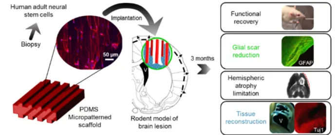
Our team has proposed a strategy for the long distance bridging of brain regions using
biomaterials seeded with neural stem cells, called neuro-implants, in collaboration with
LAAS-CNRS (Fig. 1). They are made with PDMS (polydimethylsiloxane), and
micro-structured to guide axonal growth in predefined directions (Fig. 2). We have conducted a
proof of concept study of the efficacy of neuro-implants compared to implants alone in a rat
model of corticostriatal lesion impacting the corticospinal tract, which produces loss of
forelimb strength and dexterity [75]. The implants did not increase reactive astrogliosis,
and partial tissue reconstruction within the lesion site around the implants. Reconstructed
tissue around the neuro-implants was vascularized as assessed by the HMPAO radiotracer
perfusion with SPECT imaging (Fig 3). In contrast, lesioned tissue without implants evolved
in a cystic cavity (Fig 3, red arrows). The increase in number of surviving grafted cells may
also have trophic effects on cerebral plasticity, such as growth factor and anti-inflammatory
factor secretion [76].
Fig 1: Neuro-implant concept. Guiding scaffolds located in a lesion of the corticospinal tract
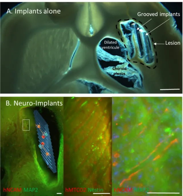
Fig. 2: Representative horizontal brain section of the lesioned area under brightfield
illumination from Implants Alone (A, Scale bar: 1 mm) and neuro-implants rats (B). The newly generated tissue was mostly located around the PDMS implants. B: Human neural stem cells were identified by a specific human marker hNCAM or hMTCO2, in combination with a marker (in green) of immature (Nestin) and mature (MAP2) neurons. Low magnification is provided on the left and higher magnifications on the right (scale bars: 100µm). Grafted cell neurites were aligned along the grooves of the implant.
Fig. 3: Measurement of cerebral blood flow by nanoSPECT Plus-CT Bioscan with
[99mTc]-HMPAO. Fifteen minutes after intravenous injection of 50 MBq of [99mTc]-HMPAO in the tail
vein of Sprague Dawley anesthetized rats, data were acquired during 7 min for SPECT (48 sec and 100 000 cps per projection, image size 276*276*164, 0.1 mm) and 1 min for CT (55 kVp, 500 msec, pitch 0,5, binning 1:4). Following the reconstruction, the CT images were spatially aligned to match the SPECT images. Processing of reconstructed images was performed with the in-house Sysiphe software [77]. Brain implants were identified on CT (blue arrows) and 3D volumes of interest (VOIs) were drawn on either side of the implants (colored rectangles) and symetric ROIs were drawn on the controlateral side as a control (not shown). Images of two rats 20 days after a corticostriatal lesion and 7 days after implantation of neuro-implants. A,E : CT-scan of brain implants (blue arrows) . One implant was inserted in rat #1 brain and 5 implants in rat #2 brain. B,F : SPECT-CT with HMPAO radiotracer on the area of brain implant. C, G : SPECT-CT with HMPAO radiotracer on the area of brain damage (located behind the implantation zone). We observed major hypoperfusion (red arrow). The presence of implants limited hypoperfusion: for rat #1, -13% in B compared to -25% in C
(ROI volume was 0.4 mm3), for rat #2, -18% in F compared to -57% in G (ROI volume was
1.5 mm3). H: sagittal view of rat #1. Coronal views B and C are located with grey and red
lines. D, I: Rat brain perfused and extracted 3 months after the lesion showing the lesion area where neuro-implants were inserted (grey arrows) or not (red arrows).
In summary, effective drug therapies are gradually becoming available to improve functional
recovery after stroke. However, these will unlikely allow spectacular gains in patients with
severe brain damage. Many research teams currently strive to demonstrate the efficacy of
stem cell transplantation, which has shown promise in many preclinical models of brain
injury. Nonetheless, stem cells alone may not repair the most extensive and debilitating
lesions. Much hope has arisen from the development of biomaterial scaffolds, a rapidly
growing field of research. These would ideally resemble the architecture of the brain in
structure [78], and be proven to allow adequate reconnections with host tissue if possible. If
not, given the complexity of this approach, they must at least provide a very high benefit
before they can be considered in a clinical setting.
Acknowledgement
We thank Carine Pestourie who carried out rat SPECT/CT experiments (Non-Invasive
Exploration Service -US006/CREFRE Inserm/UPS/ENVT Toulouse-France). We thank
Laurence Vaysse who managed cell culture and immunohistology. This work has been in part supported by a grant from the French National Agency for Research called “Investissements
References: 1. Lindvall O, Kokaia Z (2015) Neurogenesis following Stroke Affecting the Adult Brain. Cold Spring Harb Perspect Biol 7:a019034 2. Murphy TH, Corbett D (2009) Plasticity during stroke recovery: from synapse to behaviour. Nat Rev Neurosci 10:861–872 3. Jin K, Wang X, Xie L, Mao XO, Zhu W, Wang Y, Shen J, Mao Y, Banwait S, Greenberg DA (2006) Evidence for stroke-induced neurogenesis in the human brain. Proc Natl Acad Sci 103:13198– 13202 4. Hermann DM, Chopp M (2012) Promoting brain remodelling and plasticity for stroke recovery: therapeutic promise and potential pitfalls of clinical translation. Lancet Neurol 11:369–380 5. Grefkes C, Fink GR (2014) Connectivity-based approaches in stroke and recovery of function. Lancet Neurol 13:206–216 6. Liu H, Tian T, Qin W, Li K, Yu C (2016) Contrasting Evolutionary Patterns of Functional Connectivity in Sensorimotor and Cognitive Regions after Stroke. Front Behav Neurosci 10:72 7. Alia C, Spalletti C, Lai S, Panarese A, Micera S, Caleo M (2016) Reducing GABAA-mediated inhibition improves forelimb motor function after focal cortical stroke in mice. Sci Rep 6:37823 8. Chollet F, Tardy J, Albucher J-F, et al (2011) Fluoxetine for motor recovery after acute ischaemic stroke (FLAME): a randomised placebo-controlled trial. Lancet Neurol 10:123–130 9. Arvidsson A, Collin T, Kirik D, Kokaia Z, Lindvall O (2002) Neuronal replacement from endogenous precursors in the adult brain after stroke. Nat Med 8:963–970 10. Muñetón-Gómez VC, Doncel-Pérez E, Fernandez AP, Serrano J, Pozo-Rodrigálvarez A, Vellosillo-Huerta L, Taylor JS, Cardona-Gómez GP, Nieto-Sampedro M, Martínez-Murillo R (2012) Neural differentiation of transplanted neural stem cells in a rat model of striatal lacunar infarction: light and electron microscopic observations. Front Cell Neurosci. doi: 10.3389/fncel.2012.00030 11. Vaysse L, Conchou F, Demain B, Davoust C, Plas B, Ruggieri C, Benkaddour M, Simonetta-Moreau M, Loubinoux I (2015) Strength and fine dexterity recovery profiles after a primary motor cortex insult and effect of a neuronal cell graft. Behav Neurosci 129:423–434
12. Yamane J, Nakamura M, Iwanami A, et al (2010) Transplantation of galectin-1-expressing human neural stem cells into the injured spinal cord of adult common marmosets. J Neurosci Res 1481–89 13. Steinberg GK, Kondziolka D, Wechsler LR, et al (2016) Clinical Outcomes of Transplanted Modified Bone Marrow–Derived Mesenchymal Stem Cells in Stroke: A Phase 1/2a Study. Stroke 1817–24 14. Kalladka D, Sinden J, Pollock K, et al (2016) Human neural stem cells in patients with chronic ischaemic stroke (PISCES): a phase 1, first-in-man study. The Lancet 388:787–796 15. Kota DJ, Prabhakara KS, van Brummen AJ, Bedi S, Xue H, DiCarlo B, Cox CS, Olson SD (2016) Propranolol and Mesenchymal Stromal Cells Combine to Treat Traumatic Brain Injury: Propranolol and MSCs Combine to Treat TBI. Stem Cells Transl Med 5:33–44 16. Cox CS, Hetz RA, Liao GP, et al (2017) Treatment of Severe Adult Traumatic Brain Injury Using Bone Marrow Mononuclear Cells: Bone Marrow Cells for TBI. Stem Cells 35:1065–1079 17. Tabakow P, Jarmundowicz W, Czapiga B, et al (2013) Transplantation of Autologous Olfactory Ensheathing Cells in Complete Human Spinal Cord Injury. Cell Transplant 22:1591–1612 18. Boncoraglio GB, Bersano A, Candelise L, Reynolds BA, Parati EA (2010) Stem cell transplantation for ischemic stroke. In: The Cochrane Collaboration (ed) Cochrane Database Syst. Rev. John Wiley & Sons, Ltd, p CD007231 19. Burda JE, Sofroniew MV (2014) Reactive Gliosis and the Multicellular Response to CNS Damage and Disease. Neuron 81:229–248 20. Coyne TM, Marcus AJ, Woodbury D, Black IB (2006) Marrow Stromal Cells Transplanted to the Adult Brain Are Rejected by an Inflammatory Response and Transfer Donor Labels to Host Neurons and Glia. Stem Cells 24:2483–2492 21. Jablonska A, Janowski M, Lukomska B (2013) Different methods of immunosuppresion do not prolong the survival of human cord blood-derived neural stem cells transplanted into focal brain-injured immunocompetent rats. Acta Neurobiol Exp 73:88–101 22. Boisserand LSB, Kodama T, Papassin J, Auzely R, Moisan A, Rome C, Detante O (2016) Biomaterial Applications in Cell-Based Therapy in Experimental Stroke. Stem Cells Int 2016:1–14 23. Ghuman H, Massensini AR, Donnelly J, Kim S-M, Medberry CJ, Badylak SF, Modo M (2016) ECM hydrogel for the treatment of stroke: Characterization of the host cell infiltrate. Biomaterials 91:166–181 24. Emerich DF, Silva E, Ali O, Mooney D, Bell W, Yu SJ, Kaneko Y, Borlongan C (2010) Injectable VEGF Hydrogels Produce Near Complete Neurological and Anatomical Protection Following Cerebral Ischemia in Rats. Cell Transplant 19:1063–1071 25. Béduer A, Vieu C, Arnauduc F, Sol J-C, Loubinoux I, Vaysse L (2012) Engineering of adult human neural stem cells differentiation through surface micropatterning. Biomaterials 33:504–514 26. Theodore N, Hlubek R, Danielson J, Neff K, Vaickus L, Ulich TR, Ropper AE (2016) First Human Implantation of a Bioresorbable Polymer Scaffold for Acute Traumatic Spinal Cord Injury: A Clinical Pilot Study for Safety and Feasibility. Neurosurgery 79:E305–E312
27. Bible E, Dell’Acqua F, Solanky B, Balducci A, Crapo PM, Badylak SF, Ahrens ET, Modo M (2012) Non-invasive imaging of transplanted human neural stem cells and ECM scaffold remodeling in the stroke-damaged rat brain by 19F- and diffusion-MRI. Biomaterials 33:2858–2871 28. Moshayedi P, Nih LR, Llorente IL, Berg AR, Cinkornpumin J, Lowry WE, Segura T, Carmichael ST (2016) Systematic optimization of an engineered hydrogel allows for selective control of human neural stem cell survival and differentiation after transplantation in the stroke brain. Biomaterials 105:145–155 29. Cramer SC, Abila B, Scott NE, Simeoni M, Enney LA, on behalf of the MAG111539 Study Investigators (2013) Safety, Pharmacokinetics, and Pharmacodynamics of Escalating Repeat Doses of GSK249320 in Patients With Stroke. Stroke 44:1337–1342 30. Cash D, Easton AC, Mesquita M, Beech J, Williams S, Lloyd A, Irving E, Cramer SC (2016) GSK249320, A Monoclonal Antibody Against the Axon Outgrowth Inhibition Molecule Myelin-Associated Glycoprotein, Improves Outcome of Rodents with Experimental Stroke. J Neurol Exp Neurosci 2:28 31. Abe K, Yamashita T, Takizawa S, Kuroda S, Kinouchi H, Kawahara N (2012) Stem cell therapy for cerebral ischemia: from basic science to clinical applications. J Cereb Blood Flow Metab 32:1317–31 32. Bath PM, Sprigg N, England T (2013) Colony stimulating factors (including erythropoietin, granulocyte colony stimulating factor and analogues) for stroke. Cochrane Database Syst Rev. doi: 10.1002/14651858.CD005207.pub4 33. Bogousslavsky J, Victor SJ, Salinas EO, Pallay A, Donnan GA, Fieschi C, Kaste M, Orgogozo J-M, Chamorro A, Desmet A (2002) Fiblast (Trafermin) in Acute Stroke: Results of the European-Australian Phase II/III Safety and Efficacy Trial. Cerebrovasc Dis 14:239–251 34. Loubinoux I (2003) Correlation between cerebral reorganization and motor recovery after subcortical infarcts. NeuroImage 20:2166–2180 35. Tombari D, Loubinoux I, Pariente J, Gerdelat A, Albucher J-F, Tardy J, Cassol E, Chollet F (2004) A longitudinal fMRI study: in recovering and then in clinically stable sub-cortical stroke patients. NeuroImage 23:827–839 36. Loubinoux I, Dechaumont-Palacin S, Castel-Lacanal E, De Boissezon X, Marque P, Pariente J, Albucher J-F, Berry I, Chollet F (2007) Prognostic Value of fMRI in Recovery of Hand Function in Subcortical Stroke Patients. Cereb Cortex 17:2980–2987 37. Pariente J, Loubinoux I, Carel C, Albucher J-F, Leger A, Manelfe C, Rascol O, Chollet F (2001) Fluoxetine modulates motor performance and cerebral activation of patients recovering from stroke. Ann Neurol 50:718–729 38. Loubinoux I, Pariente J, Rascol O, Celsis P, Chollet F (2002) Selective serotonin reuptake inhibitor paroxetine modulates motor behavior through practice. A double-blind, placebo-controlled, multi-dose study in healthy subjects. Neuropsychologia 1815–21 39. Loubinoux I, Pariente J, Boulanouar K, Carel C, Manelfe C, Rascol O, Celsis P, Chollet F (2002) A Single Dose of the Serotonin Neurotransmission Agonist Paroxetine Enhances Motor Output: Double-Blind, Placebo-Controlled, fMRI Study in Healthy Subjects. NeuroImage 15:26–36
40. Loubinoux I, Tombari D, Pariente J, Gerdelat-Mas A, Franceries X, Cassol E, Rascol O, Pastor J, Chollet F (2005) Modulation of behavior and cortical motor activity in healthy subjects by a chronic administration of a serotonin enhancer. NeuroImage 27:299–313 41. Gerdelat-Mas A, Loubinoux I, Tombari D, Rascol O, Chollet F, Simonetta-Moreau M (2005) Chronic administration of selective serotonin reuptake inhibitor (SSRI) paroxetine modulates human motor cortex excitability in healthy subjects. NeuroImage 27:314–322 42. Loubinoux I, Chollet F (2010) Neuropharmacology in stroke recovery. In: Brain Repair Stroke. Cambridge University Press, Cambridge, pp 183–193 43. Tardy J, Pariente J, Leger A, et al (2006) Methylphenidate modulates cerebral post-stroke reorganization. NeuroImage 33:913–922 44. Chollet F (2011) Fluoxetine and motor recovery after ischaemic stroke–Author’s reply. Lancet Neurol 10:500–501 45. Chollet F, Cramer SC, Stinear C, et al (2014) Pharmacological therapies in post stroke recovery: recommendations for future clinical trials. J Neurol 261:1461–1468 46. Mead G, Hsieh C, Lee R, Kutlubaev M, Claxton A, Hankey G, Hackett M (2012) Selective serotonin reuptake inhibitors (SSRIs) for stroke recovery. - PubMed - NCBI. Cochrane Database Syst Rev 11:CD009286 47. Mead G, Hackett ML, Lundström E, Murray V, Hankey GJ, Dennis M (2015) The FOCUS, AFFINITY and EFFECTS trials studying the effect(s) of fluoxetine in patients with a recent stroke: a study protocol for three multicentre randomised controlled trials. Trials 16:369 48. Jacobs BL, Fornal CA (1997) Serotonin and motor activity. Curr Opin Neurobiol 7:820–825 49. Lim C-M, Kim S-W, Park J-Y, Kim C, Yoon SH, Lee J-K (2009) Fluoxetine affords robust neuroprotection in the postischemic brain via its anti-inflammatory effect. J Neurosci Res 87:1037–1045 50. Lee JY, Lee HE, Kang SR, Choi HY, Ryu JH, Yune TY (2014) Fluoxetine inhibits transient global ischemia-induced hippocampal neuronal death and memory impairment by preventing blood– brain barrier disruption. Neuropharmacology 79:161–171 51. Lee CH, Park JH, Yoo K-Y, Choi JH, Hwang IK, Ryu PD, Kim D-H, Kwon Y-G, Kim Y-M, Won M-H (2011) Pre- and post-treatments with escitalopram protect against experimental ischemic neuronal damage via regulation of BDNF expression and oxidative stress. Exp Neurol 229:450– 459 52. Li W-L, Cai H-H, Wang B, Chen L, Zhou Q-G, Luo C-X, Liu N, Ding X-S, Zhu D-Y (2009) Chronic fluoxetine treatment improves ischemia-induced spatial cognitive deficits through increasing hippocampal neurogenesis after stroke. J Neurosci Res 87:112–122 53. Taguchi N, Nakayama S, Tanaka M (2012) Fluoxetine has neuroprotective effects after cardiac arrest and cardiopulmonary resuscitation in mouse. Resuscitation 83:652–656 54. Buga A-M, Ciobanu O, Bădescu GM, Bogdan C, Weston R, Slevin M, Di Napoli M, Popa-Wagner A (2016) Up-regulation of serotonin receptor 2B mRNA and protein in the peri-infarcted area of aged rats and stroke patients. Oncotarget 7:17415
55. Santarelli L, Saxe M, Gross C, et al (2003) Requirement of hippocampal neurogenesis for the behavioral effects of antidepressants. science 301:805–809 56. Vermetten E, Vythilingam M, Southwick SM, Charney DS, Bremner JD (2003) Long-term treatment with paroxetine increases verbal declarative memory and hippocampal volume in posttraumatic stress disorder. Biol Psychiatry 54:693–702 57. Eriksson PS, Perfilieva E, Björk-Eriksson T, Alborn A-M, Nordborg C, Peterson DA, Gage FH (1998) Neurogenesis in the adult human hippocampus. Nat Med 4:1313–17 58. Chen J, Magavi SS, Macklis JD (2004) Neurogenesis of corticospinal motor neurons extending spinal projections in adult mice. Proc Natl Acad Sci U S A 101:16357–16362 59. Magavi SS, Leavitt BR, Macklis JD (2000) Induction of neurogenesis in the neocortex of adult mice. Nature 405:951–955 60. Dihne M, Hartung H-P, Seitz RJ (2011) Restoring Neuronal Function After Stroke by Cell Replacement: Anatomic and Functional Considerations. Stroke 42:2342–2350 61. Li Y, McIntosh K, Chen J, et al (2006) Allogeneic bone marrow stromal cells promote glial– axonal remodeling without immunologic sensitization after stroke in rats. Exp Neurol 198:313– 325 62. Lee JS, Hong JM, Moon GJ, Lee PH, Ahn YH, Bang OY (2010) A Long-Term Follow-Up Study of Intravenous Autologous Mesenchymal Stem Cell Transplantation in Patients With Ischemic Stroke. Stem Cells 28:1099–1106 63. Chen L, Zhang G, Khan AA, Guo X, Gu Y (2016) Clinical Efficacy and Meta-Analysis of Stem Cell Therapies for Patients with Brain Ischemia. Stem Cells Int 2016:1–8 64. Prasad K, Sharma A, Garg A, et al (2014) Intravenous Autologous Bone Marrow Mononuclear Stem Cell Therapy for Ischemic Stroke. Stroke 45:3618–3624 65. Hess DC, Wechsler LR, Clark WM, et al (2017) Safety and efficacy of multipotent adult progenitor cells in acute ischaemic stroke (MASTERS): a randomised, double-blind, placebo-controlled, phase 2 trial. Lancet Neurol 16:360–368 66. Kondziolka D, Steinberg GK, Wechsler L, et al (2005) Neurotransplantation for patients with subcortical motor stroke: a phase 2 randomized trial. J Neurosurg 103:38–45 67. Kondziolka D, Wechsler L, Goldstein S, et al (2000) Transplantation of cultured human neuronal cells for patients with stroke. - PubMed - NCBI. Neurology 55:565–9 68. Stilley CS, Ryan CM, Kondziolka D, Bender A, DeCesare S, Wechsler L (2004) Changes in cognitive function after neuronal cell transplantation for basal ganglia stroke. Neurology 63:1320–1322 69. Silver J, Miller JH (2004) Regeneration beyond the glial scar. Nat Rev Neurosci 5:146–156 70. Tysseling-Mattiace VM, Sahni V, Niece KL, Birch D, Czeisler C, Fehlings MG, Stupp SI, Kessler JA (2008) Self-Assembling Nanofibers Inhibit Glial Scar Formation and Promote Axon Elongation after Spinal Cord Injury. J Neurosci 28:3814–3823
71. Slotkin JR, Pritchard CD, Luque B, et al (2017) Biodegradable scaffolds promote tissue remodeling and functional improvement in non-human primates with acute spinal cord injury. Biomaterials 123:63–76 72. Park KI, Teng YD, Snyder EY (2002) The injured brain interacts reciprocally with neural stem cells supported by scaffolds to reconstitute lost tissue. Nat Biotechnol 20:1111–1117 73. Jin K, Mao X, Xie L, Galvan V, Lai B, Wang Y, Gorostiza O, Wang X, Greenberg DA (2010) Transplantation of Human Neural Precursor Cells in Matrigel Scaffolding Improves Outcome from Focal Cerebral Ischemia after Delayed Postischemic Treatment in Rats. J Cereb Blood Flow Metab 30:534–544 74. Delcroix GJ-R, Garbayo E, Sindji L, Thomas O, Vanpouille-Box C, Schiller PC, Montero-Menei CN (2011) The therapeutic potential of human multipotent mesenchymal stromal cells combined with pharmacologically active microcarriers transplanted in hemi-parkinsonian rats. Biomaterials 32:1560–1573 75. Vaysse L, Beduer A, Sol JC, Vieu C, Loubinoux I (2015) Micropatterned bioimplant with guided neuronal cells to promote tissue reconstruction and improve functional recovery after primary motor cortex insult. Biomaterials 58:46–53 76. Vaysse L, Labie C, Canolle B, Jozan S, Béduer A, Arnauduc F, Vieu C, Sol JC, Loubinoux I (2012) Adult human progenitor cells from the temporal lobe: Another source of neuronal cells. Brain Inj 26:1636–1645 77. Tensaouti F, Lotterie JA (2008) Sysiphe-Neuroimaging software toolbox. In: Eur. Soc. Magn. Reson. Med. Biol. 2008 Congr. Oct. pp 2–4 78. Álvarez Z, Castaño O, Castells AA, Mateos-Timoneda MA, Planell JA, Engel E, Alcántara S (2014) Neurogenesis and vascularization of the damaged brain using a lactate-releasing biomimetic scaffold. Biomaterials 35:4769–4781
![Fig. 3: Measurement of cerebral blood flow by nanoSPECT Plus-CT Bioscan with [99mTc]- [99mTc]-HMPAO](https://thumb-eu.123doks.com/thumbv2/123doknet/14610631.545430/17.892.121.816.122.403/fig-measurement-cerebral-blood-nanospect-plus-bioscan-hmpao.webp)
