HAL Id: hal-02472974
https://hal.sorbonne-universite.fr/hal-02472974
Submitted on 10 Feb 2020
HAL is a multi-disciplinary open access
archive for the deposit and dissemination of
sci-entific research documents, whether they are
pub-lished or not. The documents may come from
teaching and research institutions in France or
abroad, or from public or private research centers.
L’archive ouverte pluridisciplinaire HAL, est
destinée au dépôt et à la diffusion de documents
scientifiques de niveau recherche, publiés ou non,
émanant des établissements d’enseignement et de
recherche français ou étrangers, des laboratoires
publics ou privés.
Ovarian-Adnexal Reporting Data System Magnetic
Resonance Imaging (O-RADS MRI) Score for Risk
Stratification of Sonographically Indeterminate Adnexal
Masses
Isabelle Thomassin-Naggara, Edouard Poncelet, Aurelie Jalaguier-Coudray,
Adalgisa Guerra, Laure Fournier, Sanja Stojanovic, Ingrid Millet, Nishat
Bharwani, Valerie Juhan, Teresa Cunha, et al.
To cite this version:
Isabelle Thomassin-Naggara, Edouard Poncelet, Aurelie Jalaguier-Coudray, Adalgisa Guerra, Laure
Fournier, et al.. Ovarian-Adnexal Reporting Data System Magnetic Resonance Imaging (O-RADS
MRI) Score for Risk Stratification of Sonographically Indeterminate Adnexal Masses.
JAMA
Network Open, American Medical Association, 2020, 3 (1), pp.e1919896.
Ovarian-Adnexal Reporting Data System Magnetic Resonance Imaging (O-RADS MRI)
Score for Risk Stratification of Sonographically Indeterminate Adnexal Masses
Isabelle Thomassin-Naggara, MD, PhD; Edouard Poncelet, MD; Aurelie Jalaguier-Coudray, MD; Adalgisa Guerra, MD; Laure S. Fournier, MD, PhD; Sanja Stojanovic, MD, PhD; Ingrid Millet, MD, PhD; Nishat Bharwani, FRCR; Valerie Juhan, MD; Teresa M. Cunha, MD; Gabriele Masselli, MD, PhD; Corinne Balleyguier, MD, PhD; Caroline Malhaire, MD; Nicolas F. Perrot, MD; Elizabeth A. Sadowski, MD; Marc Bazot, MD; Patrice Taourel, MD, PhD; Raphaël Porcher, PhD; Emile Darai, MD, PhD; Caroline Reinhold, MD, MSC; Andrea G. Rockall, MRCP, FRCR
Abstract
IMPORTANCE Approximately one-quarter of adnexal masses detected at ultrasonography are indeterminate for benignity or malignancy, posing a substantial clinical dilemma.
OBJECTIVE To validate the accuracy of a 5-point Ovarian-Adnexal Reporting Data System Magnetic Resonance Imaging (O-RADS MRI) score for risk stratification of adnexal masses.
DESIGN, SETTING, AND PARTICIPANTS This multicenter cohort study was conducted between March 1, 2013, and March 31, 2016. Among patients undergoing expectant management, 2-year follow-up data were completed by March 31, 2018. A routine pelvic MRI was performed among consecutive patients referred to characterize a sonographically indeterminate adnexal mass according to routine diagnostic practice at 15 referral centers. The MRI score was prospectively applied by 2 onsite readers and by 1 reader masked to clinical and ultrasonographic data. Data analysis was conducted between April and November 2018.
MAIN OUTCOMES AND MEASURES The primary end point was the joint analysis of true-negative and false-negative rates according to the MRI score compared with the reference standard (ie, histology or 2-year follow-up).
RESULTS A total of 1340 women (mean [range] age, 49 [18-96] years) were enrolled. Of 1194 evaluable women, 1130 (94.6%) had a pelvic mass on MRI with a reference standard (surgery, 768 [67.9%]; 2-year follow-up, 362 [32.1%]). A total of 203 patients (18.0%) had at least 1 malignant adnexal or nonadnexal pelvic mass. No invasive cancer was assigned a score of 2. Positive likelihood ratios were 0.01 for score 2, 0.27 for score 3, 4.42 for score 4, and 38.81 for score 5. Area under the receiver operating characteristic curve was 0.961 (95% CI, 0.948-0.971) among experienced readers, with a sensitivity of 0.93 (95% CI, 0.89-0.96; 189 of 203 patients) and a specificity of 0.91 (95% CI, 0.89-0.93; 848 of 927 patients). There was good interrater agreement among both experienced and junior readers (κ = 0.784; 95% CI, 0.743-0824). Of 580 of 1130 women (51.3%) with a mass on MRI and no specific gynecological symptoms, 362 (62.4%) underwent surgery. Of them, 244 (67.4%) had benign lesions and a score of 3 or less. The MRI score correctly reclassified the mass origin as nonadnexal with a sensitivity of 0.99 (95% CI, 0.98-0.99; 1360 of 1372 patients) and a specificity of 0.78 (95% CI, 0.71-0.85; 102 of 130 patients).
CONCLUSIONS AND RELEVANCE In this study, the O-RADS MRI score was accurate when stratifying the risk of malignancy in adnexal masses.
JAMA Network Open. 2020;3(1):e1919896. doi:10.1001/jamanetworkopen.2019.19896
Key Points
Question Is the Ovarian-Adnexal
Reporting Data System Magnetic Resonance Imaging (O-RADS MRI) score accurate for stratifying the risk of malignancy of sonographically indeterminate adnexal masses?
Findings In this multicenter cohort
study that included 1340 women, the O-RADS MRI score had a sensitivity of 0.93 and a specificity of 0.91.
Meaning Applying this score in clinical
practice may allow a tailored, patient-centered approach for adnexal masses that are sonographically indeterminate, preventing unnecessary surgery, less extensive surgery, or fertility preservation when appropriate, while ensuring preoperative detection of lesions with a high likelihood of malignancy.
+
Supplemental contentAuthor affiliations and article information are listed at the end of this article.
Introduction
Adnexal masses are common, resulting in a significant clinical workload related to diagnostic imaging, surgery, and pathology.1,2
Most adnexal masses are benign, and most masses can be accurately categorized as benign or malignant on ultrasonography.3,4
However, between 18% and 31% of adnexal masses remain indeterminate following ultrasonography using International Ovarian Tumor Analysis (IOTA) Simple Rules or other ultrasonography scoring systems.3,4
Moreover, this prevalence may be an underestimation, given that many studies only report the cases with available surgical reference standards.4
There are very limited data on patients who undergo only imaging and clinical follow-up. In a prospective external validation of the IOTA Simple Rules5
among 666 women, 362 women (54.4%) underwent surgery, 309 of whom (85.4%) had benign masses. The authors reported that, among 304 patients (45.6%) who underwent expectant management, 71 patients (23.4%) experienced disappearance of the mass, and 233 (76.6%) had a persistent mass on imaging follow-up that was considered benign after 1 year of follow-up.
Percutaneous biopsy of a suspicious adnexal mass is not advised because of the risk of potentially upstaging a confined early-stage ovarian cancer or because of the risk of sampling error, resulting in a missed cancer diagnosis. As a result, despite the low rate of malignant adnexal masses discovered at ultrasonography (ie, 8%-20%),5,6
a significant number of women with sonographically indeterminate but benign adnexal masses undergo potentially unnecessary or inappropriately extensive surgical interventions.7,8
This increases the risk of loss of fertility as well as morbidity, as reported in the 2 largest ovarian cancer screening trials.7,8
Conversely, some women with an indeterminate adnexal mass undergo initial, limited, noncancer surgery and are found to have ovarian cancer, with a risk of suboptimal initial cytoreductive surgery and significantly poorer outcomes.9
Thus, preoperative characterization and risk stratification of indeterminate adnexal masses are unmet clinical needs. A validated scoring system that standardizes imaging reports and categorizes the risk of malignant neoplasm in these women would be useful as a triage test to decide whether surgery is appropriate and, if so, the extent of surgery required. This could potentially reduce unnecessary or overextensive surgery. In the literature, various scoring systems have been developed based on clinical, biochemical (eg, cancer antigen 125 [CA 125] or human epididymis protein 4 [HE 4] levels), and ultrasonographic criteria.10,11
Nevertheless, a significant subgroup of adnexal masses remain indeterminate despite optimal sonographic risk assessment, hampering treatment planning.12,13
A magnetic resonance imaging (MRI) scoring system was developed in a retrospective single-center study14
among a cohort of 497 patients with indeterminate adnexal masses at ultrasonography. This MRI-based score consisted of 5 categories according to the positive likelihood ratio for a malignant neoplasm.14
The score was based on MRI features with high positive and high negative predictive values in distinguishing benign from malignant masses that were considered indeterminate on ultrasonography. However, the score warrants validation from a multicenter study among a large cohort of women.
Therefore, the primary objective of this study was to test the score for risk stratification in women referred for an MRI of sonographically indeterminate adnexal masses in a large, prospective, multicenter clinical study. The findings provide the evidence to support the publication of the Ovarian-Adnexal Reporting Data System Magnetic (O-RADS) MRI score version 1.
Methods
This prospective multicenter cohort study was conducted between March 1, 2013, and March 31, 2018. Participant enrollment took place between March 1, 2013, and March 31, 2016, with 2-year follow-up among 362 of 1340 patients (27.0%) undergoing expectant management, which was completed by March 31, 2018. Recruitment was undertaken in 15 centers, each with a principal investigator from the European Society of Urogenital Radiology Female Pelvic Imaging working
group (eAppendix 1 in theSupplement). According to French regulations at the time of study initiation, the study was approved by the Comité Consultatif sur le Traitement de l'Information en matière de Recherche dans le domaine de la Santé. In addition, the protocol was approved by the ethics committee of each participating site. All participating women provided written informed consent. The study protocol appears in eAppendix 2 of theSupplement. This study followed the Strengthening the Reporting of Observational Studies in Epidemiology (STROBE) reporting guideline.
Study Population
Consecutive women older than 18 years who were referred to a study center for MRI to characterize a sonographically indeterminate adnexal mass were invited to participate. Exclusion criteria were pregnancy or any contraindication to MRI (eAppendix 3 in theSupplement).
MRI Acquisition and Analysis
Each patient underwent a routine pelvic MRI (1.5 T or 3 T), including morphological sequences (ie, T2-weighted; T1-weighted, with and without fat suppression; and T1-weighted after gadolinium injection) and functional sequences (ie, perfusion-weighted and diffusion-weighted sequences). If no adnexal mass was present on T2-wieghted and T1-weighted sequences, functional sequences and gadolinium injection were not mandatory (eAppendix 4 in theSupplement).
The patients’ medical records were reviewed, and gynecological symptoms and
ultrasonographic findings were recorded. The quality of the ultrasonography report was recorded using widely used standardized criteria (eAppendix 3 in theSupplement). Levels of CA 125 were recorded, if available. An experienced radiologist (ie, with >10 years of gynecological MRI expertise) (I.T.-N, E.P., A.J.-C., A.G., L.S.F., S.S., I.M., N.B, V.J., T.M.C., G.M, C.B., C.M., N.F.P., M.B., P.T., and A.G.R.) and a junior radiologist (ie, with 6-12 months of gynecological MRI expertise) read MRI scans prospectively and independently. They were unmasked to clinical and sonographic findings. Another experienced reader (with >10 years of gynecological MRI expertise) (I.T.-N., E.P., A.J.-C., V.J., and A.G.R.), masked to clinical and sonographic findings, read the MRI retrospectively. The readers characterized each mass according to a standardized lexicon and assigned a score.14
If there was no adnexal mass or if the origin of a pelvic mass was nonadnexal, readers were asked to assign a score of 1 to the adnexa and rate the nonadnexal mass as either suspicious or nonsuspicious for malignancy. The presence of solid tissue and its morphology (eg, enhancing solid papillary projection, thickened irregular septa, or the solid part of a mixed cystic solid or purely solid lesion) were evaluated. The reader then analyzed T2-weighted signal intensity within the solid tissue (ie, low or intermediate compared with the outer myometrium) and diffusion-weighted signal intensity within the solid tissue (ie, high diffusion-weighted signal intensity compared with serous fluid, eg, urine within bladder or cerebrospinal fluid). The reader classified the enhancement of the solid tissue using time intensity curve (TIC) classification.14
When TIC classification was not feasible, it was rated as TIC type 2. An MRI score of 2 was assigned if the reader diagnosed a purely cystic mass (ie, adnexal unilocular cyst with simple fluid and no solid tissue), a purely endometriotic mass (ie, adnexal unilocular cyst with endometriotic fluid and no internal enhancement), a purely fatty mass (ie, adnexal cyst with unilocular or multilocular fatty content and no solid tissue), if there was no wall enhancement, or if solid tissue was detected with homogeneous hypointense T2-weighted as well as homogeneous hypointense high b value of diffusion-weighted solid tissue (ie, a dark-dark pattern). An MRI score of 3 was assigned if the reader diagnosed an adnexal unilocular cyst with proteinaceous or hemorrhagic fluid that did not comply with endometriotic fluid signal intensity and no solid tissue, an adnexal multilocular cyst and no solid tissue, or an adnexal lesion with solid tissue that enhanced with a TIC type 1 on dynamic-contrast enhanced MRI (excluding dark-dark solid tissue). An MRI score of 4 was assigned if the reader diagnosed an adnexal lesion with solid tissue that enhanced with a TIC type 2 on dynamic-contrast enhanced MRI (excluding dark-dark solid tissue). An MRI score of 5 was assigned if the reader diagnosed an adnexal lesion with solid tissue that enhanced with a TIC type 3 on dynamic-contrast enhanced MRI or if peritoneal or omental thickening or nodules were detected.
Presence of ascites was noted. Up to 3 pelvic masses per patient were analyzed. All MRI readers were masked to the final outcome.
Reader Training
During study setup, a session of 30 anonymized MRI scans (acquired before the beginning of the study) were downloaded for a training session for all teams participating in the multicenter validation to learn how to apply the score. A standardized lexicon was used for interpretation.14
Reference Standard
Patient management was decided by a multidisciplinary team according to standard clinical practice in each center. The final diagnosis recorded for each patient was based on histology or clinical follow-up; if the lesion did not disappear or decrease at imaging follow-up, a minimum of 24 months of observation was performed (with or without imaging) from the date of the study MRI. Borderline lesions were considered malignant. In cases that underwent clinical follow-up, the origin of the pelvic mass was confirmed if there was agreement by the 2 experienced readers. In cases of disagreement, a final decision was made by a consensus panel of 5 radiologists (with >10 years of gynecological MRI expertise) (I.T.-N., I.M., and P.T.) from 2 sites.
Statistical Analysis
The study end point was the joint analysis of true-negative and false-negative rates according to the MRI score compared with the reference standard. The sample size was determined based on previous results14
to ensure that this study would have power of at least 90% to show a difference in diagnostic odds ratio between a score of 2 and 3 and between a score of 4 and 5. A total sample size of 1340 patients would ensure a probability of at least 95% to obtain the required 569, 250, 52, and 51 patients with MRI scores of 2, 3, 4, and 5, respectively,15
assuming that 6% of patients would have lesions classified as a score of 1 and 10% of patients would be lost to follow-up.
For statistical analysis, the MRI score was matched to the reference standard. These analyses used the prospective, experienced reader’s rating.
Diagnostic accuracy was evaluated both at the patient level and at the lesion level in terms of positive likelihood ratios (PLRs) and negative likelihood ratios (NLRs) for malignant masses. In addition, sensitivities, specificities, positive predictive values (PPVs), and negative predictive values (NPVs) were computed for dichotomized scores (ie, score of 2 and 3 [benign] vs score of 4 and 5 [malignant], according to predefined cutoff at 3 for the score).
To evaluate interobserver agreement, we used receiver operating characteristic curve analysis and compared the area under receiver operating characteristic curves between experienced and junior readers.16
We also computed weighted quadratic κ coefficients.17
Patients lost to follow-up (130 [9.7%]), patients for whom MRI failed to be completed (9 [0.7%]), and patients who withdrew consent (7 [0.5%]) were excluded from analyses. Among patients who were lost to follow-up, subjective assessment by experienced readers was indeterminate, borderline, or invasive in 12 of 130 patients (9.2%).
Estimates are provided with their 95% CIs. A 2-tailed P < .05 was considered statistically significant. Statistical analyses were performed using MedCalc version 9.3.0.0 (MedCalc Software) and R version 3.5.0 (R Project for Statistical Computing).
Results
Patients and Lesions
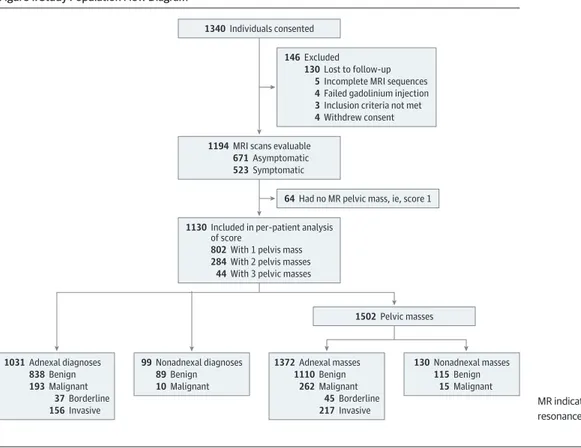
Overall, 1340 patients were enrolled in the study. The mean (range) age was 49 (18-96) years. The final, evaluable population included 1194 patients (89.1%), after 130 (9.7%) patient withdrawals (Figure 1). Of the included patients, 64 (5.4%) were found not to have a pelvic mass. The remaining 1130 patients (94.6%) had a total of 1502 pelvic masses.
Patient characteristics and clinical symptoms are described in Table 1. Patients were referred for indeterminate adnexal masses based on the results of a pelvic ultrasonograph with an issued report rated as high quality, with scores equal to or greater than 5 to 7 in 950 of 1194 patients (79.6%) (eAppendix 3 in theSupplement). Solid tissue was suspected at ultrasonography in 523 women (43.8%), including 166 malignant lesions (11.1%) and 357 benign lesions (23.8%). Ultrasonographic size of the mass was greater than 6 cm in 337 women (28.2%), including 95 malignant lesions (6.3%) and 242 benign lesions (16.1%).
Levels of CA 125 were available for 537 patients (44.9%), 398 (74.1%) of whom had benign and 139 (25.9%) of whom had malignant tumors. An elevated CA 125 level (ie,ⱖ35 U/mL; to convert to kU/L, multiply by 1.0) indicated malignant neoplasm as follows: sensitivity, 0.68 (95% CI, 0.61-0.76; 95 of 139 patients); specificity, 0.82 (95% CI, 0.78-0.86; 327 of 398 patients); PLR, 3.83 (95% CI, 3.02-4.87); NLR, 0.39 (95% CI, 0.30-0.49); PPV, 0.57 (95 of 166 patients); NPV, 0.88 (327 of 371 patients); and accuracy, 0.78 (422 of 537 patients).
A total of 915 women (76.7%) had MRI performed with a 1.5-T MRI scanner and 279 women (23.4%) with a 3-T MRI scanner using 3 different vendors (Siemens, Philips, GE Healthcare). Quality of MRI scans was considered good in 1160 of 1194 women (97.1%), with motion artifacts in 184 of 1194 (15.4%), but all scans remained diagnostic.
Of 1130 patients with a pelvic mass, 768 (67.9%) underwent surgery, and 362 (32.1%) underwent standard clinical follow-up (Table 1). Of those undergoing clinical follow-up, imaging was included for 263 women (72.6%).
There were 130 nonadnexal masses (8.6%) and 1372 adnexal masses (91.3%). The origins of nonadnexal masses are reported in Table 1. The prevalence of malignant neoplasms in the population of women with pelvic mass on MRI was 18.0% (203 of 1130).
Figure 1. Study Population Flow Diagram
1340 Individuals consented
64 Had no MR pelvic mass, ie, score 1
1502 Pelvic masses 146 Excluded
130 Lost to follow-up 5 Incomplete MRI sequences 4 Failed gadolinium injection 3 Inclusion criteria not met 4 Withdrew consent
1130 Included in per-patient analysis
of score
802 With 1 pelvis mass 284 With 2 pelvis masses
44 With 3 pelvic masses
1031 Adnexal diagnoses 838 Benign 193 Malignant 37 Borderline 156 Invasive 1372 Adnexal masses 1110 Benign 262 Malignant 45 Borderline 217 Invasive 99 Nonadnexal diagnoses 89 Benign 10 Malignant 130 Nonadnexal masses 115 Benign 15 Malignant 1194 MRI scans evaluable
671 Asymptomatic 523 Symptomatic
MR indicates magnetic resonance; MRI, magnetic resonance imaging.
Table 1. Population Characteristics
Characteristics No. (%)
Personal History (n = 1194)
Menopausal 511 (42.8)
History of pelvic surgery 364 (30.5) History of adnexal surgery 134 (11.2) History of infertility 94 (7.9) History of breast or ovarian cancer 126 (10.5) Known BRCA1/2 carriers 13 (1.1) Clinical Presentation (n = 1194)
Pelvic pain 384 (32.2)
Vaginal bleeding 60 (5.0)
Palpable mass or increasing abdominal volume 10 (0.8)
Urinary symptoms 8 (0.7)
Amenorrhea 6 (0.5)
Constipation or diarrhea 2 (0.2) Combination of previously mentioned symptoms 53 (4.4) None of these symptoms 671 (56.2) MRI Findings (n = 1194) 0 Lesions 64 (5.4) 1 Lesion 802 (67.2) 2 Lesions 284 (23.8) 3 Lesions 44 (3.7) Management (n = 1130) Primary surgery 719 (63.6) Secondary surgery
After initial follow-up 44 (3.9) After primary chemotherapy 5 (0.4) 24 mo of clinical and/or imaging follow-up 362 (32)
Imaging follow-up 263 (23.3)
Disappearance 96 (8.4)
Decrease of the mass 15 (1.3)
Stability 152 (13.4)
Clinical follow-up, ie, stabilitya 99 (8.8)
Origin of Pelvic Mass From Reference Standard
Masses, No. 1502
Adnexal
Masses, No. 1372
Malignant, No./total, No. (%)
Ovary 253/1223 (20.7)
Tubo-ovarian 9/125 (7.2)
Mesosalpinx 0/24
Nonadnexal
Masses, No. 130
Malignant, No./total, No. (%)
Uterus 4/72 (5.5)
Peritonealb 3/41 (7.3)
Colorectal 5/5 (100)
Lymph node 2/2 (100)
Other, eg, schwannoma, arterial aneurysm 0/9
Urothelial 1/1 (100)
Abbreviation: MRI, magnetic resonance imaging.
aThe 99 women without imaging follow-up had 122
lesions with the following clinical diagnoses: 23 nonadnexal masses including 14 leiomyomas, 5 peritoneal cysts, 3 hematoma, and 1 Nabothian cyst; 26 serous cystadenomas; 24 functional cysts; 23 endometrioma; 9 cystadenofibroma; 5 ovarian fibroma; 5 hydrosalpinx; 4 mature cystic teratoma; 2 mucinous cystadenoma; and 1 paraovarian cyst.
b
This category excludes tubo-ovarian peritoneal carcinoma and includes other purely peritoneal diseases, such as pseudoperitoneal cysts.
Analysis of the MRI Score at the Patient Level
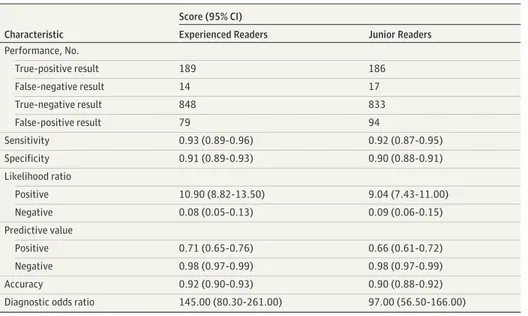
Validation per PatientDiscrimination|The score yielded an area under the receiver operating characteristic curve of 0.961 (95% CI, 0.948-0.971) among experienced readers and 0.942 (95% CI, 0.927-0.955) among junior readers, with a higher performance for the experienced readers (P = .03) (eFigure in the Supplement). Among experienced and junior readers, a score of 4 or 5 suggested a malignant mass with a sensitivity of 0.93 (95% CI, 0.89-0.96; 189 of 203 patients) and 0.92 (95% CI, 0.87-0.95; 186 of 203 patients), respectively, and a specificity 0.91 (95% CI, 0.89-0.93; 848 of 927 patients) and 0.90 (95% CI, 0.88-0.91; 833 of 927 patients), respectively. The prevalence of masses that remained indeterminate on MRI (ie, score of 4) remained low, with 122 (10.8%) among experienced readers and 141 (12.5%) among junior readers.
Performance per Patient|Among 91 women assigned a score of 1, 78 women (85.7%) with nonadnexal masses were subjectively rated nonsuspicious, and 13 women (14.3%) were subjectively rated suspicious by the readers (Table 2 and Table 3).18
All nonsuspicious masses were benign, and suspicious masses were highly indicative of a malignant tumor (PLR, 15.22; 95% CI, 4.23-54.82) (Table 3). Of the 13 women with at least 1 suspicious mass, 10 women (76.9%) had malignant tumors, and 3 (23.1%) had benign leiomyomas with degeneration.
Among 571 women assigned a score of 2, 569 (99.6%) had benign lesions (Table 3). Two premenopausal women (0.3%) had serous borderline tumors (false-negative rate, 0.3%) (Table 3).
Among 213 women assigned a score of 3, malignant tumors were found in 6 premenopausal women (2.8%) and 6 menopausal women (2.8%) (false-negative rate, 5.6%), including 4 women (33.3%) with masses containing no solid tissue (1 premenopausal woman [25.0%] and 3 menopausal women [75.0%]) (Table 3). Eight of 12 malignant tumors (75.0%) were borderline. All 12
fat-containing lesions were benign.
Among 122 women assigned a score of 4, 62 women (50.8%) had benign tumors, and 60 (49.2%) had malignant tumors, with a higher prevalence of invasive than borderline tumors (40 [32.8%] vs 20 [16.4%], respectively) (Table 3). All 14 fat-containing lesions were benign.
Among 133 women assigned a score of 5, 9 premenopausal (6.8%) and 5 menopausal women (3.8%) had benign lesions (false-positive rate, 10.5%), including 5 (35.7%) with mature teratomas, 2 (14.3%) with pelvic inflammatory disease, 2 (14.3%) with cystadenofibroma, 1 (7.1%) with Brenner tumors, 1 (7.1%) with serous cystadenoma, 1 (7.1%) with ovarian fibroma, 1 (7.1%) with struma ovarii,
Table 2. Diagnostic Performance of the Magnetic Resonance Imaging Scorea
Characteristic
Score (95% CI)
Experienced Readers Junior Readers Performance, No. True-positive result 189 186 False-negative result 14 17 True-negative result 848 833 False-positive result 79 94 Sensitivity 0.93 (0.89-0.96) 0.92 (0.87-0.95) Specificity 0.91 (0.89-0.93) 0.90 (0.88-0.91) Likelihood ratio Positive 10.90 (8.82-13.50) 9.04 (7.43-11.00) Negative 0.08 (0.05-0.13) 0.09 (0.06-0.15) Predictive value Positive 0.71 (0.65-0.76) 0.66 (0.61-0.72) Negative 0.98 (0.97-0.99) 0.98 (0.97-0.99) Accuracy 0.92 (0.90-0.93) 0.90 (0.88-0.92) Diagnostic odds ratio 145.00 (80.30-261.00) 97.00 (56.50-166.00)
aA total of 1130 magnetic resonacing imaging scans
were scored. Sensitivities, specificities, and positive and negative predictive values were computed for dichotomized scores (ie, score of 2 and 3 [benign] vs score of 4 and 5 [malignant] or score 1 [nonadnexal mass rated suspicious]). A total of 203 of 1130 patients (18.0%) had at least 1 malignant mass of adnexal or nonadnexal origin.
and 1 (7.1%) with a luteal cyst. Of the 6 fat-containing lesions, 5 (83.3%) were benign and 1 (16.7%) was a malignant germ cell (endodermal sinus) tumor (Table 3). The PLR for score 2 was 0.01; for score 3, 0.27; for score 4, 4.42; and for score 5, 38.81.
Potential Consequences for Management
In the study population, 580 of 1130 women (51.3%) with a mass on MRI and no specific
gynecological symptoms underwent surgery (362 [62.4%]) or follow-up (218 [37.6%]). Based on the standard MRI report and management, 244 women (67.4%) (121 premenopausal and 123
menopausal) with benign lesions and a score of 3 or less or a nonadnexal mass rated as nonsuspicious underwent surgery, and 1 woman (0.5%) with an invasive tumor with a score of 4 or 5 underwent initial follow-up. Moreover, 8 women (2.2%) who underwent surgery had a score of 2 (2 [25.0%] with borderline tumors) or 3 (4 [50.0%] with borderline tumors and 2 [25.0%] with invasive tumors). Reproducibility
The interrater agreement of the score between experienced and junior readers was substantial (κ = 0.784; 95% CI 0.743-0.824). Interrater agreement between experienced readers was also substantial (κ = 0.804; 95% CI, 0.764-0.844).
Analysis of the Criteria Used in the MRI Score at the Lesion Level
The overall prevalence of malignancy per lesion at histology was 18.4% (277 of 1502), 11.5% (15 of 130) for nonadnexal masses and 19.1% (262 of 1372) for adnexal masses, including 45 (3.0%) borderline tumors. Detailed analysis of imaging criteria and their diagnostic performances are available in eTable 1 and eTable 2 in theSupplement.
The origin of each pelvic mass was correctly categorized as adnexal with a sensitivity of 0.99 (95% CI, 0.98-0.99; 1360 of 1372), a specificity of 0.78 (95% CI, 0.71-0.85; 102 of 130), a PLR of 4.60 (95% CI, 3.31-6.39), an NLR of 0.01 (95% CI, 0.01-0.02), a PPV of 0.98 (95% CI, 0.97-0.99), an NPV of 0.89 (95% CI, 0.82-0.94), and an accuracy of 0.97 (95% CI, 0.96-0.98). The diagnosis of the origin was reproducible with a substantial κ of 0.68 (95% CI, 0.59-0.76) between junior and experienced readers.
Table 3. Experienced Readers’ MRI Scores Prospectively Assigned to 1130 Patients With Pelvic Masses
MRI Score Patients, No.
Positive Likelihood of Malignant Tumor (95% CI)a
Patients, No./Total No. (%)b
With Borderline Tumors With Invasive Tumors With Malignant Tumorsc
1 91 0.53 (0.30-1.07) 0 10/91 (10.9) 10/91 (10.9) Nonsuspicious nonadnexal 78 0 (0-0.16) 0 0 0 Suspicious nonadnexal 13 15.22 (4.23.54.82) 0 10/13 (76.9) 10/13 (76.9) 2 571 0.01 (0-0.04) 2/571 (0.3) 0 2 (0.3) 3 213 0.27 (0.16-0.48) 8/213 (3.7) 4/213 (1.9) 12/213 (5.6) No solid tissue 120 0.17 (0.06-0.45) 2/120 (1.7) 2/120 (1.7) 4/120 (3.3) Solid tissue 93 0.46 (0.23-0.93) 6/93 (6.4) 2/93 (2.1) 8/93 (8.6) 4 122 4.42 (3.31-6.09) 20/122 (16.4) 40/122 (32.8) 60/122 (49.2) Fatty content 14 0 0 0 No fatty content 108 20/108 (18.5) 40/108 (37.0) 60/108 (55.5) 5 133 38.81 (22.79-66.11) 7/133 (5.2) 112/133 (84.2) 119/133 (89.5) Fatty content 6 0 1/6 (16.7) 1/6 (16.7) No fatty content 127 7/127 (5.5) 111/127 (87.4) 118/127 (92.9) Abbreviation: MRI, magnetic resonance imaging.
aConfidence intervals obtained by bootstrapping.18
bA total of 64 evaluable patients did not have a pelvic mass at the time of the MRI scan,
and 91 patients had a nonadnexal mass.
c
Discussion
In this multicenter prospective cohort study, we demonstrated that a previously published14
5-point MRI score provided robust risk stratification of sonographically indeterminate adnexal masses. The study confirms a strong concordance of the PLR of malignant neoplasms for each category. Therefore, the MRI score may provide potentially crucial information for determining the therapeutic strategy, allowing the risks and benefits of expectant management or surgery to be considered case by case.19The study demonstrated the feasibility of the acquisition of the multiparametric MRI in multiple centers. Substantial interrater agreement was found, regardless of reader experience, which has been reported to be challenging in some ultrasonographic studies.20-22
External validations in smaller single-center studies have reported similar findings.23-25
The O-RADS MRI score is now proposed as the accepted score for risk assignment of sonographically indeterminate adnexal masses, supported by this strong evidence base.
The O-RADS MRI score addresses a significant clinical issue, given that approximately 18% to 31% of adnexal lesions detected on ultrasound remain indeterminate.3,4,26-29
Transvaginal sonography is accurate for detecting and characterizing adnexal lesions of classic appearance.30
However, in the 2 largest ovarian cancer screening trials,7,8
a significant number of false-positive cases underwent inappropriate surgery. Nonclassical features, such as avascular solid components, large masses, and less experienced sonographers could all contribute to lower accuracy and specificity on ultrasound examination.20,31
Several sonographic rules and scoring systems have been advocated, such as IOTA, Risk of Malignancy Index, and Risk of Ovarian Malignancy Algorithm.3,10,11,27
However, performance in real-life clinical settings has been variable, potentially because of differences in operator experience and cancer prevalence in the population being studied.27,32-34
Correctly classifying an adnexal mass as benign has positive consequences, including the potential to reduce overtreatment by unnecessary or overextensive surgery, to allow consideration of minimally invasive or fertility-preserving surgery, and to improve patient information regarding the risk of ovarian reserve alteration after surgery. The preponderant contribution of MRI in adnexal mass evaluation is its specificity, allowing confident diagnosis of many benign adnexal lesions.19
Using the O-RADS MRI score, our study demonstrated that, even in sonographically indeterminate masses, a lesion with a score of 2 has a PLR of malignant tumor of no greater than 0.01, and a lesion with a score of 3 has the PLR of malignant tumor of 0.27 among both experienced and junior readers. Thus, patients with lesions with scores of 2 or 3 can make an informed decision with the support of their physicians to undergo a minimally invasive or conservative surgical approach or expectant management. Such a high-performance clinical scoring system could allow for the development of decision-support tools, with referral of patients for appropriate follow-up vs surgery, and ensure that fertility-preserving treatment options are considered for young patients with early-stage disease.35
Our study showed that the likelihood of a borderline tumor when a lesion scores 5 was very low (<6%), as in a previous publication.14
However, as borderline tumors are a rare entity, our population included less than 3% (45 of 1502), and larger specific studies are needed.
Optimal management also relies on identifying the site of origin of a pelvic mass (ie, adnexal or nonadnexal). Our study showed that MRI helped to correctly reclassify the origin of the presumed adnexal mass on ultrasonography. In 802 women with only 1 mass described on MRI, 81 lesions (10.0%) were nonadnexal. This is particularly important for malignant nonadnexal tumors, for which initial incorrect management could adversely affect prognosis. In our population, 5.4% (15 of 277) of malignant tumors were nonadnexal lesions of uterine, colorectal, urothelial, nonepithelial peritoneal, or lymph node origin.
Limitations
This study has limitations. It was observational and without randomization, and the score was not integrated into clinical decision-making. Therefore, the clinical consequences on the number of cases in which surgery can be avoided or tailored can only be imputed. However, the validation of the score
now allows studies to test the consequences of the O-RADS MRI score in treatment planning; 2 such studies are currently underway.36,37
Furthermore, because patients were managed according to clinical recommendations, when no pelvic mass was found on MRI, no specific follow-up was undertaken in clinical care as in previous base studies.4,5
Consequently, 64 such cases were excluded from our analysis. This is a low proportion compared with the number of resolving lesions that are typically seen in general outpatient adnexal ultrasonography, given that most physiological ovarian masses are recognized and not referred for MRI. Thus, the O-RADS MRI score estimates the risk of malignancy of an existing pelvic mass detected on MRI. Magnetic resonance imaging is not recommended as a screening tool, and as such, the NPV when no mass is found is not available in the literature. In our study, 284 women had 2 lesions and 44 women had 3 lesions. As each mass was not considered independently, a potential clustering effect should be considered. In addition, 99 of 362 patients were observed during 2 years with only clinical assessment that cannot replace imaging evaluation. Furthermore, we did not include patients who were lost to follow-up in the final analysis. This could have biased the prevalence of the disease in the population, and that is why we calculated the PLR of malignant neoplasms and not the PPV. Of note, more than 90% of these patients were diagnosed with benign lesions on MRI, and this is likely to have played a part in the decision not to undertake further clinical follow-up.
Conclusions
In conclusion, this prospective multicenter cohort study confirmed the performance of a 5-point scoring system developed in a previous retrospective single-center study. The current study provides strong supporting evidence, and the score is now presented as the O-RADS MRI score. Using this score in clinical practice may allow a tailored, patient-centered approach for masses that are sonographically indeterminate, preventing unnecessary surgery, less extensive surgery, or fertility preservation when appropriate, while ensuring preoperative detection of lesions with a high likelihood of malignancy.
ARTICLE INFORMATION
Accepted for Publication: December 1, 2019.
Published: January 24, 2020. doi:10.1001/jamanetworkopen.2019.19896
Open Access: This is an open access article distributed under the terms of theCC-BY License. © 2020 Thomassin-Naggara I et al. JAMA Network Open.
Corresponding Author: Isabelle Thomassin-Naggara, MD, PhD, Service de Radiologie, Hôpital Tenon, Assistance
Publique–Hôpitaux de Paris, Hôpitaux Univesitaires Est Parisien, 58 avenue Gambetta, 75020 Paris, France (isabelle. thomassin@aphp.fr).
Author Affiliations: Service de Radiologie, Hôpital Tenon, Assistance Publique–Hôpitaux de Paris, Sorbonne
Université, Paris, France (Thomassin-Naggara, Bazot); Institute for Computing and Data Sciences, Sorbonne Université, Paris, France (Thomassin-Naggara, Bazot); American College of Radiology, Ovarian-Adnexal Reporting and Data System Magnetic Resonance Imaging Committee (Thomassin-Naggara, Sadowski, Reinhold, Rockall); Service d’Imagerie de la Femme, Centre Hospitalier de Valenciennes, Valenciennes, France (Poncelet); Institut Paoli Calmettes, Marseille, France (Jalaguier-Coudray); Hospital da Luz, Lisboa, Portugal (Guerra); Department of Radiology, Hôpital Européen Georges Pompidou, Assistance Publique-Hôpitaux de Paris, Université Paris-Descartes, Paris, France (Fournier); Centre for Radiology, Clinical Centre of Vojvodina, Medical Faculty, University of Novi Sad, Novi Sad, Serbia and Montenegro (Stojanovic); Lapeyronie Hospital, University of Montpellier, Montpellier, France (Millet, Taourel); Department of Radiology, Imperial College Healthcare NHS Trust, London, United Kingdom (Bharwani, Rockall); Hôpital de la Timone, Marseille, France (Juhan); Department of Radiology, Instituto Português de Oncologia de Lisboa Francisco Gentil, Lisboa, Portugal (Cunha); Department of Radiology, Umberto I Hospital, Sapienza University Roma, Rome, Italy (Masselli); Institut Gustave Roussy, Paris, France (Balleyguier); Institut Curie, Paris, France (Malhaire); Centre Pyramides, Paris, France (Perrot); University of Wisconsin, Madison, Wisconsin (Sadowski); Centre of Research in Epidemiology and Statistics Sorbonne Paris Cité, Institute national de la santé et de la recherche médicale, Joint Research Unit 1153, Paris, France (Porcher); Service
de Gynecologie et Obstetrique et Médecine de la Reproduction, Hôpital Tenon, Assistance Publique–Hôpitaux de Paris, Hôpitaux Univesitaires Est Parisien, Paris, France (Darai); Faculté de Médecine Pierre et Marie Curie, Sorbonne Université, Paris, France (Darai); Department of Medical Imaging, McGill University Health Centre, Montreal, Quebec, Canada (Reinhold); Division of Cancer and Surgery, Faculty of Medicine, Imperial College London, United Kingdom (Rockall).
Author Contributions: Drs Thomassin-Naggara and Porcher had full access to all of the data in the study and take
responsibility for the integrity of the data and the accuracy of the data analysis. Drs Reinhold and Rockall contributed equally.
Concept and design: Thomassin-Naggara, Guerra, Porcher, Reinhold, Rockall. Acquisition, analysis, or interpretation of data: All authors.
Drafting of the manuscript: Thomassin-Naggara, Guerra, Stojanovic, Sadowski, Darai, Reinhold, Rockall. Critical revision of the manuscript for important intellectual content: Thomassin-Naggara, Poncelet, Jalaguier-Coudray, Guerra, Fournier, Stojanovic, Millet, Bharwani, Juhan, Cunha, Masselli, Balleyguier, Malhaire, Perrot, Bazot, Taourel, Porcher, Reinhold, Rockall.
Statistical analysis: Thomassin-Naggara, Porcher, Darai. Obtained funding: Thomassin-Naggara.
Administrative, technical, or material support: Thomassin-Naggara, Poncelet, Jalaguier-Coudray, Fournier, Stojanovic, Bharwani, Masselli, Malhaire, Perrot, Bazot, Taourel, Reinhold, Rockall.
Supervision: Thomassin-Naggara, Stojanovic, Masselli, Balleyguier, Sadowski, Bazot, Taourel, Reinhold, Rockall.
Conflict of Interest Disclosures: Dr Thomassin-Naggara reported receiving personal fees and nonfinancial
support from General Electric and personal fees from Siemens, Hologic, Canon, and Guerbet outside the submitted work. Dr Fournier reported receiving grants from Invectys and speaking fees from General Electric, Novartis, Sanofi, and Janssen Pharmaceuticals outside the submitted work. Dr Balleyguier reported receiving personal fees and nonfinancial support from General Electric and personal fees from Siemens, Samsung Group, and the Bracco Group outside the submitted work. Dr Rockall reported receiving an educational speaker fee from Guerbet. No other disclosures were reported.
Funding/Support: This work was funded by a grant from the Société d’Imagerie de la Femme. Support is
acknowledged from the National Institute of Health Research Imperial Biomedical Research Centre and the Cancer Research UK Imperial Centre.
Role of the Funder/Sponsor: The funders had no role in the design and conduct of the study; collection,
management, analysis, and interpretation of the data; preparation, review, or approval of the manuscript; and decision to submit the manuscript for publication.
Additional Contributions: The European Adnex MR Score Group (EURAD) Collaborators include Asma
Bekhouche, MD, Antoine Brault, MD, Louise Gervais, MD, and Edith Kermarrec, MD (Service de Radiologie, Hôpital Tenon, Assistance Publique–Hôpitaux de Paris, Hôpitaux Univesitaires Est Parisien, Paris, France), Alexandre Bellucci, MD (Department of Radiology, Hôpital Européen Georges Pompidou, Assistance Publique-Hôpitaux de Paris, Université Paris-Descartes, Paris, France), Julien Brochet, MD (Service d’Imagerie de la Femme, Centre Hospitalier de Valenciennes, Valenciennes, France), Claudia Campos, MD (Hôpital de la Timone, Marseille, France), Danielle Donat, MD, and Marijana Basta Nikolić, MD (Centre for Radiology, Clinical Centre of Vojvodina, Medical Faculty, University of Novi Sad, Novi Sad, Serbia and Montenegro), Federica Laghi, MD (Department of Radiology, Instituto Português de Oncologia de Lisboa Francisco Gentil, Lisboa, Portugal), Emma Pages-Bouic, MD, and Cecile Verheyden, MD (Lapeyronie Hospital, University of Montpellier, Montpellier, France), Miriam Salib, FRCR, and Elena Serena, MD (Service de Gynecologie et Obstetrique et Médecine de la Reproduction, Hôpital Tenon), and Pascal Siles, MD (Department of Radiology, Imperial College Healthcare NHS Trust, London, United Kingdom). The EURAD Collaborators were not compensated for their time. The Steering Committee of Ovarian-Adnexal Reporting Data System Magnetic Resonance Imaging (O-RADS MRI) includes Caroline Reinhold, MD, MSC (Department of Medical Imaging, McGill University Health Centre, Montreal, Quebec, Canada), Andrea Rockall, MRCP, FRCR (Faculty of Medicine, Imperial College London and Department of Radiology, Imperial College Healthcare NHS Trust, London, United Kingdom), Isabelle Thomassin-Naggara, MD, PhD (Service de Radiologie, Hôpital Tenon, Assistance Publique–Hôpitaux de Paris, Hôpitaux Univesitaires Est Parisien, Paris, France), Evan Siegelman, MD (Perelman School of Medicine, University of Pennsylvania, Philadelphia), Elizabeth Sadowski, MD (University of Wisconsin, Madison, Wisconsin), Kate Maturen, MD, MS (University of Michigan Hospitals, Ann Arbor), Alberto Vargas, MD (Memorial Sloan Kettering Cancer Center, New York, New York), and Rosemary Fostner, MD (University of Salzburg, Salzburg, Austria). The members of Steering Committee of O-RADS MRI were not compensated for their time. We acknowledge the support of the study sponsor Société d’Imagerie de la Femme, support for the EURAD study from European Society for Urogenital Radiology Female Pelvic Imaging working group, and support for the O-RADS steering committee from the American College of Radiology and the European Society of
Radiology. The EURAD collaborators contributed to additional secondary image reads. The O-RADS MRI collaborators participated in collaborative work in American College of Radiology O-RADS MRI committee for discussions concerning the transition of the AdnexMR score to become the O-RADS MRI score to align the position of the score in the imaging community.
REFERENCES
1. Curtin JP. Management of the adnexal mass. Gynecol Oncol. 1994;55(3, pt 2):S42-S46. doi:10.1006/gyno. 1994.1340
2. Woo YL, Kyrgiou M, Bryant A, Everett T, Dickinson HO. Centralisation of services for gynaecological cancers:
a Cochrane systematic review. Gynecol Oncol. 2012;126(2):286-290. doi:10.1016/j.ygyno.2012.04.012 3. Meys EMJ, Kaijser J, Kruitwagen RFPM, et al. Subjective assessment versus ultrasound models to diagnose
ovarian cancer: a systematic review and meta-analysis. Eur J Cancer. 2016;58:17-29. doi:10.1016/j.ejca.2016. 01.007
4. Froyman W, Landolfo C, De Cock B, et al. Risk of complications in patients with conservatively managed ovarian
tumours (IOTA5): a 2-year interim analysis of a multicentre, prospective, cohort study. Lancet Oncol. 2019;20(3): 448-458. doi:10.1016/S1470-2045(18)30837-4
5. Alcázar JL, Pascual MA, Graupera B, et al. External validation of IOTA simple descriptors and simple rules for
classifying adnexal masses. Ultrasound Obstet Gynecol. 2016;48(3):397-402. doi:10.1002/uog.15854
6. Menon U, Gentry-Maharaj A, Hallett R, et al. Sensitivity and specificity of multimodal and ultrasound screening
for ovarian cancer, and stage distribution of detected cancers: results of the prevalence screen of the UK Collaborative Trial of Ovarian Cancer Screening (UKCTOCS). Lancet Oncol. 2009;10(4):327-340. doi:10.1016/ S1470-2045(09)70026-9
7. Buys SS, Partridge E, Black A, et al; PLCO Project Team. Effect of screening on ovarian cancer mortality: the
Prostate, Lung, Colorectal and Ovarian (PLCO) Cancer Screening randomized controlled trial. JAMA. 2011;305(22): 2295-2303. doi:10.1001/jama.2011.766
8. Jacobs IJ, Menon U, Ryan A, et al. Ovarian cancer screening and mortality in the UK Collaborative Trial of
Ovarian Cancer Screening (UKCTOCS): a randomised controlled trial. Lancet. 2016;387(10022):945-956. doi:10. 1016/S0140-6736(15)01224-6
9. Borley J, Wilhelm-Benartzi C, Yazbek J, et al. Radiological predictors of cytoreductive outcomes in patients with
advanced ovarian cancer. BJOG. 2015;122(6):843-849. doi:10.1111/1471-0528.12992
10. Timmerman D, Van Calster B, Testa A, et al. Predicting the risk of malignancy in adnexal masses based on the
Simple Rules from the International Ovarian Tumor Analysis group. Am J Obstet Gynecol. 2016;214(4):424-437. doi:10.1016/j.ajog.2016.01.007
11. Anton C, Carvalho FM, Oliveira EI, Maciel GAR, Baracat EC, Carvalho JP. A comparison of CA125, HE4, risk
ovarian malignancy algorithm (ROMA), and risk malignancy index (RMI) for the classification of ovarian masses. Clinics (Sao Paulo). 2012;67(5):437-441. doi:10.6061/clinics/2012(05)06
12. Moszynski R, Szubert S, Szpurek D, Michalak S, Krygowska J, Sajdak S. Usefulness of the HE4 biomarker as a
second-line test in the assessment of suspicious ovarian tumors. Arch Gynecol Obstet. 2013;288(6):1377-1383. doi:
10.1007/s00404-013-2901-1
13. Kaijser J, Vandecaveye V, Deroose CM, et al. Imaging techniques for the pre-surgical diagnosis of adnexal
tumours. Best Pract Res Clin Obstet Gynaecol. 2014;28(5):683-695. doi:10.1016/j.bpobgyn.2014.03.013 14. Thomassin-Naggara I, Aubert E, Rockall A, et al. Adnexal masses: development and preliminary validation of
an MR imaging scoring system. Radiology. 2013;267(2):432-443. doi:10.1148/radiol.13121161
15. Hsieh FY, Bloch DA, Larsen MD. A simple method of sample size calculation for linear and logistic regression.
Stat Med. 1998;17(14):1623-1634. doi:10.1002/(SICI)1097-0258(19980730)17:14<1623::AID-SIM871>3.0.CO;2-S 16. DeLong ER, DeLong DM, Clarke-Pearson DL. Comparing the areas under two or more correlated receiver
operating characteristic curves: a nonparametric approach. Biometrics. 1988;44(3):837-845. doi:10.2307/ 2531595
17. Landis JR, Koch GG. The measurement of observer agreement for categorical data. Biometrics. 1977;33(1):
159-174. doi:10.2307/2529310
18. Marill KA, Chang Y, Wong KF, Friedman AB. Estimating negative likelihood ratio confidence when test
sensitivity is 100%: a bootstrapping approach. Stat Methods Med Res. 2017;26(4):1936-1948. doi:10.1177/ 0962280215592907
19. Anthoulakis C, Nikoloudis N. Pelvic MRI as the “gold standard” in the subsequent evaluation of
ultrasound-indeterminate adnexal lesions: a systematic review. Gynecol Oncol. 2014;132(3):661-668. doi:10.1016/j.ygyno. 2013.10.022
20. Yazbek J, Raju SK, Ben-Nagi J, Holland TK, Hillaby K, Jurkovic D. Effect of quality of gynaecological
ultrasonography on management of patients with suspected ovarian cancer: a randomised controlled trial. Lancet Oncol. 2008;9(2):124-131. doi:10.1016/S1470-2045(08)70005-6
21. Van Holsbeke C, Van Belle V, Leone FPG, et al. Prospective external validation of the ‘ovarian crescent sign’ as a
single ultrasound parameter to distinguish between benign and malignant adnexal pathology. Ultrasound Obstet Gynecol. 2010;36(1):81-87. doi:10.1002/uog.7625
22. Faschingbauer F, Benz M, Häberle L, et al. Subjective assessment of ovarian masses using pattern recognition:
the impact of experience on diagnostic performance and interobserver variability. Arch Gynecol Obstet. 2012;285 (6):1663-1669. doi:10.1007/s00404-012-2229-2
23. Ruiz M, Labauge P, Louboutin A, Limot O, Fauconnier A, Huchon C. External validation of the MR imaging
scoring system for the management of adnexal masses. Eur J Obstet Gynecol Reprod Biol. 2016;205:115-119. doi:10. 1016/j.ejogrb.2016.07.493
24. Pereira PN, Sarian LO, Yoshida A, et al. Accuracy of the AdnexMR scoring system based on a simplified MRI
protocol for the assessment of adnexal masses. Diagn Interv Radiol. 2018;24(2):63-71. doi:10.5152/dir.2018.17378 25. Sasaguri K, Yamaguchi K, Nakazono T, et al. External validation of AdnexMR Scoring system: a single-centre
retrospective study. Clin Radiol. 2019;74(2):131-139. doi:10.1016/j.crad.2018.10.014
26. Van Calster B, Timmerman D, Valentin L, et al. Triaging women with ovarian masses for surgery: observational
diagnostic study to compare RCOG guidelines with an International Ovarian Tumour Analysis (IOTA) group protocol. BJOG. 2012;119(6):662-671. doi:10.1111/j.1471-0528.2012.03297.x
27. Meys EMJ, Jeelof LS, Achten NMJ, et al. Estimating risk of malignancy in adnexal masses: external validation
of the ADNEX model and comparison with other frequently used ultrasound methods. Ultrasound Obstet Gynecol. 2017;49(6):784-792. doi:10.1002/uog.17225
28. Sadowski EA, Paroder V, Patel-Lippmann K, et al. Indeterminate adnexal cysts at US: prevalence and
characteristics of ovarian cancer. Radiology. 2018;287(3):1041-1049. doi:10.1148/radiol.2018172271
29. Basha MAA, Refaat R, Ibrahim SA, et al. Gynecology Imaging Reporting and Data System (GI-RADS): diagnostic
performance and inter-reviewer agreement. Eur Radiol. 2019;29(11):5981-5990. doi: 10.1007/s00330-019-06181-0
30. Levine D, Brown DL, Andreotti RF, et al; Society of Radiologists in Ultrasound. Management of asymptomatic
ovarian and other adnexal cysts imaged at US Society of Radiologists in Ultrasound consensus conference statement. Ultrasound Q. 2010;26(3):121-131. doi:10.1097/RUQ.0b013e3181f09099
31. Yazbek J, Ameye L, Testa AC, et al. Confidence of expert ultrasound operators in making a diagnosis of adnexal
tumor: effect on diagnostic accuracy and interobserver agreement. Ultrasound Obstet Gynecol. 2010;35(1):89-93. doi:10.1002/uog.7335
32. Kaijser J, Van Gorp T, Van Hoorde K, et al. A comparison between an ultrasound based prediction model (LR2)
and the risk of ovarian malignancy algorithm (ROMA) to assess the risk of malignancy in women with an adnexal mass. Gynecol Oncol. 2013;129(2):377-383. doi:10.1016/j.ygyno.2013.01.018
33. Kaijser J, Sayasneh A, Van Hoorde K, et al. Presurgical diagnosis of adnexal tumours using mathematical
models and scoring systems: a systematic review and meta-analysis. Hum Reprod Update. 2014;20(3):449-462. doi:10.1093/humupd/dmt059
34. Nunes N, Ambler G, Foo X, Widschwendter M, Jurkovic D. Prospective evaluation of IOTA logistic regression
models LR1 and LR2 in comparison with subjective pattern recognition for diagnosis of ovarian cancer in an outpatient setting. Ultrasound Obstet Gynecol. 2018;51(6):829-835. doi:10.1002/uog.18918
35. Morice P, Uzan C, Fauvet R, Gouy S, Duvillard P, Darai E. Borderline ovarian tumour: pathological diagnostic
dilemma and risk factors for invasive or lethal recurrence. Lancet Oncol. 2012;13(3):e103-e115. doi: 10.1016/S1470-2045(11)70288-1
36. ClinicalTrials.gov. ADNEXMR scoring system: impact of an MR scoring system of therapeutic strategy of pelvic
adnexal masses (ASCORDIA).https://clinicaltrials.gov/ct2/show/NCT02664597?term=NCT02664597&draw= 2&rank=1. Accessed December 9, 2019.
37. ISRCTN Registry. MR in ovarian cancer.http://www.isrctn.com/ISRCTN51246892. Accessed December 9, 2019.
38. Thomassin-Naggara I, Daraï E, Cuenod CA, Rouzier R, Callard P, Bazot M. Dynamic contrast-enhanced
magnetic resonance imaging: a useful tool for characterizing ovarian epithelial tumors. J Magn Reson Imaging. 2008;28(1):111-120. doi:10.1002/jmri.21377
SUPPLEMENT.
eAppendix 1. Study Centers, Names of Principal Investigators, and Number of Patients Recruited and Included eAppendix 2. Trial Protocol
eAppendix 3. Population Selection Criteria eAppendix 4. Methodological Details
eTable 1. Lexicon and Supplementary Material with Detailed Analysis of Adnexal Lesions Based on Morphological
Characteristics
eTable 2. Diagnostic Values of Different MRI Features eFigure. Score Performance Among Senior and Junior Readers