HAL Id: inserm-00668447
https://www.hal.inserm.fr/inserm-00668447
Submitted on 9 Feb 2012
HAL is a multi-disciplinary open access
archive for the deposit and dissemination of
sci-entific research documents, whether they are
pub-lished or not. The documents may come from
teaching and research institutions in France or
abroad, or from public or private research centers.
L’archive ouverte pluridisciplinaire HAL, est
destinée au dépôt et à la diffusion de documents
scientifiques de niveau recherche, publiés ou non,
émanant des établissements d’enseignement et de
recherche français ou étrangers, des laboratoires
publics ou privés.
Corvol
To cite this version:
Annick Clément, Nadia Nathan, Ralph Epaud, Brigitte Fauroux, Harriet Corvol. Interstitial lung
diseases in children..
Orphanet Journal of Rare Diseases, BioMed Central, 2010, 5 (1), pp.22.
R E V I E W
Open Access
Interstitial lung diseases in children
Annick Clement
*†, Nadia Nathan
†, Ralph Epaud, Brigitte Fauroux, Harriet Corvol
Abstract
Interstitial lung disease (ILD) in infants and children comprises a large spectrum of rare respiratory disorders that are mostly chronic and associated with high morbidity and mortality. These disorders are characterized by inflam-matory and fibrotic changes that affect alveolar walls. Typical features of ILD include dyspnea, diffuse infiltrates on chest radiographs, and abnormal pulmonary function tests with restrictive ventilatory defect and/or impaired gas exchange. Many pathological situations can impair gas exchange and, therefore, may contribute to progressive lung damage and ILD. Consequently, diagnosis approach needs to be structured with a clinical evaluation requir-ing a careful history payrequir-ing attention to exposures and systemic diseases. Several classifications for ILD have been proposed but none is entirely satisfactory especially in children. The present article reviews current concepts of pathophysiological mechanisms, etiology and diagnostic approaches, as well as therapeutic strategies. The follow-ing diagnostic groupfollow-ing is used to discuss the various causes of pediatric ILD: 1) exposure-related ILD; 2) systemic disease-associated ILD; 3) alveolar structure disorder-associated ILD; and 4) ILD specific to infancy. Therapeutic options include mainly anti-inflammatory, immunosuppressive, and/or anti-fibrotic drugs. The outcome is highly variable with a mortality rate around 15%. An overall favorable response to corticosteroid therapy is observed in around 50% of cases, often associated with sequelae such as limited exercise tolerance or the need for long-term oxygen therapy.
Definition
Interstitial lung disease (ILD) in infants and children represents a heterogeneous group of respiratory disor-ders that are mostly chronic and associated with high morbidity and mortality (around 15%) [1,2]. These dis-orders are characterized by inflammatory and fibrotic changes that affect alveolar walls. Typical features of ILD include the presence of diffuse infiltrates on chest radiograph, and abnormal pulmonary function tests with evidence of a restrictive ventilatory defect (in older children) and/or impaired gas exchange [3].
Classification
There have been many different approaches to the clas-sification of ILD, with major shifts based on clinical investigation, improvement in chest imaging, and colla-boration with pathologists. In 1998, Katzenstein and Myers proposed four histopathologically distinct sub-groups of idiopathic interstitial pneumonias: usual
interstitial pneumonia (UIP), desquamative interstitial pneumonia (DIP) and a closely related pattern termed respiratory bronchiolitis-associated ILD, acute interstitial pneumonia (formerly Hamman-Rich syndrome), and non specific interstitial pneumonia (NSIP) [4]. In 2002, an international multidisciplinary consensus classifica-tion of idiopathic interstitial pneumonias was proposed by the American Thoracic Society (ATS)/European Respiratory Society (ERS) [5]. This classification defined a set of histologic pattern that provided the basis for clinico-radiologic-pathologic diagnosis, with the final pathologic diagnosis being made after careful correlation with clinical and radiologic features. However, as dis-cussed in several reports, the classification schemes of adult ILD are not satisfactory for the pediatric cases which seem to comprise a broader spectrum of disor-ders with a more variable clinical course [6]. In addition, pediatric histologic patterns often do not resemble pathologic features of lung tissues from adults and some forms are only observed in children younger than 2 years.
Among the proposed classifications for pediatric ILD, one strategy frequently used is to separate the primary pulmonary disorders and the systemic disorders with
* Correspondence: [email protected] † Contributed equally
Pediatric Pulmonary Department, Reference Center for Rare Lung Diseases, AP-HP, Hôpital Trousseau, Inserm UMR S-938; Université Pierre et Marie Curie-Paris 6, Paris, F-75012 France
© 2010 Clement et al; licensee BioMed Central Ltd. This is an Open Access article distributed under the terms of the Creative Commons Attribution License (http://creativecommons.org/licenses/by/2.0), which permits unrestricted use, distribution, and reproduction in any medium, provided the original work is properly cited.
pulmonary involvement. Recently, an additional group has been introduced which is based on the concept that some pediatric ILD are observed more frequently in infants, while others are more specific to older children. The last ERS monography on ILD provided a chapter on pediatric classification which is based on a clear distinction between children aged 0-2 years and children over 2 years-old [7]. Indeed the stage of lung develop-ment and maturation should be taken into consideration when approaching a diagnosis of pediatric ILD. In this view, a new term “diffuse lung disease” has recently been introduced that comprises a diverse spectrum of lung disorders with impaired gas exchange and diffuse infiltrates by imaging. These disorders, more prevalent in young children, include diffuse developmental disor-ders, lung growth abnormalities, neuroendocrine cell hyperplasia and pulmonary interstitial glycogenosis, sur-factant dysfunction disorders, disorders related to sys-temic diseases, disorders of immunocompromised host, and disorders of normal host caused by various insults such as aspiration syndrome or infections [8]. Some dis-eases are mostly observed in older children such as sys-temic diseases, idiopathic disorders as described in adults (DIP, UIP, NSIP and lymphoid interstitial pneu-monia (LIP)), unclassifiable ILD and also infectious dis-orders [9].
It is important to point out that the pathologic pro-cesses underlying the so-called diffuse lung diseases involve not only the alveolar structure but also the distal part of the small airways and the conducting zone, i.e. the terminal bronchioles. Terminal bronchioles are lined with a simple cuboidal epithelium containing Clara cells, basal cells and a limited number of ciliated cells. Clara cells secrete nonsticky proteinaceous compounds to maintain the airway in the smallest bronchioles, which constitute the quiet zone between the conducting and the respiratory lung zones [10]. The terminal bronch-ioles are surrounded by a spiral of smooth muscle. Each of the terminal bronchioles divides to form respiratory bronchioles which contain a small number of alveoli. Consequently, the term of diffuse lung disease refers to disorders that can affect both the distal part of the con-ducting and the respiratory lung zones, and include ILD as well as pathological processes leading to obstruction/ obliteration of small airways [8]. Therefore, diffuse lung diseases encompass a broader group of diseases than ILD which refers to disorders that affect the respiratory function of the lung and consequently the pulmonary structure responsible of the diffusion of gases between blood and air (i.e. the alveolar epithelium, the intersti-tium, and the pulmonary capillary endothelium).
The present review focuses on ILD in immunocompe-tent children, and excludes pulmonary consequences of previous lung injury in situations of chronic aspiration
syndromes, resolving acute respiratory distress syn-drome, and bronchopulmonary dysplasia.
Epidemiology
An estimated prevalence of 3.6 per million has been reported by Dinwiddie and coworkers through a national survey of chronic ILD in immunocompetent children in the United Kingdom and Ireland over a three year period (1995-1998) [1]. This prevalence is certainly under-estimated due to the lack of standar-dized definitions and the absence of organized reporting systems. From the limited published data composed mainly of case reports and small series, it seems that pediatric ILD occurs more frequently in the younger age and in boys [11]. In addition, nearly 10% of cases appear to be familial [12].
Pathophysiology
Critical role of the alveolar epithelium
The understanding of the mechanisms underlying the development and progression of ILD remains elusive [13,14]. Indeed, for a long time, chronic ILD and pul-monary fibrosis were believed to result mainly from chronic inflammation following an initial injury to the alveolar epithelial lining [15,16]. In cases of limited injury, it was thought that the reparative attempt could reverse the trend toward fibrosis. By contrast, in situa-tions of continuing injury, the repair process driven by inflammatory molecules produced by the local cells will result in scarring and structural changes. Therefore, by targeting the inflammatory response, the belief was that fibrosis could be prevented or controlled. This theory explains the large use of anti-inflammatory therapy with, however, limited clinical efficacy.
Based on clinical and experimental observations, a new paradigm has progressively emerged with the alveo-lar epithelium being viewed as a key actor in the devel-opment of ILD [17-19]. Following injury, alveolar epithelial cells (AEC) may actively participate in the restoration of a normal alveolar architecture through a coordinated process of re-epithelialization, or in the development of fibrosis through a process known as epithelial-mesenchymal transition (EMT) [20]. Complex networks orchestrate EMT leading to changes in cell architecture and behaviour, loss of epithelial characteris-tics and gain of mesenchymal properties. The reasons for epithelial cell loss and inappropriate re-epithelialisa-tion are still debated, but ongoing apoptosis is believed to be a key component in the progression of the disor-der [21]. Prolonged denudation of the basement mem-brane contributes to altered interactions and cross-talk between AECs and mesenchymal cells, resulting in pro-found modifications of cell functions with imbalanced production of oxidants, proteases, and polypeptide
mediators including cytokines and growth factors such as Transforming Growth Factor (TGF)-b and Endothelin (ET)-1. A consequence is the perpetuation of a vicious cycle with TGF-b promoting epithelial cell apoptosis, which in turn increases the local production of TGF-b [22]. ET-1 is also considered to be an important actor, based on the current knowledge of its numerous func-tions including fibroblast and smooth muscle cell mito-gen, and stimulant of collagen synthesis [23,24]. Recent studies showed that ET-1 is produced by AEC, and could induce alveolar EMT via stimulation of endogen-ous TGF-b production.
Multiple causes and pathways
ILD may be caused by myriad etiologies with differing prognoses and natural history. Indeed, multiple factors may injure the alveolar epithelium and initiate the devel-opment of ILD [25]. The initiating injury can be intro-duced through the airways and the circulation, or can occur as a result of sensitization. Consequently, the mechanisms underlying disease progression will be influenced by the causative event as well as by the host and the environment. These mechanisms are developed through interactions of multiple pathways, which include apoptotic pathways, developmental pathways, and endoplasmic reticulum (ER) associated pathways (Figure 1).
Apotosis plays a central role in lung remodeling asso-ciated with ILD [26]. An important molecule in the events associated with epithelial cell apoptosis is TGF-b, which is overexpressed in ILD. Downstream events linked to upregulation of TGF-b include modifications in the expression of various components of the cell cycle machinery, mainly the cyclin-dependent kinases (CDK) system that plays an essential role in ensuring proper cell cycle progression. Recently, much work has been focused on the protein p21cip1, a member of the CDK inhibitor family. This protein promotes cell cycle arrest to apoptosis in cases of cellular DNA damage. Interestingly, upregulation of p21cip1 has been reported in the lung tissues of patients with pulmonary fibrosis, primarily in hyperplastic alveolar epithelial cells [27] The increased expression of p21cip1 can favour the pro-cess of epithelial cell apoptosis. Apoptotic cells can also produce TGF-b. A consequence would be the perpetua-tion of a vicious cycle with TGF-b promoting epithelial cell apoptosis, which in turn increases the local produc-tion of TGF-b.
Recently, it has been suggested that genes associated with lung development and embryonic pathways could be involved in aberrant epithelium-mesenchymal cross-talk and epithelial plasticity, and could therefore partici-pate in the development of chronic ILD. Selman and coworkers reported that lung fibrosis is characterized by
Figure 1 Mechanisms and pathways involved in the response of the alveolar structure of the lung to injury. Abbreviation: Transforming Growth Factor (TGF)-b.
enrichment for genes associated with cell adhesion, extracellular matrix, smooth muscle differentiations, and genes associated with lung development [28-31]. During EMT in the embryonic period, cells undergo a switch from a polarized epithelial phenotype to a highly motile mesenchymal phenotype [32]. Molecular processes gov-erning EMT are induced by a cooperation of receptor tyrosine kinases or oncogenic Ras (RTK/Ras) pathway and TGF-b signaling [33]. Recently, additional pathways and effectors have been reported to play a role in the induction of EMT, such as Wnt//b-catenin, Notch and Sonic hedgehog signalling [34].
Recent reports strongly suggest that the ER stress may represent an important mechanism of the altered repair process observed in the alveolar epithelium of fibrotic lung [35]. Situations associated with abnormal regulation
of the cascade of events leading to the formation of mature protein result in either misfolding or mistarget-ing of the protein. These events trigger induction of intracellular aggregate formation and ER stress, which can lead to cell death through apoptosis and autophagic pathways [36,37]. Several stimuli including oxidant-anti-oxidant imbalance, viral proteins, inflammatory mole-cules, nutrient deprivation may induce ER stress [38,39] (Figure 2). Among the cytoprotective mechanisms avail-able are the ER chaperones such as binding immunoglo-bulin protein (BiP). Interestingly, mutant BiP mice have been reported to die within several hours of birth from respiratory failure due to impaired secretion of pulmon-ary surfactant by type 2 AEC. In these animals, expres-sion of surfactant protein (SP)-C was reduced and the lamellar bodies were malformed, indicating that BiP
Figure 2 Alveolar structure disorder-associated ILD and ER stress. The (Endoplasmic Reticulum) ER and its protein maturation machinery allow the synthesis of mature secretory and membrane proteins with specific folded conformation. In situations of stress induced by genetic mutations or environmental factors, unfolded or misfolded proteins are retained in the ER and induce a defence mechanism called the ER stress response. The induction of ER chaperones is critical to increase the ER folding capacity allowing the production of correctly folded protein. When this defence mechanism is impaired, the misfolded proteins can either be degraded by the proteasome or form protein aggregates. Protein aggregates are toxic and can cause conformational diseases. Within the alveolar epithelium, misfolding of SP-C could trigger induction of intra-cellular aggregate formation and ER stress, with consequently development of alveolar structure disorder-associated ILD and conformational disease.
plays a critical role in the biosynthesis of surfactant [40]. Several recent reports suggest the possible implication of ER stress in ILD, with activation of stress response markers in fibrotic lung tissues.
Surfactant deficiency and stem cell dysfunction
It is now well established that surfactant dysfunction plays an important role in the development and progres-sion of ILD. Pulmonary surfactant is a multimolecular complex constituted of phospholipids and proteins secreted by type 2 AEC into the alveolar space. It assures alveolar stability by reducing surface tension along the epithelial lining and this role involves mainly the lipids and the specific hydrophobic SP, SP-B and SP-C. Other important players in surfactant metabolism include the ATP-binding cassette, sub-family A, member 3 (ABCA3) and the thyroid transcription factor 1 (TTF-1).
Surfactant deficiency can be induced by a number of primitive and secondary mechanisms. Among them are genetic defects with mutations in SP-B gene (SFTPB) as well as genes coding for SP-C (SFTPC), ABCA3, and TTF-1 [41-43]. More than 30 SFTPB (located on chro-mosome 2) mutations have been identified among patients with a congenital deficiency in SP-B. For SFTPClocated on chromosome 8, at least 35 mutations have been described, localized primarily in the COOH-terminal Brichos domain [44,45]. A proposed function of the Brichos domain is a chaperone-like activity, which could prevent misfolding and aggregation of the parent protein. Alterations in the Brichos domain could therefore lead to diseases through mechanisms related to abnormal protein processing and cell toxicity [46]. Recently, several studies have also documented the role of ABCA3 deficiency in ILD. ABCA3 functions in the transport of surfactant lipids into lamellar bodies and is required to maintain pulmonary surfactant phospho-lipid homeostasis. Another contributor of ILD is TTF-1 (NK2 homeobox 1) dysfunction. TTF-1 is a critical reg-ulator of transcription for the surfactant protein SP-B and SP-C. It is encoded by a gene located on chromo-some 14q13 and is composed of three exon and two introns [47]. It is expressed in the thyroid, brain and lung.
Stem cell dysfunction represents a new domain of investigation. Alveolar epithelium regeneration and repair requires activation and proliferation of tissue-resi-dent (progenitor) cells and their differentiation to replace the damaged cells [48]. However, unlike cancer cells, stem cells are not immortal and display decreasing telomere length with aging [49]. Telomere shortening has been documented to be associated with reduced capacity for stem cell renewal, and decreased activity of telomerase, the polymerase responsible for telomere
maintenance. The stem cells of the alveolar epithelium are the type 2 AEC, and expression of telomerase has been documented in these cells [48]. Experimental stu-dies have also indicated that telomerase is expressed mainly during lung development with a peak expression before birth followed by a decrease to nearly undetect-able levels in mature alveolar epithelium. Interestingly, telomerase expression and activity could be reinduced in normal quiescent type 2 AEC exposed to oxidative stress [50]. The current understanding is that a population of type 2 AEC may have the capacity to survive injury through telomerase activation, and consequently may be responsible for repopulation of the damaged alveolar epithelium. On the basis of reports of pulmonary disor-ders in dyskeratosis congenita (a rare hereditary disease of poor telomere maintenance), recent and exciting find-ings have documented mutations in the telomerase gene in familial idiopathic pulmonary fibrosis [51]. In addi-tion, it is likely that environmental factors such as inflammation, oxidative stress, or virus infection may modify telomerase activity and account for the develop-ment of organ-specific disease associated with telomer-ase dysfunction. In this view, new data in chronic respiratory diseases support the concept that alveolar stem cell dysfunction may play an important role in the rate of progression or severity in ILD [52]. The question whether telomerase mutations or telomere dysfunction may be implicated in pediatric ILD needs to be addressed in prospective studies, one possible tool being determination of telomere length in circulating leukocytes.
Role of age
The frequency of lung fibrotic disorders is much lower in children than in adults. Some clinical situations have features certainly unique to children, but many of these diseases overlap their adult counterparts with the pri-mary event being injury and damage of the alveolar epithelium [11,13]. Yet, the overall outcome and prog-nosis of the diseases in children are thought to be less severe than in adult patients. In addition, pediatric ILD is more responsive to therapeutic strategies than adult ILD [9]. These differences may be explained by the types of initial injury, which may not be similar due to changes in the host environment. Another explanation is the modifications of the process of wound healing with age. Comparison of the response to injury in foetal and adult skin shows clear differences [53]. Skin wound healing in the foetus is characterized by complete regen-eration of the skin and the absence of scar formation. Progressively with age, the skin looses the capacity to regenerate the original tissue architecture with the result being scar formation that extends outside the wound bed. The process of healing involves the coordinated
regulation of cell proliferation and migration and tissue remodeling, predominantly by polypeptide growth fac-tors [54]. The slowing of wound healing that occurs in the aged may be related to changes in the activity of these various regulatory factors. In a study on the role of aging in the development of cardiac fibrosis in the rabbit, differences in the cascade of events leading to myocardial remodeling were observed, with mainly the presence of more myofibroblasts synthesising collagen and expressing high levels of TGF-b in older animals [55]. A study of growth factors involved in skin wound healing in young and aged mice also showed age-depen-dent changes. Expression of all the fibroblast growth factors was diminished in aged mice, even in healthy skin. In addition, the post-wound regulation of expres-sion of these factors and of TGF-b was less pronounced and slower than in young mice. These findings are in agreement with data observed in muscle that indicated significant alterations in the TGF-b production with age [56,57]. Other potential mechanism is linked to the observation that injury in adult tissues does in certain circumstances stimulate tissue regeneration, depending on the presence of small subsets of primitive stem cells. Stem cells are the self-renewing, primitive, undifferen-tiated, multipotent source of multiple cell lineages [49]. While such cells are critical for development and growth through childhood, residual pools of adult stem cells are hypothesized to be the source of the frequently limited tissue regeneration and repair that occurs in adults [58]. Unlike embryonic stem cells, adult stem cells are not immortal, and show decreasing telomere length with increasing age. The naturally limited replacement capa-city of such endogenous stem cell pools may occur via exhaustion of the stem cell pool or arise as a conse-quence of inherited or acquired mutations that alter proper stem cell function [59]. The limited life span of cells may result from replicative senescence in response to various stresses including DNA damage, oxidants, and telomere erosion [52]. All these forms of injury have been documented in the lung from adult patients with ILD.
Diagnosis of ILD
Clinical presentation
The prevalence of children ILD is higher in the younger patients: more than 30% of patients are less than 2 years at diagnosis, as recorded by the recent ERS Task Force. 7% have parental consanguinity and nearly 10% of case siblings were affected by similar diseases. There is a male predominance with a sex ratio of 1.4. The present-ing clinical manifestations are often subtle and non-specific. The onset of symptoms is, in most cases, insi-dious and many children may have had symptoms for years before the diagnosis of ILD is confirmed. However,
the majority of patients has symptoms for less than one year at the time of initial evaluation. The clinical mani-festations vary from asymptomatic presentation with radiological features suggestive of ILD to more charac-teristic presence of respiratory symptoms and signs such as cough, tachypnea and exercise intolerance [9,60]. These varying presentations are also reflected in the report published by Fan et al. who systematically evalu-ated the clinical symptoms and physical findings of 99 consecutive children with ILD [2]. Common symptoms at presentation included cough, dyspnea, tachypnea and chest wall retraction, exercise limitation and frequent respiratory infections. Cough is observed in almost 75% of the patients, is normally non-productive and does not disturb sleep. Tachypnea is observed in 80% of patients and is usually the earliest and most common respiratory symptom. Unexplained fever is also reported in almost one third of infants. Failure to thrive (37%), tiring dur-ing feeddur-ing and weight loss are also common symptoms, mainly in young patients. Although a history of wheez-ing may be elicited in almost 50% of the patients, wheezing is documented by physical examination in only 20% of the cases.
The frequent clinical findings are inspiratory crackles (44%), tachypnea and retraction. In a child with a nor-mal birth history, these are strongly suggestive of ILD. Other findings associated with an advanced stage of lung disease include finger clubbing (13%) and cyanosis during exercise or at rest (28%) [9,61]. During physical examination it is essential to check the presence of asso-ciated non-respiratory symptoms such as joint disease, cutaneous rashes, and recurrent fever suggestive of col-lagen-vascular disorders. Details should also be obtained on precipiting factors such as feeding history, infections, or exposure to dust and drugs. In addition, information on relatives or siblings with similar lung conditions should be gathered.
Chest imaging
Plain radiographs are usually performed in a child sus-pected of ILD at first presentation, but the information provided is often limited and the key chest imaging tool for diagnosis is the High Resolution Computed Tomo-graphy (HRCT), which can visualize the parenchymal structure to the level of the secondary pulmonary lobule. HRCT technique for ILD diagnosis has been exten-sively discussed [62-64]. To optimise spatial resolution, there is a general agreement to use thin sections, the smallest field of view and a sharp resolution algorithm. The most common HRCT feature of ILD is widespread ground-glass attenuation. Intralobular lines, irregular interlobular septal thickening and honeycombing are less common findings. Large subpleural air cysts in the upper lobes adjacent to areas of ground-glass opacities
have been also reported in young children with ILD. These cysts are interpreted as paraseptal or irregular emphysema. HRCT is useful for ILD diagnosis and selection of lung area to be biopsied. It is proposed that it also may contribute to monitor disease activity and/or severity. However, evaluation is still needed to support a role of HRCT as a follow up tool in pediatric patients.
Pulmonary function testing
Pulmonary function testing (PFT) techniques are well established in children and adolescents. However, chil-dren aged 2-6 years represent a real challenge in pul-monary function assessment as they cannot be sedated and find it difficult to cooperate with all respiratory manoeuvres. In 2007, an ATS and ERS statement on PFT in preschool children summarized the current knowledge on the PFT techniques suitable for young children [65,66].
Although PFT does not provide specific information, it represents a useful investigation for both the diagnosis and the management of ILD [11]. Generally, in ILD, pulmonary function abnormalities reflect a restrictive ventilatory defect with reduced lung compliance and decreased lung volumes [67-69]. Vital capacity (VC) is variably diminished; the decrease in total lung capacity (TLC) in general is relatively less than in VC. Functional residual capacity (FRC) is also reduced but relatively less than VC and TLC, and residual volume (RV) is generally preserved; thus the ratios of FRC/TLC and RV/TLC are often increased. Airway involvement is observed only in a minority of patients. Lung diffusing capacity of carbon monoxide (DLCO) or transfer factor (TLCO) is often markedly reduced and may be abnormal before any radiological findings. However, DLCO corrected for lung volume may also be normal in many children. Hypoxemia as defined by a reduced resting arterial oxy-gen saturation (SaO2) or a reduced resting arterial
oxy-gen tension is often present. Hypercarbia occurs only late in the disease course. During exercise the above described dysfunctions become even more pronounced. Thus, gas exchange during exercise might be a more consistent and sensitive indicator of early disease [3].
Bronchoalveolar lavage
Bronchoalveolar lavage (BAL) usefully provides speci-mens for cytological examination, microbial cultures, and molecular analysis. Besides infections, BAL can be of diagnostic value in several situations. In the context of pulmonary alveolar proteinosis, BAL abnormalities are characterized by milky appearance fluid, abundant proteinaceous periodic acid schiff positive material, and presence of foamy alveolar macrophages (AM) [70]. BAL can also be diagnostic for pulmonary alveolar hae-morrhage [11]. This diagnostic is easy when the BAL
fluid has a bloody or pink color, but its gross appear-ance may be normal. Microscopic analysis may then be of value by documenting the presence of red blood cells in AM or haemosiderin laden AM [71]. Among other situations, the diagnosis of Langerhans cell histiocytosis can be performed with the use of the monoclonal anti-bodies revealing the presence of CD1a positive cells (in more than 5% of the BAL cells) [72]. Lipid disorders with lung involvement represent another indication of BAL. This includes congenital lipid-storage diseases (Gaucher’s disease and Niemann-Pick disease) or chronic lipid pneumonia due to chronic aspiration [73,74]. However, in cases of aspiration syndromes, the presence of lipid laden AM is sensitive but not specific [75].
In other pathological situations, BAL can usefully serve to direct further investigations. Accumulation of BAL T-lymphocytes with prevalence of CD4+ cells is suggestive of sarcoidosis, whilst prevalence of CD8+ cells is suggestive of hypersensitivity pneumonitis [76]. Also, an increase in BAL eosinophils suggests pulmon-ary infiltrates associated with eosinophilia syndromes [77]. Depending of the underlying diseases, a number of cellular and molecular investigations can be proposed including the studies of various surfactant components, phospholipids and apoproteins [78].
Tissue biopsies
With increasing recognition of the different patterns of ILD and their clinical significance, histological investiga-tion has become increasingly important. Depending on disorder presentation, biopsy may concern more accessi-ble organs than the lung such as the skin or the liver in sarcoidosis. Histological evaluation of lung tissue usually represents the final step in a series of diagnostic approaches.
Different methods may be used to obtain lung tissue. The major difference between individual methods lies mainly in balancing invasiveness against the potential for obtaining adequate and sufficient tissue for diagno-sis. The techniques of choice are open lung biopsy and video assisted thoracoscopy biopsy. In children, open lung biopsy usually provides sufficient tissue with few complications related directly to the biopsy procedure [79]. Video assisted thoracoscopy biopsy is an alternative to open lung biopsy, and it has been shown that the procedure can be safely performed, even in small chil-dren [80]. The place of other methods like transbron-chial lung biopsy and percutaneous needle lung biopsy in appropriate diagnosis of pediatric patients with ILD has to be established [81-83].
The lung histological patterns that can be observed in ILD have been reviewed by the ATS/ERS [5]. In children, they include mainly: DIP, NSIP, and LIP. DIP
is characterized by airspaces filled with AM, thickened alveolar septa, scattered mixed inflammatory cells and minimal fibrosis. Many alveolar spaces are lined by hyperplastic type 2 AEC. Recently, association with surfactant disorders has been reported [41,84-86]. NSIP encompasses a broad spectrum of abnormalities with varying degrees of alveolar wall inflammation or fibrosis. The cellular pattern of NSIP is characterized by mild to moderate interstitial chronic inflammation and type 2 AEC hyperplasia in inflammation areas. It has been reported in a variety of underlying conditions including connective tissues diseases and surfactant disorders. LIP features include a marked diffuse infil-trate of mature lymphocytes, plasma cells and histio-cytes within the pulmonary interstitium, particularly the alveolar walls. They are often associated with either connective tissues disorders or immunodeficiency states, both congenital and acquired [9]. Another pat-tern described mainly in adults is diffuse alveolar damage (DAD), which includes diffuse homogeneous thickening of alveolar interstitial walls with myofibro-blast accumulation, prominent type 2 AEC hyperplasia and atypia, and hyaline membranes containing surfac-tant proteins and cellular debris [87]. Usual interstitial pneumonia (UIP) is rare in children [88]. It is charac-terized by severe remodeling of the alveolar structure with heterogeneous appearance consisting of contigu-ous areas of normal lung, dense scarring, and bronch-iolar abnormal proliferation. Interstitial inflammation is usually mild to moderate. Histologic patterns of ILD unique to infancy are described below.
Other tests
Laboratory tests are used to exclude a number of respiratory diseases in childhood that does not typically present with ILD such as chronic aspiration syndromes, resolving acute respiratory distress syndrome, tuberculo-sis, cystic fibrotuberculo-sis, bronchopulmonary dysplasia and dif-fuse pulmonary disease such as cystic fibrosis. Laboratory tests also verify the absence of immunodefi-ciencies [3].
When these conditions have been eliminated, the spectrum of investigations that should be performed for the diagnostic approach will be guided by the history and clinical presentation in each individual child. These investigations are discussed below for the various disor-ders. In addition, an increasing number of blood and BAL biomarkers for evaluation of disease severity and progression is currently investigated. The studied mole-cules include various cytokines and chemokines, surfac-tant protein D, Krebs von den Lungen-6 antigen (KL-6), matrix metalloproteinases MMP1 and MMP7 and defensins [89-92].
Etiological diagnosis of ILD
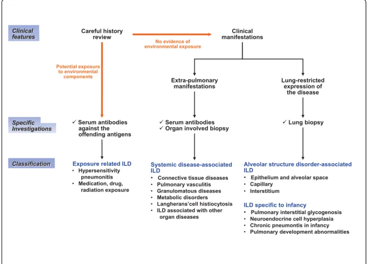
A large number of pathological situations can impair gas exchange and contribute to progressive lung damage and ILD. Consequently, diagnosis approaches need to be organized by cause, with a clinical evaluation requiring a careful history paying attention to exposures and sys-temic diseases. Indeed, in a number of pathological situations, no final diagnosis is proposed and the con-clusion reported by the physician in charge of the patient is ILD of unknown cause. However, information from recent studies highlights the concept that lung insults caused by substances from the environment or in the context of systemic diseases are largely under-esti-mated and should be more often discussed considered in the diagnostic process. Based on this consideration, the following diagnostic grouping for pediatric ILD can be considered 1) exposure-related ILD; 2) systemic dis-ease-associated ILD; 3) alveolar structure disorder-asso-ciated ILD; and 4) ILD specific to infancy.
Accordingly, a step-by-step etiological diagnostic approach is required and is summarized in Figure 3. Once the diagnosis of ILD is established on clinical, radiological, and functional findings, a careful history should be obtained for potential exposure-related dis-eases leading to discuss the need for specific serum anti-bodies against offending antigens. The following step focuses on the search for systemic disease associated ILD, oriented by the presence of clinical and functional extra-pulmonary manifestations. In such situations, additional investigations should include specific serum antibodies and possibly tissue biopsies in organs other than the lung. Finally, elimination of these 2 groups of causes with a lung restricted expression of the disease allows discussing the potential interest of a lung biopsy.
Exposure-related ILD
Exposure-related disease refers to diseases caused by a sufficient level of exposure (dose) to components with target organ contact, and subsequent biologic changes and clinical expression. Many agents have been asso-ciated with pulmonary complications of various types including ILD. The adult literature has provided exten-sive lists of candidate molecules [93]. In children, the potential involvement of these molecules is not similar as the environmental conditions and the use of thera-peutic drugs differ. It is important to point out that exposure-related diseases are certainly under-estimated in the pediatric age. One reason is linked to the fact that the diagnosis is less often discussed than in adults as pediatricians and other child health care providers do not usually have the expertise necessary to take an environmental history. In this review, the most frequent causes of exposure-related ILD are discussed.
Hypersensitivity pneumonitis
Hypersensitivity pneumonitis (HP) is a cell-mediated immune reaction to inhaled antigens in susceptible per-sons [94,95]. In children, HP is often associated with exposure to antigens in the home environment as well as with certain hobbies. The most frequent types of HP include bird fancier’s diseases, humidifier lung diseases, and chemical lung diseases. Bird fancier’s diseases are induced by exposure to birds with the antigens being glycoproteins in avian droppings, and on feathers. Importantly, respiratory symptoms in exposed patients who have only one pet bird at home should raise the suspicion of HP [96]. Humidifier lung diseases (air con-ditioner lung, misting fountain lung, basement lung dis-eases) are caused mainly by free-living amoeba and nematodes, as well as bacteria and fungi. Chemical lung diseases can be induced by various inorganic antigens such as those from vaporized paints and plastics.
Low-molecular-weight chemicals may react with proteins in the airways, thus forming complete antigens. Once exposure history is obtained, additional information is required and includes biologic tests allowing measure-ments of environmental contaminants and interpretation of the results by environmental medicine experts.
As HP is believed to be an adult disease, children are often diagnosed at the chronic stage of the disease resulting of a long-term exposure to low levels of inhaled antigens. Children can develop subtle interstitial inflammatory reactions in the lung without noticeable symptoms for months [97]. Clinical features in the clas-sic form include non productive cough, dyspnea, malaise, asthenia and occasional cyanosis [95]. Lung function abnormalities are not specific and appear simi-lar to changes observed in other ILD. HRCT abnormal-ities vary from ground glass attenuation predominantly in the mid-upper zone to nodular opacities with signs of
Figure 3 Search for ILD etiology in children. ILD is defined by the presence of diffuse infiltrates on chest radiographs or chest high resolution computed tomography, and abnormal pulmonary function tests with evidence of a restrictive ventilatory defect (in older children) and/or impaired gas exchange. The search for etiology requires a systematic step-by-step diagnostic strategy for identifying: exposure-related ILD; systemic disease-associated ILD; alveolar structure disorder-associated ILD; and ILD specific to infancy.
air-trapping [62,63,98]. Laboratory tests focus mainly on the search for serum-precipitating IgG antibodies against the offending antigen [95]. However, the pre-sence of these antibodies is considered to be of ques-tionable clinical relevance for diagnosis, as it is observed in up to 50% of serum samples of exposed but asympto-matic individuals. BAL cell profile study typically shows an increase in total cell count with a remarkable eleva-tion in the percentage of lymphocytes often over 50% with a decreased CD4/CD8 ratio [95,97]. However, in contrast to studies in adults, the CD4/CD8 ratio could be within the normal range for children [76]. Histo-pathologic evaluation of lung tissue is usually not neces-sary for the diagnosis of HP.
At the present time, there is no diagnostic test that is pathognomonic for HP, and only significant predictors of HP are identified. The most significant diagnostic tool is a detailed environmental exposure history. Other diagnostic features include: positive precipitating antibo-dies to the offending antigen; recurrent episodes of symptoms; symptoms occurring 4-8 h after exposure; occurrence of diffuse parenchymal lung disease by lung function and HRCT; BAL abnormalities with lymphocy-tic alveolitis and increased CD8+ T cells.
Medication, drug, radiation and tobacco exposure
Drugs used in inflammatory or cancer pediatric diseases can cause ILD. They include anti-inflammatory agents (e.g. aspirin, etanercept), immunosuppressive and che-motherapeutic agents (e.g. azathioprine, methotrexate, cyclophosphamide), antibiotics, cardiovascular agents, and, for teenagers, illicit drugs [99,100]. There are no distinct clinical, radiographic or pathologic patterns, and the diagnosis is usually made when a patient is exposed to medication known to result in lung disease, with a timing of exposure appropriate for disease development and elimination of other causes of ILD. Treatment relies on avoidance of further exposure and corticosteroids in markedly impaired patients.
Exposure to therapeutic radiation in the management of pediatric cancer may also results in ILD. Patients pre-senting within 6 months of therapy generally have radio-graphic abnormalities with ground glass patterns in both radiation-exposed and unexposed tissue [101].
The association between tobacco use and ILD is less well appreciated than the relation with chronic obstruc-tive pulmonary disease (COPD). In addition, pediatric patients do not usually have a significant smoking his-tory to develop respirahis-tory disorders [102].
Systemic disease-associated ILD Connective tissues disease
Connective tissues disorders (CTD) are a heterogeneous group of immunologically mediated inflammatory dis-eases. Their origins are multifactorial with genetic,
constitutional and environmental elements contributing to their development. CTD refers to any disease that has the connective tissues of the body as a primary tar-get of pathology. The connectives tissues are composed of two major structural proteins, elastin and collagen, with different types of collagen proteins in each tissue [103]. Many CTD feature abnormal immune system activity associated with inflammation. Pulmonary mani-festations of CTD may include both vascular and inter-stitial components. From recent reports, the incidence of ILD in the context of CTD appears to be higher than previously appreciated [104,105]. Importantly, ILD may precede the development of clinically obvious CTD, sometimes by months or years. Table 1 provides infor-mation on suggestive clinical and serological features in selected conditions. The main disorders to be consid-ered in childhood are rheumatoid arthritis, systemic sclerosis, and systemic lupus erythematosus. The other include Sjögren syndrome, dermatomyositis and poly-myositis, ankylosing spondylitis, and mixed connective tissue disease.
Rheumatoid arthritis Rheumatoid arthritis (RA) is an inflammatory disorder defined by its characteristic di-arthroidal joint involvement. It is the most common CTD in children, but pulmonary involvement is less fre-quent than in adults. Genetic and environmental factors seem to be important contributors of disease progres-sion, with influence of sex (more frequent in male), pre-sence of two copies of the HLA-DRB1“shared epitope” (HLA-DR SE) and anticyclic citrullinated peptide anti-body (anti-CCP), and possibly tobacco exposure [106,107].Almost 50% of patients with RA have specific serologic abnormalities several years before the onset of joint symptoms, and the findings of elevated serum levels of IgM rheumatoid factor or anti-CCP is asso-ciated with a high risk for the development of RA [107]. Systemic sclerosisSystemic sclerosis (SSc) is character-ized by a progressive dermatologic abnormality [108]. Its etiology remains unknown; it is believed to be a com-plex disease in which interactions between environmen-tal, auto-immune, and genetic factors result in various disease phenotypes [109]. Although it is a rare disease in childhood, the diagnosis is based on skin disease. Cardiopulmonary complications are common and have been associated with death in young patients. Almost all patients with SSc have serum antinuclear antibodies. The other autoantibody markers are listed in table 1. Recently, the presence of anti-DNA topoisomerase II autoantibody has been reported to be a key factor in the development of ILD, in association with class II MHC status (HLA-DR3, HLA-DPBI) [110].
Systemic lupus erythematosusSystemic lupus erythe-matosus (SLE) is an auto-immune disorder characterized by the involvement and dysfunction of multiple organ
systems. The mechanisms of tissue injury involve auto-antibody production and immunocomplex formation leading to an inflammatory process. Diverse clinical phe-notypes are observed, including a variety of mucocuta-neous lesions, non erosive arthropathy, renal disease (glomerulonephritis and interstitial nephritis), lung dis-ease, pericarditis, and a spectrum of neurologic disor-ders. Laboratory abnormalities are characterized by the presence of antibodies reactive to nuclear (ANA) and cytoplasmic antigens.
Pulmonary vasculitis
Pulmonary vasculitis are observed in vasculitic syn-dromes that preferentially affect small vessels (arterioles, venules, and capillaries). They include the anti-neutro-phil cytoplasmic antibody (ANCA)-associated vasculitis (Wegener’s granulomatosis, Churg-Strauss syndrome, and microscopic polyangitis) that share histologic simila-rities without immune deposits; anti-glomerular base-ment membrane (GBM) disease; Henoch-Schönlein purpura and cryoglobulinemia vasculitis. Vasculitic
Table 1 Systemic disease-associated interstitial lung diseases: suggested clinical features and serotypes
CONNESTIVE TISSUE DISEASE MAIN CLINICAL FEATURES
MAIN SEROLOGICAL FEATURES
HLA
PREDISPOSITION
Rheumatoid arthritis Arthralgia RF IgM and IgA
Anti-CCP Anti-keratin
HLA-DR SE
Systemic sclerosis Sclerodactyly ANA-SSc
Anti-centromere Anti-topoisomerase I (Scl70) and II Anti-RNA polymerase HLA-DR3, HLA-DPB1 Systemic lupus erythematosus Skin rash Arthralgia Glomerulonephritis ANA Anti-native DNA Anti-nucleosome Anti-Sm, RNP, SSA, SSB Anti-ribosome CIC
Sjögren syndrome Xerostomia
Serophtalmia ANA Anti-SSA, SSB RF Anti-RNP Dermatomyositis and polymyositis Muscle weakness Skin rash ANA Anti-Jo1 Anti-Mi2 Anti-SRP C-ADM-140 Ankylosing Spondylitis
Bony ankylosis HLA-B27
Mixed connective tissue disease
Raynaud phenomenon Anti-U1-RNP
PULMONARY VASCULITIS MAIN CLINICAL FEATURES MAIN SEROLOGICAL FEATURES HLA PREDISPOSITION Wegener’s granulomatosis Glomerulonephritis Sinusitis c-ANCA PR3 Churg-Strauss syndrome Asthma p-ANCA Microscopic polyangitis Glomerulonephritis Sinusitis Skin involvement p-ANCA Goodpasture syndrome
Glomerulonephritis anti-GMB HLA-DRB1*1501
Henoch-Schönlein purpura
Purpura
Glomerulonephritis
IgA deposition HLA-DRB1
Cryoglobulinemic vasculitis Skin involvment Hepatitis Glomerulonephritis Cryoglobulin
Abbreviations: Rheumatoid factor (RF), Immunoglobulin (Ig), Human leucocyte Antigen (HLA), Anticyclic citrullinated peptide (anti-CCP), Antinuclear antibodies (ANA), Systemic sclerosis (SSc), Smith (Sm), ribonucleoprotein (RNP), circulating immune complex (CIC), anti-histidyl-t-RNA synthetase (Jo1), signal recognition particle (SRP), anti-U1-ribonucleoprotein antibody (anti-U1-RNP Ab); Cytoplasmic-staining (c) or Perinuclear-staining (p) anti-neutrophil cytoplasmic antibody (ANCA), anti-glomerular basement membrane (anti-GBM)
syndromes that affect large/medium vessels (such as Kawasaki’s disease, polyarteritis nodosa) only occasion-ally affect the lung [111].
Wegener’s granulomatosis Wegener’s granulomatosis (WG) is a rare disease of uncertain cause. It seems to affect children as much as adults with an increasing reported incidence around 2.75 cases/million/year, mostly in teenagers with a reported median age of 14.2 years (4-17 years) [112,113]. It is characterized by inflammation in a variety of tissues including blood ves-sels (vasculitis). WG primarily affects the upper respira-tory tract, lung, and kidneys. The diagnosis is based on the combination of symptoms and a biopsy of affected tissue with necrotising granulomatous vasculitis in the absence of an infectious etiology. The diagnosis is further supported by positive blood tests for cytoplas-mic-staining (c)-ANCA PR3 type [114].
Churg-Strauss syndrome Churg-Strauss syndrome (CSS) is a granulomatous small-vessel vasculitis. The cause of this allergic angiitis and granulomatosis is not known, but autoimmunity is evident with the presence of hypergammaglobulinemia, increased levels of immu-noglobulin E (IgE), and perinuclear-staining (p)-ANCA. The diagnosis relies on biopsy evidence for vasculitis and at least 4 criteria among the following: moderate to severe asthma, blood eosinophilia (at least 10%), and nonfixed pulmonary infiltrates with extravascular eosi-nophils on biopsy [115]. Twenty-nine pediatric cases have been reported so far in the literature, with lung involvement in 72% of [116].
Anti-glomerular basement membrane disease Good-pasture syndrome is a rare disease that involves rapidly progressive kidney failure along with lung disease and is characterized by the deposition of anti-GBM antibodies. Several cases have been reported in the pediatric litera-ture. The autoantibodies mediate tissue injury by bind-ing to their reactive epitopes in the basement membranes. This binding can be visualized as the linear deposition of immunoglobulin along the glomerular basement membrane. The principle component of the basement membrane is type IV collagen which can be expressed as 6 different chains, from alpha1 to alpha6. The Goodpasture antigen has been localized to the car-boxyl terminus of the noncollagenous domain of the alpha3 chain of type IV collagen. The GBM anti-body can usually be found in serum [117]. Strong evi-dence exists that genetics play an important role. Patients with Goodpasture disease have an increased incidence of HLA-DRB1 compared to control popula-tions [118].
Granulomatous diseases
Granulomatous disorders are characterized by the pre-sence of granulomas defined as a focal, compact collec-tion of inflammatory cells in which mononuclear cells
predominate. Granulomas form as a result of tissue injury by a wide variety of agents including micro-organisms, antigens, chemical, drugs and other irritants. In other situations including sarcoidosis, the etiologic factors remain to be determined.
SarcoidosisSarcoidosis is a chronic inflammatory dis-ease in which granulomatous lesions can develop in many organs, mainly the lung. Its cause remains obscure, and most likely involves environmental and host factors [119]. The current concept is that a still unknown stimulus activates quiescent T cells and macrophages leading to recruitment and activation of mononuclear cells, with, as a consequence, granuloma formation, alveolitis, and in some cases interstitial lung fibrosis [120]. Sarcoidosis is relatively uncommon among children. Its diagnosis is based on a combination of suggestive clinical features, with histologically-docu-mented noncaseating granuloma, in the absence of other known causes of granuloma formation [121].
The incidence and prevalence of sarcoidosis are reported to be influenced by age, race and geographic localization [122]. Although the youngest patients reported were infants 2 and 3-months old, most of the cases in children occur in preadolescents and adoles-cents. From the national patient registry on patients with sarcoidosis in Denmark during the period 1979-1994, 81 patients with a confirmed diagnosis were ≤16 years of age [123]. The calculated incidence was 0.29 per 100.000 person-years. In children ≤4 years of age, the incidence was 0.06; it increased gradually to 1.02 in children aged 14-15 years. Marked racial differ-ences in the incidence and prevalence of sarcoidosis have been reported by many authors [122]. Various reports in the literature also indicate that race and eth-nicity affect both the patterns of organ involvement and disease severity. In a follow-up study we have conducted in 21 children with pulmonary sarcoidosis, 12 children were Black [124]. Also the number of organs involved was higher in the Black than in the Caucasian children.
Clinical manifestations in sarcoidosis are the conse-quences of local tissue infiltration with sarcoid granu-loma. Therefore, disease expression depends on the organ or system involved and a variety of symptoms and physical findings can be observed [125]. The modes of presentation include non-specific constitutional symp-toms, alone or associated with symptoms related to spe-cific organ involvement. In the report of children with sarcoidosis in Denmark, the most common non specific symptoms were asthenia, weight loss, and fever [123]. Clinical findings mainly include respiratory manifesta-tions, lymphadenopathy, skin lesions, ocular and central nervous system abnormalities. The most common radio-graphic findings are hilar lymph node enlargements, with or without lung changes. Lung function abnormalities are
frequently observed in children with restrictive pulmon-ary pattern and abnormal diffusing capacity [126]. Other investigations such as BAL documenting a lymphocytic alveolitis with increased CD4/CD8 ratio, and elevated serum angiotensin-converting enzyme may provide addi-tional evidence of sarcoidosis [127].
Other granulomatous disorders in childrenA number of pathological situations are associated with granuloma-tous disorders defined by the presence of non-caseating granuloma in biopsied tissues. Infections are the main causes of other granulomatous diseases, and are in some cases related to disorders of neutrophil function such as chronic granulomatous disease (CGD) [128]. Most chil-dren with CGD present with recurrent bacterial and fungal infections. The most frequently encountered pathogens are Staphylococcus aureus, Aspergillus, Bur-kholderia cepacia, and enteric gram negative bacteria [129]. The most prominent pulmonary lesions include an extensive infiltration of the lung parenchyma and hilar adenopathy. In some situations, a homogeneous distribution of small granulomatous lesions can occur, with a radiological appearance of miliary tuberculosis.
The other granulomatous diseases can be seen in other described diseases, such as immune disorders (including Crohn’s disease and histiocytosis X), HS pneumonitis, vasculitis disorders or neoplasms.
Metabolic disorders
Lysosomal diseases Gaucher’s disease is an autosomal recessive disease and the most common of the lysosomal storage diseases. It is caused by a genetic deficency of the enzyme lysosomal gluco-cerebrosidase that catalyses the breakdown of glucocerebroside, a cell membrane consti-tuent of red and white blood cells. The consequence is an accumulation of glucocerebroside in reticuloendothe-lial cells, leading to excessive deposition of fatty material in the spleen, liver, kidneys, lung, brain and bone mar-row. Pulmonary expression is mainly characterized by physiologic involvement (reduction in lung the diffusion capacity and the functional residual volume). Lung imaging may show interstitial changes [130].
Niemann-Pick diseases are genetic diseases primarily due to deficiency of sphingomyelinase resulting in the accumulation of sphingomyelin within lysosomes in the macrophage-monocyte phagocyte system, mainly the brain, spleen, liver, lung, and bone marrow. Histol-ogy demonstrates lipid laden macrophages in the mar-row, as well as “sea-blue histiocytes” on pathology. The infantile form with a dominant neurologic expression is rapidly fatal. In older patients, cases of ILD have been reported [131].
Hermansky-Pudlak syndrome is a heterogeneous group of autosomal recessive disorders associated with accumulation of a ceroid-like substance in lysosomes of a variety of tissues. It is characterized by albinism,
bleeding tendency associated to poor platelet aggrega-tion and systemic complicaaggrega-tions associated to lysosomal dysfunction. A chronic inflammatory process may explain the progressive development of ILD and fibrosis [132].
Familial hypercalcemia with hypocalciuria Familial hypercalcemia with hypocalciuria is caused by autosomal dominant loss-of-function mutations in the gene encod-ing the calcium-sensencod-ing receptor (CASR), a G-protein coupled membrane receptor expressed in many tissues [133]. Loss-of-function mutations in CASR impair the feedback inhibition of parathyroid hormone secretion in response to a rise in the blood calcium concentration. The result is hypercalcemia associated with inappropri-ately normal or mildly elevated levels of parathyroid hormone. In the kidneys, mutations in CASR prevent the feedback inhibition of calcium reabsorption in situa-tion of hypercalcemia, leading to relative hypocalciuria. Respiratory symptoms are usually mild and associated with reduction in the lung diffusion capacity. Lung histology indicates the presence of foreign body giant cells and mononuclear cells infiltrating the alveolar interstitium, without circumscribed granulomas.
Langerhans’-cell histiocytosis
Langerhans’-cell histiocytosis is part of the histiocytosis syndromes, which are characterized by an abnormal proliferation of Langerhans’ cells [134]. The Langerhans cells are differentiated cells of monocyte-macrophage lineage that function as antigen-presenting cells. The origin of the expanded population of Langerhans’ cells is unknown; in adults, the only consistent epidemiologic association is with cigarette smoking. These cells may form tumors, which may affect various parts of the body. Most cases of pediatric Langerhans’-cell histiocy-tosis are observed in children between ages 1 and 15 years, with usually bone involvement (80%) including the skull. The tumors produce a punched-out appear-ance on bone X-ray, and can cause fracture without apparent traumatism. Langerhans’-cell histiocytosis can also affects various organs including the lung [135].
Children with pulmonary Langerhans’-cell histiocytosis present in a variety of ways. They can be asymptomatic or present common symptoms such as nonproductive cough and dyspnea. HRCT of the chest is a useful and sensitive tool for the diagnosis. Indeed, the combination of diffuse, irregularly shaped cystic spaces with small peribronchiolar nodular opacities, predominantly in the middle and upper lobe, is highly suggestive of pulmon-ary Langerhans’-cell histiocytosis [63]. Other abnormal-ities include ground-glass attenuation. The presence of increased numbers of Langerhans’ cells in BAL fluid (identified by staining with antibodies against CD1a) with a proportion greater than 5 percent is also strongly suggestive of pulmonary Langerhans’-cell histiocytosis.
Histologically, the cellular lesions forms nodules con-taining a mixed population of cells with variable num-bers of Langerhans’ cells, eosinophils, lymphocytes, plasma cells, fibroblasts, and pigmented alveolar macrophages.
ILD associated with other organ diseases
Several forms of ILD have been reported to occur with inflammatory bowel diseases (Crohn’s disease) and celiac disease [136]. Primary biliary cirrhosis and chronic hepatitis have also been reported to be associated with parenchymal lung dysfunction [137,138]. In addition, there are reports on ILD in association with neurocuta-neous disorders (tuberous sclerosis, neurofibromatosis, ataxia-telangiectasia) and amyloidosis [139].
Alveolar structure disorder-associated ILD
Depending on the causes, the components of the alveo-lar structure (the epithelium and the alveoalveo-lar space, the interstitium, and the pulmonary capillary endothelium) can be involved differently and can serve as primary tar-gets of the underlying pathological processes. Based on history, clinical presentation, BAL data, and, most important, on information from lung tissue studies, the disorders can be gathered in groups according to predo-minant structural targets (Figure 4).
Disorders affecting primarily the alveolar epithelium and the alveolar space
The disorders affecting primarily the alveolar epithelium and the alveolar space share common histopathological
description, with preserved pulmonary architecture, hyperplasia of type 2 AEC, interstitial infiltrates com-posed of immuno/inflammatory cells and scattered myo-fibroblasts, and the alveolar space filled with either immuno/inflammatory cells, desquamated materials, or components derived from surfactant lipid and protein complex. In the coming years, it is likely that the list of disorders will expand rapidly with the availability of spe-cific tissue markers. Currently, the following grouping can be proposed: infections, surfactant disorders, and eosinophilic lung diseases.
Infections The role of infection, mainly viral, in the development and progression of ILD is sustained by a number of human and experimental reports. From recent knowledge, it is strongly suggested that latent viral infections may be involved in the pathogenesis of ILD, through targeting of the alveolar epithelium. The main virus implicated include adenovirus, members of human herpes virus family (Epstein-Barrr virus and cytomegalovirus), and respiratory syncitial virus [140]. Number of other viruses can also be involved such as Influenza A, hepatitis C, or even Human Immunodefi-ciency Virus (HIV) in immunocompetent children [141-144].
Human adenovirus being predominantly respiratory pathogens, adenovirus infections can cause a variety of pulmonary symptoms and can persist for long periods of time. Several studies in adult patients have indicated that the adenovirus gene product E1A could be detected
Figure 4 Alveolar structure disorder-associated ILD. Depending on the causes, the alveolar structure components can be involved differently and serve as primary targets of the underlying pathological processes. Based on history, clinical presentation, BAL and lung tissue information, the disorders can be gathered in groups according to the predominant alveolar targets: epithelium, vascular or interstitial components.
in lung tissues by in situ hybridization in up to 16% of cases of idiopathic pulmonary fibrosis. The causative role of the virus in the initiation of the disease remains uncertain, but it may be an important factor in its pro-gression as treatment with corticosteroids may make patients more susceptible to adenovirus infection or reactivation from latency. E1A has been shown to increase the production of TGF-b and to induce lung epithelial cells to express mesenchymal markers, thereby contributing to remodeling of the alveolar structure [145]. Isolation of the virus from the throat and serolo-gic studies are diagnostic supportive, but the diagnosis is confirmed by the detection of the virus in lung tissues.
Epstein-Barrr virus (EBV) and cytomegalovirus (CMV) are widespread pathogens that share the characteristic ability of herpesviruses to remain latent within the body over long periods. In mice, the control of herpesviruses replication have also been reported to be associated with the arrest of lung fibrosis [146]. EBV is present in all populations, infecting more than 95% of individuals within the first decades of life. Infection by CMV is reported in 60% of individuals aged 6 and older and more than 90% of aged individuals have antibodies against CMV. In addition, CMV is also the virus most frequently transmitted to a developing fetus. Most healthy people who are infected by EBV and CMV after birth have no symptoms, but infection is important to certain high-risk groups of infants and immunocompro-mised individuals. Several studies in the adult literature have reported an increased incidence of EBV and CMV infection in patients with pulmonary fibrosis, associated with virus DNA-positive lung tissue biopsies in several cases [147]. However, so far, no evidence of causal rela-tionship between viruses and pulmonary fibrosis has been provided.
Respiratory syncytial virus (RSV) is the most common cause of viral lower respiratory tract infection. It affects people of all ages, and can cause severe disease in infants, in older immunodeficient children and the elderly. An intriguing feature of RSV infection is the susceptibility of previously infected individuals to rein-fection with antigenically closely related viruses or the identical virus strain. Recently, increased interest has been focused on the contribution of persistent RSV in several chronic lung diseases including chronic obstruc-tive pulmonary disease [148]. The role of RSV in the physiopathology of theses disorders as well as and the mechanisms of its persistence remain to be elucidated [149]. Interestingly, in a recent work on the histopathol-ogy of untreated human RSV infection, the presence of the virus in AEC has been documented [150]. From these various data, a role of RSV in the development of ILD needs to be investigated. Immunostaining with
RSV-specific antibodies of tissues from lung biopsy should be proposed.
Among the other pathogens, Chlamydophila pneumo-niaeand Mycoplasma pneumoniae are currently draw-ing increasdraw-ing consideration. They are frequent causes of community acquired pneumonia in children. Before the age of 10 years, almost 70% of children have had Chlamydophila pneumoniae infection based on serologi-cal studies [151]. These pathogens are intracellular organisms that primarily infect respiratory epithelial cells and alveolar macrophages and have the propensity to persist within several cell types such as macrophages. They are well known to cause a wide variety of respira-tory manifestations, with possible progression towards diffuse parenchymal diseases associated with interstitial infiltrates on chest imaging and reduction in the lung diffusion capacity [152]. Regarding Legionella pneumo-philiainfection, progression towards ILD has been infre-quently reported in adult patients.
Results from recent studies provided evidence that viruses can infect the alveolar epithelium and may be documented in lung tissues from patients using virus DNA detection and immunohistochemistry. A number of specific antibodies are currently available and should prompt to investigate the presence of the above cited viruses in the lung tissues from children with ILD. Surfactant disorders Surfactant disorders include mainly genetic surfactant protein disorders and pulmon-ary alveolar proteinosis
The deficiency in SP-B is a rare autosomal recessive condition known to be responsible for lethal neonatal respiratory distress. Rare survivals have been described in partial deficiencies [153,154]. The SFTPC mutation I73T (c.218 T > C) is the more prevalent mutation. Others are described in only one family. The phenotype associated with SFTPC mutations is extremely heteroge-neous leading from neonatal fatal respiratory failure to children and adults chronic respiratory disease with ILD [45]. Recessive mutations in the ABCA3 gene were first attributed to fatal respiratory failure in term neonates but are increasingly being recognized as a cause of ILD in older children and young adults. Over 100 ABCA3 mutations have been identified in neonates with respira-tory failure and in older children with ILD [86,155-161]. Mutations in the TTF-1 gene are associated with “brain-lung-thyroid syndrome” which combines congenital hypothyroidism, neurological symptoms (hypotonia, chorea), and ILD of variable intensity [162-168]. So far, few mutations have been reported, mostly in exon 3 [169,170].
Pulmonary alveolar proteinosis (PAP) is a rare lung disorder characterized by alveolar filling with floccular material derived from surfactant phospholipids and pro-tein components. PAP is described as primary or
secondary to lung infections, hematologic malignancies, and inhalation of mineral dusts. Recently, the impor-tance of granulocyte/macrophage colony-stimulating fac-tor (GM-CSF) in the pathogenesis of PAP has been documented in experimental models and in humans. GM-CSF signaling is required for pulmonary alveolar macrophage catabolism of surfactant. In PAP, disruption of GM-CSF signaling has been shown, and is usually caused by neutralizing autoantibodies to GM-CSF. Therefore, the emerging concept is that PAP is an auto-immune disorder resulting in macrophage and neutro-phil dysfunction. In a recent report, it has been reported that GM-CSF autoantibodies are normally present in healthy individuals, but at lower levels than in PAP patients [171]. In addition, in vitro experiments indi-cated that these autoantibodies reduce GM-CSP signal-ing similarly in healthy individuals and in PAP patients. At levels above a critical threshold, GM-CSF autoantibo-dies are associated with multiple impaired GM-CSF dependent myeloid function [172]. Several cases of genetic defects in the common beta chain for the GM-CSF receptor have been documented [173].
Eosinophilic lung diseases Eosinophilic lung diseases constitute a diverse group of disorders of various ori-gins. The diagnosis is suggested by the presence of pul-monary infiltrates on chest imaging and peripheral eosinophilia. It is confirmed by the presence of increased amounts of eosinophils in BAL and/or lung tissue eosinophilia. In this section, eosinophilic vasculitis will not be discussed (see chapter 6.2.2). The search for an etiology includes a combination of clinical and laboratory investigations. Eosinophilic lung diseases of known cause in children include mainly allergic bronch-opulmonary aspergillosis, parasitic infections and drug reactions. Eosinophilic lung diseases of unknown cause comprise Loeffler syndrome (characterized by migrating pulmonary opacities), acute eosinophilic pneumonia, and chronic eosinophilic pneumonia [174,175]. The idiopathic hyper-eosinophilic syndrome is a rare disor-der observed mainly in adults; it is characterized by pro-longed eosinophilia and a multiorgan system dysfunction due to eosinophil infiltration with pulmon-ary involvement documented in almost half of the patients [176,177].
Disorders affecting primarily the alveolar vascular components
Alveolar capillary dysplasia and pulmonary capillary hemangiomatosis The pulmonary capillaries form a dense sheet-like meshwork composed of short intercon-nected capillary segments. The capillary meshes are wrapped over the alveoli, with only a single sheet of capillaries between adjacent alveoli on the same alveolar duct. Impaired development of this vascular network can be caused by genetic defects, prematurity or injury.
Aberrant angiogenesis documented in pediatric patients include mainly alveolar capillary dysplasia, and pulmon-ary capillpulmon-ary hemangiomatosis [178]. Alveolar capillpulmon-ary dysplasia is a rare disorder, presenting with persistent pulmonary hypertension of the newborn [179]. The strongest diagnostic features are poor capillary apposi-tion and density, allied with medial arterial hypertrophy and misalignment of pulmonary vessels [180]. Pulmon-ary capillPulmon-ary hemangiomatosis is also a rare disease that is characterized by proliferation of capillary-sized vessels within the alveolar walls of the lung [181]. Intimal thick-ening and medial hypertrophy of the small muscular pulmonary arteries are present resulting in elevated pul-monary vascular resistance. Most cases appear sporadic. Chest imaging shows nodular pulmonary infiltrates and septal lines. A definitive diagnosis can be made only by histologic examination. Interestingly, capillary prolifera-tion in the alveolar wall has been reported in hereditary haemorrhagic telangiectasia [182].
Lymphatic disorders Alveolar structure formation is characterized by refinement of the gas exchange unit and functional adaptation of endothelial cells into ves-sels including pulmonary lymphatics. The pulmonary lymphatic network promotes efficient gas exchange through maintaining interstitial fluid balance. Lymphatic disorders can be classified as primary or secondary.
Congenital errors of lymphatic development can lead to primary pulmonary lymphatic disorders that include lymphangiomas and lymphangiomatosis, lymphangiecta-sis, and lymphatic dysplasia syndrome [183,184]. Lym-phangiomas are focal proliferations of well differentiated lymphatic tissue, and lymphangiomatosis describes the presence of multiple lymphangiomas. Most of these disorders are discovered in fetuses or during the early postnatal period. Lymphangiectasis is characterized by pathologic dilation of lymphatics. The term“lymphatic dysplasia syndrome” includes congenital chylothorax, and the yellow nail syndrome (a triad of idiopathic pleural effusions, lymphedema, and dystrophic nails) [185]. Secondary forms of lymphatic disorders result from a variety of processes such as chronic airway inflammation that impair lymph drainage and increase lymph production [186].
Diffuse alveolar hemorrhage syndromesDiffuse alveo-lar hemorrhage (DAH) syndromes are caused by the dis-ruption of alveolar-capillary basement membrane as a consequence of injury to the alveolar septal capillaries, and less commonly to the arterioles and veinules. The hallmarks are intra-alveolar accumulation of red blood cells, fibrin, and hemosiderin-laden macrophages. It is important to point out that approximately one third of patients with DAH do not manifest hemoptysis, and BAL can be extremely helpful if this entity is suspected by showing the presence of siderophages or red blood