HAL Id: hal-02790966
https://hal.inrae.fr/hal-02790966
Preprint submitted on 5 Jun 2020
HAL is a multi-disciplinary open access
archive for the deposit and dissemination of sci-entific research documents, whether they are pub-lished or not. The documents may come from teaching and research institutions in France or abroad, or from public or private research centers.
L’archive ouverte pluridisciplinaire HAL, est destinée au dépôt et à la diffusion de documents scientifiques de niveau recherche, publiés ou non, émanant des établissements d’enseignement et de recherche français ou étrangers, des laboratoires publics ou privés.
perch, Perca flavescens
Romain Feron, Margot Zahm, Cédric Cabau, Christophe Klopp, Céline
Roques, Olivier Bouchez, Camille Eché, Sophie Valière, Cécile Donnadieu,
Pierrick Haffray, et al.
To cite this version:
Romain Feron, Margot Zahm, Cédric Cabau, Christophe Klopp, Céline Roques, et al.. Characteriza-tion of a Y-specific duplicaCharacteriza-tion/inserCharacteriza-tion of the anti-Mullerian hormone type II receptor gene based on a chromosome-scale genome assembly of yellow perch, Perca flavescens. 2019. �hal-02790966�
1
Characterization of a Y-specific duplication/insertion of the anti-Mullerian
1hormone type II receptor gene based on a chromosome-scale genome
2assembly of yellow perch, Perca flavescens.
34
Romain Feron1,2, Margot Zahm3, Cédric Cabau4, Christophe Klopp3,4, Céline Roques5, Olivier
5
Bouchez5, Camille Eché5, Sophie Valière5, Cécile Donnadieu5, Pierrick Haffray6, Anastasia
6
Bestin6, Romain Morvezen6, Hervé Acloque7, Peter T. Euclide8, Ming Wen1, Elodie Jouano1,
7
Manfred Schartl9, John H., Postlethwait10, Claire Schraidt11, Mark R. Christie11,12, Wes
8
Larson8, Amaury Herpin1, Yann Guiguen1*.
9 10
AFFILIATIONS
11 12
1 INRA, UR 1037 Fish Physiology and Genomics, F-35000 Rennes, France. 2 Department of
13
Ecology and Evolution, University of Lausanne, and Swiss Institute of Bioinformatics, 1015 14
Lausanne, Switzerland. 3 Plate-forme bio-informatique Genotoul, Mathématiques et
15
Informatique Appliquées de Toulouse, INRA, Castanet Tolosan, France. 4 SIGENAE,
16
GenPhySE, Université de Toulouse, INRA, ENVT, Castanet Tolosan, France. 5 INRA, US
17
1426, GeT-PlaGe, Genotoul, Castanet-Tolosan, France. 6 SYSAAF, French poultry and
18
aquaculture breeders, 35042, Rennes Cedex, France. 7 GenPhySE, Université de Toulouse,
19
INRA, INPT, ENVT, Castanet-Tolosan, France. 8 U. S. Geological Survey, Wisconsin
20
Cooperative Fishery Research Unit, University of Wisconsin-Stevens Point, 800 Reserve St., 21
Stevens Point, WI 54481, USA. 9 Developmental Biochemistry, Biozentrum, University of
22
Würzburg, Würzburg, Germany and The Xiphophorus Genetic Stock Center, Department of 23
Chemistry and Biochemistry, Texas State University, San Marcos, Texas, USA. 10 Institute of
24
Neuroscience, University of Oregon, Eugene, Oregon, USA. 11 Department of Forestry and
25
Natural Resources, Purdue University; 715 W. State St., West Lafayette, Indiana 47907-2054 26
USA. 12 Department of Biological Sciences, Purdue University; 915 W. State St., West
27
Lafayette, Indiana 47907-2054 USA. 28
29
*corresponding author (yann.guiguen@inra.fr) 30
31 32
2
ABSTRACT
33 34
Background: Yellow perch, Perca flavescens, is an ecologically and commercially important
35
species native to a large portion of the northern United States and southern Canada. It is also a 36
promising candidate species for aquaculture. No yellow perch reference genome, however, has 37
been available to facilitate improvements in both fisheries and aquaculture management 38
practices. 39
Findings: By combining Oxford Nanopore Technologies long-reads, 10X genomics Illumina
40
short linked reads and a chromosome contact map produced with Hi-C, we generated a high-41
continuity chromosome scale yellow perch genome assembly of 877.4 Mb. It contains, in 42
agreement with the known diploid chromosome yellow perch count, 24 chromosome-size 43
scaffolds covering 98.8% of the complete assembly (N50 = 37.4 Mb, L50 = 11). Genome 44
annotation identified 41.7% (366 Mb) of repeated elements and 24,486 genes including 16,579 45
genes (76.3%) significantly matching with proteins in public databases. We also provide a first 46
characterization of the yellow perch sex determination locus that contains a male-specific 47
duplicate of the anti-Mullerian hormone type II receptor gene (amhr2by) inserted at the 48
proximal end of the Y chromosome (chromosome 9). Using this sex-specific information, we 49
developed a simple PCR genotyping test which accurately differentiates XY genetic males 50
(amhr2by+) from XX genetic females (amhr2by-).
51
Conclusions: Our high-quality genome assembly is an important genomic resource for future
52
studies on yellow perch ecology, toxicology, fisheries, and aquaculture research. In addition, 53
the characterization of the amhr2by gene as a candidate sex determining gene in yellow perch 54
provides a new example of the recurrent implication of the transforming growth factor beta 55
pathway in fish sex determination, and highlights gene duplication as an important genomic 56
mechanism for the emergence of new master sex determination genes. 57
58
KEYWORDS: Yellow perch, evolution, whole genome sequencing, long-reads,
sex-59
determination, transforming growth factor beta, amhr2 60
61 62
3
DATA DESCRIPTION
63 64
Introduction and background
6566 67
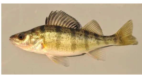
Yellow perch, Perca flavescens (Figure 1), is an ecologically and economically important 68
species native to a large portion of the northern United States and southern Canada. Yellow 69
perch supports recreational and commercial fisheries and is a major component of the food web 70
in many inland lakes, where they are often the most abundant prey for larger species such as 71
walleye (Sander vitreus), northern pike (Esox lucius), muskellunge (Esox masquinongy), and 72
lake trout (Salvelinus namaycush) [1]. In the Laurentian Great Lakes, yellow perch are an 73
important native species that has been heavily impacted by fishing pressure and environmental 74
changes over the last century [2,3]. Yellow perch is consistently among the most valuable 75
commercially harvested fish species in the Great Lakes ($2.64/lb. dockside value in 2000 [4]), 76
with fillets selling as high as $12/lb). However, many yellow perch fisheries have been forced 77
to close due to substantial population declines [5]. The mechanisms underlying these declines 78
are not fully understood but could be investigated using a combination of ecological and genetic 79
studies if adequate genomic information were available. 80
From an aquaculture perspective, yellow perch has many desirable attributes. For example, 81
yellow perch can tolerate high stocking densities, are relatively disease resistant, and can be 82
raised successfully under a variety of temperature and water conditions [6,7]. Furthermore, 83
yellow perch can be reared from hatching to marketable size in a relatively short period of time 84
(~1 year vs. 2+ years for most salmonids). Because yellow perch eat a diverse array of prey 85
items [8], their feed can be obtained from ecologically sustainable sources while remaining cost 86
effective (in contrast salmon are often fed a diet consisting primarily of other wild-caught 87
fishes, known as fish meal). Lastly, yellow perch fillets have a firm texture and a mild flavor 88
yielding a high market value. 89
The challenges faced by the yellow perch aquaculture include: increasing the spawning window 90
for broodstock, reducing early life stage mortality, and developing large-bodied strains with 91
faster growth rates [6]. Yellow perch spawn seasonally (typically in late spring to early summer) 92
during a relatively narrow period of time (1-2 weeks). From an aquaculture perspective, it can 93
be challenging to find males and females that are ready to spawn at the same time and, if the 94
fish are not monitored daily, the peak spawning period can be missed entirely [9]. Compared to 95
4 other aquaculture species, yellow perch also have a protracted free-swimming larval stage (~30 96
days), during which the fish require precise food and water conditions for optimal survival. 97
Developing broodstock that produce offspring with a shorter larval stage or that produce larger, 98
more robust offspring would allow perch to be successfully reared in a broader array of 99
facilities. Lastly, while yellow perch can already be grown to marketable size relatively quickly, 100
the relative lack of selective breeding means that there is considerable room for developing 101
yellow perch strains with faster growth rates and larger body sizes [6]. 102
These challenges, which currently limit the wide-scale adoption of yellow perch as an 103
aquaculture species, can be addressed using cutting edge genomic resources, such as the 104
genome assembly described here. For example, one straightforward step towards obtaining fish 105
with faster growth rates and larger body size would be to produce genetically all-female 106
populations, as females grow considerably faster and larger than males [10–12]. More 107
generally, sequencing and characterizing the yellow perch genome will facilitate improvements 108
in both aquaculture and fisheries management practices. 109
110
Results and Discussion
111112
Genome characteristics
113 114
By a combination of three approaches -- Oxford Nanopore Technologies (ONT) long-reads, 115
10X genomics Illumina short linked reads (PE150 chemistry), and a chromosome contact map 116
(Hi-C) -- we generated a high-continuity, chromosome length de novo genome assembly of the 117
yellow perch. Before the Hi-C integration step, the assembly yielded a genome size of 877 Mb 118
with 879 contigs, a N50 contig size of 4.3 Mb, and a L50 contig number of 60 (i.e. half of the 119
assembled genome is included in the 60 longest contigs). After Hi-C integration, the genome 120
assembled into 269 fragments with a total length of 877.4 Mb, including 24 chromosome-length 121
scaffolds representing 98.78 % of the complete genome sequence (N50 = 37.4 Mb, L50 = 11) 122
(see Table 1). Genome sizes are both very close to the 873 Mbp GenomeScope [13] estimation 123
based on short-read analysis with a repeat length of 266 Mbp (30.5%) and slightly lower than 124
the estimation of P. flavescens genome sizes based on C-values (900 Mbp and 1200 Mbp 125
records in the Animal Genome Size Database [14]). The 24 chromosome-length scaffolds 126
obtained after Hi-C integration are consistent with the diploid chromosome (Chr) number of 127
yellow perch (2n = 48) [15]. The genome completeness of these assemblies was estimated using 128
5 Benchmarking Universal Single-Copy Orthologs (BUSCO) v3.0 [16] based on the 129
Actinopterygii database. BUSCO scores (see Table 1) of the pre-Hi-C and post-Hi-C assemblies 130
were roughly similar (Complete BUSCOs between 97.6% and 97.8%) and with small values 131
for both fragmented (< 1%) and missing (< 1.5%) BUSCO genes. 132
Repeated elements accounted for 41.71% (366 Mbp) of our chromosomal assembly and these 133
regions were soft masked before gene annotation. Using protein, transcript, and de novo gene 134
prediction evidence we annotated 24,486 genes, including 16,579 (76.3%) that significantly 135
matched with a protein hit in the non-redundant NCBI database (Table 2). Our yellow perch 136
genome was also annotated with the NCBI Eukaryotic Genome Annotation Pipeline (NCBI 137
Perca flavescens Annotation Release 100 [17]), leading to a higher gene count (28,144) with
138
possibly multiple transcripts per gene (Table 2). 139
140
Yellow perch sex-determination
141 142
Yellow perch has a male monofactorial heterogametic sex determination system (XX/XY) [18] 143
with undifferentiated sex chromosomes [19]. Using a male-versus-female pooled gDNA whole 144
genome sequencing strategy [20], we identified a relatively small region of 100 kb localized at 145
the proximal end of chromosome 9 (Chr09:0-100,000 bp) with a complete absence of female 146
reads, excluding repeated elements (Fig. 2.A-B). This coverage bias strongly supports the 147
hypothesis that Chr09 is the yellow perch sex chromosome and contains a small Y-specific 148
region in phenotypic males that is completely absent from phenotypic females. Genome 149
annotation shows that this Y-specific insertion on Chr09 contains a duplicate copy (amhr2by) 150
of the autosomal anti-Mullerian hormone receptor gene located on Chr04 (amhr2a). The amhr2 151
gene has previously been characterized as a master sex-determining gene in some pufferfishes 152
[21,22] and the hotei mutation in the medaka amhr2 gene induces a male-to-female sex-reversal 153
of genetically XY fish [23]. However, in contrast to pufferfishes, in which the differentiation 154
of X and Y chromosomes is extremely limited and originated from an allelic diversification 155
process, the yellow perch amhr2by sequence is quite divergent from its amhr2a autosomal 156
counterpart. Specifically, the amhr2by gene shows only 88.3 % identity with amhr2a in the 157
aligned coding sequence and 89.1 % in the aligned parts of the introns, but with many long gaps 158
and indels in the introns (Fig. 2C-D). This nucleotide sequence divergence impacts the protein 159
sequence of the yellow perch amhr2by gene (Fig. 2D-2E), but due to a complete absence of 160
exons 1 & 2 (Fig. 2C-2E) compared to its autosomal counterpart, the yellow perch Amhr2by 161
6 protein translates as a N-terminal-truncated type II receptor that lack most of the cysteine-rich 162
extracellular part of the receptor, which is crucially involved in ligand binding specificity [24]. 163
164
To validate the male specificity of this potential Y-specific insertion, we designed primers 165
specific for both amhr2by and amhr2a and genotyped 25 male and 25 female yellow perch 166
collected from a Southeastern Lake Michigan population, which is geographically isolated from 167
the Plum Lake (Wisconsin) population of the 30 males and 30 females used for initial analysis 168
with pool-sequencing. The presence/absence of the amhr2by PCR product was perfectly 169
correlated with the determined phenotypic sex, with the amplification of an amhr2by fragment 170
only in the 25 males and no amplification in the 25 females (see Fig. 2F for 18 of the 50 171
individuals tested). The simultaneous amplification of the amhr2a fragment in both males and 172
in females provided an internal control preventing single-locus dropout in such a multiplexed 173
PCR reaction. 174
175
This complete sex-linkage result makes the yellow perch amhr2by an obvious candidate as a 176
sex determining gene. Interestingly, anti-Mullerian hormone (Amh) has been also characterized 177
as a male-promoting gene in zebrafish [25] and as a master sex determining gene both in 178
Patagonian pejerrey [26], Nile tilapia [27] and Northern pike [28]. The role of transforming 179
growth factor beta (TGF-ß) members in sex determination is not limited to the Amh pathway; 180
additional TGF-ß family genes have also been characterized as master sex determining genes, 181
including growth differentiation factor 6 (gdf6) in the turquoise killifish [29] and gonadal soma 182
derived factor (gsdf) in the Luzon medaka and the sablefish [30,31]. Additional evidence, 183
including loss of amhr2by function experiments in XY males and gain of amhr2by function 184
experiments in XX females, is necessary to critically test the hypothesis that this male-specific 185
amhr2by duplication really functions as a master sex determining gene in yellow perch.
186
However, given the known importance of the Amh pathway in fish sex determination, and that 187
no other gene in that small sex locus is known to play a role in sex differentiation, amhr2by is 188
a prime candidate for the yellow perch master sex determining gene. This finding provides 189
another example of the recurrent utilization of the TGF-ß pathway in fish sex determination, 190
and thus supports the ‘limited option’ hypothesis [32], which states that some genes are more 191
likely than others to be selected as master sex determining genes. How this N terminal truncated 192
Amhr2 could trigger its function as a master sex determining gene is as yet unknown, but one 193
hypothesis is that this truncation constitutively activates the Amh receptor causing it to signal 194
in the absence of Amh ligand. 195
7 196
However, regardless of the precise role of the structural variation of amhr2 in sex determination, 197
we have developed a simple molecular protocol for genotypically sexing perch of any life stage 198
and produced a fully annotated, chromosome-scale genome assembly that will undoubtedly aid 199
in the conservation and management of this species. 200
201
MATERIAL AND METHODS
202 203
Sampling and genomic DNA extraction
204 205
The male yellow perch used for whole genome sequencing was sampled from Plum Lake, Vilas 206
County, Wisconsin, USA. A 0.5 ml blood sample was taken from this animal and immediately 207
put in a TNES-Urea lysis buffer (TNES-Urea: 4 M urea; 10 mM Tris-HCl, pH 7.5; 125 mM 208
NaCl; 10 mM EDTA; 1% SDS) [33]. High molecular weight genomic DNA (gDNA) was then 209
purified by phenol-chloroform extraction. For the chromosome contact map (Hi-C), 1.5 ml of 210
blood was taken from a different male from a domesticated line of yellow perch raised at the 211
Farmory, an aquaculture facility in Green Bay, Wisconsin, USA. The fresh blood sample was 212
slowly cryopreserved with 15 % Dimethyl sulfoxide (DMSO) in a Mr. Frosty Freezing 213
Container (Thermo Scientific) at -80°C. Fin clip samples (30 males and 30 females) for whole-214
genome sequencing of pools of individuals (Pool-seq) were collected from wild yellow perch 215
in Green Bay, Lake Michigan, Wisconsin, USA, placed in 90% ethanol and then stored dried 216
until gDNA extraction was performed using the NucleoSpin Kit for Tissue (Macherey-Nagel, 217
Duren, Germany). Genomic DNAs from individual fish were then quantified using a Qubit 218
fluorometer (Thermofisher) and pooled in equimolar ratios by individual and sex, resulting in 219
one gDNA pool for males and one gDNA pool for females. For validation of amhr2by sex-220
linkage, 50 phenotypically sexed individuals (25 males and 25 females) wild perch from Lake 221
Michigan were collected in May of 2018 using gill net sets off the shore of Michigan City, 222
Indiana (41°42.5300'N, 86°57.5843'W). Upon collection, each individual fish was euthanized, 223
phenotypic sex was determined by visual inspection of gonads during necropsy, and caudal fin 224
clips were taken from each yellow perch individual and stored in 95% non-denatured ethanol. 225
Genomic DNA was extracted using the DNeasy extraction kit and protocol (Qiagen). 226
227
DNA library construction and sequencing
8
Nanopore sequencing
229
The quality and purity of gDNA was assessed using spectrophotometry, fluorometry and 230
capillary electrophoresis. Additional purification steps were performed using AMPure XP 231
beads (Beckman Coulter). All library preparations and sequencing were performed using 232
Oxford Nanopore Ligation Sequencing Kits SQK-LSK108 (Oxford Nanopore Technology) (14 233
flowcells) or SQK-LSK109 (2 flowcells) according to the manufacturer’s instructions. For the 234
SQK-LSK108 sequencing Kit, 140 µg of DNA was purified and then sheared to 20 kb using 235
the megaruptor system (Diagenode). For each library, a DNA-damage-repair step was 236
performed on 5 µg of DNA. Then an END-repair+dA-tail-of-double-stranded-DNA-fragments 237
step was performed and adapters were ligated to DNAs in the library. Libraries were loaded 238
onto two R9.5 and twelve R9.4 flowcells and sequenced on a GridION instrument at a 239
concentration of 0.1 pmol for 48 h. For the SQK-LSK109 sequencing Kit, 10 µg of DNA was 240
purified and then sheared to 20 kb using the megaruptor system (Diagenode). For each library, 241
a one-step-DNA-damage repair+END-repair+dA-tail-of-double-stranded-DNA-fragments 242
procedure was performed on 2 µg of DNA. Adapters were then ligated to DNAs in the library. 243
Libraries were loaded on R9.4.1 flowcells and sequenced on either a GridION or PromethION 244
instrument at a concentration of 0.05 pmol for 48h or 64h respectively. The 15 GridION 245
flowcells produced 69.4 Gb of data and the PromethION flowcell produced 65.5 Gb of data. 246
10X Genomics sequencing
247
The Chromium library was prepared according to 10X Genomics’ protocols using the Genome 248
Reagent Kit v2. Sample quantity and quality controls were validated by Qubit, Nanodrop and 249
Femto Pulse machines. The library was prepared from 10 ng of high molecular weight (HMW) 250
gDNA. Briefly, in the microfluidic Genome Chip, a library of Genome Gel Beads was 251
combined with HMW template gDNA in master mix and partitioning oil to create Gel Bead-252
In-EMulsions (GEMs) in the Chromium apparatus. Each Gel Bead was then functionalized with 253
millions of copies of a 10x™ barcoded primer. Dissolution of the Genome Gel Bead in the 254
GEM released primers containing (i) an Illumina R1 sequence (Read 1 sequencing primer), (ii) 255
a 16 bp 10x Barcode, and (iii) a 6 bp random primer sequence. R1 sequence and the 10x™ 256
barcode were added to the molecules during the GEM incubation. P5 and P7 primers, R2 257
sequence, and Sample Index were added during library construction. 10 cycles of PCR were 258
applied to amplify the library. Library quality was assessed using a Fragment Analyser and 259
9 library was quantified by qPCR using the Kapa Library Quantification Kit. The library was then 260
sequenced on an Illumina HiSeq3000 using a paired-end read length of 2x150 nt with the 261
Illumina HiSeq3000 sequencing kits and produced 315 million read pairs. 262
Hi-C sequencing
263
In situ Hi-C was performed according to previously described protocols [34]. Cryopreserved
264
blood cells were defrosted, washed with PBS twice and counted. 5 million cells were then cross-265
linked with 1% formaldehyde in PBS, quenched with Glycine 0.125M and washed twice with 266
PBS. Membranes were then disrupted with a Dounce pestle, nuclei were permeabilized using 267
0.5% SDS and then digested with HindIII endonuclease. 5’-overhangs at HindIII-cut restriction 268
sites were filled-in, in the presence of biotin-dCTP with the Klenow large fragment, and then 269
re-ligated at a NheI restriction site. Nuclei were lysed and DNA was precipitated and then 270
purified using Agencourt AMPure XP beads (Beckman Coulter) and quantified using the Qubit 271
fluorometric quantification system (Thermo). T4 DNA polymerase was used to remove un-272
ligated biotinylated ends. Then the Hi-C library was prepared according to Illumina’s protocols 273
using the Illumina TruSeq Nano DNA HT Library Prep Kit with a few modifications: 1.4µg 274
DNA was fragmented to 550nt by sonication. Sheared DNA was then sized (200-600pb) using 275
Agencourt AMPure XP beads, and biotinylated ligation junctions were captured using M280 276
Streptavidin Dynabeads (Thermo) and then purified using reagents from the Nextera Mate Pair 277
Sample preparation kit (Illumina). Using the TruSeq nano DNA kit (Illumina), the 3’ ends of 278
blunt fragments were adenylated. Next, adaptors and indexes were ligated and the library was 279
amplified for 10 cycles. Library quality was assessed by quantifying the proportion of DNA cut 280
by endonuclease NheI using a Fragment Analyzer (Advanced Analytical Technologies, Inc., 281
Iowa, USA). Finally, the library was quantified by qPCR using the Kapa Library Quantification 282
Kit (Roche). Sequencing was performed on an Illumina HiSeq3000 apparatus (Illumina, 283
California, USA) using paired-end 2x150 nt reads. This produced 128 million read pairs (38.4 284
Gb of raw nucleotides). 285
Pool sequencing
286
Pool-sequencing libraries were prepared according to Illumina’s protocols using the Illumina 287
TruSeq Nano DNA HT Library Prep Kit (Illumina, California, USA). In short, 200 ng of each 288
gDNA pool (males and females pools) was fragmented to 550 bp by sonication on M220 289
Focused-ultrasonicator (COVARIS). Size selection was performed using SPB beads (kit beads) 290
10 and the 3’ ends of blunt fragments were mono-adenylated. Then, adaptors and indexes were 291
ligated and the construction was amplified with Illumina-specific primers. Library quality was 292
assessed using a Fragment Analyzer (Advanced Analytical Technologies, Inc., Iowa, USA) and 293
libraries were quantified by qPCR using the Kapa Library Quantification Kit (Roche). 294
Sequencing was performed on a NovaSeq (Illumina, California, USA) using a paired-end read 295
length of 2x150 nt with Illumina NovaSeq Reagent Kits. Sequencing produced 119 million 296
paired reads for the male pool library and 132 million paired reads for the female pool library. 297
298
Genome assembly and analysis
299
Genome size estimation
300
K-mer-based estimation of the genome size was carried out with GenomeScope [13]. 10X reads 301
were processed with Jellyfish v1.1.11 [35] to count 17-, 19-, 21-, 23- and 25-mers with a max 302
k-mer coverage of 10,000. 303
Genome assembly
304
GridION and PromethION data were trimmed using Porechop v0.2.1 [36], corrected using 305
Canu v1.6 [37] and filtered to keep only reads longer than 10 kbp. Corrected reads were then 306
assembled using SmartDeNovo version of May-2017 [38] with default parameters. The 307
assembly base pair quality was improved by several polishing steps including two rounds of 308
long read alignment to the draft genome with minimap2 v2.7 [39] followed by Racon v1.3.1 309
[40], as well as three rounds of 10X genomics short read alignments using Long Ranger v2.1.1 310
(10x Genomics 2018) followed by Pilon v1.22 [41]. The polished genome assembly was then 311
scaffolded using Hi-C as a source of linking information. Reads were aligned to the draft 312
genome using Juicer [42] with default parameters. A candidate assembly was then generated 313
with 3D de novo assembly (3D-DNA) pipeline [43] with the -r 0 parameter. Finally, the 314
candidate assembly was manually reviewed using Juicebox Assembly Tools [42]. Genome 315
completeness was estimated using Benchmarking Universal Single-Copy Orthologs (BUSCO) 316
v3.0 [16] based on 4,584 BUSCO orthologs derived from the Actinopterygii lineage. 317
Genome annotation
11 The first annotation step was to identify repetitive content using RepeatMasker v4.0.7 [43], 319
Dust (Kuzio et al., unpublished but described in [44]), and TRF v4.09 [45]. A species-specific 320
de novo repeat library was built with RepeatModeler v1.0.11 [46] and repeated regions were
321
located using RepeatMasker with the de novo and Danio rerio libraries. Bedtools v2.26.0 [47] 322
was used to merge repeated regions identified with the three tools and to soft mask the genome. 323
The MAKER3 genome annotation pipeline v3.01.02-beta [48] combined annotations and 324
evidence from three approaches: similarity with fish proteins, assembled transcripts (see 325
below), and de novo gene predictions. Protein sequences from 11 fish species (Astyanax 326
mexicanus, Danio rerio, Gadus morhua, Gasterosteus aculeatus, Lepisosteus oculatus,
327
Oreochromis niloticus, Oryzias latipes, Poecilia formosa, Takifugu rubripes, Tetraodon
328
nigroviridis, Xiphophorus maculatus) found in Ensembl were aligned to the masked genome
329
using Exonerate v2.4 [49]. As Perca fluviatilis is a relatively closely related species from P. 330
flavescens (divergence time is estimated to be 19.8 million years ago according to [50]),
RNA-331
Seq reads of P. fluviatilis (NCBI BioProject PRJNA256973) from the PhyloFish project [51] 332
were used for genome annotation and aligned to the chromosomal assembly using STAR 333
v2.5.1b [52] with outWigType and outWigStrand options to output signal wiggle files. 334
Cufflinks v2.2.1 [53] was used to assemble the transcripts which were used as RNA-seq 335
evidence. Braker v2.0.4 [54] provided de novo gene models with wiggle files provided by 336
STAR as hint files for GeneMark and Augustus training. The best supported transcript for each 337
gene was chosen using the quality metric called Annotation Edit Distance (AED) [55]. Genome 338
annotation gene completeness was assessed by BUSCO using the Actinopterygii group. Finally, 339
predicted genes were subjected to similarity searches against the NCBI NR database using 340
Diamond v0.9.22 [56]. The top hit with a coverage over 70% and identity over 80% was 341
retained. 342
Pool-sequencing analysis
343
Reads from the male and female pools were aligned to the chromosomal assembly with BWA 344
mem (version 0.7.17, [57]), and the resulting BAM files were sorted and PCR duplicates 345
removed using Picard tools (version 2.18.2). A file containing the nucleotide composition of 346
each pool for each genomic position was generated using samtools mpileup (version 1.8, [58]) 347
and popoolation2 mpileup2sync (version 1201, [59]). This file was then analyzed with custom 348
software (PSASS version 2.0.0 [60]) to compute 1) the position and density of sex-specific 349
SNPs, defined as SNPs heterozygous in one sex while homozygous in the other sex, 2) absolute 350
12 and relative read depth for the male and female pools along the genome, and 3) FST between
351
males and females in windows along the genome. PSASS was run with default parameters 352
except --window-size which was set to 5000 and --output-resolution which was set to 1000. 353
Validation of amhr2by sex-linkage
354 355
To validate the sex-linkage of amhr2by in males suggested by the pool-sequencing results, two 356
primer sets were designed based on the alignment of yellow perch amhr2a and amhr2by genes 357
with one primer pair specific for the autosomal amhr2a gene (forward: 5’-358
GGGAAACGTGGGAAACTCAC-3’, and reverse: 5’-AGCAGTAGTTACAGGGCACA-3’, 359
expected fragment size: 638 bp) and one primer pair specific for the Y chromosomal amhr2by 360
gene (forward: 5’-TGGTGTGTGGCAGTGATACT-3’, and reverse: 5’-361
ACTGTAGTTAGCGGGCACAT-3’, expected fragment size: 443 bp). Gene alignments were 362
run with mVISTA [61]. Primers were sourced from Integrated Data Technologies (IDT). All 363
samples were run blind with respect to phenotypic sex; the male and female samples were 364
randomized, and their phenotypic sex was not cross referenced with field data until gel 365
electrophoresis was run on the final PCR products. Genotyping was carried out on each gDNA 366
sample using a multiplexed PCR approach. The PCR reaction solution was composed of 50 µl 367
of PCR Master Mix (Quiagen), 10 µl of each primer (40 µl total), and 10 µl of gDNA 368
(concentrations of gDNA ranging from 150 to 200 ng/ µl) for a total reaction volume of 100 µl. 369
Thermocycling conditions were 1 cycle of 3 min at 94°C, followed by 35 cycles of 30 sec at 370
94°C, 30 sec at 51°C, and 1 min at 72°C, and finishing with 10 min incubation at 72°C. PCR 371
products were loaded on a 1.5 % agarose gel, run at 100V for 45 minutes and visualized with a 372
UVP UVsolo touch UV box. 373
374
Availability of supporting data
375 376
This Whole Genome Shotgun project has been deposited at DDBJ/ENA/GenBank under the 377
accession SCKG00000000. The version described in this paper is version SCKG01000000. Hi-378
C, 10X genomics and pool-sequencing Illumina reads, and Oxford Nanopore Technologies 379
genome raw reads are available in the Sequence Read Archive (SRA), under BioProject 380 reference PRJNA514308. 381 382 Author contributions 383
13 384
YG, MS, and JHP designed the project. WL, CS and MC collected the samples, EJ, MW, CR, 385
OB, SV and HA extract the gDNA, made the genomic libraries and sequenced them. RF, CC, 386
CK, MZ, PE, AH and YG processed the genome assemblies and / or analyzed the results. CS 387
and MC checked sex-linkage of amhr2by on yellow perch samples. YG, RF, WL, MC, JHP, 388
CC, CK, and CR wrote the manuscript. MS, JHP, CD, PH, AB, RM, MC and YG, supervised 389
the project administration and raised funding. All the authors read and approved the final 390 manuscript. 391 392 Competing interests 393 394
All authors declare no competing interests. 395
396
Acknowledgements
397 398
This project was supported by funds from the “Agence Nationale de la Recherche” and the 399
“Deutsche Forschungsgemeinschaft” (ANR/DFG, PhyloSex project, 2014-2016), the CRB-400
Anim “Centre de Ressources Biologiques pour les animaux domestiques” project 401
PERCH’SEX, the FEAMP “Fonds européen pour les affaires maritimes et la pêche” project 402
SEX’NPERCH, and R01 GM085318 from the National Institutes of Health, USA. Additional 403
funding was provided to MRC from the Great Lakes Fishery Commission, Project ID: 404
2018_CHR_44072. The GeT core facility, Toulouse, France was supported by France 405
Génomique National infrastructure, funded as part of “Investissement d’avenir” program 406
managed by Agence Nationale pour la Recherche (contract ANR-10-INBS-09). We are grateful 407
to the Genotoul bioinformatics platform Toulouse Midi-Pyrenees (Bioinfo Genotoul) for 408
providing computing and/or storage resources. Any use of trade, product, or company name is 409
for descriptive purposes only and does not imply endorsement by the U.S. Government. 410 411 412 REFERENCES 413 414
1. Becker GC. Fishes of Wisconsin. Madison, Wisconsin: University of Wisconsin Press, 1983, 415
1052 p. http://digicoll.library.wisc.edu/cgi-bin/EcoNatRes/EcoNatRes-416
idx?id=EcoNatRes.FishesWI. Accessed 7 Feb 2019. 417
14 2. Evans MS. Recent Major Declines in Zooplankton Populations in the Inshore Region of Lake 418
Michigan: Probable Causes and Implications. Can J Fish Aquat Sci. 1986;43:154–9. 419
3. Baldwin N, Saalfeld R, Dochoda M, Buettner H, Eshenroder L. Commercial Fish Production 420
in the Great Lakes 1867–2009. Great Lakes Fishery Commission, Ann Arbor, MI. 2009. 421
4. Kinnunen RE. Great Lakes Commercial Fisheries. 2003. Michigan Sea Grant Extension, 422 Marquette. 423 https://pdfs.semanticscholar.org/d243/74bc18e240e29a8418e96e766ea8e8be50ed.pdf. 424 Accessed 19 Jul 2019. 425
5. Marsden JE, Robillard SR. Decline of Yellow Perch in Southwestern Lake Michigan, 1987– 426
1997. North American Journal of Fisheries Management. 2004;24:952–66. 427
6. Malison JA. A white paper on the status and needs of yellow perch aquaculture in the North 428
Central Region. North Central Regional Aquaculture Center, Michigan State University, East 429
Lansing. 2003. 430
7. Malison JA, Held JA. Effects of fish size at harvest, initial stocking density and tank lighting 431
conditions on the habituation of pond-reared yellow perch (Perca flavescens) to intensive 432
culture conditions. Aquaculture. 1992;104:67–78. 433
8. Keast A. Diet overlaps and feeding relationships between the year classes in the yellow perch 434
(Perca flavescens). Environ Biol Fish. 1977;2:53–70. 435
9. Żarski D, Horváth A, Held JA, Kucharczyk D. Artificial Reproduction of Percid Fishes. In: 436
Kestemont P, Dabrowski K, Summerfelt RC, editors. Biology and Culture of Percid Fishes: 437
Principles and Practices. Dordrecht: Springer Netherlands; 2015. p. 123–61. 438
10. Malison JA, Garcia-Abiado M a. R. Sex control and ploidy manipulations in yellow perch 439
(Perca flavescens) and walleye (Stizostedion vitreum). Journal of Applied Ichthyology. 440
1996;12:189–94. 441
11. Rougeot C. Sex and Ploidy Manipulation in Percid Fishes. In: Kestemont P, Dabrowski K, 442
Summerfelt RC, editors. Biology and Culture of Percid Fishes: Principles and Practices. 443
Dordrecht: Springer Netherlands; 2015. p. 625–34. 444
12. Malison JA, Kayes TB, Wentworth BC, Amundson CH. Growth and Feeding Responses of 445
Male versus Female Yellow Perch (Perca flavescens) Treated with Estradiol-17β. Can J Fish 446
Aquat Sci. 1988;45:1942–8. 447
13. Vurture GW, Sedlazeck FJ, Nattestad M, Underwood CJ, Fang H, Gurtowski J, et al. 448
GenomeScope: fast reference-free genome profiling from short reads. Bioinformatics. 449
2017;33:2202–4. 450
14. Animal Genome Size Database. http://www.genomesize.com/index.php. Accessed 7 Feb 451
2019. 452
453
15. Ráb P, Roth P, Mayr B. Karyotype Study of Eight Species of European Percid Fishes 454
(Pisces, Percidae). Caryologia. 1987;40:307–18. 455
16. Simão FA, Waterhouse RM, Ioannidis P, Kriventseva EV, Zdobnov EM. BUSCO: 456
15 assessing genome assembly and annotation completeness with single-copy orthologs. 457
Bioinformatics. 2015;31:3210–2. 458
17. Perca flavescens Annotation Report. 459
https://www.ncbi.nlm.nih.gov/genome/annotation_euk/Perca_flavescens/100/. Accessed 5 Jul 460
2019. 461
18. Malison JA, Kayes TB, Best CD, Amundson CH, Wentworth BC. Sexual Differentiation 462
and Use of Hormones to Control Sex in Yellow Perch (Perca flavescens). Can J Fish Aquat 463
Sci. 1986;43:26–35. 464
19. Beçak ML, Beçak W, Roberts FL, Shoffner RN, Volpe EP. Perca flavescens (Mitchill) 465
(Yellow perch) 2n = 48. In: Beçak ML, Beçak W, Roberts FL, Shoffner RN, Volpe EP, editors. 466
Chromosome Atlas: Fish, Amphibians, Reptiles, and Birds: Volume 2. Berlin, Heidelberg: 467
Springer Berlin Heidelberg; 1973. 468
20. Gammerdinger WJ, Conte MA, Baroiller J-F, D’Cotta H, Kocher TD. Comparative analysis 469
of a sex chromosome from the blackchin tilapia, Sarotherodon melanotheron. BMC Genomics. 470
2016;17:808. 471
21. Kamiya T, Kai W, Tasumi S, Oka A, Matsunaga T, Mizuno N, et al. A trans-species 472
missense SNP in Amhr2 is associated with sex determination in the tiger pufferfish, Takifugu 473
rubripes (fugu). PLoS Genet. 2012;8:e1002798.
474
22. Ieda R, Hosoya S, Tajima S, Atsumi K, Kamiya T, Nozawa A, et al. Identification of the 475
determining locus in grass puffer (Takifugu niphobles) provides evidence for sex-476
chromosome turnover in a subset of Takifugu species. PLOS ONE. 2018;13:e0190635. 477
23. Morinaga C, Saito D, Nakamura S, Sasaki T, Asakawa S, Shimizu N, et al. The hotei 478
mutation of medaka in the anti-Mullerian hormone receptor causes the dysregulation of germ 479
cell and sexual development. Proc Natl Acad Sci USA. 2007;104:9691–6. 480
24. Heldin C-H, Moustakas A. Signaling Receptors for TGF-β Family Members. Cold Spring 481
Harb Perspect Biol. 2016;8:a022053. 482
25. Yan Y-L, Batzel P, Titus T, Sydes J, Desvignes T, Bremiller R, et al. The roles of Amh in 483
zebrafish gonad development and sex determination. bioRxiv. 2019;650218. 484
26. Hattori RS, Murai Y, Oura M, Masuda S, Majhi SK, Sakamoto T, et al. A Y-linked anti-485
Müllerian hormone duplication takes over a critical role in sex determination. Proc Natl Acad 486
Sci USA. 2012;109:2955–9. 487
27. Li M, Sun Y, Zhao J, Shi H, Zeng S, Ye K, et al. A Tandem Duplicate of Anti-Müllerian 488
Hormone with a Missense SNP on the Y Chromosome Is Essential for Male Sex Determination 489
in Nile Tilapia, Oreochromis niloticus. PLOS Genetics. 2015;11:e1005678. 490
28. Pan Q, Feron R, Yano A, Guyomard R, Jouanno E, Vigouroux E, et al. Identification of the 491
master sex determining gene in Northern pike (Esox lucius) reveals restricted sex chromosome 492
differentiation. bioRxiv. 2019;549527. 493
29. Reichwald K, Petzold A, Koch P, Downie BR, Hartmann N, Pietsch S, et al. Insights into 494
Sex Chromosome Evolution and Aging from the Genome of a Short-Lived Fish. Cell. 495
16 2015;163:1527–38.
496
30. Myosho T, Otake H, Masuyama H, Matsuda M, Kuroki Y, Fujiyama A, et al. Tracing the 497
emergence of a novel sex-determining gene in medaka, Oryzias luzonensis. Genetics. 498
2012;191:163–70. 499
31. Rondeau EB, Messmer AM, Sanderson DS, Jantzen SG, von Schalburg KR, Minkley DR, 500
et al. Genomics of sablefish (Anoplopoma fimbria): expressed genes, mitochondrial phylogeny, 501
linkage map and identification of a putative sex gene. BMC Genomics. 2013;14:452. 502
32. Marshall Graves JA, Peichel CL. Are homologies in vertebrate sex determination due to 503
shared ancestry or to limited options? Genome Biol. 2010;11:205. 504
33. Asahida T, Kobayashi T, Saitoh K, Nakayama I. Tissue Preservation and Total DNA 505
Extraction form Fish Stored at Ambient Temperature Using Buffers Containing High 506
Concentration of Urea. Fisheries science. 1996;62:727–730. 507
34. Foissac S, Djebali S, Munyard K, Vialaneix N, Rau A, Muret K, et al. Transcriptome and 508
chromatin structure annotation of liver, CD4+ and CD8+ T cells from four livestock species. 509
bioRxiv. 2019;316091. 510
35. Marçais G, Kingsford C. A fast, lock-free approach for efficient parallel counting of 511
occurrences of k-mers. Bioinformatics. 2011;27:764–70. 512
36. Wick R. adapter trimmer for Oxford Nanopore reads. https://github.com/rrwick/Porechop. 513
Accessed 7 Feb 2019. 514
37. Koren S, Walenz BP, Berlin K, Miller JR, Bergman NH, Phillippy AM. Canu: scalable and 515
accurate long-read assembly via adaptive k-mer weighting and repeat separation. Genome Res. 516
2017;27:722–36. 517
38. Ruan J. Ultra-fast de novo assembler using long noisy reads: ruanjue/smartdenovo. 518
https://github.com/ruanjue/smartdenovo. Accessed 7 Feb 2019. 519
39. Li H. Minimap2: pairwise alignment for nucleotide sequences. Bioinformatics. 520
2018;34:3094–100. 521
40. Vaser R, Sović I, Nagarajan N, Šikić M. Fast and accurate de novo genome assembly from 522
long uncorrected reads. Genome Res. 2017;27:737–46. 523
41. Walker BJ, Abeel T, Shea T, Priest M, Abouelliel A, Sakthikumar S, et al. Pilon: an 524
integrated tool for comprehensive microbial variant detection and genome assembly 525
improvement. PLoS ONE. 2014;9:e112963. 526
42. Durand NC, Shamim MS, Machol I, Rao SSP, Huntley MH, Lander ES, et al. Juicer 527
Provides a One-Click System for Analyzing Loop-Resolution Hi-C Experiments. Cell Syst. 528
2016;3:95–8. 529
43. Dudchenko O, Batra SS, Omer AD, Nyquist SK, Hoeger M, Durand NC, et al. De novo 530
assembly of the Aedes aegypti genome using Hi-C yields chromosome-length scaffolds. 531
Science. 2017;356:92–5. 532
17 44. Morgulis A, Gertz EM, Schäffer AA, Agarwala R. A fast and symmetric DUST 533
implementation to mask low-complexity DNA sequences. J Comput Biol. 2006;13:1028–40. 534
45. Benson G. Tandem repeats finder: a program to analyze DNA sequences. Nucleic Acids 535
Res. 1999;27:573–80. 536
46. RepeatModeler Download Page. http://www.repeatmasker.org/RepeatModeler/. Accessed 537
23 Mar 2019. 538
47. Quinlan AR, Hall IM. BEDTools: a flexible suite of utilities for comparing genomic 539
features. Bioinformatics. 2010;26:841–2. 540
48. Holt C, Yandell M. MAKER2: an annotation pipeline and genome-database management 541
tool for second-generation genome projects. BMC Bioinformatics. 2011;12:491. 542
49. Slater GSC, Birney E. Automated generation of heuristics for biological sequence 543
comparison. BMC Bioinformatics. 2005;6:31. 544
50. Couture P, Pyle G, Pyle G. Evolutionary Relationships, Population Genetics, and Ecological 545
and Genomic Adaptations of Perch (Perca). Biology of Perch. 2015. Available from: 546
https://www.taylorfrancis.com/. Accessed 19 July 2019. 547
51. Pasquier J, Cabau C, Nguyen T, Jouanno E, Severac D, Braasch I, et al. Gene evolution and 548
gene expression after whole genome duplication in fish: the PhyloFish database. BMC 549
Genomics. 2016;17:368. 550
52. Dobin A, Davis CA, Schlesinger F, Drenkow J, Zaleski C, Jha S, et al. STAR: ultrafast 551
universal RNA-seq aligner. Bioinformatics. 2013;29:15–21. 552
53. Trapnell C, Williams BA, Pertea G, Mortazavi A, Kwan G, van Baren MJ, et al. Transcript 553
assembly and quantification by RNA-Seq reveals unannotated transcripts and isoform 554
switching during cell differentiation. Nat Biotechnol. 2010;28:511–5. 555
54. Hoff KJ, Lange S, Lomsadze A, Borodovsky M, Stanke M. BRAKER1: Unsupervised 556
RNA-Seq-Based Genome Annotation with GeneMark-ET and AUGUSTUS. Bioinformatics. 557
2016;32:767–9. 558
55. Eilbeck K, Moore B, Holt C, Yandell M. Quantitative measures for the management and 559
comparison of annotated genomes. BMC Bioinformatics. 2009;10:67. 560
56. Buchfink B, Xie C, Huson DH. Fast and sensitive protein alignment using DIAMOND. Nat 561
Methods. 2015;12:59–60. 562
57. Li H. Aligning sequence reads, clone sequences and assembly contigs with BWA-MEM. 563
arXiv:13033997 [q-bio]. http://arxiv.org/abs/1303.3997. 2013. Accessed 24 May 2019. 564
58. Li H, Handsaker B, Wysoker A, Fennell T, Ruan J, Homer N, et al. The Sequence 565
Alignment/Map format and SAMtools. Bioinformatics. 2009;25:2078–9. 566
59. Kofler R, Pandey RV, Schlötterer C. PoPoolation2: identifying differentiation between 567
populations using sequencing of pooled DNA samples (Pool-Seq). Bioinformatics. 568
2011;27:3435–6. 569
18 60. Feron R. RomainFeron/PSASS: Version 2.0.0. Zenodo; 2019. 570
https://zenodo.org/record/2615936#.XTyIS3s6_AI. Accessed 27 Jul 2019. 571
61. Frazer KA, Pachter L, Poliakov A, Rubin EM, Dubchak I. VISTA: computational tools for 572
comparative genomics. Nucleic Acids Res. 2004;32:W273-279. 573
62. Higgins DG, Sharp PM. CLUSTAL: a package for performing multiple sequence alignment 574 on a microcomputer. Gene. 1988;73:237–44. 575 576 577
Tables
578 579Table 1. Yellow perch assembly statistics.
580
Assembly metrics Pre Hi-C Post Hi-C
Number of reads 3,118,677 3,118,677
Total size of reads 49,450,446,732 49,450,446,732
Number of contigs 879 269
Total size of the assembly 877,025,633 877,440,133
Longest fragment 18,280,501 44,580,961
Shortest fragment 160 160
Mean fragment size 997,754 3,261,859
Median fragment size 216,440 15,167
N50 fragment length 4,304,620 37,412,490
L50 fragment count 60 11
Assembly completeness Pre Hi-C Post Hi-C
Complete BUSCOs 4,482 (97.8%) 4,472 (97.6%)
Complete and single-copy BUSCOS 4,371 (95.4%) 4,363 (95.2%) Complete and duplicated BUSCOS 111 (2.4%) 109 (2.4%)
Fragmented BUSCOs 47 (1%) 41 (0.9%)
Missing BUSCOs 55 (1.2%) 71 (1.5%)
581 582
19
Table 2. Yellow perch annotation statistics.
583
Gene annotation This paper NCBI
Number of genes 24,486 28,144
Number of mRNA 21,723 42,926
Number of tRNA 2,763 1,250
Transcriptome size 56,137,542 bp 138,437,341 bp
Mean transcript length 2,292 bp 2,938 bp
Longest transcript 67,783 bp 94,494 bp
Number of coding genes with significant hit against NCBI NR
16,579 (76.3%) 20,992 (88,4%)
Gene completeness (Actinopterygii dataset)
Complete BUSCOs 4,287 (93.5%) 4,555 (99.4%) Fragmented BUSCOs 87 (1.9%) 18 (0.4%) Missing BUSCOs 210 (4.6%) 11 (0.2%) 584 585 586
20 587
Figures
588 589 590Figure 1: Adult yellow perch (Perca flavescens).
21 592
593
Figure 2: Characterization of a Y-specific duplication/insertion of the anti-Mullerian
594
hormone receptor gene (amhr2by) in yellow perch. A. Pool-seq data illustrating relative read
595
depth across chromosome 9 (Chr09) for the male (blue line) and female (red line) pools showing 596
a coverage difference between males (blue area) and females (red area) in the first 100 kb of 597
Chr09. B. Zoom-in on the read depth difference between males and females in the first 150 kb 598
of Chr09. Gene annotation is represented by blue boxes with arrows to indicate transcript 599
orientation (NCBI Perca flavescens Annotation Release 100 [13]). Abbreviations: slob1 600
(probable inactive serine/threonine-protein kinase slob1, LOC114561790), amhr2by (anti-601
Mullerian hormone type-2 receptor-like, LOC114561927), gja1l (gap junction alpha-1 protein-602
like, LOC114562210), gjcl1 (gap junction gamma-1 protein-like, LOC114562012), rfc2l 603
22 (replication factor C subunit 2-like, LOC114561955). C. Identity plot of the alignment of 604
amhr2by gene sequence (Chr09, bottom panel) with the autosomal amhr2a gene sequence
605
(Chr04, top panel). The structure of the amhr2a gene is depicted with blue boxes (exons, E1 to 606
E12) and blue lines (introns) with arrows indicating the transcription orientation. The solid line 607
on the identity plot (bottom panel) represents 50% nucleotide identity between the two 608
sequences. D. ClustalW [62] alignment of Amhr2a and Amhr2by proteins. Identical amino-609
acids are shaded and the cysteines in the extracellular domain of Amhr2 are shown with bolded 610
black asterisks. Additional cysteines specific to Amhr2by are highlighted in blue. The different 611
domains of the receptor are boxed. E. Schematic representation of the two yellow perch Amhr2 612
proteins showing that the main differences impact the extracellular domain with parts missing 613
in Amhr2by represented in white and the two additional cysteines represented by blue asterisks. 614
F. Validation of amhr2by sex linkage in yellow perch. Agarose gel electrophoresis of multiplex
615
PCR of amhr2a (higher size PCR fragment, 638 bp), and amhr2by (lower size PCR fragment, 616
443 bp) in nine females (F, left side) and nine males (M, right side) genomic DNA. 617
618 619