HAL Id: hal-01320073
https://hal.sorbonne-universite.fr/hal-01320073
Submitted on 23 May 2016
HAL is a multi-disciplinary open access
archive for the deposit and dissemination of
sci-entific research documents, whether they are
pub-lished or not. The documents may come from
L’archive ouverte pluridisciplinaire HAL, est
destinée au dépôt et à la diffusion de documents
scientifiques de niveau recherche, publiés ou non,
émanant des établissements d’enseignement et de
the hippocampus: an associative network
Caroline Le Duigou, Jean Simonnet, Maria T. Teleñczuk, Desdemona Fricker,
Richard Miles
To cite this version:
Caroline Le Duigou, Jean Simonnet, Maria T. Teleñczuk, Desdemona Fricker, Richard Miles.
Recur-rent synapses and circuits in the CA3 region of the hippocampus: an associative network. Frontiers
in Cellular Neuroscience, Frontiers, 2014, 7, pp.262. �10.3389/fncel.2013.00262�. �hal-01320073�
Recurrent synapses and circuits in the CA3 region of the
hippocampus: an associative network
Caroline Le Duigou, Jean Simonnet, Maria T. Teleñczuk, Desdemona Fricker and Richard Miles*
Centre de Recherche de l’Institut du Cerveau et de la Moelle, INSERM U975, CHU Pitié-Salpêtrière, Université Pierre et Marie Curie, Paris, FranceEdited by:
Enrico Cherubini, International School for Advanced Studies, Italy
Reviewed by:
Dominique Debanne, Aix-Marseille, France
Katalin Toth, Universite Laval, Canada
*Correspondence:
Richard Miles, Centre de Recherche de l’Institut du Cerveau et de la Moelle, INSERM U975, CHU Pitié-Salpêtrière, Université Pierre et Marie Curie, 47 Boulevard de l’Hôpital, Paris 75013, France e-mail: [email protected]
In the CA3 region of the hippocampus, pyramidal cells excite other pyramidal cells and
interneurons. The axons of CA3 pyramidal cells spread throughout most of the region to
form an associative network. These connections were first drawn by Cajal and Lorente
de No. Their physiological properties were explored to understand epileptiform discharges
generated in the region. Synapses between pairs of pyramidal cells involve one or few
release sites and are weaker than connections made by mossy fibers on CA3 pyramidal
cells. Synapses with interneurons are rather effective, as needed to control unchecked
excitation. We examine contributions of recurrent synapses to epileptiform synchrony, to
the genesis of sharp waves in the CA3 region and to population oscillations at theta and
gamma frequencies. Recurrent connections in CA3, as other associative cortices, have a
lower connectivity spread over a larger area than in primary sensory cortices. This sparse,
but wide-ranging connectivity serves the functions of an associative network, including
acquisition of neuronal representations as activity in groups of CA3 cells and completion
involving the recall from partial cues of these ensemble firing patterns.
Keywords: CA3, recurrent, synapse, circuit, hippocampus, associative
RECURRENT EXCITATORY SYNAPSES BETWEEN CA3 CELLS:
EMERGENCE
Recurrent connections between CA3 cells in the hippocampus can
be seen in early drawings of Golgi stained neurons.
Schaffer (1892)
and
Ramón y Cajal (1899)
drew pyramidal cell processes that
ram-ify extensively in the CA3 region as well as projecting into CA1.
Later, but still before cellular physiology,
Lorente de Nó (1934)
drew axonal terminals of a CA3 cell contacting mid-apical
den-drites of a nearby pyramidal cell and a basket cell (
Figure 1). So
a basis for recurrent excitation existed before synaptic operations
were fully accepted. The absence of this detail did not impede
spec-ulation. Recurrent connections between cells of the same region
were linked to feedback in chains of connected neurons.
Lorente
de No (1938)
and later
Hebb (1949)
proposed they might
gener-ate reverberating neuronal discharges as an immedigener-ate electrical
memory.
Intracellular electrophysiology began for the hippocampus
with the work of Spencer and Kandel. Initial results dampened
the excitation somewhat. They showed that stimulating CA3 cell
axons induced dominant inhibitory actions mediated by
pyrami-dal cell excitation of interneurons (
Spencer and Kandel, 1961
).
However recurrent actions were soon linked to reverberation and
epileptic synchrony (
Kandel and Spencer, 1961
). This link was later
strengthened by work on epileptiform synchrony induced by
peni-cillin an early antagonist of inhibitory synaptic actions (
Lebovitz
et al., 1971
). Explicitly combining computer simulations and in
vitro physiology,
Traub and Wong (1982)
and
Wong and Traub
(1983)
showed how recurrent excitatory synapses might underly
delayed all-or-nothing population bursts induced by
disinhibi-tion. Physiological support for recurrent synaptic actions came
from records of synaptic interactions between CA3 pyramidal cells
in slices (
Miles and Wong, 1986
). Recurrent synapses together
with the modeling work could explain the unexpected
find-ing that stimulatfind-ing a sfind-ingle cell could initiate interictal-like
bursts of much larger neuronal populations (
Miles and Wong,
1983
).
AXONAL DISTRIBUTIONS OF CA3 PYRAMIDAL CELLS
Axons of single CA3 pyramidal cells of the rat (
Figure 1) and
guinea-pig have been traced from neurons filled with biocytin
or horseradish peroxidase (
Ishizuka et al., 1990
;
Sik et al., 1993
;
Li et al., 1994
;
Wittner et al., 2006a
;
Wittner and Miles, 2007
).
Before projecting out of the region, axons ramify in stratum oriens
and radiatum of CA3 contacting apical and basilar dendrites of
other pyramidal cells as well as interneurons. Typically they divide
into 5–10 collaterals projecting in different directions but rarely
returning towards their parent neuron. Longitudinal projections
of single axons (
Lorente de Nó, 1934
) can extend for
∼70% of
the dorso-ventral extent of rodent hippocampus (
Sik et al., 1993
;
Li et al., 1994
). A significant proportion of synapses made by a
CA3 pyramidal cell may contact other CA3 cells. The
Li et al.
(1994)
estimated 30–70%. Other connections are made onto CA1
neurons, while there is also a strong commissural projection.
The total axonal length of well-filled CA3 pyramidal cell
arbors is estimated as 150–300 mm in the rat with about 30%
of the ramification within CA3 (
Ishizuka et al., 1990
;
Li et al.,
1994
). Terminals are present along all of this distance and a
single pyramidal cell is estimated to form 30,000 to 60,000
ter-minals. Terminals have been thought to target pyramidal cells
and interneurons with a frequency similar to the presence of
these neuronal types. Recent data suggest some interneuron
subtypes may be selectively innervated (
Wittner et al., 2006b
).
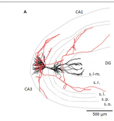
FIGURE 1 | CA3 pyramidal cell axon and targets. (A) Reconstruction
of a CA3 pyramidal cell dendrites, in black, and partial reconstruction of the axon, in red. Adapted from a cell filled byIshizuka et al.(1995; published as cell c12866 on neuromorpho.org). The CA3, CA1, and dentate gyrus (DG) regions are indicated as are the layers lacunosum-moleculare (s. l-m.), radiatum (s. r.), lucidum (s.l.), pyramidale (s.p.), and oriens (s.o.).
(B) Drawing of putative axo-dendritic connexions between pyramidal cells
(Py. 1 and 2) and interneurons with somata in different layers (B.c., Str. o.c., Str. r.c., Str. l.c., Str. m.c.). The axon of Py. 2 may contact the dendrites of Py. 1, in red, and the interneuron of stratum oriens, in blue. The axon of Py. 1 is drawn contacting the basket cell, in blue (drawing adapted from
Lorente de Nó, 1934).
Intra-regional differences exist: CA3b pyramidal cells tend to
innervate targets in stratum oriens and radiatum about equally,
while CA3a pyramidal cell axons target stratum oriens
tar-gets more than those in stratum radiatum (
Wittner and Miles,
2007
).
CA3 PYRAMIDAL CELL AXON PHYSIOLOGY
Axon collaterals of CA3 pyramidal cells are un-myelinated. They
include Schaffer collaterals that project to CA1 as well as those
that ramify within the CA3 region. Action potentials are
initi-ated at
∼30–40 μm from the soma, where sodium (Na) channel
density reaches a peak according to physiology and
immunos-taining (
Meeks and Mennerick, 2007
). In regions beyond the
action potential initiation site, recurrent axons of CA3 pyramidal
cells conduct at velocities of 0.2–0.4 mm/ms (
Soleng et al., 2003b
;
Meeks and Mennerick, 2007
).
The Na channels expressed by CA3 recurrent collaterals
seem likely to be Nav1.2 and Nav1.6 (
Royeck et al., 2008
;
Debanne et al., 2011
). These axons express multiple
voltage-gated potassium (K) channels including Kv1.1, Kv1.2, and
Kv1.4 (
Lorincz and Nusser, 2008
), ID (
Saviane et al., 2003
) the
M-channel (Kv7/KCNQ
Vervaeke et al., 2006
), and the
hyper-polarization activated h-current (
Soleng et al., 2003a
). This
diversity of channel expression provides multiple means to
mod-ulate action potential shape and so control transmitter release
(
Bischofberger et al., 2006
).
Action potential modulation by
axonal K-channels may become a total suppression of transmission
when an IA-like K-current is fully activated (
Debanne et al., 1997
;
Kopysova and Debanne, 1998
).
CA3 PYRAMIDAL CELL TERMINALS: NUMBERS, FORM,
CONTENTS, CHANNELS AND RELEASE
Varicosities are formed at distances of 2–5
μm all along CA3
recurrent axons. They often have an ovoid form of diameter
∼0.4 μm compared to an axonal diameter of ∼0.2 μm (
Sik
et al., 1993
;
Li et al., 1994
;
Wittner and Miles, 2007
).
Elec-tron microscopy (EM;
Figure 2) indicates they possess attributes
of pre-synaptic boutons with active zones and synaptic
vesi-cles and they face densities at post-synaptic sites (
Schikorski
and Stevens, 1997
;
Shepherd and Harris, 1998
;
Holderith et al.,
2012
). While varicosities may contain up to three to four active
sites, typically they have just one. Synaptic vesicles in
recur-rent terminals have diameters of 20–40 nm. A terminal may
contain up to 800 vesicles with a mean number of 150–270
vesicles.
A small proportion of vesicles are so close (∼5 nm) to
pre-synaptic membrane that they are considered to be “docked” or
available for release. The number of docked vesicles is estimated
at 1–15 per terminal (
Schikorski and Stevens, 1997
;
Shepherd
and Harris, 1998
;
Holderith et al., 2012
). Vesicles in terminals
of CA3 pyramidal cell axons express the transporters, VGLUT1
and 2, and so presumably contain glutamate (
Herzog et al.,
2006
). EM studies on CA3 axon terminals have not revealed a
distinct population of large dense-core vesicles, which might
con-tain peptides or other co-transmitters. About half of recurrent
terminals contain one mitochondrion (
Shepherd and Harris,
1998
) and smooth endoplasmic reticulum is typically present:
both organelles contribute to calcium (Ca) homeostasis (
Sheng
and Cai, 2012
).
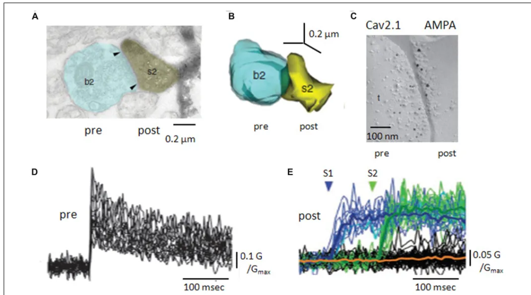
FIGURE 2 | Anatomy and Ca handling at recurrent synapses between CA3 pyramidal cells. (A) Electron microscopy of a recurrent terminal, b2,
apposed to a CA3 pyramidal cell dendritic spine, s2. (B) Three-dimensional reconstruction of the contact. The area of the active zone [arrows in (A)] was 0.10μm2. (C) Double immuno-staining of SDS-digested freeze fracture replica of a recurrent synapse. The smaller gold particles label Cav2.1
molecules (pre) and the larger gold particles recognize a pan-AMPA antibody (post). (D) Pre-synaptic Ca transients, measured as changes in fluorescent intensity, for 25 axon terminals of a CA3 pyramidal cell. (E) Post-synaptic Ca transients, in response to two pre-synaptic stimuli. Note the occurrence of failures in both post-synaptic responses but their absence from pre-synaptic signals (adapted with permission fromHolderith et al., 2012).
Ca entry into presynaptic terminals triggers transmitter release.
CA3 axonal terminals express multiple Ca channel subtypes
including Cav2.1, Cav2.2, Cav2.3 (
Holderith et al., 2012
), as do
the mossy fiber terminals that also terminate on CA3 pyramidal
cells (
Li et al., 2007
). Freeze-fracture replica gold immuno-labeling
(
Figure 2) suggests a single terminal expresses several tens of
Cav2.1 channels (
Holderith et al., 2012
). This is more, but
not many more, than estimates of the number of Ca-channels
needed to trigger release from hippocampal inhibitory
termi-nals (
Bucurenciu et al., 2010
). Possibly, an elevated Na channel
density in terminals enhances Ca entry by boosting
depolariza-tion due to axonal spikes (
Engel and Jonas, 2005
). Certainly,
recurrent terminals express various types of K channel which
control transmitter release by limiting terminal depolarization.
They may include the delayed rectifier type channels Kv1.1
and Kv1.2, the fast-inactivating A-type channel Kv1.4 (
Debanne
et al., 1997
;
Kopysova and Debanne, 1998
;
Lorincz and Nusser,
2008
;
Palani et al., 2010
) as well as K-channels sensitive to both
Ca and voltage (
Saviane et al., 2003
;
Raffaelli et al., 2004
) and
the muscarine sensitive M-channel Kv7/KCNQ (
Vervaeke et al.,
2006
).
Ca changes induced in local recurrent terminals by
pyrami-dal cell firing have been resolved by imaging (
Holderith et al.,
2012
;
Sasaki et al., 2012
). A single action potential induces a
Ca signal of rise time less than 1 ms that decays over several
10 s of ms (
Figure 2). Ca entry occurs without failure even
if it varies between trials at the same terminal and Ca
ele-vations at neighboring terminals are poorly correlated. For a
given terminal, the mean amplitude of Ca-signals is better
correlated with the area of the active zone than terminal volume
(
Holderith et al., 2012
).
CA3 axon terminals express receptors for transmitters which
modulate Ca entry or later steps in release processes (
Figure 2).
Receptors for the metabotropic glutamate receptor, mGluR7,
expressed at active zones facing interneurons but not principal
cells (
Shigemoto et al., 1996
) specifically control the excitation
of inhibitory cells (
Scanziani et al., 1998
). The kainate
recep-tor GluK1, reduces release by effects on both Ca entry and
on G-protein mediated stages in transmitter release (
Salmen
et al., 2012
). In contrast, presynaptic NMDA receptors enhance
Ca entry and facilitate release at some synapses made by CA3
collaterals(
McGuinness L et al., 2010
).
PRE- MEETS POST: SYNAPSES MADE BY CA3 PYRAMIDAL
CELLS WITH OTHER CA3 CELLS
When a single spike induces Ca entry into a CA3 axon
termi-nal, one, or none, or several vesicles of the excitatory transmitter
glutamate are liberated. Release fails, when Ca enters a
ter-minal but no transmitter is liberated, as shown by imaging
Ca-entry (
Figure 2) via post-synaptic glutamate receptors (
Koester
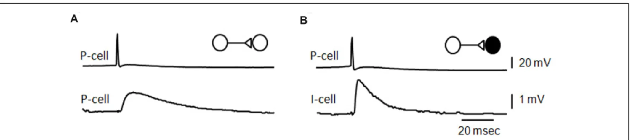
FIGURE 3 | Unitary effects of recurrent excitatory synapses. (A) Average of EPSPs initiated in a CA3 pyramidal cell by single
action potentials in a pre-synaptic pyramidal cell (B), average of
EPSPs elicited in a fast-spiking CA3 interneuron by action potentials in a pyramidal cell [an unpublished data Miles and Wong (B) adapted from Miles, 1990].
and Johnston, 2005
;
Holderith et al., 2012
). Multi-vesicular
release following a single action potential is most convincingly
demonstrated when two distinct post-synaptic events can be
resolved in time, as at some inhibitory synapses in the
cere-bellum (
Auger et al., 1998
). Analysis of variations in synaptic
events over a range of liberation probabilities supports
multi-vesicular liberation (
Conti and Lisman, 2003
;
Christie and Jahr,
2006
).
Glutamate, released from a pre-synaptic terminal, binds to
post-synaptic receptors. The number of receptors per site has been
estimated with physiological, imaging, and anatomical techniques.
Post-synaptic sites facing terminals of CA3 pyramidal cell axons
in young animals, all express NMDA (N -methyl-
D-aspartate)
receptors (
Takumi et al., 1999
). Glutamate uncaging onto
post-synaptic sites activates 3–10 NMDA receptors (
Nimchinsky et al.,
2004
). Semi-quantitative immunostaining studies and imaging
agree that about 30% of post-synaptic sites possess no AMPA (
α-amino-3-hydroxy-5-methyl-4-isoxazolepropionic acid) receptors
(
Nusser et al., 1998
;
Takumi et al., 1999
;
Nimchinsky et al., 2004
).
At synapses where AMPA receptors are expressed, about 10 of
them (
Figure 2) are estimated to be activated after a single
pre-synaptic spike in acute slices (
Nimchinsky et al., 2004
), 40–150 in
culture (
Matsuzaki et al., 2001
). AMPA receptors are present at
recurrent synapses with most types of interneuron (
Nusser et al.,
1998
). NMDA receptors are less frequently expressed at synapses
with interneurons and may be absent at contacts with fast-spiking,
parvalbumin containing cells (
Nyiri et al., 2003
).
There are two other important differences between synapses
made with interneurons and with pyramidal cells. First,
recur-rent contacts tend to innervate pyramidal cell spines, while those
with most types of inhibitory cell innervate dendritic shafts
(
Gulyas et al., 1993
;
Freund and Buzsáki, 1996
). Second, the
AMPA receptor isoforms involved are different. AMPA
recep-tor complexes at synapses formed with interneurons do not
include the GluR2 subunit (
Bochet et al., 1994
;
Geiger et al.,
1995
), resulting in faster kinetics (
Miles, 1990
), Ca-permeability,
and
a
block
by
endogenous
intraneuronal
polyamines
(
Isaac, 2007
).
PRE- MEETS POST IN DUAL RECORDS
Double records from pre- and post-synaptic neurones at recurrent
synapses between CA3 cells were first made to prove their existence
directly. They remain the most persuasive means to examine how
one neuron influences another. They have permitted definition
of the number of synaptic contacts involved in a unitary
connec-tion and assessment of variability and changes in synaptic efficacy
(
Debanne et al., 2008
).
Records from pairs of CA3 pyramidal cells in acute slices
(
Figure 3) suggest one pyramidal cell excites 2–3% of
possi-ble pyramidal cell targets in a slice (
Miles and Wong, 1986
;
Miles and Wong, 1987b
). Odds are more favorable in
organ-otypic slices. Connectivities are 30–60% (
Debanne et al., 1995
;
Pavlidis and Madison, 1999
). The number of release sites
involved in a connection may also be higher in organotypic
cultures. One to three contacts have been validated by EM
for synapses between pyramidal cells and interneurons recorded
and filled with biocytin in slices. In contrast, light microscopy
suggests 14–19 putative contacts may be involved in
connec-tions between CA3 pyramidal cells in organotypic culture
(
Pavlidis and Madison, 1999
).
The mean amplitude of synaptic potentials is about 1 mV
at connections between pyramidal cells in acute slices (
Miles
and Wong, 1986
) and in culture (
Debanne et al., 1995
). EPSPs
induced in fast-spiking interneurons (
Figure 3) are larger and
faster than those initiated in pyramidal cells. Unitary synaptic
cur-rent amplitude at connections made in culture can vary in the
range 10–200 pA with an average near 30 pA (
Pavlidis and
Madi-son, 1999
;
Sasaki et al., 2012
). In records from both acute slices
and culture, events initiated successively at the same connection
vary in amplitude. Transmission can fail, more often at
connec-tions with smaller averaged events. However pre-synaptic Ca entry
never fails, even though it varies between successive action
poten-tials (
Holderith et al., 2012
;
Sasaki et al., 2012
) and Ca signals are
higher at terminals with a higher release probability (
Koester and
Johnston, 2005
).
Synaptic events initiated sequentially at the same site vary in
amplitude. This variability may have both pre- and post-synaptic
components (
Silver et al., 2003
;
Biró et al., 2005
). Clear data on
post-synaptic variability, is facilitated at connections with a single
identified release site. At such a synapse, the variability in size of
post-synaptic events was estimated at 20–50% (
Gulyas et al., 1993
).
This variability might emerge from differences in the number of
transmitter molecules released or in the activation of post-synaptic
receptors.
The properties of recurrent synapses differ quite markedly from
those of mossy fiber inputs, the other major source of
excita-tion of CA3 pyramidal cells. A mossy fiber may make 10–20
connections with different CA3 pyramidal cells (
Claiborne et al.,
1986
). A recurrent collateral makes several thousand contacts
with a much larger target population. Mossy fiber boutons
con-tact proximal apical dendrites of CA3 pyramidal cells and have
a diameter of 4–8
μm. Each bouton may include 20–30 active
zones, whereas a recurrent synapse may make one to three
ter-minals on a post-synaptic cell. Finally mossy fibers contact apical
dendrites near the CA3 soma, while recurrent synapses
termi-nate at more distant dendritic sites resulting in smaller, slower
somatic synaptic events. A mossy fiber input from one dentate
granule cell can induce CA3 pyramidal cell firing and can so
be termed a “detonator” synapse (
Henze et al., 2002
), whereas
multiple spikes are needed to induce firing at recurrent synapses
(
Miles and Wong, 1987a
).
SHORT-TERM AND LONG-TERM SYNAPTIC PLASTICITY IN
DOUBLE RECORDINGS
Records from pre- and post-synaptic cells at recurrent synapses
have offered novel insights into activity dependent changes
in synaptic strength over times lasting from milliseconds to
hours.
Short-term plasticity (milliseconds to seconds) results from at
least two functionally opposing processes. First, a single spike
may facilitate transmission when the same synapse is activated
again (
Ddel Castillo and Katz, 1954
). An enhanced release
prob-ability over several tens of milliseconds is ascribed to a residual
elevation of intra-terminal Ca (
Holderith et al., 2012
;
Sasaki
et al., 2012
). Second, and inversely, depression may result if few
vesicles are available for release (
Schikorski and Stevens, 1997
;
Shepherd and Harris, 1998
). If they are replaced slowly (
Stevens
and Tsujimoto, 1995
;
Staley et al., 1998
) the probability of a
sec-ond release may be reduced by depletion. Both processes occur
at connections between CA3 pyramidal cells (
Debanne et al.,
1996
;
Pavlidis and Madison, 1999
). When a first spike induces
a large event, a second synaptic response tends to be smaller
due to depletion. Inversely a second EPSP tends to be larger
after a small first event due to the residual Ca enhancement
of release probability. Reflecting the underlying mechanisms,
facilitation is maximal at 20–70 ms and terminates at about
500 ms, while depression can take several seconds to recover
completely.
Long-term plasticity (minutes to hours) at different synapses
varies in mechanisms of induction and expression. One of the
most studied forms, long-term synaptic potentiation at Schaffer
collateral synapses made by CA3 pyramidal cells with CA1 cells,
is induced via the activation of NMDA receptors and expressed
as the post-synaptic recruitment of AMPA receptors (
Kerchner
and Nicoll, 2008
). Long-term changes in synaptic efficacy seem
to depend on similar mechanisms at recurrent synapses between
CA3 pyramidal cells. Paired records from coupled CA3 cells
have revealed some unitary details of this synaptic plasticity. The
same connection can be potentiated or depotentiated (
Debanne
et al., 1998
) by different temporal patterns of paired pre- and
post-synaptic firing. About 20% of unitary interactions depend
exclusively on NMDA receptors before potentiation (
Montgomery
et al., 2001
), while both AMPA and NMDA receptors are activated
after potentiation. Weak connections potentiate to a larger degree
than initially strong connections (
Debanne et al., 1999
;
Mont-gomery et al., 2001
). Finally some connections between CA3
pyra-midal cells do not seem to potentiate at all (
Debanne et al., 1999
;
Montgomery and Madison, 2002
).
TRANSMISSION OF RECURRENT EXCITATORY SIGNALS ON
THE MEMBRANE OF A POST-SYNAPTIC CELL
Activation of membrane currents intrinsic to a post-synaptic cell
by recurrent EPSPs affects how they sum, spread and eventually
initiate firing. Initial evidence came from a prolongation of the
decay of unitary EPSPs induced by pyramidal cell depolarization
at subthreshold membrane potentials (
Miles and Wong, 1986
).
In contrast unitary EPSPs initiated in fast-spiking inhibitory cells
were not prolonged at depolarised subthreshold potentials (
Miles,
1990
). Work combining somatic records and synaptic stimuli with
cell-attached records from dendrites, showed the activation of both
inward currents, probably persistent Na channels, low-threshold
Ca channels (
Magee and Johnston, 1995
), and outward currents,
both inactivating and persistent (
Hoffman et al., 1997
). These
cur-rents have been more precisely described for EPSPs initiated by
Schaffer collaterals (
Lipowsky et al., 1996
;
Andreasen and Lambert,
1999
;
Perez-Rosello et al., 2011
), as has evidence for a dendritic
expression of the I–h current (
Magee, 1999
).
Distinct currents have been associated with specific effects
on EPSP shape, summation, and spread. Na-channel activation
near the peak of an EPSP tends to increase amplitude, while
Ca-channels activated during the decay phase act to prolong
EPSPs. The striking increase in dendritic expression of the I–
h channel with distance from the soma (
Lörincz et al., 2002
)
tends to equalize EPSPs impinging at proximal and distal sites
(
Magee, 1999
). Dendritically expressed inactivating K-channels
have been linked to less-than-linear summation of paired EPSPs
impinging on different dendrites (
Urban and Barrionuevo, 1998
).
Dual records from the soma and apical dendrites of CA3
pyra-midal cells disclose two distinct regions of dendritic
excitabil-ity (
Kim et al., 2012
). Fast Na-spikes are more easily initiated
at distant sites corresponding to zones of recurrent synaptic
inputs, while excitability of more proximal dendritic sites is
lower.
The role of intrinsic currents in shaping interneuron EPSPs
may be quite different to that in pyramidal cells. Simulated
EPSPs induce purely inward currents in pyramidal cells but
rather induce inward-outward current sequences in interneurons
(
Fricker and Miles, 2000
). So, while, EPSPs in pyramidal cells are
prolonged, EPSPs in interneurons may decay more rapidly due to
the activation of an outward current at subthreshold potentials.
Synaptic inputs to a neuron are significant to surrounding cells
when they initiate firing. Summed EPSPs initiated by repetitive
firing of a single CA3 pyramidal cell sometimes induce cause
a post-synaptic pyramidal cell to fire (
Miles and Wong, 1986
).
Spike-to-spike latencies are 10–15 ms, consistent with a role
for recurrent excitatory synapses in the genesis of delayed (50–
100 ms) population bursts (
Traub and Wong, 1982
;
de la Prida
et al., 2006
). Recent work suggests spike-to-spike transmission
may be limited to a few strong connections (
Ikegaya et al.,
2013
).
Pyramidal cells induce interneuron firing more effectively
and at shorter latencies of 1–3 ms (
Miles, 1990
;
Csicsvari et al.,
1998
;
Cohen and Miles, 2000
). Interneuron EPSPs are larger and
faster than recurrent EPSPs in pyramidal cells, and
interneu-ron firing threshold is lower (
Figure 4). When interneurons are
excited to fire, pyramidal cells may trigger di-synaptic IPSPs
(Figure 4) with high probability and considerable divergence
(
Miles, 1990
;
Csicsvari et al., 1998
;
Bazelot et al., 2010
). While
EPSP boosting mechanisms in interneuron dendrites are not
clear, it is surprising that EPSPs induced from a single site
(
Gulyas et al., 1993
) can induce firing. Even so, EPSP-spike
coupling at single release site excitatory synapses with some
cerebellar interneurons (
Carter and Regehr, 2002
) is also
suf-ficiently strong that EPSPs control the timing of interneuron
firing.
RECURRENT EXCITATORY CONTRIBUTIONS TO POPULATION
ACTIVITIES IN THE CA3 REGION
Recurrent synapses transmit excitation from CA3 pyramidal cells
to other pyramidal cells and to interneurons. They play a key role
in operations and functions of the CA3 region, including the
gen-eration of physiological and pathological synchronous population
activities.
INTERICTAL EPILEPTIFORM RHYTHM
A key finding linking recurrent excitation to epileptiform activity
was that stimulating any afferent pathway induced epileptiform
firing in CA3 (
Ayala et al., 1973
). Population bursts occurred with
a variable delay of 20–100 ms after the afferent response.
Traub and
Wong (1982)
suggested that during the delay recurrent synaptic
interactions within the CA3 population generate a population
syn-chrony. Synchrony induced in disinhibited slices is complete in that
all neurons tend to fire together with a field potential decorated
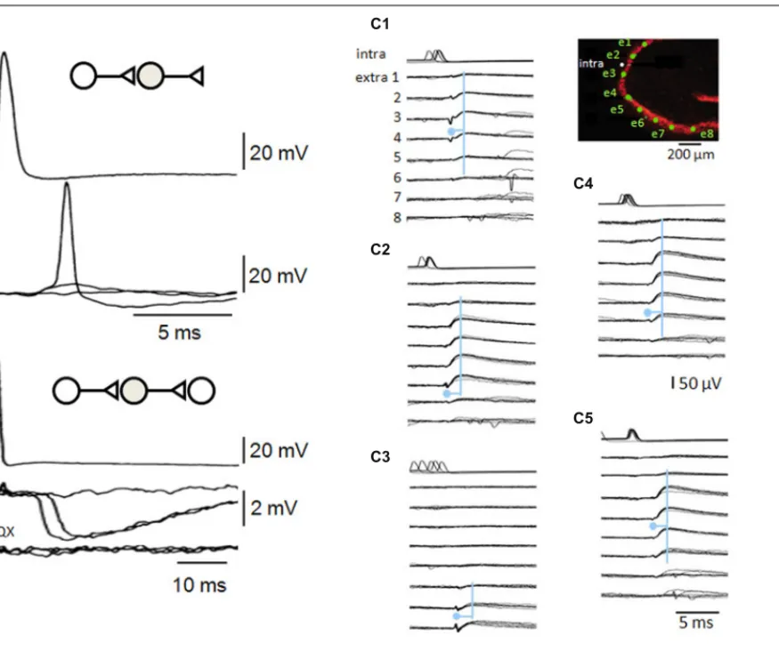
FIGURE 4 | Recurrent inhibitory circuits in the CA3 region. (A)
Post-synaptic responses of a fast-spiking interneuron to single pre-synaptic action potentials in a CA3 pyramidal cell. Responses include a failure of transmission, an EPSP and an EPSP that initiates interneuron firing. (B) Di-synaptic inhibitory interactions between two CA3 pyramidal cells. Single action potentials in one cell induce IPSPs at variable latencies consistent with that of firing in (A), as well as some failures. Di-synaptic IPSPs were suppressed by the glutamate receptor blocker CNQX. (C) A single pyramidal cell can initiate multiple di-synaptic IPSPs via firing in distinct interneurons.
Records from a pyramidal cell (intra) and extracellular records from eight sites in st. pyramidale (extra 1–8, the diagram shows st. pyramidale in red and electrode sites in green). Field IPSPs were detected on electrodes 1–6 (C1), 2–7 (C2), 6–8 (C3), 1–7 (C4), and 2–6 (C5) repeatably following single action potentials (traces are aligned on six overlapping field IPSPs for each trace). Field IPSPs are preceded by extracellular action potentials of short duration on electrodes 2–3 (C1), 6 (C2), 7–8 (C3), 6–7 (C4), and 5–6 (C5). The pyramidal cell may have initiated five distinct di-synaptic inhibitory interactions in these slice records (seeBazelot et al., 2010).
with high frequency oscillations (
Jefferys et al., 2012
). Traub and
Wong suggested recurrent circuits should possess two properties
to generate such an event. Recurrent contacts should be
diver-gent and one cell could cause more than one target neuron to fire.
These points were verified with the demonstration that some
sin-gle pyramidal cells could induce or entrain inter-ictal-like events
(
Miles and Wong, 1983
,
1986
,
1987a
;
de la Prida et al., 2006
).
Di-synaptic feedback inhibition via CA3 pyramidal cell excitation of
feedback interneurons, was shown to prevent the spread of firing
by recurrent excitatory pathways (
Miles and Wong, 1986
,
1987a
,
b
).
Recurrent synaptic function controls several features of the
epileptiform activity induced by disinhibition. The duration of
the population burst (20–80 ms) has been shown to result from
transmitter depletion (
Staley et al., 1998
). The delay from one burst
to the next (1–10 s) depends on the time for docked vesicles to be
replenished (
Staley et al., 1998
;
Staley et al., 2001
). Procedures that
induce persistent synaptic changes have persistent effects on the
strength and frequency of network burst firing (
Bains et al., 1999
;
Behrens et al., 2005
).
Cellular properties also affect disinhibition induced synchrony
by controlling transmission in chains of connected neurons.
In slices, population bursts tend to be initiated in the CA3a
region, where cellular excitability and recurrent connectivity are
high (
Wittner and Miles, 2007
). In CA3a, spontaneous events
are preceded by a field potential of duration about 50 ms
(
Wittner and Miles, 2007
) during which excitatory synaptic
events occur with increasing frequency. This delay is
simi-lar to that between single cell firing and a population event
(
Miles and Wong, 1983
;
de la Prida et al., 2006
). Modeling
work suggested that during this time activity in the
pyra-midal cell population increases in non-linear fashion (
Traub
and Wong, 1982
). An epileptiform burst occurs when
popula-tion activity exceeds a threshold frequency (
de la Prida et al.,
2006
).
SHARP-WAVE RHYTHM
Sharp waves (
O’Keefe and Nadel, 1978
;
Buzsáki et al., 1992
) are
field potentials of duration 100–150 ms, corresponding to a
partial neuronal synchrony during behaviors including
immobil-ity and slow wave sleep. They are initiated in the CA3 region
(
Csicsvari et al., 2000
) and have been associated with the
consol-idation of acquired events (
Girardeau et al., 2009
;
Jadhav et al.,
2012
) represented as firing in specific groups of neurons.
Both recurrent excitatory interactions and the actions of
spe-cific interneurons have been implicated in the genesis of sharp
waves (
Buzsáki et al., 1992
;
Csicsvari et al., 2000
). Sharp wave
fields are enhanced by inducing long-term changes at recurrent
synapses (
Behrens et al., 2005
). And yet, sharp waves are not
iden-tical with epileptiform events and do not depend on recurrent
excitation alone (
Liotta et al., 2011
). Repetitive firing of
peri-somatic interneurons may be a crucial element in sharp wave
generation (
Buzsáki et al., 1992
;
Klausberger et al., 2003
).
Gap-junctions have also been associated with sharp-waves, with the
observation of “spikelets” in pyramidal cells and a blockade by
gap-junction antagonists (
Draguhn et al., 1998
). However sharp
waves persist, at reduced strength, in animals where the gap
junction protein connexin 36 is genetically deleted (
Pais et al.,
2003
). Possibly then, recurrent excitation of both pyramidal cells
and interneurons (
Hájos et al., 2013
) may suffice to generate
sharp waves.
THETA AND GAMMA RHYTHMS
In contrast to sharp waves, theta fields (4–12 Hz) are generated
when spatial memory representations are first acquired
dur-ing movements (
Vanderwolf, 1969
;
O’Keefe and Nadel, 1978
).
Place-cells fire with theta oscillations and theta waves are also
detected in rapid eye movement sleep.
Theta oscillations probably depend on signals generated
out-side the CA3 region. Signals from the septum may provide a
sustained cholinergic excitation as well as glutamatergic (
Huh
et al., 2010
) and inhibitory signals which selectively targeting
hip-pocampal interneurons to disinhibit pyramidal cells (
Freund and
Antal, 1988
;
Tóth et al., 1997
;
King et al., 1998
). Synaptic
con-nections within the CA3 region probably reinforce the rhythm
via reciprocal interactions between pyramidal cells and some,
probably peri-somatic, interneurons (
Soltesz and Deschênes,
1993
).
Gamma oscillations at 30–70 Hz may be superimposed on theta
rhythmicity (
Bragin et al., 1995
;
Csicsvari et al., 2003
;
Hasselmo,
2005
). They are suggested to bind, or coordinate, activity of
spa-tially dispersed neurons due to a single stimulus (
Gray et al., 1989
).
In contrast to theta, gamma oscillations are generated within
the CA3 region. Reciprocal synaptic interactions between
peri-somatic inhibitory cells and CA3 pyramidal cells via recurrent
synapses are suggested to contribute both in vivo (
Csicsvari et al.,
2003
) and in slice models of gamma induced by cholinergic
ago-nists (
Oren et al., 2006
) or kainate (
Fisahn, 2005
). Gap junctions
that transmit excitation between CA3 pyramidal cell axons may
be another crucial factor in gamma generation (
Traub and Bibbig,
2000
;
Traub et al., 2003
).
COMPARISON OF RECURRENT CONNECTIVITY IN CA3 AND
OTHER CORTICAL REGIONS
The hippocampal treatment of events, memories or
representa-tions may depend in part on the associative nature of the recurrent
excitatory network between CA3 pyramidal cells. How do
recur-rent circuits in CA3 compare to those in other associative or
sensory cortical regions?
The spatial extent of excitatory terminals seems to differ for
recurrent synapses in associative, allocortical regions, such as
CA3 and the olfactory cortex, and in six-layered primary
sen-sory neocortex. CA3 pyramidal cell axons project longitudinally
through most of the hippocampus (
Lorente de Nó, 1934
;
Li et al.,
1994
). Local axons diffusely cover most of the olfactory cortex
(
Haberly, 2001
;
Franks et al., 2011
;
Poo and Isaacson, 2011
).
Con-nectivity within a six-layered cortex is certainly more complex,
but overall may be more restrained in space. For instance, axons
of layer IV pyramidal cells from sensory cortices tend to
ram-ify locally within modules such as a single somatosensory barrel
(
Petersen and Sakmann, 2000
;
Feldmeyer, 2012
). Superficial or
deep layer pyramidal cells of primary visual or somatosensory
cortex make longer range but often patchy projections
termi-nating in regions occupied by cell groups of similar function
(
Gilbert and Wiesel, 1989
;
Holmgren et al., 2003
;
Ko et al., 2011
; cf
Feldmeyer, 2012
)
The density of excitatory connections between pyramidal cells
may be somewhat higher in sensory cortical modules than in
asso-ciative allocortex such as CA3 or piriform cortex. Paired records
from acute slices gave a value of 0.02–0.03 for the probability of
a connection between two CA3 pyramidal cells (
Miles and Wong,
1986
) and recurrent connectivity in piriform cortex is estimated
at 0.002–0.01 (
Franks et al., 2011
;
Hagiwara et al., 2012
). Estimates
of connectivity are somewhat higher from paired records in slices
of sensory cortex. The probability of connection between cells in
different cortical layers ranges from 0.1 to 0.3 (0.2–0.3 in layer 4
of barrel cortex,
Lefort et al., 2009
;
Feldmeyer, 2012
; 0.1 in layer
2/3 of neocortex,
Holmgren et al., 2003
; 0.1 in layer 5 neocortex,
Markram et al., 1997
).
An alternative way to define connectivity could be to
mea-sure the spatial distribution of terminals formed by the axon
of a single cell. Terminals of some pyramidal cells in sensory
cortex (
Petersen and Sakmann, 2000
) seem likely to show a
more focal topology than those of the CA3 region (
Ishizuka
et al., 1990
;
Li et al., 1994
). Data from paired records in
slices indicates a lower local connectivity in CA3 than in
sen-sory cortex. Lower values for recurrent connectivity may be a
design feature to ensure sparse representations in an associative
region.
Recurrent excitatory synapses may contact cortical
interneu-rons selectively in both associative and sensory cortices. Paired
records suggest connectivity from pyramidal cells to fast-spiking
interneurons is higher than onto pyramidal cells (0.5–0.7 in
neocortex layer 2,
Holmgren et al., 2003
; in barrel cortex layer 2
∼0.6,
Avermann et al., 2012
; 0.2 in piriform cortex layer 3,
Stokes
and Isaacson, 2010
). A higher connectivity as well as stronger
signaling at single connections with GABAergic interneurons
(
Helmstaedter et al., 2008
) protects against excessive synchrony,
maintains stable population firing and sharpens signaling by
imposing a sparse coding.
The strength of afferent and recurrent synapses may differ
in both associative and sensory cortices. Mossy fiber synapses
with CA3 pyramidal cells have more release sites (
Claiborne
et al., 1986
) and stronger actions (
Henze et al., 2002
). Synapses
from olfactory bulb onto piriform cortex cells are both stronger
and less numerous that recurrent synapses (
Franks et al., 2011
;
Poo and Isaacson, 2011
). In barrel cortex however,
recur-rent connections between layer 4 pyramidal cells seem to be
stronger (
Feldmeyer et al., 1999
;
Feldmeyer, 2012
) than
thala-mic synapses which excite the same cells (
Bruno and Sakmann,
2006
).
Thus recurrent networks of associative cortical regions have a
wider spatial extent and a lower probability of connection between
pyramidal cells than those in sensory cortices.
THE CA3 RECURRENT SYSTEM AS AN ASSOCIATIVE
NETWORK
Associative synaptic networks have been linked to the processes
of completion and recall of stored information (
Figure 5).
McNaughton and Morris (1987)
noted that similar hypotheses
have often been discovered. What do they assume? And how might
they be tested?
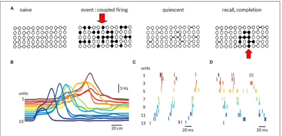
FIGURE 5 | Recurrent excitatory networks. (A) Possible schema of
connectivity and operations in a recurrent neuronal network. Some neurons are connected in the naïve network. Coupled firing in a subset of neurons during an event reinforces synapses between them. Reinforcement persists during quiescence, until partial activation recalls or completes firing of the
neuronal subset associated with the original event. (B) Sequential firing of 13 pyramidal place cells as an animal passes through a space (horizontal axis is distance). Reactivation of sequential firing of these cells as (C) forward replay or (D) backward replay (adapted with permission fromDiba and Buzsáki, 2007).
Such hypotheses suppose that information, or a
representa-tion, or an event, or a memory, has a distributed existence as
the correlated, or synchronous, discharge of a group of neurons
(
Hebb, 1949
;
Marr, 1971
). Different informations presumably
involve different groups, raising the question of how
repre-sentations are constrained to be neuronally orthogonal (
Marr,
1971
;
Rolls and Treves, 1998
). They suppose that a way exists
to associate or strengthen synaptic relations within such a group
or ensemble of synchronously active neurons. It might
cor-respond to the persistent synaptic potentiation which occurs
when pre- and post-synaptic cells fire together (
Hebb, 1949
;
Bliss and Lomo, 1973
). They suppose that a full
representa-tion of an event can be recalled from some of its elements
(
Gardner-Medwin, 1976
;
McNaughton and Morris, 1987
). The
CA3 recurrent network where activity in some single cells can
trig-ger population activities (
Miles and Wong, 1983
;
Fujisawa et al.,
2006
) might be capable of operations similar to a cued recall
(
Figure 5). The spatially widespread but lower connectivity of
associative recurrent networks may favor this form of information
storage.
Improved techniques to record and manipulate activity in
large groups of neurons begin to suggest distributed ensembles
may contribute to storage and recall. Using tetrodes to separate
firing in 50–100 single units,
Wilson and McNaughton (1994)
showed that CA1 place-sensitive neurons that fired together
dur-ing a spatial behavior, discharged synchronously again durdur-ing
the following episode of sleep. Correlated firing in cell pairs
was increased as animals learned a task and maintained
dur-ing replay. A specific role for recurrent synapses was established
by genetically deleting NMDA receptor expression at
recur-rent synapses of CA3 pyramidal cells (
Nakazawa et al., 2002
,
2003
). With the basis for persistent changes abolished, recall
of spatial memories from partial cues was suppressed. Optical
stimulation has recently been used to re-activate neurons
asso-ciated with a representation (
Liu et al., 2012
). An ensemble of
granule cells active during fear conditioning was labeled with
a construction including c-fos which also induced expression
of a light-sensitive opsin. Re-activating the sparse granule cell
ensemble optically later, induced a fear response in a different
context.
These data point to distinct neuronal operations associated
with acquisition and recall. A two-stage memory system has often
been postulated (
James, 1890
;
Buzsáki, 1989
). The two stages may
occur during distinct brain and behavioral states. External
rep-resentations, especially those associated with space (
O’Keefe and
Nadel, 1978
) and possibly also time (
Huxter et al., 2003
;
Kraus
et al., 2013
) are acquired during theta activity. In contrast, recall
or consolidation is linked with sharp-waves generated in CA3
(
Buzsáki, 1989
). Switching between these opposing behaviors
might be achieved with distinct modulatory transmitters (
Has-selmo et al., 1995
) or, perhaps more economically, by external
control of specific interneurons (
Viney et al., 2013
).
Acquisition and replay of ensemble activity were first
described during theta and sharp waves respectively (
Wilson
and McNaughton, 1994
). Several variants of the exact replay
of neuronal firing sequences have now been distinguished
most often in CA1 during sleep (
Lee and Wilson, 2002
;
cf
Matsumoto et al., 2013
) and the awake state (
Foster and
Wilson, 2006
;
Diba and Buzsáki, 2007
). Firing replay
dur-ing sharp waves is increasdur-ingly linked to the consolidation of
a memory or representation by transfer from the
hippocam-pus to a more permanent storage in cortex (
Rasch and Born,
2007
;
Nakashiba et al., 2009
;
O’Neill et al., 2010
). During sharp
waves of slow-wave sleep, similar firing sequences are detected
in hippocampus and cortex (
Ji and Wilson, 2007
) and
sup-pressing sharp waves during sleep interferes with consolidation
(
Girardeau et al., 2009
).
The data on these forms of replay raises questions for future
work. It needs to be re-examined in CA3. Many, but not all (
Diba
and Buzsáki, 2007
), papers report data from CA1 with the caveat
that the activity is likely to have originated in CA3. How is the
apparent precision in firing maintained during the translation
from CA3 to CA1? How is an appropriate sequence initiated in
CA3? What neuronal and synaptic mechanisms can explain how a
specific sharp wave is chosen, define the inhibitory and pyramidal
cells that fire during it, and permit reversal of this sequence?
Bet-ter techniques to define cellular and synaptic physiology in context
of data on the activity of large numbers of neurons (
Matsumoto
et al., 2013
) will be needed for the next steps to uncover the role of
recurrent synapses and the functions of the CA3 region.
ACKNOWLEDGMENTS
We would like to thank Gyuri Buzsaki, Attila Gulyas, Kai Kaila,
and Liset Menendez de la Prida for comments. This work was
supported by ERC-senior award 322721 and ERA-net to R. Miles.
REFERENCES
Andreasen, M., and Lambert, J. D. C. (1999). Somatic amplification of distally generated subthreshold EPSPs in rat hippocampal pyramidal neurones. J. Physiol. 519, 85–100. doi: 10.1111/j.1469-7793.1999.0085o.x
Auger, C., Kondo, S., and Marty, A. (1998). Multivesicular release at single func-tional synaptic sites in cerebellar stellate and basket cells. J. Neurosci. 18, 4532– 4547.
Avermann, M., Tomm, C., Mateo, C., Gerstner, W., and Petersen, C. C. (2012). Microcircuits of excitatory and inhibitory neurons in layer 2/3 of mouse barrel cortex. J. Neurophysiol. 107, 3116–3134. doi: 10.1152/jn.00917.2011
Ayala, G. F., Dichter, M., Gumnit, R. J., Matsumoto, H., and Spencer, W. A. (1973). Genesis of epileptic interictal spikes. New knowledge of cortical feedback systems suggests a neurophysiological explanation of brief paroxysms. Brain Res. 52, 1–17. doi: 10.1016/0006-8993(73)90647-1
Bains, J. S., Longacher, J. M., and Staley, K. J. (1999). Reciprocal interactions between CA3 network activity and strength of recurrent collateral synapses. Nat. Neurosci. 2, 720–726. doi: 10.1038/11184
Bazelot, M., Dinocourt, C., Cohen, I., and Miles, R. (2010). Unitary inhibitory field potentials in the CA3 region of rat hippocampus. J. Physiol. 588, 2077–2090. doi: 10.1113/jphysiol.2009.185918
Behrens, C. J., van den Boom, L. P., de Hoz, L., Friedman, A., and Heinemann, U. (2005). Induction of sharp wave-ripple complexes in vitro and reorgani-zation of hippocampal networks. Nat. Neurosci. 8, 1560–1567. doi: 10.1038/ nn1571
Biró, A. A., Holderith, N. B., and Nusser, Z. (2005). Quantal size is independent of the release probability at hippocampal excitatory synapses. J. Neurosci. 25, 223–232. doi: 10.1523/JNEUROSCI.3688-04.2005
Bischofberger, J., Engel, D., Frotscher, M., and Jonas, P. (2006). Timing and effi-cacy of transmitter release at mossy fiber synapses in the hippocampal network.
Pflugers. Arch. 453, 361–372. doi: 10.1007/s00424-006-0093-2
Bliss, T. V., and Lomo, T. (1973). Long-lasting potentiation of synaptic transmis-sion in the dentate area of the anaesthetized rabbit following stimulation of the perforant path. J. Physiol. 232, 331–356.
Bochet, P., Audinat, E., Lambolez, B., Crépel, F., Rossier, J, Iino, M., et al. (1994). Subunit composition at the single-cell level explains functional prop-erties of a glutamate-gated channel. Neuron 12, 383–388. doi: 10.1016/0896-6273(94)90279-8
Bragin, A., Jandó, G., Nádasdy, Z., Hetke, J., Wise, K., and Buzsáki, G. (1995). Gamma (40-100 Hz) oscillation in the hippocampus of the behaving rat. J.
Neurosci. 15, 47–60.
Bruno, R. M., and Sakmann, B. (2006). Cortex is driven by weak but synchronously active thalamocortical synapses. Science 312, 1622–1627. doi: 10.1126/science. 1124593
Bucurenciu, I., Bischofberger, J., and Jonas, P. (2010). A small number of open Ca2++ channels trigger transmitter release at a central GABAergic synapse. Nat.
Neurosci. 13, 19–21. doi: 10.1038/nn.2461
Buzsáki, G., Horváth, Z., Urioste, R., Hetke, J., and Wise, K. (1992). High-frequency network oscillation in the hippocampus. Science 256, 1025–1027. doi: 10.1126/science.1589772
Buzsáki, G. (1989). Two-stage model of memory trace formation: a role for “noisy” brain states. Neuroscience 31, 551–570. doi: 10.1016/0306-4522(89)90423-5 Carter, A. G., and Regehr, W. G. (2002). Quantal events shape cerebellar interneuron
firing. Nat. Neurosci. 5, 1309–1318. doi: 10.1038/nn970
Christie, J. M., and Jahr, C. E. (2006). Multivesicular release at schaffer collateral – CA1 hippocampal synapses. J. Neurosci. 26, 210–216. doi: 10.1523/JNEUROSCI.4307-05.2006
Claiborne, B. J., Amaral, D. G., and Cowan, W. M. (1986). A light and electron microscopic analysis of the mossy fibers of the rat dentate gyrus. J. Comp. Neurol. 246, 435–458. doi: 10.1002/cne.902460403
Cohen, I., and Miles, R. (2000). Contributions of intrinsic and synaptic activities to the generation of neuronal discharges in in vitro hippocampus. J. Physiol. 524, 485–502. doi: 10.1111/j.1469-7793.2000.00485.x
Conti, R., and Lisman, J. (2003). The high variance of AMPA receptor- and NMDA receptor-mediated responses at single hippocampal synapses: evidence for multiquantal release. Proc. Natl. Acad. Sci. U .S.A. 100, 4885–4890. doi: 10.1073/pnas.0630290100
Csicsvari, J., Hirase, H., Czurko, A., and Buzsáki, G. (1998). Reliability and state dependence of pyramidal cell-interneuron synapses in the hippocam-pus: an ensemble approach in the behaving rat. Neuron 21, 179–189. doi: 10.1016/S0896-6273(00)80525-5
Csicsvari, J., Hirase, H., Mamiya, A., and Buzsáki, G. (2000). Ensemble patterns of hippocampal CA3-CA1 neurons during sharp wave-associated population events.
Neuron 28, 585–594. doi: 10.1016/S0896-6273(00)00135-5
Csicsvari, J., Jamieson, B., Wise, K. D., and Buzsáki, G. (2003). Mechanisms of gamma oscillations in the hippocampus of the behaving rat. Neuron 37, 311–322. doi: 10.1016/S0896-6273(02)01169-8
de la Prida, L. M., Huberfeld, G., Cohen, I., and Miles, R. (2006). Threshold behavior in the initiation of hippocampal population bursts. Neuron 49, 131–142. doi: 10.1016/j.neuron.2005.10.034
Debanne, D., Boudkkazi, S., Campanac, E., Cudmore, R. H., Giraud, P., Fronzaroli-Molinieres, L., et al. (2008). Paired-recordings from synaptically coupled cortical and hippocampal neurons in acute and cultured brain slices. Nat. Protoc. 3, 1559–1568. doi: 10.1038/nprot.2008.147
Debanne, D., Campanac, E., Bialowas, A., Carlier, E., and Alcaraz, G. (2011). Axon physiology. Physiol. Rev. 91, 555–602. doi: 10.1152/physrev.00048.2009 Debanne, D., Gähwiler, B. H., and Thompson, S. M. (1998). Long-term synaptic
plasticity between pairs of individual CA3 pyramidal cells in rat hippocampal slice cultures. J. Physiol. 507, 237–247. doi: 10.1111/j.1469-7793.1998.237bu.x Debanne, D., Gähwiler, B. H., and Thompson, S. M. (1999). Heterogeneity of
synap-tic plassynap-ticity at unitary CA3-CA1 and CA3-CA3 connections in rat hippocampal slice cultures. J. Neurosci. 19, 10664–10671.
Debanne, D., Guérineau, N. C., Gähwiler, B. H., and Thompson, S. M. (1995). Physiology and pharmacology of unitary synaptic connections between pairs of cells in areas CA3 and CA1 of rat hippocampal slice cultures. J. Neurophysiol. 73, 1282–1294.
Debanne, D., Guérineau, N. C., Gähwiler, B. H., and Thompson, S. M. (1996). Paired-pulse facilitation and depression at unitary synapses in rat hippocampus: quantal fluctuation affects subsequent release. J. Physiol. 491, 163–176. Debanne, D., Guérineau, N. C., Gähwiler, B. H., and Thompson, S. M. (1997).
Action-potential propagation gated by an axonal I(A)-like K++ conductance in hippocampus. Nature 389, 286–289. doi: 10.1038/38502
Ddel Castillo, J., and Katz, B. (1954). Statistical factors involved in neuromuscular facilitation and depression. J. Physiol. 124, 574–585.
Diba, K. and Buzsáki, G. (2007). Forward and reverse hippocampal place-cell sequences during ripples. Nat. Neurosci. 10, 1241–1242. doi: 10.1038/ nn1961
Draguhn, A., Traub, R. D., Schmitz, D., and Jefferys, J. G. (1998). Electrical coupling underlies high-frequency oscillations in the hippocampus in vitro. Nature 394, 189–192. doi: 10.1038/28184
Engel, D., and Jonas, P. (2005). Presynaptic action potential amplification by voltage-gated Na++ channels in hippocampal mossy fiber boutons. Neuron 45, 405–417. doi: 10.1016/j.neuron.2004.12.048
Feldmeyer, D., Egger, V., Lübke, J., and Sakmann, B. (1999). Reliable synap-tic connections between pairs of excitatory layer 4 neurones within a single ‘barrel’ of developing rat somatosensory cortex. J. Physiol. 521, 169–190. doi: 10.1111/j.1469-7793.1999.00169.x
Feldmeyer, D. (2012). Excitatory neuronal connectivity in the barrel cortex. Front.
Neuroanat. 6:24. doi: 10.3389/fnana.2012.00024
Fisahn, A. (2005). Kainate receptors and rhythmic activity in neuronal net-works: hippocampal gamma oscillations as a tool. J. Physiol. 562, 65–72. doi: 10.1113/jphysiol.2004.077388
Foster, D. J., and Wilson, M. A. (2006). Reverse replay of behavioural sequences in hippocampal place cells during the awake state. Nature 440, 680–683. doi: 10.1038/nature04587
Franks, K. M., Russo, M. J., Sosulski, D. L., Mulligan, A. A., Siegelbaum, S. A., and Axel, R. (2011). Recurrent circuitry dynamically shapes the activation of piriform cortex. Neuron 72, 49–56. doi: 10.1016/j.neuron.2011.08.020
Freund, T. F., and Antal, M. (1988). GABA-containing neurons in the septum control inhibitory interneurons in the hippocampus. Nature 336, 170–173. doi: 10.1038/336170a0
Freund, T. F., and Buzsáki, G. (1996). Interneurons of the hippocampus.
Hippocampus 6, 347–470.
Fricker, D., and Miles, R. (2000). EPSP amplification and the precision of spike timing in hippocampal neurons. Neuron 28, 559–569. doi: 10.1016/S0896-6273(00)00133-1
Fujisawa, S., Matsuki, N., and Ikegaya, Y. (2006). Single neurons can induce phase transitions of cortical recurrent networks with multiple internal states. Cereb.
Cortex 16, 639–654. doi: 10.1093/cercor/bhj010
Gardner-Medwin, A. R. (1976). The recall of events through the learning of asso-ciations between their parts. Proc. R. Soc. Lond. B Biol. Sci. 194, 375–402. doi: 10.1098/rspb.1976.0084
Geiger, J. R., Melcher, T., Koh, D. S., Sakmann, B., Seeburg, P. H., Jonas, P., et al. (1995). Relative abundance of subunit mRNAs determines gat-ing and Ca2++ permeability of AMPA receptors in principal neurons and interneurons in rat CNS. Neuron 15, 193–204. doi: 10.1016/0896-6273(95) 90076-4
Gilbert, C. D., and Wiesel, T. N. (1989). Columnar specificity of intrinsic hor-izontal and corticocortical connections in cat visual cortex. J. Neurosci. 9, 2432–2442
Girardeau, G., Benchenane, K., Wiener, S. I., Buzsáki, G., and Zugaro, M. B. (2009). Selective suppression of hippocampal ripples impairs spatial memory.
Nat. Neurosci. 12, 1222–1223. doi: 10.1038/nn.2384
Gray, C. M., König, P., Engel, A. K., and Singer, W. (1989). Oscillatory responses in cat visual cortex exhibit inter-columnar synchronization which reflects global stimulus properties. Nature 338, 334–337. doi: 10.1038/338 334a0
Gulyas, A. I., Miles, R., Sik, A., Tamamaki, N., and Freund, T. F. (1993). Hippocampal pyramidal cells excite basket cells via a single release site. Nature 366, 683–687. doi: 10.1038/366683a0
Haberly, L. B. (2001). Parallel-distributed processing in olfactory cortex: new insights from morphological and physiological analysis of neuronal circuitry.
Chem. Senses 26, 551–576. doi: 10.1093/chemse/26.5.551
Hagiwara, A., Pal, S. K., Sato, T. F., Wienisch, M., and Murthy, V. N. (2012). Optophysiological analysis of associational circuits in the olfactory cortex. Front.
Neural Circuits 6:18. doi: 10.3389/fncir.2012.00018
Hájos, N., Karlócai, M. R., Németh, B., Ulbert, I., Monyer, H., Szabó, G., et al. (2013). Input-output features of anatomically identified CA3 neurons during hippocampal sharp wave/ripple oscillation in vitro. J. Neurosci. 33, 11677–11691. doi: 10.1523/JNEUROSCI.5729-12.2013