HAL Id: hal-03113204
https://hal.uvsq.fr/hal-03113204
Submitted on 20 Jan 2021
HAL is a multi-disciplinary open access
archive for the deposit and dissemination of sci-entific research documents, whether they are pub-lished or not. The documents may come from teaching and research institutions in France or abroad, or from public or private research centers.
L’archive ouverte pluridisciplinaire HAL, est destinée au dépôt et à la diffusion de documents scientifiques de niveau recherche, publiés ou non, émanant des établissements d’enseignement et de recherche français ou étrangers, des laboratoires publics ou privés.
Lessons on SpA pathogenesis from animal models
Maxime Breban, Simon Glatigny, Bilade Cherqaoui, Marie Beaufrère, Marc
Lauraine, Aurore Rincheval-Arnold, Sébastien Gaumer, Isabelle Gueńal, Luiza
Araujo
To cite this version:
Maxime Breban, Simon Glatigny, Bilade Cherqaoui, Marie Beaufrère, Marc Lauraine, et al.. Lessons on SpA pathogenesis from animal models. Seminars in Immunopathology, Springer Verlag, 2021, �10.1007/s00281-020-00832-x�. �hal-03113204�
Lessons on SpA Pathogenesis from Animal Models Maxime Breban, M.D., Ph.D. 1,2,3 Simon Glatigny, Ph.D. 1,2 Bilade Cherqaoui, M.D. 1,2 Marie Beaufrère, M.D. 1,2 Marc Lauraine, M.Sc. 1,2 Aurore Rincheval-Arnold, Ph.D. 4 Sébastien Gaumer, Ph. D. 4 Isabelle Guénal, Ph. D. 4 Luiza M. Araujo, Ph.D. 1,2
1- Infection & Inflammation, UMR 1173, Inserm, UVSQ/Université Paris Saclay, 2 ave de la Source de la Bièvre, 78180 Montigny-le-Bretonneux, France
2-Laboratoire d’Excellence Inflamex, Université Paris Descartes, Sorbonne-Paris-Cité, Paris, France
3- Service de Rhumatologie, Hôpital Ambroise Paré, AP-HP, 9 ave Charles de Gaulle, 92100 Boulogne, France
4- LGBC, EA4589, UVSQ/Université Paris-Saclay, EPHE/PSL Research University, 2 ave de la Source de la Bièvre, 78180 Montigny-le-Bretonneux, France
Corresponding author: Maxime Breban, M.D., Ph.D. Phone number: 33-1490945672
Fax number: 33-149095865 Email: [email protected] ORCID ID: 0000-0002-6932-9395
Abstract
Understanding the complex mechanisms underlying a disorder such as spondyloarthritis (SpA) may benefit from studying animal models. Several suitable models have been developed, in particular to investigate the role of genetic factors predisposing to SpA, including HLA-B27, ERAP1 and genes related to the interleukin (IL)-23/IL-17 axis. One of the best examples of such research is the HLA-B27 transgenic rat model that fostered the emergence of original theories regarding HLA-B27 pathogenicity, including dysregulation of innate immunity, contribution of the adaptive immune system to chronic inflammation and influence of the microbiota on disease development. Very recently, a new model of HLA-B27 transgenic Drosophila helped to expand further some of those theories in an unexpected direction involving the TGF/BMP family of mediators. On the other hand, several
spontaneous, inducible and/or genetically modified mouse models - including SKG mouse, TNFare mouse and IL-23-inducible mouse model of SpA- have highlighted the importance of
TNF and IL-23/IL-17 axis in the development of SpA manifestations. Altogether, those animal models afford not only to study disease mechanism but also to investigate putative therapeutic targets.
Keywords: Ankylosing spondylitis, spondyloarthritis, animal models, HLA-B27, TNF
Interleukin-23; Interleukin-17.
Declarations:
Funding: Maxime Breban's laboratory work on animal models of spondyloarthritis is
currently supported by grants from FOREUM, ANR (French National Agency for Research), SATT-Paris-Saclay, PFIZER and NOVARTIS.
Conflicts of interest/Competing interests: There are no conflicts of interest pertaining to this work
Ethics approval: not applicable Consent to participate: not applicable Consent for publication: not applicable
Availability of data and material: not applicable Code availability: not applicable
Introduction
Spondyloarthritis (SpA) is one of the most common groups of chronic inflammatory joint disorders, affecting between 0.2% and 1.61% of the general adult population [1, 2]. The hallmark of SpA is articular inflammation which may affect both axial (i.e. spinal, pelvic and thoracic wall joints) and peripheral skeleton structures, including arthritis, enthesitis and dactylitis. In addition, the spectrum of SpA comprises frequent extra-articular features, including anterior uveitis, psoriasis, and inflammatory bowel disease (IBD, i.e. Crohn's disease (CD) or ulcerative colitis (UC)). A major characteristic of SpA is the variability of its clinical presentation. This led initially to the recognition of distinct entities which were separated altogether from rheumatoid arthritis as "seronegative" spondylarthritides, given their lack of association with autoantibodies, such as rheumatoid factor or anti-citrullinated peptides antibodies. They comprise ankylosing spondylitis (AS), psoriatic arthritis (PsA), reactive arthritis (ReA) and arthritis associated with idiopathic inflammatory bowel diseases (AIBD). Their grouping as SpA was further proposed, based on the observation of frequent overlap between these disorders, that may arise consecutively in the same patient (e.g. AS "secondary" to PsA, ReA, or AIBD), be virtually indistinguishable from each other or affect different individuals within a same family [3, 4]. Classification criteria were then developed to account for the unified concept of SpA [4].
Another well-established characteristic of SpA is its strong heritability that is in large part explained by its association with the major histocompatibility (MHC) antigen HLA-B27, discovered 47 years ago [5, 6]. However, the MHC region itself has been estimated to account for merely half of the whole genetic predisposition [7]. Indeed, over the last decade, multiple polymorphisms associated with increased disease susceptibility have been identified in genes outside of the MHC [8]. Among them, two closely related endoplasmic reticulum (ER) aminopeptidases genes, ERAP1 and ERAP2 are of particular interest, as they may influence HLA-B27 effect [9]. Such interpretation is supported by the evidence for genetic interaction between ERAP1 and HLAB loci in determining disease susceptibility [10]. Moreover, several other disease-associated genes code for cytokines, their receptors or downstream effectors that are involved in the type 3 immune response, including IL12B, IL27A, IL1R2, IL6R, IL23R, JAK2, TYK2, STAT3 and CARD9 [8]. Thus, such wealth of recent findings opened new tracks for mechanistic research on SpA pathophysiology. Noteworthy however, the contribution of each of these new loci to the whole genetic susceptibility is relatively sparse, especially as
compared to that of HLA-B27. Therefore, trying to explain the predisposing role of HLA-B27 in SpA has remained a major focus of research in the field for decades.
Animal models have improved our understanding of HLA-B27 implication in SpA
The association between HLA-B27 and SpA is one of the first described between an MHC allele and a human disorder. However, despite its remarkable strength and consistency across populations, there is currently no consensus on its significance and the role played by this HLA molecule in disease pathogenesis remains largely unexplained.
HLA-B27 is a protein encoded by the most polymorphic human gene., i.e. the HLAB locus. As for other classical class I MHC molecule, its main function is to bind short peptides spanning 8 to 10 amino acids in large in and to present them in combination with the non-covalently-associated invariant light chain2-microglobulin (2m), at the cell surface, where the trimolecular complex is specifically recognized by CD8+ T cells. The allelic diversity of HLA-B molecules mainly concerns their peptide-binding groove, resulting in distinctive capacities to bind defined peptides. Thus, the collection of peptides that a given HLA-B molecule could bind and present -the peptidome- is dictated by its particular structure. This has been clearly shown in the case of HLA-B27, which selectively presents peptides bearing an Arg residue as a second anchor position, a quite unique property among class I MHC alleles [11]. Thus, the specificity of its antigen-presenting function is at the origin of one of the earliest theories regarding HLA-B27 pathogenicity, the "arthritogenic peptide" hypothesis which speculates on an auto-immune attack driven by cytotoxic CD8+ T cells and targeting a self-peptide exquisitely presented by HLA-B27 [12]. Complementary to the foregoing, as a possible explanation for breaking of tolerance, immune response initiated against an
infectious agent could secondarily shift to self-antigen attack by virtue of molecular mimicry. This could notably explain occurrence of ReA in the weeks following an intestinal or uro-genital bacterial infection [13]. Several arguments indirectly support those theories, including evidence for recurrent CD8+ T cell clonotypes and for cytotoxic CD8+ T cell lines displaying cross-reactivity for Epstein-Barr viral epitope and vasointestinal peptide receptor-derived self-peptide, in HLA-B27+ SpA patients [14, 15]. Nevertheless, the pathogenicity of such CD8+ T cells remains to be demonstrated. The lack of proof for a direct implication of the canonical antigen-presenting function of HLA-B27 in its pathogenicity fostered the emergence of alternative theories based on peculiar biochemical behaviors of the molecule. The most popular alternate hypotheses proposed over the last two decades speculated either on a
remarkable propension of the HLA-B27 heavy chain to misfold during its synthesis in the endoplasmic reticulum (ER) and to trigger an unfolded protein response (UPR) or on the formation of aberrant heavy chains homodimers expressed at the surface of the cell where they may interact with killer immunoglobulin-like receptors (KIR) expressed by natural killer (NK) cells and CD4+ T cells, both of which could trigger an inflammatory response [16, 17]. HLA-B27 transgenic rat model of SpA
Several lines of HLA-B27 transgenic mice were initially produced that remained healthy, except for the development of arthritis when crossed with 2m-deficient mice [18]. However, such phenotype appeared later not to be specific for the HLA-B27 transgene but rather for the mouse genetic background [19].
In contrast, several lines of HLA-B27/human 2m (h2m) double transgenic lines of rats develop a spontaneous phenotype that resembles SpA (rat-SpA). This striking observation established in the early 90's has remained until now the strongest argument supporting a direct implication of HLA-B27 itself -rather than nearby genetic factor- in the pathogenesis of SpA [20]. The effect of HLA-B27 is overwhelming since it is observed on several genetic
backgrounds and in different lines of rats harboring distinct HLA-B27/h2m transgenic loci (each locus consisting in various copy numbers of both transgenes inserted in a unique genomic site). It is also specific since it is not observed in a rat line transgenic for HLA-B7/h2m control allele, structurally close to HLA-B27 and negatively associated with SpA [10]. Importantly, it is dependent on a large number of copies of both HLA-B27 and h2m transgenes. Hence, the respective numbers of each transgene -dictating the amount of the corresponding transcript/protein that is produced- condition the phenotypic expression of rat-SpA [21]. In lines bearing the highest numbers of copies of HLA-B27 transgene, the
phenotype affects both sexes and mimics severe form of AIBD, by combining peripheral arthritis -more prevalent in males- with UC and psoriatic skin changes. Moreover, increasing the number of h2m transgene copies resulted in heightened prevalence and severity of arthritis [22, 23]. Other line with fewer copies of HLA-B27 but a high number of h2m transgene copies develop a phenotype more reminiscent of AS, restricted to males and to osteo-articular tissue, including spinal and sacroiliac joint inflammation with
develop early epididymoorchitis that seems to be a critical event, at least for the second phenotype since orchiectomy prevents its development [25].
Several key experiments have allowed to identify major factors driving spontaneous SpA development in this HLA-B27 transgenic rat model and their conclusions have significantly influenced our understanding of HLA-B27 pathogenicity.
a- The critical implication of myeloid-derived antigen-presenting cells in rat-SpA Rat-SpA could be induced in irradiated wild-type rats by the transfer of immature
haematopoietic stem cells originating from the bone marrow or fetal liver from disease-prone HLA-B27 transgenic donor rats, but not by mature lymphocytes contained in lymph nodes or spleen. Reciprocally, HLA-B27 transgenic rats were cured from established disease by irradiation followed by transplantation of bone marrow cells from wild-type donor [26]. This indicated that expression of the HLA-B27/h2m transgene in myeloid-derived cell -suspected to be an antigen-presenting cell (APC)- was necessary and sufficient to induce rat-SpA. This observation fostered an extensive characterization of APCs -with a particular interest for conventional dendritic cells (cDCs), which are potent professional APCs bridging innate to adaptive immunity- evidencing functional alterations that could contribute to disease pathogenesis. First of all, APCs from disease-prone HLA-B27 transgenic rats showed a decreased capacity to stimulate T cells -even in antigen-independent assays- and this correlated strongly with disease susceptibility in a wide variety of transgenic lines [27–29]. Contrasting with such functional impairment, cDCs from HLA-B27 transgenic rats are prone to drive a T-helper 17 (Th17) bias during CD4+ T cell differentiation and/or activation, an observation consistent with the accumulation of Th17 cells seen in this model [29–31]. A consistent biased impact was also observed on regulatory T cells (Treg) that produced more IL-17A and less IL-10 when activated with DCs from HLA-B27 transgenic rat [32]. At the molecular level, HLA-B27 transgenic DCs displayed altered cytoskeleton dynamic and were impaired in their capacity to engage costimulatory molecules -in particular CD28- and to form a functional immunological synapse with T cells [27, 33, 34]. This could contribute to both the stimulatory inefficacy and the Th17 bias -given that CD86-CD28 signaling impairment seems to favor Th17 bias [35].
Looking in more details at cDC subpopulations, an imbalance was found between the cDC2 subset -implicated in T cell activation and cytokines production- and cDC1 -critical for
tolerance induction- marked by a decreased proportion of the latter in the spleen, the
mesenteric lymph nodes and the lymph draining the gut [29, 36]. This could at least in part be related to the defective migration of cDC1 from the colonic mucosa to mesenteric lymph nodes, possibly due to cytoskeleton alteration [36]. Of particular interest was a relative decrease in all the organs studied and an altered capacity to support in vitro Treg
differentiation, of the XCR1+ cDC1 subset, known to promote the induction of peripheral Treg by the production of transforming growth factor-β1 (TGF1) and retinoic acid from vitamin A [36, 37]. Besides, the cDC2 subset from disease-prone HLA-B27 transgenic lines displayed a down-regulation of Interferon gamma (Ifng) and IFN-regulated genes at the transcriptomic level that could be secondary to an induction of suppressor of cytokine
signaling (Socs)3 gene. Interestingly, such "reverse" IFN signature appeared to be shared with APCs from AS patients [38, 39]. As predicted, down-regulation of the IFN pathway in B27 transgenic rat cDC2 was associated with a decreased expression of IL-27, a cytokine
belonging to the IL-12 family and known to oppose IL-17 and to favor IL-10 production from T cells, as well as Treg activation [38, 40]. IL-10 expression by cDC2 was concomitantly decreased, potentially contributing to a pro-inflammatory profile [38].
b- The pathogenic role of CD4+ T cells in rat-SpA
Rat-SpA fails to develop in athymic nude HLA-B27 transgenic rats, unless reconstituted with T cells. This indicated that thymically-derived T cells are required for disease expression [41]. The transfer of purified CD4+ T cells to nude B27 rats, even from disease-resistant donors, was associated with vigorous disease induction, whereas CD8+ T cells were relatively inefficient [41]. Importantly, rat-SpA was unaffected by CD8+ T cells depletion [42]. The foregoing results obtained in lines developing the AIBD phenotype are consistent with the observation that both phenotypes of rat-SpA remained unchanged in rats lacking the CD8 molecule, despite a profound alteration of their capacity to raise a cytotoxic CD8+ T cell-mediated response [43]. Additionally, HLA-B27 transgenic rats were shown as defective in their capacity to mount a CD8+ T cell-mediated cytotoxic response [44 and Breban M., unpublished observation], an observation possibly related to altered XCR1+ cDC1 subset. Altogether, those observations do not support the implication of canonical antigen-presenting function of HLA-B27 to CD8+ T cells as the mechanism of its pathogenicity and appear therefore inconsistent with the "arthritogenic peptide" hypothesis.
Concerning the nature of pathogenic CD4+ T cells, Th17 cells are the most likely involved, considering the selective expansion of those cells concomitant with disease development, as mentioned above [30]. Nevertheless, anti-IL-17 antibody treatment was inefficient at
preventing disease onset in a rat line developing the AIBD phenotype [30]. This suggests the critical involvement of other pro-inflammatory mediator(s) among which TNF is a
compelling candidate, since APCs from HLA-B27 transgenic rat boost its production by CD4+ T cells and it is co-expressed by Th17 cells [30]. Consistently, anti-TNF treatment was effective in reducing gut and joint manifestations in HLA-B27 transgenic rat [45].
c- Gut and joint inflammation are dependent on the gut microbiota
The role of gut microbiota in HLA-B27 transgenic rat has recently been reviewed elsewhere and will only be summarized herein [46]. When raised under germ-free conditions, HLA-B27 transgenic rats from the lines developing an AIBD phenotype in conventional conditions, were protected from gut and joint inflammation, both of which were triggered upon recolonization with commensal microbiota. This showed the dependence of SpA
manifestations on the presence of regular microbiota. Selective recolonization of the germ-free rats showed that strictly anaerobic bacteria, especially Bacteroides spp. (and particularly B. vulgatus) were the most pro-inflammatory but without absolute specificity [47]. The triggering of gut inflammation in germ-free HLA-B27 transgenic rats mono-associated with distinct commensal bacteria indicated that expression of HLA-B27 was facilitating an aberrant pro-inflammatory response to various resident bacteria. In line with such
interpretation, APCs from HLA-B27 transgenic rats exhibited decreased IL-10 production in response to Toll-like receptor (TLR) stimuli, that could contribute to exaggerate the immune response to bacterial components. Indeed, CD4+ T cells from those rats were overproducing IFN- in response to antigens derived from commensal enteric micro-organisms, suggesting a loss of tolerance towards regular microbiota [48–50]. Additionally, dysregulated immune response in HLA-B27 transgenic rats could contribute to disease predisposition by favoring an imbalance between pro-inflammatory and anti-inflammatory microbiota. In favor of such possibility, microbiota composition is altered in the gut from HLA-B27 transgenic rats -a situation referred to as dysbiosis. However, the implication of this finding remains difficult to interpret, since the nature of dysbiosis appears fairly variable, depending on the rat genetic background and/or the animal facility, with little overlap between different disease-prone strains [51, 52].
The foregoing results were obtained in rat lines developing an AIBD phenotype in which gut inflammation may strongly influence dysbiosis and reciprocally, but the influence of gut microbiota has not been directly examined in the line developing an isolated articular phenotype. However, splenocytes from the latter line were shown to overproduce pro-inflammatory cytokines -i.e. TNF, IL-1, and IL-6- in response to TLR and/or dectin-1 ligands. Furthermore, in this line, arthritis and spondylitis could be triggered by using suboptimal dose of Freund's adjuvant -an emulsion of heat-killed Mycobacterium tuberculosis-, showing again that HLA-B27 expression amplifies the immune response towards stimuli of microbial origin [53].
d- Involvement of non-canonical molecular behavior of HLA-B27 in rat-SpA The above results indicate that HLA-B27/h2m expression may drive rat-SpA by
dysregulating APCs function, resulting in biased CD4+ T cell response to stimuli, such as microbial components. This happens in conditions of high expression levels of both
transgenes. However, the precise molecular mechanism by which HLA-B27 expression may affect APC biology remains an unsolved issue.
The tendency of HLA-B27 to misfold in the ER could be a critical event, by triggering a UPR, leading to type 3 immune response driven by IL-23 production [54]. Indeed, such consequences have been shown to happen in bone marrow-derived macrophages from HLA-B27 transgenic rats, upon LPS activation [31, 55]. Other experiments carried with mature cDCs revealed also some possible consequences of a UPR, such as increased expression of ER Protein-disulfide isomerases A3, A4 and A6 [33, 38]. Therefore, experiments were carried out to functionally test UPR implication. Their principle was to facilitate HLA-B27 folding by increasing h2m expression, using genetic cross with a line carrying a multicopy h2m transgenic locus. As expected, the result was a decrease of UPR markers, but contrary to the prediction that UPR was a triggering event in rat-SpA, disease was not attenuated. Even more, both severity and frequency of arthritis worsened -as already mentioned above [23, 38]. Therefore, UPR may not be the critical molecular mechanism by which HLA-B27 triggers rat-SpA.
One of the remarkable non-canonical features of HLA-B27 is its propensity to form
homodimers/oligomers of heavy chain [56]. Intriguingly, despite reducing UPR, increasing h2m level in HLA-B27 transgenic rats favored an intracellular accumulation of misfolded
HLA-B27 heavy chain in combination with h2m and ER chaperones, i.e. BiP and Glucose regulated protein-94, in ER-derived vesicles [57]. Similar phenomenon was observed in human cell line transfected with HLA-B27 subtypes differently associated with AS
predisposition, the extent of which correlated positively with disease predisposition. It was also observed in lymphoid cells from SpA patients in greater amount than in cells from control individuals [57]. Such accumulation could possibly be due to an impaired ubiquitination of HLA-B27 heavy chain dimers/oligomers, a process that facilitates the disposal of misfolded protein [58]. How such phenomenon could contribute to cellular disturbance is still unclear. However, it indicates that the pathogenicity of HLA-B27 may be due to an accumulation of unusual conformers, likely devoid of canonical peptide, in
conjunction with 2m and ER chaperones. With this regard, it is interesting that the
peptidome of HLA-B27 transgenic rats did not differ between disease-susceptible and healthy lines and that a deletion of Erap1 -an ER aminopeptidase involved in generating peptides suitable for class I MHC loading- did not affect rat-SpA expression despite a marked impact on HLA-B27 peptidome [59]. This fits with the hypothesis that well-folded HLA-B27 molecules bound to canonical peptides are not involved in disease pathogenesis.
Homodimers/oligomers of HLA-B27 heavy chain have also been detected at the surface of human or HLA-B27 transgenic rat cells [56, 60, 61]. These non-canonical conformers could bind to innate immune receptors that include KIRs on human cells and paired
immunoglobulin receptors expressed by rodent APCs, with putative pro-inflammatory consequences, including TNF production [16, 60]. However, the relevance of this phenomenon to HLA-B27 pathogenicity has recently been challenged, given its lack of correlation with disease susceptibility among HLA-B27 subtypes [62].
HLA-B27 transgenic Drosophila, a new model to study HLA-B27 pathogenicity
Deciphering the non-canonical consequences of HLA-B27 expression could benefit from a simplified model. This was the major reason to use Drosophila, an organism originally devoid of MHC molecule and of several key elements of the class I MHC peptide-loading complex, to further address such issue [63]. Several HLA-B transgenes were inserted as single copy in a predefined Drosophila genomic site. They were expressed either alone or in combination with h2m, using tissue-specific drivers. Two distinct SpA-associated HLA-B27 subtypes, i.e. HLA-B*27:04 and HLA-B*27:05 were studied in comparison with the SpA-negatively-associated HLA-B*07:02 allele.
Remarkably, when expressed in the wing or eye, both SpA-associated HLA-B27 transgenes induced striking phenotypes, i.e. a loss of the crossveins and a small-eye, respectively [63]. Those phenotypes required the presence of h2m and were absent in the HLA-B*07:02 transgenic Drosophila. Noteworthy, in the presence of h2m, HLA-B27 conformers, presumably loaded with peptides (as recognized by ME1 antibody, a murine monoclonal antibody specific for well-folded HLA-B27 and B7 molecules) were expressed at the cell surface despite the lack of peptide-loading complex, consistently with previous observation made in Drosophila cell line [63, 64].
Crossing the HLA-B27 transgenic Drosophila line with mutant flies or flies which missexpress specific gene, then analyzing phenotypes of offspring, allowed to pinpoint affected signaling pathway responsible for the observed phenotypes. They showed that HLA-B27 expression interfered with bone morphogenetic protein (BMP) signaling, a
developmental pathway involved in the formation of Drosophila wing and eye. More specifically, HLA-B27 exerted a dominant negative effect on the type I BMP receptor (BMPR1) Saxophone. Saxophone, like its mammalian orthologs, activin receptor-like kinase 1 (ALK1) and ALK2 associates with other BMPR1 and type II BMP receptors to form heterotetramer. In this complex, they can exert either agonistic or antagonistic effect on signaling, depending on the context [65]. In Drosophila, expression of the HLA-B27/h2m transgenes, resulted in a loss of the antagonistic function of Saxophone on BMP signaling. Consistently, physical interaction was shown at the cell surface between folded HLA-B27/h2m complexes and Saxophone [63].
Thus, the HLA-B27 transgenic Drosophila model unraveled a novel molecular mechanism that could explain HLA-B27 pathogenicity. Accordingly, HLA-B27 may exert a dominant negative effect on ALK1 and/or ALK2, that could be relevant to human SpA. Those receptors are involved in the negative regulation of BMP and Activin//transforming growth factor (TGF signaling in mammals and their inhibition may result in excessive ossification and inflammation, two hallmarks of SpA. Indeed, excessive ossification in response to Activin is characteristic of fibrodysplasia ossificans progressiva, a rare autosomal dominant mendelian disorder due to mutations of ALK2 that reverse its inhibitory function [66].
Those intriguing results prompted us to examine whether they could be extrapolated to cells from SpA patients. Indeed, a close physical interaction was evidenced between well-folded HLA-B27 molecules and ALK2 at the surface of B-lymphoblastoid cell lines from AS
patients, that was not detected with other HLA-B molecules in control lines [63]. Moreover, increased signaling in response to Activin-A and TGF -two ligands that may be antagonized by ALK1 or ALK2- was shown in T cells from HLA-B27+ SpA patients, as compared to control subjects [63].
How ERAPs polymorphisms may influence SpA susceptibility
Several polymorphisms of ERAP1 and ERAP2 genes affect SpA susceptibility [10, 67, 68]. The products of these two neighboring genes are structurally related aminopeptidases located in the ER, where they trim peptides to an optimal length for binding to class I MHC
molecules [69, 70]. The association of ERAP1 with SpA is conditional on HLA-B allele, principally HLA-B27, whereas this is not the case for ERAP2 which is disease-associated independently of HLA-B27 status [10, 68]. Functionally, the ERAP1 disease-associated alleles confer increased enzymatic activity, generating peptides with lower affinity for HLA-B27, which could favor their dissociation and contribute to the accumulation of misfolded HLA-B heavy chain in the ER-derived vesicles described above [59, 71]. As for ERAP1, SpA risk is favored by ERAP2 polymorphisms conferring increased enzymatic activity, albeit their role may not be identical, given the lack of interaction with HLA-B27 [70].
An implication of ERAP1 in SpA, independent of HLA-B27, was suggested by recent study of an ERAP1-/- mouse model -of note, mouse is devoid of ERAP2- that develops several
features of AS, including inflammation of spine, bony ankylosis of the lumbo-sacral joint, erosions of the sacroiliac joint, osteoporosis, dysbiosis and increased susceptibility to colitis [72]. This phenotype could be related to a decrease of tolerogenic DC and
CD3+CD4+Foxp3−IL10+ Tr1-like cells, an inducible regulatory T cell population. Noteworthy, this observation does not corroborate the increased aminopeptidase activity conferred by ERAP1 polymorphisms associated with SpA in human, nor their interaction with HLA-B27. It could rather be linked to a decreased expression by tolerogenic DC and
macrophages of Qa-2, the murine ortholog of HLA-G, a non-classical class I MHC molecule, thought to be important for Tr1 induction via its interaction with Ig-like transcript 2 (ILT-2) and ILT-4 receptors on naïve CD4+ T cells.
Animal models addressing the role of cytokine signaling pathways in SpA
The tremendous efficacy of anti-TNF therapies established the critical role of this pro-inflammatory cytokine in SpA [73]. Its cellular source and targets in the context of SpA need to be characterized. Mouse models of deregulated TNF production helped to clarify those questions, in addition to the HLA-B27 transgenic rat model discussed above, in which CD4+ T cells were shown to over-produce TNF.
a- The TNFARE mouse
Deletion of the adenylate-uridylate-rich elements in the murine TNF locus (TNFARE mice) leads to spontaneous and inducible overexpression of mouse TNF by stromal cells and hemopoietic cells, including macrophages, T and B cells [74]. Those mice develop a spontaneous phenotype reminiscent of SpA by combining a destructive polyarthritis with sacroiliitis, enthesitis and a CD-like ileitis [74, 75]. Joint and gut inflammation exhibited both common and distinct requirements [75, 76]. Regarding IBD, TNF produced by myeloid cells or T cells -but not B cell, nor stromal cells- exhibited pathogenic capacity and both
hematopoietic and tissue stromal cells were equally important and sufficient targets for pathogenic TNF. Ultimately, IBD resulted from an IL-12- and IFN--driven Th1-like response and on the activation of pathogenic CD8+ T cell, whereas CD4+ T cells were protective [76]. In contrast, adaptive immunity was dispensable for the induction of arthritis whereas tissue stroma-residing cells were required TNF targets [74, 76, 77].
b- The tmTNF-overexpressing mouse
Biologically active TNF exists either in a secreted form or as a transmembrane (tm)-bound cytokine, the former being released upon cleavage of the latter by ADAM17 (A disintegrin and metalloproteinase 17, also known as TNF converting enzyme, TACE). In SpA, tmTNF appears to be more expressed in the synovial membrane than in rheumatoid arthritis, and this could be related to lower ADAM17 activity, raising the intriguing possibility that tmTNF could be specially involved in SpA [78]. Indeed, the TgA86 mouse that overexpresses tmTNF -due to defective ADAM17- develops axial and peripheral joint synovitis, enthesitis, and osteitis, with endochondral new bone formation ultimately leading to complete axial and peripheral joint ankylosis, typical of SpA [78, 79]. In this model, tmTNF expression by stromal cells appears critical, being required for peripheral arthritis and more pathogenic than tmTNF expressed by hematopoietic cells as for spondylitis. Moreover, as in the TNFARE model, joint inflammation required the ubiquitous TNF receptor I, whereas TNF receptor II,
specifically expressed by hematopoietic cells, appeared to play a secondary role, notably in new bone formation.
Implication of the IL-23/IL-17 cytokines axis
23 is a heterodimeric cytokine of the 12/6 family, consisting of 23p19 and IL-12/23p40 subunits. Produced by APCs, it exerts an essential role in driving type 3 immune response involving Th17 cells and other sources of IL-17, including / T cells, innate lymphoid cells 3, mucosal associated invariant T cells and invariant natural killer T cells (iNKT) as effector cells. Several animal models have implicated the IL-23/IL-17 signaling cascade in SpA, along with other experimental evidence, including numerous polymorphisms affecting genes within this axis that have been associated with SpA susceptibility, as
mentioned above.
a- IL-23 expression triggers SpA in mouse
Sustained expression of IL-23 in mouse was achieved by the delivery of a minicircle DNA construct. This resulted in the development of enthesitis and entheseal new bone formation of the paws as well as the axial skeleton at the sites of attachment of the spinal ligaments to bone and the sacroiliac joint. Characteristic anatomic/clinical features of SpA also included
psoriasis and inflammation in the aortic root and valve [80]. In this model, mouse entheses and aortic root were shown to contain a subset of resident IL-23 receptor-positive, retinoic acid receptor-related orphan nuclear receptor (ROR)t+ CD4- CD8- T cells that belong mainly to the / T cells population. These cells could respond to IL-23 by producing IL-22 and IL-17 that mediated entheseal inflammation and osteoproliferation [80, 81]. Silencing SOCS3, a negative regulator of cytokine/signal transducers and activators of transcription signaling potentiated IL-23-inducible SpA development [82]. The relevance of this model to the human context has been reinforced by the detection of comparable / T cells residing in human entheses and the evidence for an enrichment of RORγt+ iNKT and / T cells
exhibiting skewed Th17 phenotype in the joint of SpA patients [83, 84].
This model relies on an artificial source of IL-23, but other murine models have also
highlighted the importance of the IL-23/IL-17 axis in more physiological situation. This is the case of the SKG strain variant of the BALB/c mouse which harbors a W163C mutation of ZAP-70, an adaptor protein signaling downstream of the T cell receptor, that results in
weakened signal. This mutation affects thymic selection, enriching the peripheral repertoire with IL-17-skewed autoreactive T cells and also altering the function of Foxp3+ Tregs that are inefficient to suppress arthritis development [85]. Intraperitoneal injection of -glucan-containing products such as curdlan in this mouse raised in specific pathogen-free conditions, was shown to induce a phenotype similar to SpA, consisting of dactylitis, Achilles tendon enthesitis, plantar fasciitis, destructive arthritis of ankles and wrists, spondylitis and
sacroiliitis. Extra-articular features comprised uveitis and ileitis resembling CD [85]. The -glucan is a molecular pattern that is present in plants, fungal cell walls and bacteria. It mimics a microbial stimulation by binding to the dectin-1 receptor that signals through CARD9 in APCs and activates the production of IL-12 and IL-23. The development of this inflammatory disease was shown to be dependent on IL-23 production in the ileum, acting through IL-17, and the production of those cytokines was dependent on microbiota because it was absent in germ-free mice [85]. Thus the production of IL-23 that can take place in the gut or even in the entheses appears as an essential cytokine in the development of SpA in some circumstances [86, 87].
b- Murine model of SpA implicates IL-17 production independent of IL-23
In patients, inhibition of IL-23 appeared not to be efficacious in all situations, with striking differences between PsA that responds well to anti-IL-23 treatment and axial SpA that does not [88, 89]. This observation differs from the broader success of IL-17 inhibitors in SpA which is consistent with experimental evidence in HLA-B27 transgenic rat and with the existence of IL-23-independent pathways of IL-17 production [90–92].
A model of SpA driven by IL-23-independent production of IL-17 was obtained by injecting intra-peritoneally in inbred mouse strains, mannan, a cell wall polysaccharide from the fungus Saccharomyces cerevisiae. It induced an acute inflammation resembling human PsA by combining hyperkeratinous skin predominantly on the ears and paws with arthritis in the articular joints, enthesitis and periostitis. In this model, adaptive immunity was not
implicated. Mannan injection stimulated macrophages to produce TNF-α, which triggered γδ T cell activation and IL-17A production, which, in turn, were involved in recruiting
neutrophils to the joints and skin, leading to disease phenotypes [93]. c- The protective role of IL-27 in mouse model of SpA
IL-27 belongs to the same family of cytokines as IL-12 and IL-23 [94]. It is active as a heterodimer consisting of the IL-27p28 -subunit, coded by the IL27A gene, and the Epstein-Barr virus-induced protein 3 -subunit. Like IL-12 and IL-23, it is produced by APCs. It was initially reported to play a critical role in promotion of Th1 differentiation and IFN
production. However, subsequent studies revealed that IL-27 has broader stimulatory and inhibitory roles by inducing IL-10-producing Treg cells and opposing Th17 differentiation [40]. Interestingly, IL27A gene polymorphisms are associated with SpA predisposition and IL-27 production by DCs was altered in the HLA-B27 transgenic rat model, as described above. Genetic ablation of IL-27 receptor in mouse leads to axial bone changes reminiscent of AS by combining bone loss with ossification of the intervertebral disks. A small proportion of those mice developed neutrophilic dermatitis in the tail that progressed into the
subcutaneous tissues and vertebral body of the tail, resulting in neutrophilic periostitis and osteitis [95]. This complex model also required a heterozygous deletion of P53 and was not associated with inflammation. However, it highlights the putative role of IL-27 to antagonize the bone consequences of SpA, i.e. osteoporosis and excessive ossification.
Spontaneous ankylosing enthesitis (ANKENT), a mouse model of PsA
Aging male DBA/1 mice develop spontaneous dactylitis and nail disease, which sometimes develops into onychoperiostitis, reminiscent of PsA [96, 97]. Interestingly, occurrence of this ANKENT is increased by stress, a feature similar to SpA [98, 99]. By histology, the affected paws exhibit dactylitis and enthesitis with infiltration by polymorphonuclear cells and
mononuclear cells in the subcutaneous tissue, tendon sheath and enthesis, as well as cartilage hyperplasia at the entheseal insertion followed by endochondral bone formation leading to joint space bridging.
This disease was shown as independent of T cells and TNF inhibition did not affect its development [96, 100]. In contrast, it was strongly inhibited by forcing the expression of noggin, a broad BMP antagonist. This therapeutic efficacy highlights the critical involvement of BMP signaling in the process, including the inflammatory changes, which may implicate Activin signaling antagonism as well as BMPs [101, 102].
Studies of the HLA-B27/h2m transgenic rat model of SpA have linked the
pathogenicity of HLA-B27 to non-canonical properties of this molecule resulting in deregulated immune response, notably to stimuli of microbial origin.
HLA-B27/h2m transgenic Drosophila unraveled a dominant negative interaction of HLA-B27 with BMPR1, which could account for increased signaling in response to BMP/TGF family ligands, leading to excessive inflammation and ossification.
Several mouse models have highlighted the prominent roles of TNF signaling and IL-23/IL-17 axis in the development of SpA manifestations.
References:
1. Taurog JD, Chhabra A, Colbert RA (2016) Ankylosing Spondylitis and Axial Spondyloarthritis. N Engl J Med 374:2563–2574. https://doi.org/10.1056/NEJMra1406182 2. Stolwijk C, van Onna M, Boonen A, van Tubergen A (2016) Global Prevalence of Spondyloarthritis: A Systematic Review and Meta-Regression Analysis: Analysis of SpA Prevalence. Arthritis Care Res 68:1320–1331. https://doi.org/10.1002/acr.22831
3. Wright V (1978) Seronegative polyarthritis: a unified concept. Arthritis Rheum 21:619–633. https://doi.org/10.1002/art.1780210603
4. Baeten D, Breban M, Lories R, et al (2013) Are spondylarthritides related but distinct conditions or a single disease with a heterogeneous phenotype? Arthritis Rheum 65:12–20. https://doi.org/10.1002/art.37829
5. Breban M, Miceli-Richard C, Zinovieva E, et al (2006) The genetics of spondyloarthropathies. Jt Bone Spine Rev Rhum 73:355–362.
https://doi.org/10.1016/j.jbspin.2005.11.010
6. Caffrey MFP, James DCO (1973) Human Lymphocyte Antigen Association in Ankylosing Spondylitis. Nature 242:121–121. https://doi.org/10.1038/242121a0 7. Dernis E, Said-Nahal R, D’Agostino M-A, et al (2009) Recurrence of
spondylarthropathy among first-degree relatives of patients: a systematic cross-sectional study. Ann Rheum Dis 68:502–507. https://doi.org/10.1136/ard.2008.089599
8. Costantino F, Breban M, Garchon H-J (2018) Genetics and Functional Genomics of Spondyloarthritis. Front Immunol 9:2933. https://doi.org/10.3389/fimmu.2018.02933 9. López de Castro JA, Alvarez-Navarro C, Brito A, et al (2016) Molecular and
pathogenic effects of endoplasmic reticulum aminopeptidases ERAP1 and ERAP2 in MHC-I-associated inflammatory disorders: Towards a unifying view. Mol Immunol 77:193–204. https://doi.org/10.1016/j.molimm.2016.08.005
10. Cortes A, Pulit SL, Leo PJ, et al (2015) Major histocompatibility complex associations of ankylosing spondylitis are complex and involve further epistasis with ERAP1. Nat
Commun 6:7146. https://doi.org/10.1038/ncomms8146
11. Schittenhelm RB, Sian TCCLK, Wilmann PG, et al (2015) Revisiting the
Arthritogenic Peptide Theory: Quantitative Not Qualitative Changes in the Peptide Repertoire of HLA-B27 Allotypes. Arthritis Rheumatol Hoboken NJ 67:702–713.
https://doi.org/10.1002/art.38963
12. Benjamin R, Parham P (1990) Guilt by association: HLA-B27 and ankylosing spondylitis. Immunol Today 11:137–142
13. Scofield RH, Warren WL, Koelsch G, Harley JB (1993) A hypothesis for the HLA-B27 immune dysregulation in spondyloarthropathy: contributions from enteric organisms, B27 structure, peptides bound by B27, and convergent evolution. Proc Natl Acad Sci 90:9330–9334. https://doi.org/10.1073/pnas.90.20.9330
14. Fiorillo MT, Rückert C, Hülsmeyer M, et al (2005) Allele-dependent Similarity between Viral and Self-peptide Presentation by HLA-B27 Subtypes. J Biol Chem 280:2962– 2971. https://doi.org/10.1074/jbc.M410807200
15. Hanson AL, Nel HJ, Bradbury L, et al (2020) T‐ cell receptor immunosequencing reveals altered repertoire diversity and disease‐ associated clonal expansions in ankylosing spondylitis patients. Arthritis Rheumatol art.41252. https://doi.org/10.1002/art.41252 16. Bowness P (2015) HLA-B27. Annu Rev Immunol 33:29–48.
https://doi.org/10.1146/annurev-immunol-032414-112110
17. Colbert RA, Tran TM, Layh-Schmitt G (2014) HLA-B27 misfolding and ankylosing spondylitis. Mol Immunol 57:44–51. https://doi.org/10.1016/j.molimm.2013.07.013
18. Khare SD, Luthra HS, David CS (1995) Spontaneous inflammatory arthritis in HLA-B27 transgenic mice lacking beta 2-microglobulin: a model of human spondyloarthropathies.
J Exp Med 182:1153–1158. https://doi.org/10.1084/jem.182.4.1153
19. Kingsbury DJ, Mear JP, Witte DP, et al (2000) Development of spontaneous arthritis in beta2-microglobulin-deficient mice without expression of HLA-B27: association with deficiency of endogenous major histocompatibility complex class I expression. Arthritis Rheum 43:2290–2296. https://doi.org/10.1002/1529-0131(200010)43:10<2290::AID-ANR17>3.0.CO;2-6
20. Hammer RE, Maika SD, Richardson JA, et al (1990) Spontaneous inflammatory disease in transgenic rats expressing HLA-B27 and human beta 2m: an animal model of HLA-B27-associated human disorders. Cell 63:1099–1112
21. Taurog JD, Maika SD, Simmons WA, et al (1993) Susceptibility to inflammatory disease in HLA-B27 transgenic rat lines correlates with the level of B27 expression. J Immunol Baltim Md 1950 150:4168–4178
22. Taurog JD, Maika SD, Satumtira N, et al (1999) Inflammatory disease in HLA-B27 transgenic rats. Immunol Rev 169:209–223
23. Tran TM, Dorris ML, Satumtira N, et al (2006) Additional human β2-microglobulin curbs HLA–B27 misfolding and promotes arthritis and spondylitis without colitis in male HLA–B27–transgenic rats. Arthritis Rheum 54:1317–1327. https://doi.org/10.1002/art.21740 24. van Duivenvoorde LM, Dorris ML, Satumtira N, et al (2012) Relationship between inflammation, bone destruction, and osteoproliferation in the HLA-B27/human β 2
-microglobulin-transgenic rat model of spondylarthritis. Arthritis Rheum 64:3210–3219. https://doi.org/10.1002/art.34600
25. Taurog JD, Rival C, van Duivenvoorde LM, et al (2012) Autoimmune
epididymoorchitis is essential to the pathogenesis of male-specific spondylarthritis in HLA-B27-transgenic rats. Arthritis Rheum 64:2518–2528. https://doi.org/10.1002/art.34480 26. Breban M, Hammer RE, Richardson JA, Taurog JD (1993) Transfer of the
inflammatory disease of HLA-B27 transgenic rats by bone marrow engraftment. J Exp Med 178:1607–1616
27. Hacquard-Bouder C, Falgarone G, Bosquet A, et al (2004) Defective costimulatory function is a striking feature of antigen-presenting cells in an HLA-B27-transgenic rat model of spondylarthropathy. Arthritis Rheum 50:1624–1635. https://doi.org/10.1002/art.20211 28. Fert I, Glatigny S, Poulain C, et al (2008) Correlation between dendritic cell functional defect and spondylarthritis phenotypes in HLA-B27/human beta2-microglobulin-transgenic rat lines. Arthritis Rheum 58:3425–3429. https://doi.org/10.1002/art.24023
29. Utriainen L, Firmin D, Wright P, et al (2012) Expression of HLA-B27 causes loss of migratory dendritic cells in a rat model of spondylarthritis. Arthritis Rheum 64:3199–3209. https://doi.org/10.1002/art.34561
30. Glatigny S, Fert I, Blaton MA, et al (2012) Proinflammatory Th17 cells are expanded and induced by dendritic cells in spondylarthritis-prone HLA-B27-transgenic rats. Arthritis Rheum 64:110–120. https://doi.org/10.1002/art.33321
31. DeLay ML, Turner MJ, Klenk EI, et al (2009) HLA-B27 misfolding and the unfolded protein response augment interleukin-23 production and are associated with Th17 activation in transgenic rats. Arthritis Rheum 60:2633–2643. https://doi.org/10.1002/art.24763
32. Araujo LM, Fert I, Jouhault Q, et al (2014) Increased production of interleukin-17 over interleukin-10 by treg cells implicates inducible costimulator molecule in experimental spondyloarthritis. Arthritis Rheumatol Hoboken NJ 66:2412–2422.
https://doi.org/10.1002/art.38737
33. Dhaenens M, Fert I, Glatigny S, et al (2009) Dendritic cells from spondylarthritis-prone HLA-B27-transgenic rats display altered cytoskeletal dynamics, class II major histocompatibility complex expression, and viability. Arthritis Rheum 60:2622–2632. https://doi.org/10.1002/art.24780
34. Hacquard-Bouder C, Chimenti M-S, Giquel B, et al (2007) Alteration of antigen-independent immunologic synapse formation between dendritic cells from HLA-B27-transgenic rats and CD4+ T cells: selective impairment of costimulatory molecule engagement by mature HLA-B27. Arthritis Rheum 56:1478–1489.
https://doi.org/10.1002/art.22572
35. Paulos CM, Carpenito C, Plesa G, et al (2010) The inducible costimulator (ICOS) is critical for the development of human T(H)17 cells. Sci Transl Med 2:55ra78.
https://doi.org/10.1126/scitranslmed.3000448
36. Ermoza K, Glatigny S, Jah N, et al (2019) Tolerogenic XCR1+ dendritic cell population is dysregulated in HLA-B27 transgenic rat model of spondyloarthritis. Arthritis Res Ther 21:46. https://doi.org/10.1186/s13075-019-1827-9
37. Denning TL, Norris BA, Medina-Contreras O, et al (2011) Functional Specializations of Intestinal Dendritic Cell and Macrophage Subsets That Control Th17 and Regulatory T Cell Responses Are Dependent on the T Cell/APC Ratio, Source of Mouse Strain, and Regional Localization. J Immunol 187:733–747. https://doi.org/10.4049/jimmunol.1002701 38. Fert I, Cagnard N, Glatigny S, et al (2014) Reverse interferon signature is
characteristic of antigen-presenting cells in human and rat spondyloarthritis. Arthritis Rheumatol Hoboken NJ 66:841–851. https://doi.org/10.1002/art.38318
39. Smith JA, Barnes MD, Hong D, et al (2008) Gene expression analysis of macrophages derived from ankylosing spondylitis patients reveals interferon-γ dysregulation. Arthritis Rheum 58:1640–1649. https://doi.org/10.1002/art.23512
40. Yoshida H, Hunter CA (2015) The Immunobiology of Interleukin-27. Annu Rev Immunol 33:417–443. https://doi.org/10.1146/annurev-immunol-032414-112134
41. Breban M, Fernández-Sueiro JL, Richardson JA, et al (1996) T cells, but not thymic exposure to HLA-B27, are required for the inflammatory disease of HLA-B27 transgenic rats. J Immunol Baltim Md 1950 156:794–803
42. May E, Dorris ML, Satumtira N, et al (2003) CD8 alpha beta T cells are not essential to the pathogenesis of arthritis or colitis in HLA-B27 transgenic rats. J Immunol Baltim Md 1950 170:1099–1105
43. Taurog JD, Dorris ML, Satumtira N, et al (2009) Spondylarthritis in HLA-B27/human beta2-microglobulin-transgenic rats is not prevented by lack of CD8. Arthritis Rheum
60:1977–1984. https://doi.org/10.1002/art.24599
44. Falgarone G, Blanchard HS, Riot B, et al (1999) Cytotoxic T-cell-mediated response against Yersinia pseudotuberculosis in HLA-B27 transgenic rat. Infect Immun 67:3773–3779 45. Milia AF, Ibba-Manneschi L, Manetti M, et al (2011) Evidence for the prevention of enthesitis in HLA-B27/hβ2m transgenic rats treated with a monoclonal antibody against TNF-α. J Cell Mol Med 15:270–279. https://doi.org/10.1111/j.1582-4934.2009.00984.x
46. Breban M, Beaufrère M, Glatigny S (2020) The microbiome in spondyloarthritis. Best Pract Res Clin Rheumatol 101495. https://doi.org/10.1016/j.berh.2020.101495
47. Rath HC, Wilson KH, Sartor RB (1999) Differential induction of colitis and gastritis in HLA-B27 transgenic rats selectively colonized with Bacteroides vulgatus or Escherichia coli. Infect Immun 67:2969–2974
48. Qian B-F, Tonkonogy SL, Sartor BR (2008) Aberrant innate immune responses in TLR-ligand activated HLA-B27 transgenic rat cells: Inflamm Bowel Dis 14:1358–1365. https://doi.org/10.1002/ibd.20502
49. Qian B-F, Tonkonogy SL, Balfour Sartor R (2006) Luminal bacterial antigen-specific CD4+ T-cell responses in HLA-B27 transgenic rats with chronic colitis are mediated by both major histocompatibility class II and HLA-B27 molecules. Immunology 117:319–328. https://doi.org/10.1111/j.1365-2567.2005.02303.x
proinflammatory cytokine responses to commensal bacteria in HLA-B27 transgenic rats. Clin Exp Immunol 136:30–39. https://doi.org/10.1111/j.1365-2249.2004.02410.x
51. Asquith MJ, Stauffer P, Davin S, et al (2016) Perturbed Mucosal Immunity and Dysbiosis Accompany Clinical Disease in a Rat Model of Spondyloarthritis: IMPACT OF HLA-B27 ON MUCOSAL IMMUNITY AND INTESTINAL MICROBIOTA. Arthritis Rheumatol 68:2151–2162. https://doi.org/10.1002/art.39681
52. Gill T, Asquith M, Brooks SR, et al (2018) Effects of HLA-B27 on Gut Microbiota in Experimental Spondyloarthritis Implicate an Ecological Model of Dysbiosis. Arthritis
Rheumatol Hoboken NJ 70:555–565. https://doi.org/10.1002/art.40405
53. van Tok MN, Satumtira N, Dorris M, et al (2017) Innate Immune Activation Can Trigger Experimental Spondyloarthritis in HLA-B27/Huβ2m Transgenic Rats. Front Immunol 8:920. https://doi.org/10.3389/fimmu.2017.00920
54. Navid F, Colbert RA (2017) Causes and consequences of endoplasmic reticulum stress in rheumatic disease. Nat Rev Rheumatol 13:25–40.
https://doi.org/10.1038/nrrheum.2016.192
55. Turner MJ, Sowders DP, DeLay ML, et al (2005) HLA-B27 Misfolding in Transgenic Rats Is Associated with Activation of the Unfolded Protein Response. J Immunol 175:2438– 2448. https://doi.org/10.4049/jimmunol.175.4.2438
56. Jeanty C, Sourisce A, Noteuil A, et al (2014) HLA-B27 subtype oligomerization and intracellular accumulation patterns correlate with predisposition to spondyloarthritis. Arthritis Rheumatol Hoboken NJ 66:2113–2123. https://doi.org/10.1002/art.38644
57. Jah N, Jobart-Malfait A, Ermoza K, et al (2020) HLA-B27 subtypes predisposing to ankylosing spondylitis accumulate in endoplasmic reticulum-derived compartment apart from the peptide-loading complex. Arthritis Rheumatol. https://doi.org/10.1002/art.41281
58. Navid F, Layh-Schmitt G, Sikora KA, et al (2018) The Role of Autophagy in the Degradation of Misfolded HLA-B27 Heavy Chains. Arthritis Rheumatol 70:746–755. https://doi.org/10.1002/art.40414
59. Barnea E, Melamed Kadosh D, Haimovich Y, et al (2017) The Human Leukocyte Antigen (HLA)-B27 Peptidome in Vivo, in Spondyloarthritis-susceptible HLA-B27 Transgenic Rats and the Effect of Erap1 Deletion. Mol Cell Proteomics MCP 16:642–662. https://doi.org/10.1074/mcp.M116.066241
60. Kollnberger S, Bird LA, Roddis M, et al (2004) HLA-B27 Heavy Chain Homodimers Are Expressed in HLA-B27 Transgenic Rodent Models of Spondyloarthritis and Are Ligands for Paired Ig-Like Receptors. J Immunol 173:1699–1710.
https://doi.org/10.4049/jimmunol.173.3.1699
61. Tran TM, Satumtira N, Dorris ML, et al (2004) HLA-B27 in Transgenic Rats Forms Disulfide-Linked Heavy Chain Oligomers and Multimers That Bind to the Chaperone BiP. J Immunol 172:5110–5119. https://doi.org/10.4049/jimmunol.172.8.5110
62. Lim Kam Sian TCC, Indumathy S, Halim H, et al (2019) Allelic association with ankylosing spondylitis fails to correlate with human leukocyte antigen B27 homodimer formation. J Biol Chem 294:20185–20195. https://doi.org/10.1074/jbc.RA119.010257 63. Grandon B, Rincheval-Arnold A, Jah N, et al (2019) HLA-B27 alters BMP/TGFβ signalling in Drosophila , revealing putative pathogenic mechanism for spondyloarthritis. Ann Rheum Dis 78:1653–1662. https://doi.org/10.1136/annrheumdis-2019-215832
64. Jackson MR, Song ES, Yang Y, Peterson PA (1992) Empty and peptide-containing conformers of class I major histocompatibility complex molecules expressed in Drosophila melanogaster cells. Proc Natl Acad Sci U S A 89:12117–12121
65. Yadin D, Knaus P, Mueller TD (2016) Structural insights into BMP receptors: Specificity, activation and inhibition. Cytokine Growth Factor Rev 27:13–34.
66. Hatsell SJ, Idone V, Wolken DMA, et al (2015) ACVR1R206H receptor mutation causes fibrodysplasia ossificans progressiva by imparting responsiveness to activin. Sci Transl Med 7:303ra137-303ra137. https://doi.org/10.1126/scitranslmed.aac4358
67. Kadi A, Izac B, Said-Nahal R, et al (2013) Investigating the genetic association between ERAP1 and spondyloarthritis. Ann Rheum Dis 72:608–613.
https://doi.org/10.1136/annrheumdis-2012-201783
68. Robinson PC, Costello M-E, Leo P, et al (2015) ERAP2 is associated with ankylosing spondylitis in HLA-B27-positive and HLA-B27-negative patients. Ann Rheum Dis 74:1627– 1629. https://doi.org/10.1136/annrheumdis-2015-207416
69. Paladini F, Fiorillo MT, Tedeschi V, et al (2020) The Multifaceted Nature of
Aminopeptidases ERAP1, ERAP2, and LNPEP: From Evolution to Disease. Front Immunol 11:1576. https://doi.org/10.3389/fimmu.2020.01576
70. López de Castro JA (2018) How ERAP1 and ERAP2 Shape the Peptidomes of Disease-Associated MHC-I Proteins. Front Immunol 9:2463.
https://doi.org/10.3389/fimmu.2018.02463
71. Costantino F, Talpin A, Evnouchidou I, et al (2015) ERAP1 Gene Expression Is Influenced by Nonsynonymous Polymorphisms Associated With Predisposition to Spondyloarthritis. Arthritis Rheumatol 67:1525–1534. https://doi.org/10.1002/art.39072 72. Pepelyayeva Y, Rastall DPW, Aldhamen YA, et al (2018) ERAP1 deficient mice have reduced Type 1 regulatory T cells and develop skeletal and intestinal features of Ankylosing Spondylitis. Sci Rep 8:12464. https://doi.org/10.1038/s41598-018-30159-5
73. Braun J, Sieper J, Breban M, et al (2002) Anti-tumour necrosis factor alpha therapy for ankylosing spondylitis: international experience. Ann Rheum Dis 61 Suppl 3:iii51-60. https://doi.org/10.1136/ard.61.suppl_3.iii51
74. Kontoyiannis D, Pasparakis M, Pizarro TT, et al (1999) Impaired On/Off Regulation of TNF Biosynthesis in Mice Lacking TNF AU-Rich Elements. Immunity 10:387–398. https://doi.org/10.1016/S1074-7613(00)80038-2
75. Armaka M, Apostolaki M, Jacques P, et al (2008) Mesenchymal cell targeting by TNF as a common pathogenic principle in chronic inflammatory joint and intestinal diseases. J Exp Med 205:331–337. https://doi.org/10.1084/jem.20070906
76. Kontoyiannis D, Boulougouris G, Manoloukos M, et al (2002) Genetic Dissection of the Cellular Pathways and Signaling Mechanisms in Modeled Tumor Necrosis Factor– induced Crohn’s-like Inflammatory Bowel Disease. J Exp Med 196:1563–1574. https://doi.org/10.1084/jem.20020281
77. Jacques P, Lambrecht S, Verheugen E, et al (2014) Proof of concept: enthesitis and new bone formation in spondyloarthritis are driven by mechanical strain and stromal cells. Ann Rheum Dis 73:437–445. https://doi.org/10.1136/annrheumdis-2013-203643
78. Kaaij MH, van Tok MN, Blijdorp IC, et al (2020) Transmembrane TNF drives osteoproliferative joint inflammation reminiscent of human spondyloarthritis. J Exp Med 217:e20200288. https://doi.org/10.1084/jem.20200288
79. Vafeiadou EC, Geka C, Ntari L, et al (2020) Ectopic bone formation and systemic bone loss in a transmembrane TNF-driven model of human spondyloarthritis. In Review 80. Sherlock JP, Joyce-Shaikh B, Turner SP, et al (2012) IL-23 induces
spondyloarthropathy by acting on ROR-γt+ CD3+CD4−CD8− entheseal resident T cells. Nat Med 18:1069–1076. https://doi.org/10.1038/nm.2817
81. Reinhardt A, Yevsa T, Worbs T, et al (2016) Interleukin-23-Dependent γ/δ T Cells Produce Interleukin-17 and Accumulate in the Enthesis, Aortic Valve, and Ciliary Body in Mice: Enthesis-Resident γ/δ T Cells. Arthritis Rheumatol 68:2476–2486.
https://doi.org/10.1002/art.39732
Spondyloarthritis in Mice Induced by Minicircle DNA Expressing IL23. Front Immunol 9:2641. https://doi.org/10.3389/fimmu.2018.02641
83. Cuthbert RJ, Watad A, Fragkakis EM, et al (2019) Evidence that tissue resident human enthesis γδT-cells can produce IL-17A independently of IL-23R transcript expression. Ann Rheum Dis 78:1559–1565. https://doi.org/10.1136/annrheumdis-2019-215210
84. Venken K, Jacques P, Mortier C, et al (2019) RORγt inhibition selectively targets IL-17 producing iNKT and γδ-T cells enriched in Spondyloarthritis patients. Nat Commun 10:9. https://doi.org/10.1038/s41467-018-07911-6
85. Rahman MA, Thomas R (2017) The SKG model of spondyloarthritis. Best Pract Res Clin Rheumatol 31:895–909. https://doi.org/10.1016/j.berh.2018.07.004
86. Ciccia F, Bombardieri M, Principato A, et al (2009) Overexpression of interleukin-23, but not interleukin-17, as an immunologic signature of subclinical intestinal inflammation in ankylosing spondylitis. Arthritis Rheum 60:955–965. https://doi.org/10.1002/art.24389 87. Bridgewood C, Watad A, Russell T, et al (2019) Identification of myeloid cells in the human enthesis as the main source of local IL-23 production. Ann Rheum Dis 78:929–933. https://doi.org/10.1136/annrheumdis-2018-214944
88. Sakkas LI, Zafiriou E, Bogdanos DP (2019) Mini Review: New Treatments in Psoriatic Arthritis. Focus on the IL-23/17 Axis. Front Pharmacol 10:872.
https://doi.org/10.3389/fphar.2019.00872
89. Baeten D, Østergaard M, Wei JC-C, et al (2018) Risankizumab, an IL-23 inhibitor, for ankylosing spondylitis: results of a randomised, double-blind, placebo-controlled, proof-of-concept, dose-finding phase 2 study. Ann Rheum Dis 77:1295–1302.
https://doi.org/10.1136/annrheumdis-2018-213328
90. Tok MN, Duivenvoorde LM, Kramer I, et al (2019) Interleukin‐ 17A Inhibition Diminishes Inflammation and New Bone Formation in Experimental Spondyloarthritis. Arthritis Rheumatol 71:612–625. https://doi.org/10.1002/art.40770
91. van Tok MN, Na S, Lao CR, et al (2018) The Initiation, but Not the Persistence, of Experimental Spondyloarthritis Is Dependent on Interleukin-23 Signaling. Front Immunol 9:1550. https://doi.org/10.3389/fimmu.2018.01550
92. Lee JS, Tato CM, Joyce-Shaikh B, et al (2015) Interleukin-23-Independent IL-17 Production Regulates Intestinal Epithelial Permeability. Immunity 43:727–738.
https://doi.org/10.1016/j.immuni.2015.09.003
93. Khmaladze I, Kelkka T, Guerard S, et al (2014) Mannan induces ROS-regulated, IL-17A-dependent psoriasis arthritis-like disease in mice. Proc Natl Acad Sci 111:E3669–E3678. https://doi.org/10.1073/pnas.1405798111
94. Hasegawa H, Mizoguchi I, Chiba Y, et al (2016) Expanding Diversity in Molecular Structures and Functions of the IL-6/IL-12 Heterodimeric Cytokine Family. Front Immunol 7:. https://doi.org/10.3389/fimmu.2016.00479
95. Dibra D, Xia X, Gagea M, et al (2018) A spontaneous model of spondyloarthropathies that develops bone loss and pathological bone formation: A process regulated by IL27RA-/- and mutant-p53. PLOS ONE 13:e0193485. https://doi.org/10.1371/journal.pone.0193485 96. Corthay A, Hansson AS, Holmdahl R (2000) T lymphocytes are not required for the spontaneous development of entheseal ossification leading to marginal ankylosis in the DBA/1 mouse. Arthritis Rheum 43:844–851.
https://doi.org/10.1002/1529-0131(200004)43:4<844::AID-ANR15>3.0.CO;2-B
97. Lories RJU (2004) Ankylosing enthesitis, dactylitis, and onychoperiostitis in male DBA/1 mice: a model of psoriatic arthritis. Ann Rheum Dis 63:595–598.
https://doi.org/10.1136/ard.2003.013599
98. Holmdahl R, Jansson L, Andersson M, Jonsson R (1992) Genetic, hormonal and behavioural influence on spontaneously developing arthritis in normal mice. Clin Exp
Immunol 88:467–472. https://doi.org/10.1111/j.1365-2249.1992.tb06473.x
99. Luo G, Boelle PY, Turbelin C, et al (2019) Abrupt and unexpected stressful life events are followed with increased disease activity in spondyloarthritis: A two years web-based cohort study. Joint Bone Spine 86:203–209. https://doi.org/10.1016/j.jbspin.2018.05.009 100. Lories RJU, Derese I, de Bari C, Luyten FP (2007) Evidence for uncoupling of inflammation and joint remodeling in a mouse model of spondylarthritis. Arthritis Rheum 56:489–497. https://doi.org/10.1002/art.22372
101. Lories RJU, Derese I, Luyten FP (2005) Modulation of bone morphogenetic protein signaling inhibits the onset and progression of ankylosing enthesitis. J Clin Invest 115:1571– 1579. https://doi.org/10.1172/JCI23738
102. Bayramov AV, Eroshkin FM, Martynova NY, et al (2011) Novel functions of Noggin proteins: inhibition of Activin/Nodal and Wnt signaling. Development 138:5345–5356. https://doi.org/10.1242/dev.068908
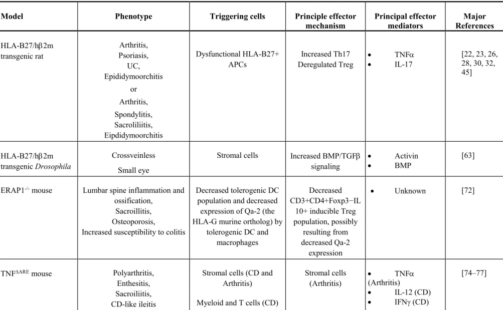
Table 1. Major characteristics of animal models used to study SpA mechanism.*
Model Phenotype Triggering cells Principle effector
mechanism Principal effector mediators Major References HLA-B27/h2m transgenic rat Arthritis, Psoriasis, UC, Epididymoorchitis or Arthritis, Spondylitis, Sacroliliitis, Eipdidymoorchitis Dysfunctional HLA-B27+ APCs Increased Th17 Deregulated Treg TNF IL-17 [22, 23, 26, 28, 30, 32, 45] HLA-B27/h2m transgenic Drosophila Crossveinless Small eye
Stromal cells Increased BMP/TGF
signaling
Activin
BMP
[63]
ERAP1-/- mouse Lumbar spine inflammation and
ossification, Sacroillitis, Osteoporosis,
Increased susceptibility to colitis
Decreased tolerogenic DC population and decreased
expression of Qa-2 (the HLA-G murine ortholog) by
tolerogenic DC and macrophages Decreased CD3+CD4+Foxp3−IL 10+ inducible Treg population, possibly resulting from decreased Qa-2 expression Unknown [72]
TNFARE mouse Polyarthritis,
Enthesitis, Sacroiliitis, CD-like ileitis
Stromal cells (CD and Arthritis)
Myeloid and T cells (CD)
Stromal cells (Arthritis) TNF (Arthritis) IL-12 (CD) IFN (CD) [74–77]
Th1 and CD8+ T cells (CD) tmTNF overexpressing mouse (TgA86) Arthritis, Enthesitis, Osteitis,
Bony ankylosis (axial and peripheral joints) Stromal cells ± haematopoietic cells TNFRI ± TNFRII signaling tmTNF [78, 79] Minicircle IL-23 DNA-inducible SpA Enthesitis,
Entheseal ossification (paw, spine, sacroiliac joint),
Psoriasis, Aortic root and valve
inflammation
IL-23R+ RORt+ CD4-
CD8- /T cells residing in enthesis and aortic root
Il-23-dependent type 3 immune response IL-17 IL-22 [80, 81] Curdlan-induced SpA in SKG mouse (ZAP-70 mutation) Arthritis, Enthesitis, Spondylitis, Sacroiliitis, Ileitis Uveitis
Curdlan-activated gut APCs (via Dectin 1 receptor)
IL-23-dependent type 3 immune response IL-17 [85] Fungus mannan inducible PsA Arthritis, Enthesistis, Osteitis, Periostitis, Hyperkeratinous skin TNF-producing macrophages / T cells-dependent type 3 immune response IL-17 [93]
Spine ossification, Neutrophilic dermatitis (rare),
Osteitis and periostitis (rare) Ankylosing enthesitis (ANKENT) mouse Dactylitis Onychoperiosteitis Unknown Increased BMP signaling BMP/TGF family ligands [97, 101]
* hm: human 2-microglobulin; UC: ulcerative colitis; APC: antigen-presenting cell; Th17: T-helper 17 cell; Treg: regulatory T cell; TNF: tumor necrosis factor-; IL-17: interleukin-17; BMP: bone morphogenic protein; TGF: transforming growth factor ; DC: dendritic cell; CD: Crohn's disease; PsA: psoriatic arthritis; IL27RA: IL27 receptor .