. . . .
. . . .
Sirt1 inhibition promotes in vivo arterial thrombosis
and tissue factor expression in stimulated cells
Alexander Breitenstein
1,2,3†, Sokrates Stein
1,2†, Erik W. Holy
1,2,3, Giovanni G. Camici
1,2,
Christine Lohmann
1,2, Alexander Akhmedov
1,2, Remo Spescha
1,2, Peter J. Elliott
4,
Christoph H. Westphal
4, Christian M. Matter
1,2,3, Thomas F. Lu¨scher
1,2,3,
and Felix C. Tanner
1,2,3*
1
Cardiovascular Research, Physiology Institute, University of Zurich, 8057 Zurich, Switzerland;2
Center for Integrative Human Physiology (ZHIP), University of Zurich, Zurich, Switzerland;
3
Cardiology, Cardiovascular Center, University Hospital Zurich, Ra¨mistrasse 100, 8091 Zurich, Switzerland; and4Sirtris, a GSK company, 200 Technology Square, Cambridge, MA 02139,
USA
Received 17 March 2010; revised 7 October 2010; accepted 21 October 2010; online publish-ahead-of-print 26 October 2010 Time for primary review: 32 days
Aims The mammalian silent information regulator-two 1 (Sirt1) blunts the noxious effects of cardiovascular risk factors such as type 2 diabetes mellitus and obesity. Nevertheless, the role of Sirt1 in regulating the expression of tissue factor (TF), the key trigger of coagulation, and arterial thrombus formation remains unknown.
Methods and results
Human as well as mouse cell lines were used for in vitro experiments, and C57Bl/6 mice for in vivo procedures. Sirt1 inhibition by splitomicin or sirtinol enhanced cytokine-induced endothelial TF protein expression as well as surface activity, while TF pathway inhibitor protein expression did not change. Sirt1 inhibition further enhanced TF mRNA expression, TF promoter activity, and nuclear translocation as well as DNA binding of the p65 subunit of nuclear factor-kappa B (NFkB/p65). Sirt1 siRNA enhanced TF protein and mRNA expression, and this effect was reduced in NFkB/p652/2 mouse embryonic fibroblasts reconstituted with non-acetylatable Lys310-mutant NFkB/p65. Acti-vation of the mitogen-activated protein kinases p38, c-Jun NH2-terminal kinase, and p44/42 (ERK) remained
unaf-fected. In vivo, mice treated with the Sirt1 inhibitor splitomicin exhibited enhanced TF activity in the arterial vessel wall and accelerated carotid artery thrombus formation in a photochemical injury model.
Conclusion We provide pharmacological and genetic evidence that Sirt1 inhibition enhances TF expression and activity by increasing NFkB/p65 activation in human endothelial cells. Furthermore, Sirt1 inhibition induces arterial thrombus formation in vivo. Hence, modulation of Sirt1 may offer novel therapeutic options for targeting thrombosis.
-Keywords Tissue factor † Sirt1 † Thrombosis † NFkB
1. Introduction
Cardiovascular diseases represent a major health burden. Acute vas-cular events such as myocardial infarction and ischaemic stroke account for the majority of deaths in Western countries.1Formation of an arterial thrombus is the central event in such acute vascular syn-dromes. Tissue factor (TF) is the key trigger of the coagulation cascade and thereby crucially involved in arterial thrombosis.2–4 Its impact on thrombus formation may be enhanced in atherosclerosis since TF expression is induced in the inflammatory environment of atherosclerotic plaques.5 In line with this notion, clinical studies demonstrate higher TF levels in the culprit lesion of patients with
acute coronary syndromes.6 Numerous inflammatory mediators
such as tumour necrosis factor alpha (TNF-a7) or histamine,8 but
also pro-thrombotic mediators like thrombin,9 induce endothelial TF expression by activating the MAP kinases p38, ERK, and c-Jun NH2-terminal kinase (JNK), and consequently transcription factors
such as nuclear factor-kappa B (NFkB).2
Silent information regulator-two (Sir2) is an NAD+-dependent class III histone deacetylase.10The mammalian sirtuins are
evolutiona-rily conserved homologues of the yeast Sir2,11and silent information regulator-two 1 (Sirt1) is the closest orthologue.12 In addition to
maintaining chromatin structure,10Sirt1 has been shown to regulate
transcription factors such as forkhead box class O (FOXO),13
†These authors contributed equally to this work.
*Corresponding author. Tel:+41 44 255 11 11; fax: +41 44 635 68 27, Email: [email protected]
Published on behalf of the European Society of Cardiology. All rights reserved.&The Author 2010. For permissions please email: [email protected].
p53,14 peroxisome proliferator-activated receptor-g,15 endothelial nitric oxide synthase (eNOS),16 and p65 subunit of nuclear factor-kappa B (NFkB/p65).17 Hence, Sirt1 is critically involved in cellular responses to stress,18 senescence,19 and mitochondrial function.20 Furthermore, endothelial overexpression of Sirt1 diminishes plaque formation in a mouse model of atherosclerosis,21and pharmacological activation of Sirt1 improves glucose homeostasis in mice and humans.22Based on these findings, Sirt1 modulators are under inves-tigation in clinical trials for the treatment of patients with cardiovascu-lar risk factors.
Since downstream targets of Sirt1 such as NFkB are involved in the regulation of TF expression, this study was designed to investigate the effect of Sirt1 on TF expression and arterial thrombus formation.
2. Methods
2.1 Cell culture
Human aortic endothelial cells (HAECs; Clonetics, Allschwil, Switzerland) were cultured as described.23Briefly, adhering HAECs were grown to con-fluence and rendered quiescent for 24 h in medium containing 0.5% FCS before stimulation. Sirt12/2 mouse embryonic fibroblasts (MEFs) were kindly provided by David Sinclair (Harvard Medical School, Boston, MA, USA) and were grown to confluence in Dulbecco’s modified Eagle’s medium supplemented with 10% FCS. Similarly, NFkB/p652/2MEFs with
reconstituted wild-type NFkB/p65 or non-acetylatable Lys310-mutant NFkB/p65 were used as described previously.24Cells were pre-treated
with splitomicin (Sigma, Buchs, Switzerland), sirtinol (Calbiochem, Lucerne, Switzerland), or resveratrol (Sigma) for 1 h before stimulation with 5 or 10 ng/mL TNF-a (R&D Systems, Minneapolis, MN, USA), 1 U/ mL thrombin (R&D Systems), or 1025mol/L histamine (Sigma), respect-ively. Transient transfection with pcDNA3.1-SIRT1 or Sirt1 siRNA (Sirt1 siRNA oligonucleotide sequence: 5′-GATGAAGTTGACCTCCTCA-3′) was performed using Lipofectamine Reagent (Invitrogen, Basel, Switzer-land) or Lipofectamine RNAi MAX (Invitrogen), respectively, as described previously.15,25Cytotoxicity was assessed by a colorimetric assay to detect lactate dehydrogenase (LDH; Roche, Basel, Switzerland).
2.2 Western blot analysis
Protein expression was determined as described.8 Antibodies against human TF, tissue factor pathway inhibitor (TFPI; both from American Diagnostica, Stamford, CT, USA), and Sirt1 (Santa Cruz Biotechnology, Santa Cruz, CA, USA) were used at 1:2000 dilution. Antibodies against phosphorylated p38 mitogen-activated protein (MAP) kinase (p38), p44/ 42 MAP kinase (ERK), and JNK (all from Cell Signaling, Danvers, MA, USA) were used at 1:1000, 1:5000, and 1:1000 dilution, respectively. Anti-bodies against total p38, ERK, and JNK (all from Cell Signaling) were diluted to 1:3000, 1:10 000, and 1:1000, respectively. The antibody against IkB-a (Santa Cruz Biotechnology) was applied at a 1:1000 dilution. Alpha-tubulin (Sigma) were applied to control protein loading (1:10 000 dilution). Primary antibodies were detected with a horseradish peroxidase-linked secondary antibody (Amersham, Munich, Germany).
2.3 Real-time PCR
Total RNA was extracted from HAECs with 1 mL TRIzol Reagent (Invi-trogen) as described.23Conversion of total cellular RNA to cDNA was carried out with Moloney murine leukaemia virus reverse transcriptase and random hexamer primers (Amersham) in a final volume of 33 mL using 4 mg of RNA. The total cDNA pool obtained served as template for subsequent PCR amplification with primers specific for full-length TF (sense primer: 5′-TCCCCAGAGTTCACACCTTACC-3′, antisense primer: 5′-CCTTTCTCCTGGCCCATACAC-3′; bases 508 – 529 of F3 cDNA; NCBI no. NM 001993). Real-time PCR amplification was
performed in an MX3000P PCR cycler (Stratagene) using the SYBR Green JumpStart kit (Sigma) in 25 mL final reaction volume containing 2 mL cDNA, 10 pmol of each primer, 0.25 mL of internal reference dye, and 12.5 mL of JumpStart Taq ReadyMix (buffer, dNTP, stabilizers, SYBR Green, Taq polymerase, and JumpStart Taq antibody). A melting curve analysis was performed after amplification to verify the accuracy of the amplicon. Ribosomal L28 RNA in HAECs or S12 RNA in MEFs served as loading control.
2.4 TF activity in vitro
TF surface activity in HAECs was analysed using a colorimetric assay (American Diagnostica). Cells were incubated at 378C with human FVIIa and FX, allowing for the formation of the TF/FVIIa complex at the cell surface. Conversion of FX to FXa was measured by the ability of FXa to cleave a chromogenic substrate. A standard curve was established with lipidated human TF to assure that the results were in the linear range of detection.
2.5 Histone deacetylase activity
Cell-based histone deacetylase activity (HDAC) assay was performed according to the manufacturer’s instructions (BIOMOL, Hamburg, Germany). Briefly, HAECs were starved in phenol-red-free media contain-ing 0.5% FCS. After 24 h, cells were incubated with 200 mmol/L Fluor de Lys substrate with or without trichostatin A (TSA; 1 mmol/L, BIOMOL), and with or without splitomicin (100 mmol/L, Sigma). After 2 h, cells were incubated with Fluor de Lys developer, lysed after 30 min, and equal amount of lysates were analysed for enzyme activity using a fluor-escence reader (Ex. 360 nm, Em. 460 nm).
2.6 TF promoter activity
An adenoviral vector (Ad5/hTF/Luc) containing the minimal TF promoter (2227 to +121 bp) upstream of the Luciferase cDNA and the SV40 PolyA signal was prepared as described.26 For viral transfection, the vector was added to HAEC at 100 pfu/cell for 1 h. HAEC were kept in growth medium for 24 h and then serum-starved for 24 h prior to TNF-a stimulation with or without Sirt1 inhibitor pre-treatment. Cells were stimulated with TNF-a for 30 min. Firefly luciferase activity was determined in cell lysates using a luminometer (Bertholg Technologies, Bad Wildbad, Germany).
2.7 NFkB DNA binding assay and NFkB/p65
immunofluorescence
Adhering HAECs were pre-treated with Sirt1 inhibitors for 1 h, followed by stimulation with 10 ng/mL TNF-a for additional 30 min. Nuclear protein was obtained by using a nuclear extraction kit (Active Motif, Rix-ensart, Belgium). Cells were harvested in hypotonic buffer for 15 min before centrifugation, isolated nuclei were resuspended in a hypertonic buffer, and nuclear protein was extracted by incubation on a rotator for 30 min. The supernatant containing the nuclear protein was collected after centrifugation. The DNA binding reaction was carried out with 5 mg of nuclear protein in a 96-well plate coated with consensus sequences for NFkB (GGGACTTTCC) for 1 h at room temperature. After washing, NFkB/p65 antibody (Active Motif) was added and incubated for 1 h, fol-lowed by incubation with a horseradish peroxidase-conjugated secondary antibody. Finally, NFkB/p65 DNA binding was assessed spectrophotome-trically at 450 nm.
HAECs were stained with FITC-labelled mouse anti-NFkB/p65 (Santa Cruz Biotechnology). Cytoplasmic NFkB/p65 was analysed using an SP2 confocal microscope (Leica), and quantification performed with the open-source software CellProfiler.27 Intensity of the green channel was measured in the cytoplasm (nuclear area subtracted from the total cell area) of at least 150 cells per treatment group at the Z-section where the nuclei had their largest diameter.
2.8 NFkB/p65 immunoprecipitation
HAECs were treated with 50 mM SIRT1 or scrambled siRNA overnight, followed by stimulation with TNF-a (10 ng/mL) for 20 min. Cells were then harvested and protein extracted in lysis buffer (20 mM HEPES, pH 7.5, 80 mM NaCl, 2.5 mM MgCl2, 1 mM EDTA, 0.5% NP-40, 1 mM
phenyl-methylsulfonyl fluoride, 10 mg/mL aprotinin, 10 mg/mL leupeptin, and 100 mM splitomicin). One milligram whole-cell lysates were immunopre-cipitated with rabbit anti-NFkB/p65 (Santa Cruz) using Protein G agarose (Millipore, Zug, Switzerland). Immunoprecipitated samples were immuno-blotted with rabbit anti-acLys310NFkB/p65 (Abcam, Cambridge, UK), the total lysates (5% input) with rabbit anti-SIRT1 and rabbit anti-NFkB/p65 (both from Santa Cruz Biotechnology).
2.9 Carotid artery thrombosis model and TF
activity in vivo
The investigation conforms to the Guide for the Care and Use of Laboratory Animals published by the US National Institutes of Health (NIH Publication no. 85-23, revised 1996). All animal procedures were approved by the local animal committee (Kantonales Veterina¨ramt Zurich, Switzerland) and performed in accordance with our institutional guidelines. C57BL/6 mice aged 12 – 14 weeks weighing on average 27 g were anaesthetized by intraperitoneal injection of 87 mg/kg sodium pentobarbital (Butler, Columbus, OH, USA). Rose Bengal (Fisher Scientific, Fair Lawn, NJ, USA) was diluted to 12 mg/mL in phosphate-buffered saline and then injected into the tail vein at a concentration of 63 mg/kg. Mice were secured in a supine position, placed under a dissecting microscope, and the right common carotid artery was exposed following a midline cervical incision. A Doppler flow probe (Model 0.5 VB, Transonic Systems, Ithaca, NY, USA) was applied and connected to a flowmeter (Transonic, Model T106). Six minutes after Rose Bengal injection, a 1.5 mW green light laser (540 nm; Melles Griot, Carlsbad, CA, USA) was applied to the site of injury at a distance of 6 cm for 60 min or until thrombosis occurred. From the onset of injury, blood flow was monitored up to 120 min, at which time the experiment was terminated.7Occlusion was defined as flow ≤0.1 mL/min for at least 1 min. Mice were divided into two groups: splitomicin (80 mg/kg with an intraperitoneal injection every 24 h for 5 days), or vehicle control (0.5% methylcellulose).
Right carotid arteries were homogenized in 50 mL of lysis buffer and left to stand on ice for 30 min. TF activity was measured by using the colori-metric assay as described above.
2.10 Statistical analysis
Data are indicated as mean + SEM. Unpaired Student’s t-test was used to evaluate differences between two groups. For statistical analysis of data from multiple groups, one-way ANOVA was performed. A P value ,0.05 denoted a significant difference. All statistical values are additionally summarized in the Supplementary material online, Table S1.
3. Results
3.1 Sirt1 inhibition enhances TF expression
and activity
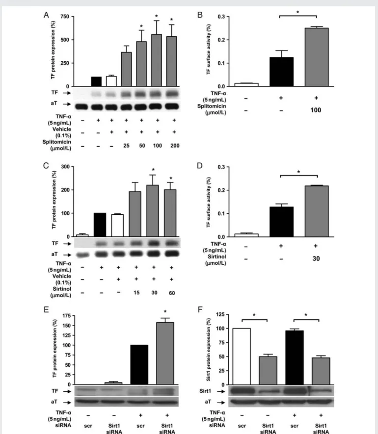
TF protein expression was determined in TNF-a (5 ng/mL for 5 h) stimulated HAECs in the presence or absence of increasing concen-trations of splitomicin (25 – 200 mmol/L) or sirtinol (15 – 60 mmol/L), respectively. Both inhibitors enhanced TNF-a-induced TF protein expression in a concentration-dependent manner; maximal activation occurred at 100 mmol/L in splitomicin and at 30 mmol/L in sirtinol-treated cells, respectively (n ¼ 4; P , 0.05; Figure1A and C ). These effects were paralleled by an increased TF surface activity in cells pre-treated with either Sirt1 inhibitor (n ¼ 4; P , 0.01, Figure1B and D).
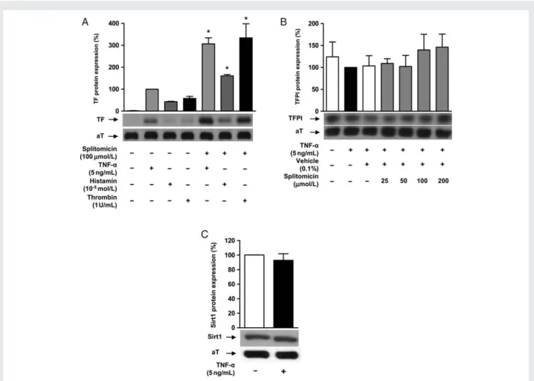
Sirt1 knockdown using specific siRNA enhanced TNF-a-induced TF protein expression (n ¼ 4; P , 0.01; Figure1E); western blot analysis confirmed reduced Sirt1 expression in cells transfected with Sirt1 siRNA (n ¼ 4; P , 0.01; Figure1F). Sirt1 inhibition also enhanced TF protein expression in response to stimulation with histamine (1025mol/L) or thrombin (1 U/mL), respectively (n ¼ 4; P , 0.05; Figure 2A). The expression of TFPI, the physiological antagonist of TF, remained unaffected (n ¼ 4; P ¼ NS; Figure 2B). TNF-a did not alter endogenous Sirt1 protein expression (n ¼ 4; P ¼ NS; Figure 2C), and no changes in cell morphology nor LDH release were detected by any of these treatments (n ¼ 4 for each; P ¼ NS; Supplementary material online, Figure S1).
3.2 TF mRNA expression is induced
by Sirt1 inhibition
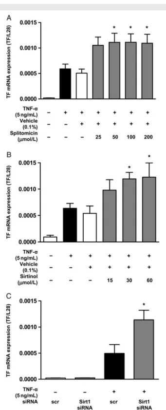
Real-time rtPCR revealed that TNF-a (5 ng/mL) induced TF mRNA expression within 1 h (n ¼ 4; P , 0.05; Figure 3A). Both splitomicin and sirtinol enhanced TF mRNA expression in stimulated endothelial cells (splitomicin: n ¼ 4; P , 0.05; Figure3A, sirtinol: n ¼ 4; P , 0.05; Figure 3B). In parallel, Sirt1 knockdown using siRNA enhanced TNF-a-stimulated TF mRNA expression (n ¼ 4; P , 0.01; Figure3C).
3.3 Sirt1 inhibition reduces HDAC
class III activity
A cell-based HDAC assay was performed in HAECs to confirm that the Sirt1 inhibitor splitomicin diminishes intracellular deacetylase activity. Cells were treated with 1 mmol/L TSA to inactivate HDAC classes I and II in the presence or absence of splitomicin (100 mmol/L). Consistent with inhibition of HDAC class III, splitomicin reduced deacetylase activity (n ¼ 4; P , 0.05; Supplementary material online, Figure S2A) as compared with TSA alone. In contrast, splitomi-cin did not affect TF protein expression in Sirt12/2MEFs (n ¼ 4; P ¼ NS; Supplementary material online, Figure S2B).
3.4 Activation of Sirt1 impairs TF
expression and activity
Pharmacological activation of Sirt1 by resveratrol, a commonly used, but less specific Sirt1 activator, impaired TNF-a-induced TF protein and mRNA expression (n ¼ 4; P , 0.01 for TF protein, and P , 0.05 for TF mRNA; Supplementary material online, Figure S3A and B). In parallel, overexpression of Sirt1 in Sirt12/2 MEFs reduced TNF-a-induced TF expression (n ¼ 4; P , 0.01; Supplementary material online, Figure S3C and D).
3.5 Sirt1 inhibition enhances TF promoter
activity
To assess whether Sirt1 inhibition enhances TF promoter activity, the impact of splitomicin and sirtinol on the TF promoter was analysed. HAECs were transfected with a luciferase plasmid under control of the human minimal TF promoter (2221 up to+121 bp). Splitomicin and sirtinol enhanced stimulated TF promoter activity as compared with TNF-a alone (n ¼ 5; P , 0.01; Figure4A and B).
3.6 MAP kinase activation is not affected by
Sirt1 inhibition
To assess whether modulation of Sirt1 activity alters MAP kinase acti-vation, HAECs were examined at different time points after TNF-a stimulation. The MAP kinases p38, ERK, and JNK were transiently
Figure 1 Sirt1 inhibition enhances endothelial TF expression and activity. (A and B) Splitomicin enhances TNF-a-induced TF protein expression (*P , 0.05 vs. TNF-a alone) and surface activity (*P , 0.01 vs. TNF-a alone) in human endothelial cells. (C and D) Sirtinol exerts similar effects on TF protein expression (*P , 0.05 vs. TNF-a alone) and activity (*P , 0.01 vs. TNF-a alone). (E and F) Sirt1 siRNA enhances TNF-a-induced TF protein expression (*P , 0.01 vs. TNF-a alone).
activated by TNF-a (n ¼ 3; Supplementary material online, Figures S4 and S5). Phosphorylation of p38, ERK, and JNK remained unaffected in cells pre-treated with either splitomicin (n ¼ 3; P ¼ NS; Supplemen-tary material online, Figure S4) or sirtinol, respectively (n ¼ 3; P ¼ NS; Supplementary material online, Figure S5). Total expression of MAP kinases remained unchanged at any time point with or without Sirt1 inhibitors.
3.7 Sirt1 inhibition enhances NFkB/p65
DNA binding via deacetylation of Lys
310of NFkB/p65
NFkB/p65 is a transcription factor that regulates TF expression. Thus, the effect of Sirt1 inhibition on NFkB/p65 activation was investigated. TNF-a (10 ng/mL) induced a significant increase in NFkB/p65 DNA binding as compared with control (n ¼ 5; P , 0.01; Figure 5A and B). Sirt1 inhibition with splitomicin (100 mmol/L) or sirtinol (30 mmol/L) enhanced NFkB/p65 DNA binding (n ¼ 5; P , 0.01; Figure5A and B). In line with this, translocation of NFkB/p65 from the cytoplasm to the nucleus was increased after TNF-a stimulation as confirmed by NFkB/p65 immunofluorescence and pre-treatment
with splitomicin further enhanced NFkB/p65 nuclear translocation (n ¼ 5; P , 0.01; Supplementary material online, Figure S6).
Since degradation of the inhibitory protein of NFkB, IkB-a, is an early step in activation of NFkB/p65, the effect of Sirt1 inhibition on IkB-a degradation was investigated. TNF-a induced a transient degra-dation of IkB-a (n ¼ 3; Supplementary material online, Figures S4 and S5). Neither splitomicin nor sirtinol altered the degradation pattern of IkB-a as compared with TNF-a alone (n ¼ 3; P ¼ NS; Supplementary material online, Figures S4 and S5).
For further analysis, NFkB/p652/2 MEFs were used and reconsti-tuted with either wild-type NFkB/p65 or a non-acetylatable Lys310 -mutant NFkB/p65. The effect of Sirt1 siRNA on TF expression was less pronounced in MEFs reconstituted with the non-acetylatable Lys310-mutant NFkB/p65 as compared with the wild-type NFkB/p65 (n ¼ 5; P , 0.01; Figure 5C), although both types of reconstituted cells exhibited enhanced TF mRNA expression after TNF-a stimulation when Sirt1 was knocked down by siRNA (n ¼ 5; P , 0.05; Figure5C). Western blot analysis confirmed reduced Sirt1 expression in all the cells transfected with Sirt1 siRNA (n ¼ 4; P , 0.05; Figure5D).
Sirt1 was also silenced in HAECs using siRNA. The cells were stimulated with TNF-a (10 ng/mL) and a NFkB/p65 Figure 2 Splitomicin induce TF expression in response to different mediators, but does not alter TFPI expression. TNF-a does not alter Sirt1 expression. (A) TNF-a, histamine, and thrombin induce TF protein expression. Splitomicin up-regulates TF expression in response to each stimulus (*P , 0.05 vs. each stimulation factor). (B) Splitomicin does not alter TFPI expression (P ¼ NS). (C) TNF-a stimulation does not change endogenous expression of Sirt1 (P ¼ NS).
immunoprecipitation was performed. There was a higher extent of Lys310NFkB/p65 acetylation in cells with impaired Sirt1 expression (n ¼ 3; Figure5E).
3.8 Sirt1 inhibition induces TF activity and
arterial thrombosis in vivo
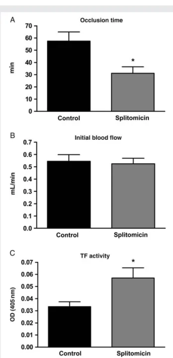
C57Bl/6 mice were treated with splitomicin (80 mg/kg body weight, intraperitoneal injection every 24 h for 5 days) or vehicle (0.5% methylcellulose). Vehicle-treated mice developed carotid artery thrombosis within a mean occlusion time of 57.8. + 7.5 min, while splitomicin-treated mice occluded within a mean time period of 31.2 + 5.3 min (n ¼ 7; P , 0.05; Figure 6A). Initial blood flow in carotid artery did not differ between vehicle- and splitomicin-treated mice (0.54+0.05 vs. 0.52+0.05 mL/min; n ¼ 7; P ¼ NS; Figure6B). Splitomicin treatment increased TF activity in mouse carotid artery in vivo as compared with the controls (n ¼ 6; P , 0.05; Figure6C). Figure 3 Sirt1 inhibition induces endothelial TF expression at the
transcriptional level. (A and B) Real-time rtPCR reveals that splitomi-cin and sirtinol enhance TNF-a-induced TF mRNA expression (*P , 0.05 vs. TNF-a alone for splitomicin; *P , 0.05 vs. TNF-a alone for sirtinol). (C) Sirt1 siRNA enhances TNF-a-induced TF mRNA expression (*P , 0.01 vs. TNF-a alone).
Figure 4 Sirt1 inhibition increases TF promoter activity. (A and B) TNF-a increases the activity of the minimal TF promoter. Splitomicin (A) and sirtinol (B) enhance TF promoter activity under cytokine-induced conditions (*P , 0.01 vs. TNF-a alone). AU, arbi-trary units.
4. Discussion
The present study demonstrates that Sirt1 inhibits TF expression at the transcriptional level via NFkB/p65 in human vascular cells. Further-more, it shows that inhibition of Sirt1 induces thrombus formation and arterial TF activity in vivo.
To inhibit Sirt1 activity in human vascular cells, two different pharmacological agents were applied. Both splitomicin and sirtinol are established inhibitors of Sirt1.11,28–30 It is still debated to what extent these drugs specifically inhibit Sirt1; indeed, sirtinol may also inhibit Sirt2.30Nevertheless, the concentrations of both substances applied in this study are within the established range.17,19Moreover, splitomicin did not alter TF expression in Sirt12/2 MEFs, and TF was increased when Sirt1 was down-regulated by siRNA. Hence,
these data support the conclusion that Sirt1 regulates the expression of TF.
Cytokine-mediated TF expression is mainly regulated at the tran-scriptional level, where NFkB/p65 is importantly involved.2 Transcrip-tionally active NFkB consists of a heterodimeric complex mainly composed of a p65 and a p50 subunit. In quiescent cells, NFkB is retained in the cytoplasm by its inhibitor IkB. Upon cytokine stimu-lation, IkB becomes degraded allowing NFkB to translocate to the nucleus and to stimulate gene transcription. In this study, both Sirt1 inhibitors enhanced nuclear translocation and DNA binding of NFkB/ p65, identifying NFkB/p65 as a downstream target of Sirt1 in the context of TF expression. Since the Sirt1 inhibitors did not affect MAP kinases nor IkB degradation, an involvement of these mediators can be ruled out; thus, a direct effect of Sirt1 on NFkB/p65 seems Figure 5 Sirt1 inhibition enhances NFkB/p65 activation via acetylation of Lys310of NFkB/p65. (A and B) TNF-a stimulates NFkB/p65 DNA binding activity as compared with control conditions. Splitomicin and sirtinol both enhance NFkB/p65 DNA binding (*P , 0.01 vs. TNF-a alone). (C) Sirt1 siRNA induces TF mRNA up-regulation in the presence of wild-type NFkB/p65, whereas its expression is reduced by a non-acetylatable Lys310-mutant NFkB/p65 (*P , 0.01 vs. wild-type NFkB/p65). (D) Sirt1 protein down-regulation by specific siRNA is demonstrated. (E) NFkB/p65 immunoprecipita-tion in HAECs reveals more acetylated Lys310NFkB/p65 upon Sirt1 siRNA treatment.
likely. Acetylation of Lys310and Lys221residues of NFkB/p65 impairs its association with IkB and increases its DNA-binding capacity.31Sirt1 deacetylates Lys310 of the NFkB/p65 subunit in different cell types and thereby blunts NFkB/p65-mediated gene expression.17,32 Exper-iments involving NFkB/p652/2 MEFs reconstituted with either wild-type NFkB/p65 or non-acetylatable Lys310-mutant NFkB/p65, respect-ively, demonstrate that regulation of TF mRNA expression by Sirt1 indeed depends on Lys310 acetylation of NFkB/p65. Nevertheless, other downstream targets of Sirt1, such as eNOS or p53, are also known to regulate TF expression,33,34and a role of these regulators
in Sirt1-mediated TF expression in addition to that of NFkB/p65 cannot be ruled out by the current data.
TF is the key initiator of coagulation and therefore an important trigger of thrombosis.35 Exposure of TF to the circulating blood results in acute thrombosis and eventually vascular occlusion; in fact, reducing TF expression impairs thrombus formation.36To inves-tigate arterial thrombosis in vivo, a photochemical vascular injury model was used, since it is an established method to study TF-dependent thrombus formation.36 Mean occlusion time in splitomicin-treated mice was reduced by nearly 50%, supporting the concept that inhibition of Sirt1 induces arterial thrombosis. The increased TF activity in mouse carotid artery indicates that Sirt1 inhi-bition regulates thrombosis at least in part via activation of TF in vivo. TFPI, the physiological inhibitor of TF, was not affected by splitomicin treatment excluding compensatory effects on TF activity. Given the importance of the balance between TF and TFPI for thrombosis,37 these findings underscore a role for TF in the modulation of thrombus formation by Sirt1. Hence, Sirt1 activators, which are currently under investigation for the treatment of type 2 diabetes mellitus, may possess additional protective cardiovascular effects by inhibiting arter-ial thrombus formation.
Sirt1 inhibition resulted in an enhanced TF expression after stimu-lation with different mediators. Hence, Sirt1 may suppress TF expression especially in the inflammatory environment observed in patients exposed to cardiovascular risk factors and with advanced atherosclerotic lesions.38 Indeed, elevated levels of soluble TF are observed in patients with atherosclerosis as compared with con-trols.39 Furthermore, even higher concentrations are measured in the area around the culprit lesion in patients with unstable angina or acute myocardial infarction as compared with patients with stable angina.6,40 Taken together, pharmacological or genetic acti-vation of Sirt1 could be a promising therapeutic target in these conditions.
A recent report described that endothelial overexpression of Sirt1 diminishes atherogenesis in ApoE-deficient mice, suggesting an anti-atherosclerotic effect of Sirt1.21 In addition, Sirt1 exerts beneficial effects on cardiovascular risk factors such as type 2 diabetes mellitus and arterial hypertension.22,41,42 Sirt1 also mediates the effects of caloric restriction on life-span extension,43 which may in turn improve endothelial function and blood pressure regulation.44 Since TF expression and activity is increased by cardiovascular risk factors such as type 2 diabetes mellitus45,46 and arterial hypertension,47 Sirt1 activators, which are currently under investigation in clinical trials for the treatment of cardiovascular risk factors, could exert a dual beneficial effect preventing arterial thrombosis not only by down-regulating TF expression, but also by interfering with the risk factors inducing it.
In summary, this study demonstrates that Sirt1 inhibition enhances TF in vitro as well as in vivo, and accelerates arterial thrombus for-mation. Sirt1 exerts these effects at the transcriptional level by mod-ulating NFkB/p65 DNA binding without affecting MAP kinase activation. These findings reveal a novel action of Sirt1 and suggest that Sirt1 activators may be applied for the prevention of thrombosis.
Acknowledgements
We thank the Center for Microscopy and Image Analysis (University of Zurich, Switzerland) for using their resources.
Figure 6 Sirt1 inhibition accelerates arterial thrombus formation. (A) Time to thrombotic occlusion after mouse carotid artery photo-chemical injury in vivo. Splitomicin promotes thrombus formation (*P , 0.05 vs. vehicle alone). (B) Initial blood flow in the carotid artery is unchanged (P ¼ NS). (C) Splitomicin increases TF activity in mouse carotid artery in vivo. Values are indicated as absorbance at 405 nm (*P , 0.05 vs. vehicle).
Conflict of interest: none declared.
Funding
This study was supported by the Swiss National Science Foundation (grant no. 3200B0-113328/1 to F.C.T., grant no. 3100-068118.02/1 to T.F.L., and grant no. 31-114094/1 to C.M.M.; Berne, Switzerland), the Bonizzi-Theler Foundation (Zurich, Switzerland), Velux Foundation (Zurich, Switzerland), Wolfermann Na¨geli Foundation (Zurich, Switzerland), MERCATOR Foundation (Essen, Germany), and the Swiss Heart Foundation (Berne, Switzerland).
References
1. Rosamond W, Flegal K, Furie K, Go A, Greenlund K, Haase N et al. Heart disease and stroke statistics – 2008 update: a report from the American Heart Association Stat-istics Committee and Stroke StatStat-istics Subcommittee. Circulation 2008;117:e25 – e146. 2. Steffel J, Luscher TF, Tanner FC. Tissue factor in cardiovascular diseases: molecular
mechanisms and clinical implications. Circulation 2006;113:722 – 731.
3. Tilley R, Mackman N. Tissue factor in hemostasis and thrombosis. Semin Thromb Hemost 2006;32:5 – 10.
4. Toschi V, Gallo R, Lettino M, Fallon JT, Gertz SD, Fernandez-Ortiz A et al. Tissue factor modulates the thrombogenicity of human atherosclerotic plaques. Circulation 1997;95:594 – 599.
5. Wilcox JN, Smith KM, Schwartz SM, Gordon D. Localization of tissue factor in the normal vessel wall and in the atherosclerotic plaque. Proc Natl Acad Sci USA 1989; 86:2839 – 2843.
6. Ardissino D, Merlini PA, Ariens R, Coppola R, Bramucci E, Mannucci PM. Tissue-factor antigen and activity in human coronary atherosclerotic plaques. Lancet 1997; 349:769 – 771.
7. Camici GG, Steffel J, Akhmedov A, Schafer N, Baldinger J, Schulz U et al. Dimethyl sulfoxide inhibits tissue factor expression, thrombus formation, and vascular smooth muscle cell activation: a potential treatment strategy for drug-eluting stents. Circulation 2006;114:1512 – 1521.
8. Steffel J, Akhmedov A, Greutert H, Luscher TF, Tanner FC. Histamine induces tissue factor expression: implications for acute coronary syndromes. Circulation 2005;112: 341 – 349.
9. Steffel J, Arnet C, Akhmedov A, Iseli SM, Luscher TF, Tanner FC. Histamine differen-tially interacts with tumor necrosis factor-alpha and thrombin in endothelial tissue
factor induction: the role of c-Jun NH2-terminal kinase. J Thromb Haemost 2006;4:
2452 – 2460.
10. Gasser SM, Cockell MM. The molecular biology of the SIR proteins. Gene 2001;279: 1 – 16.
11. Lavu S, Boss O, Elliott PJ, Lambert PD. Sirtuins – novel therapeutic targets to treat age-associated diseases. Nat Rev Drug Discov 2008;7:841 – 853.
12. Imai S, Armstrong CM, Kaeberlein M, Guarente L. Transcriptional silencing and long-evity protein Sir2 is an NAD-dependent histone deacetylase. Nature 2000;403: 795 – 800.
13. Motta MC, Divecha N, Lemieux M, Kamel C, Chen D, Gu W et al. Mammalian SIRT1 represses forkhead transcription factors. Cell 2004;116:551 – 563.
14. Luo J, Nikolaev AY, Imai S, Chen D, Su F, Shiloh A et al. Negative control of p53 by Sir2alpha promotes cell survival under stress. Cell 2001;107:137 – 148.
15. Picard F, Kurtev M, Chung N, Topark-Ngarm A, Senawong T, Machado De Oliveira R et al. Sirt1 promotes fat mobilization in white adipocytes by repressing PPAR-gamma. Nature 2004;429:771 – 776.
16. Mattagajasingh I, Kim CS, Naqvi A, Yamamori T, Hoffman TA, Jung SB et al. SIRT1 pro-motes endothelium-dependent vascular relaxation by activating endothelial nitric oxide synthase. Proc Natl Acad Sci USA 2007;104:14855 – 14860.
17. Yeung F, Hoberg JE, Ramsey CS, Keller MD, Jones DR, Frye RA et al. Modulation of NF-kappaB-dependent transcription and cell survival by the SIRT1 deacetylase. EMBO J 2004;23:2369 – 2380.
18. Alcendor RR, Gao S, Zhai P, Zablocki D, Holle E, Yu X et al. Sirt1 regulates aging and resistance to oxidative stress in the heart. Circ Res 2007;100:1512 – 1521. 19. Ota H, Akishita M, Eto M, Iijima K, Kaneki M, Ouchi Y. Sirt1 modulates premature
senescence-like phenotype in human endothelial cells. J Mol Cell Cardiol 2007;43: 571 – 579.
20. Lagouge M, Argmann C, Gerhart-Hines Z, Meziane H, Lerin C, Daussin F et al. Resveratrol improves mitochondrial function and protects against metabolic disease by activating SIRT1 and PGC-1alpha. Cell 2006;127:1109 – 1122.
21. Zhang QJ, Wang Z, Chen HZ, Zhou S, Zheng W, Liu G et al. Endothelium-specific overexpression of class III deacetylase SIRT1 decreases atherosclerosis in apolipopro-tein E-deficient mice. Cardiovasc Res 2008;80:191 – 199.
22. Milne JC, Lambert PD, Schenk S, Carney DP, Smith JJ, Gagne DJ et al. Small molecule activators of SIRT1 as therapeutics for the treatment of type 2 diabetes. Nature 2007; 450:712 – 716.
23. Breitenstein A, Stampfli SF, Camici GG, Akhmedov A, Ha HR, Follath F et al. Amio-darone inhibits arterial thrombus formation and tissue factor translation. Arterioscler Thromb Vasc Biol 2008;28:2231 – 2238.
24. Buerki C, Rothgiesser KM, Valovka T, Owen HR, Rehrauer H, Fey M et al. Functional relevance of novel p300-mediated lysine 314 and 315 acetylation of RelA/p65. Nucleic Acids Res 2008;36:1665 – 1680.
25. Stein S, Schafer N, Breitenstein A, Besler C, Winnik S, Lohmann C et al. SIRT1 reduces endothelial activation without affecting vascular function in ApoE-/-mice. Aging (Albany NY) 2010;2:353 – 360.
26. Holy EW, Akhmedov A, Luscher TF, Tanner FC. Berberine, a natural lipid-lowering drug, exerts prothrombotic effects on vascular cells. J Mol Cell Cardiol 2009;46: 234 – 240.
27. Carpenter AE, Jones TR, Lamprecht MR, Clarke C, Kang IH, Friman O et al. CellPro-filer: image analysis software for identifying and quantifying cell phenotypes. Genome Biol 2006;7:R100.
28. Araki T, Sasaki Y, Milbrandt J. Increased nuclear NAD biosynthesis and SIRT1 acti-vation prevent axonal degeneration. Science 2004;305:1010 – 1013.
29. Fulco M, Schiltz RL, Iezzi S, King MT, Zhao P, Kashiwaya Y et al. Sir2 regulates skeletal muscle differentiation as a potential sensor of the redox state. Mol Cell 2003;12: 51 – 62.
30. Grubisha O, Smith BC, Denu JM. Small molecule regulation of Sir2 protein deacety-lases. FEBS J 2005;272:4607 – 4616.
31. Chen LF, Greene WC. Shaping the nuclear action of NF-kappaB. Nat Rev Mol Cell Biol 2004;5:392 – 401.
32. Chen J, Zhou Y, Mueller-Steiner S, Chen LF, Kwon H, Yi S et al. SIRT1 protects against microglia-dependent amyloid-beta toxicity through inhibiting NF-kappaB signaling. J Biol Chem 2005;280:40364 – 40374.
33. Yang Y, Loscalzo J. Regulation of tissue factor expression in human microvascular endothelial cells by nitric oxide. Circulation 2000;101:2144 – 2148.
34. Yu JL, May L, Lhotak V, Shahrzad S, Shirasawa S, Weitz JI et al. Oncogenic events regu-late tissue factor expression in colorectal cancer cells: implications for tumor pro-gression and angiogenesis. Blood 2005;105:1734 – 1741.
35. Nemerson Y. Tissue factor and hemostasis. Blood 1988;71:1 – 8.
36. Day SM, Reeve JL, Pedersen B, Farris DM, Myers DD, Im M et al. Macrovascular thrombosis is driven by tissue factor derived primarily from the blood vessel wall. Blood 2005;105:192 – 198.
37. Pedersen B, Holscher T, Sato Y, Pawlinski R, Mackman N. A balance between tissue factor and tissue factor pathway inhibitor is required for embryonic development and hemostasis in adult mice. Blood 2005;105:2777 – 2782.
38. Libby P, Ridker PM, Maseri A. Inflammation and atherosclerosis. Circulation 2002;105: 1135 – 1143.
39. Reilly MP, Rohatgi A, McMahon K, Wolfe ML, Pinto SC, Rhodes T et al. Plasma cyto-kines, metabolic syndrome, and atherosclerosis in humans. J Investig Med 2007;55: 26 – 35.
40. Annex BH, Denning SM, Channon KM, Sketch MH Jr., Stack RS, Morrissey JH et al. Differential expression of tissue factor protein in directional atherectomy specimens from patients with stable and unstable coronary syndromes. Circulation 1995;91: 619 – 622.
41. Banks AS, Kon N, Knight C, Matsumoto M, Gutierrez-Juarez R, Rossetti L et al. SirT1 gain of function increases energy efficiency and prevents diabetes in mice. Cell Metab 2008;8:333 – 341.
42. Lee JH, Song MY, Song EK, Kim EK, Sung Moon W, Han MK et al. Overexpression of SIRT1 protects pancreatic {beta}-cells against cytokine toxicity through suppressing NF-{kappa}B signaling pathway. Diabetes 2009;58:344 – 351.
43. Guarente L, Picard F. Calorie restriction– the SIR2 connection. Cell 2005;120: 473 – 482.
44. Fontana L, Klein S. Aging, adiposity, and calorie restriction. JAMA 2007;297:986 – 994. 45. Diamant M, Nieuwland R, Pablo RF, Sturk A, Smit JW, Radder JK. Elevated numbers of tissue-factor exposing microparticles correlate with components of the metabolic syndrome in uncomplicated type 2 diabetes mellitus. Circulation 2002;106:2442 – 2447. 46. Lim HS, Blann AD, Lip GY. Soluble CD40 ligand, soluble P-selectin, interleukin-6, and tissue factor in diabetes mellitus: relationships to cardiovascular disease and risk factor intervention. Circulation 2004;109:2524 – 2528.
47. Felmeden DC, Spencer CG, Chung NA, Belgore FM, Blann AD, Beevers DG et al. Relation of thrombogenesis in systemic hypertension to angiogenesis and endothelial damage/dysfunction (a substudy of the Anglo-Scandinavian Cardiac Outcomes Trial [ASCOT]). Am J Cardiol 2003;92:400 – 405.